Article

Effects of Tissue Preservation on Carbon and Nitrogen Stable Isotope Signatures in Syngnathid Fishes and Prey

by
Miquel Planas
1,*,
Alex Paltrinieri
1,2,
Mario Davi Dias Carneiro
1,3 and
Jorge Hernández-Urcera
1
1
Institute of Marine Research (CSIC), 36208 Vigo, Spain
2
Dipartimento di Scienze della Vita e dell’Ambiente, Università Politecnica delle Marche, 60131 Ancona, Italy
3
Laboratório de Piscicultura Estuarina e Marinha, Instituto de Oceanografia, Universidade Federal do Rio Grande—FURG, Rio Grande, RS 96203-900, Brazil
*
Author to whom correspondence should be addressed.
Submission received: 28 October 2020 / Revised: 25 November 2020 / Accepted: 2 December 2020 / Published: 4 December 2020
(This article belongs to the Section Aquatic Animals)

Abstract

:

Simple Summary

Stable isotope analysis (SIA) was used to assess the influence of various preservation methods (freezing, ethanol and formaldehyde) on syngnathid (seahorses and pipefishes) fins, seahorse newborns (seahorses), and prey (copepods and Artemia). The first available conversion models for syngnathids are provided, enabling their application to isotopic studies in the field and in the laboratory.

Abstract

Isotopic stable analysis (SIA) is a powerful tool in the assessment of different types of ecological and physiological studies. For that, different preservation methods for sampled materials are commonly used prior to isotopic analysis. The effects of various preservation methods (freezing, ethanol and formaldehyde) were analyzed for C:N, and δ13C and δ15N signals on a variety of tissues including dorsal fins (three seahorse and two pipefish species), seahorse newborns (three seahorses species), and prey (copepods and different stages of Artemia) commonly used to feed the fishes under rearing conditions. The aims of the study were: (i) to evaluate isotopic effects of chemical preservation methods across different types of organisms and tissues, using frozen samples as controls, and (ii) to construct the first conversion models available in syngnathid fishes. The chemical preservation in ethanol and, to a lesser extent, in formaldehyde significantly affected δ13C values, whereas the effects on δ15N signatures were negligible. Due to their low lipid content, the isotopic signals in fish fins was almost unaffected, supporting the suitability of dorsal fins as the most convenient material in isotopic studies on vulnerable species such as syngnathids. The regression equations provided resulted convenient for the successful conversion of δ13C between preservation treatments. Our results indicate that the normalization of δ15N signatures in preserved samples is unnecessary. The conversion models should be applicable in isotopic field studies, laboratory experiments, and specimens of historical collections.

1. Introduction

Stable isotope compositions of carbon and nitrogen (typically expressed as δ13C and δ15N, respectively), are used in a variety of studies, including tracing trophic chains [1,2], estimation of trophic enrichment factors [1,2,3], diet reconstruction [4,5], tissue turnover rates [6,7], discrimination between hatchery-reared and wild spawned individuals, or migrations [8,9].
The isotopic composition of animal tissues reflects the dietary isotopic composition, especially for C and N, within a difference of a few units (discrimination factor) [10,11]. Isotopic signatures are species- and tissue-specific [12,13] and might vary depending on several factors such as dietary isotopic values or developmental stage [14,15,16]. However, some important issues on the application of stable isotope analysis (SIA) need to be properly addressed, particularly on type of tissue, sample preservation method, and lipid correction.
Whenever possible, SIA is performed on muscle tissue due to its relative isotopic stability [12]. Where lethal sampling is not desirable, other fish tissues such as fins and scales are non-lethal alternatives to muscle [15,17,18,19,20], especially in threatened and endangered species [21,22,23,24]. The isotopic signals from those surrogate tissues can be converted to muscle values by means of mathematical corrections obtained from inter-tissue comparisons.
The methods commonly used for tissue preservation include drying, freezing, ethanol or formaldehyde, depending on the objectives, limitations of the study and sampling conditions [25,26,27,28,29]. However, the results of inter-methodological comparisons might be unpredictable and vary among taxa, suggesting the need to analyze zoological groups separately [26,28].
Lipids are depleted (more negative values) in δ13C when compared to other biochemical compounds (e.g., proteins, carbohydrates), affecting bulk tissue signatures [10,11,30,31,32]. Lipid extraction may also result in changes in δ15N values of the lipid-free sample [33]. Alternatively, mathematical normalization techniques should be applied for lipid normalization when C:N ratios are high [34,35]. Ideally, corrections should be applied using model estimates on the same or similar type of organisms.
In the present study, we assessed the effects of freezing and solvent preservatives (ethanol and formaldehyde) on C:N ratios and δ13C and δ15N signatures both in syngnathid fishes, including adults (fin clipping) and newborns (bulk specimens), and in prey (copepods and different stages of Artemia) commonly used to raise those fishes. Syngnathids are a family of fishes including endangered and vulnerable species [36,37]. For that reason, the use of lethal sampling should be avoided whenever possible, and fin tissue is an excellent and suitable material for isotopic analyses [22]. Since tissue conversion models specific for syngnathid fishes were lacking, the main aim of this study was to provide for the first time mathematical corrections for δ13C and δ15N in syngnathid species considering different types of specimens and preservation methods. The models provided would be helpful in field, experimental and natural history collections.

2. Materials and Methods

2.1. Live Prey

We analyzed different sources of prey commonly used in the rearing of syngnathids: calanoid copepods (Acartia tonsa) and Artemia (nauplii, metanauplii enriched for 24 and 72 h, enriched adults and unenriched adults). Microalgae (Phaeodactylum tricornutum, Rhodomonas lens and Isochrysis galbana) were cultivated at 22 ± 1 °C on F2P media to feed copepods Acartia tonsa and Artemia until the adult stage. The copepods were fed on the microalgae R. lens (26–27 °C and 38 salinity). Only copepods retained on a 180 µm mesh (copepodites and adults) were analyzed.
Artemia nauplii and metanauplii were produced to feed seahorse juveniles, whereas adult Artemia were delivered to adult seahorses. Artemia cysts (EG MC450 and AF; Ocean Nutrition, San Diego, CA, USA) were hatched at 28 °C and newly hatched nauplii were collected to produce enriched metanauplii (from AF cysts) and adults (from EG cysts). For metanauplii, the nauplii were enriched (2–3 days at 100 Artemia mL−1) twice daily on a mixture including live microalgae P. tricornutum, Red Pepper (Bernaqua, Belgium) and dried Spirulina.
The production of adult Artemia was carried out at 26–28 °C. The adults were long-time enriched (3–6 days) or unenriched. The enrichment was carried out as previously reported [38].
All samples were rinsed with distilled water, preserved according to the established procedures (see below), dried for 48 h (60 °C) and manually homogenized using a mortar and pestle.

2.2. Fishes

The following five species of Syngnathidae were analyzed: pipefishes Syngnathus acus Linnaeus, 1758 and Syngnathus typhle Linnaeus, 1758, and seahorses Hippocampus guttulatus Cuvier, 1829, Hippocampus hippocampus Linnaeus, 1758 and Hippocampus reidi Ginsburg, 1933. The pipefishes were captured in Arcade Cove (Ría de Vigo, NW Spain) in March–April 2016, transferred to the laboratory and fin clipped for further sampling. The seahorses were reared in captivity and sampled for dorsal fin tissue (fin clipping) and bulk newborn. The adults were fed on mixtures of adult Artemia and mysidaceans (commercial frozen Neomysis sp., and live wild caught Siriella armata and Leptomysis sp. Further details on the maintenance and rearing conditions for the three seahorse species were provided in [39,40].
A variable number (n > 5 per sample) of bulk juveniles were sampled for C:N and stable isotope analysis (SIA), and pooled prior to conservation. Newborn seahorses were sampled after the male’s pouch release (prior to first feeding) and euthanized with Tricaine MS-222 (0.1 mg L-1, Sigma Aldrich, Darmstadt, Germany).

2.3. Experimental Preservation Procedures

Tissue samples were submitted to direct freezing at −80 °C (control) or preserved in 95% ethanol (EtOH) or 4% formaldehyde (CH2O) (Merck, Germany) for comparisons on C:N ratios, and δ13C and δ15N values. Samples included prey (see above) and dorsal fins in five species of adult syngnathids (H. guttulatus, H. hippocampus, H. reidi, S. acus and S. typhle). Additionally, the bulk newborn of three seahorse species (H. abdominalis, H. guttulatus and H. reidi) were also collected and preserved and analyzed as for dorsal fins.
Seahorse breeders were fed on different types of prey (mysidaceans and adult Artemia), which affected isotopic signals in both fins and newborn. Consequently, samples tissues comprised a wide range in isotopic values (especially for δ13C) and C:N ratios, as shown in Figure 1.
All collected samples were filtered, rinsed with distilled water and stored using the different preservation procedures for 3–4 months prior to analysis.

2.4. Isotope Ratio Mass Spectrometry

Samples for stable isotope analyses (SIA) were homogenized and aliquots were transferred to preweighted tin capsules (ø 3.3 × 5 mm, 0.03 mL) (Lüdiswiss, Flawil, Switzerland). The analyses were made on sub-samples of 0.5–1 mg dry weight biomass. δ13C and δ15N values and elemental composition (total C and N percentage) were analyzed at Servizos de Apoio á Investigación (SAI) of the University of A Coruña (Spain) [22]. The samples were measured by continuous flow isotope ratio mass spectrometry using a FlashEA1112 elemental analyzer (ThermoFinnigan, San José, CA, USA) coupled to a Delta Plus mass spectrometer (FinniganMat, Bremen, Germany) through a Conflo II interface. Carbon and nitrogen stable isotope abundance was expressed as permil (‰) relative to VPDB (Vienna Pee Dee Belemnite) and Atmospheric Air, according to the following equation:
δX = (Rsample/Rreference) − 1,
where X is 13C or 15N and R is the corresponding ratio of 13C/12C or 15N/14N. As part of an analytical batch run, a set of international reference materials for δ15N values (IAEA-N-1, IAEA-N-2, USGS25) and δ13C values (NBS 22, IAEA-CH-6, USGS24) were analyzed. The range of C:N ratios in sampled tissues (2.9–6.3) were within the range (0.4–6.9) of reference materials used. The precision (standard deviation) for the analysis of δ13C and δ15N of the laboratory standard (acetanilide) was ±0.15‰ (1-sigma, n = 10). Standards were run every 10 biological samples. The isotopic analysis procedure fulfils the requirements of the ISO 9001 standard. The laboratory is submitted to annual intercalibration exercises (e.g., Forensic isotope ratio mass spectrometry scheme—FIRMS, LGC Standards, UK).

2.5. Data Analysis

The analyses were performed with R v.3.6.1 [41] and Statistica 8.0 packages (StatSoft, USA). The significance level was set at p < 0.05. The datasets on C:N and isotopic values in prey and seahorses (fins and newborn) were submitted for a Shapiro-Wilk test to confirm the assumption that the data were normally distributed, followed by analysis of covariance (ANCOVA) with organisms as covariates [42]. Covariates included in the analyses were prey type (copepods and several stages of Artemia), syngnathid genera (Syngnathus and Hippocampus), or seahorse species (H. guttulatus, H. abdominalis and H. reidi), depending on the analysis performed. Adjusted group means were obtained after partialing out the effects of the covariate using the Effect package in R. When significant, differences of means were submitted to multiple mean comparisons [43]. Least squares linear regression with 95% confidence intervals corrected against control values was used to assess the efficacy of the chemical preservation treatments. Regression models obtained with or without interception were compared using AIC (Arkaike Information Criterion). The models with the smallest AIC values were retained.

2.6. Bioethical Approval
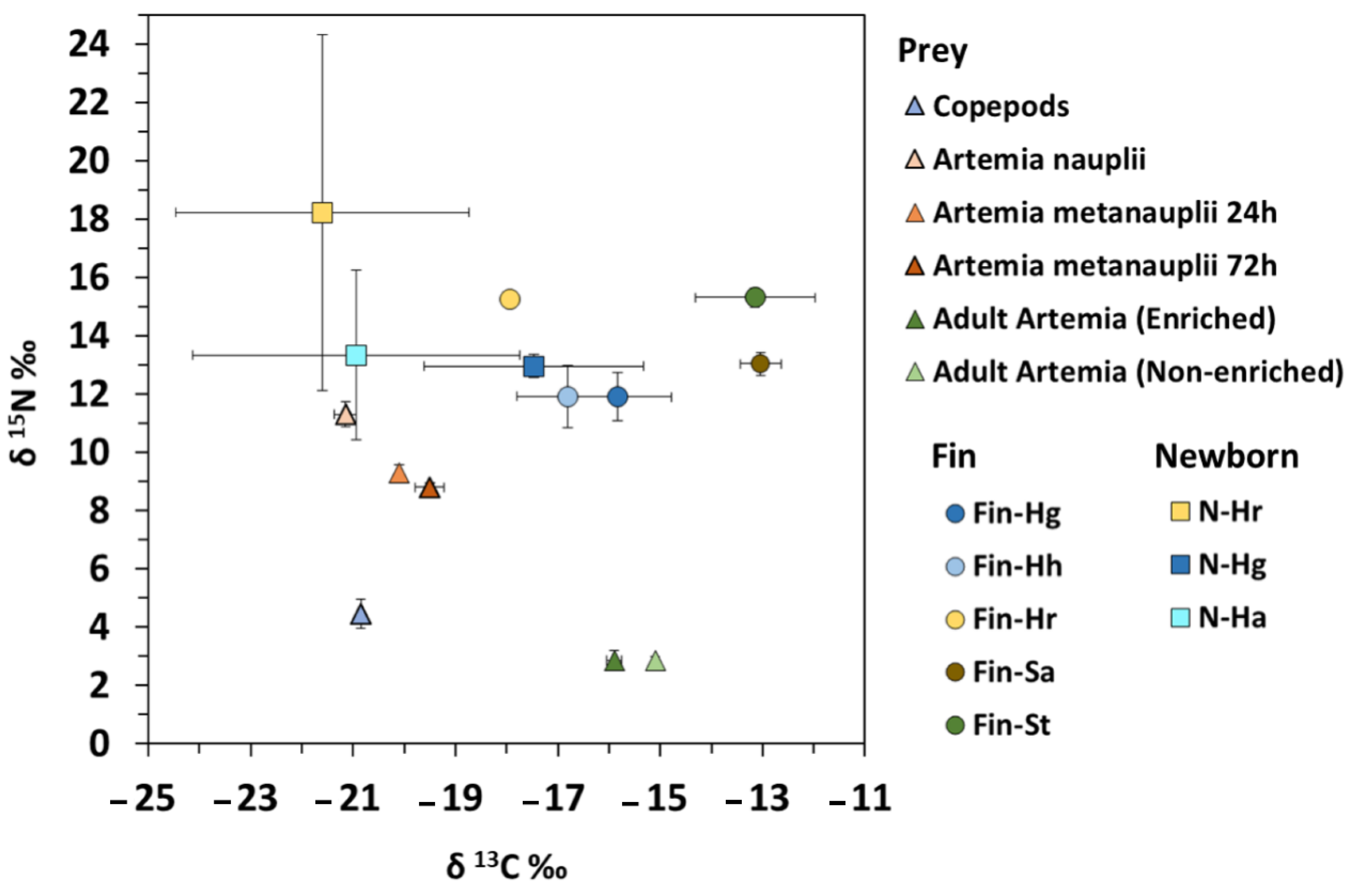
Animal capture, handling and sampling were conducted in compliance with all bioethics standards on animal experimentation of the Spanish Government (Real Decreto 1201/2005, 10th October) and the Regional Government Xunta de Galicia (REGA ES360570202001/15/FUN/BIOL.AN/MPO01 and ES360570202001/16/EDU-FOR07/MPO01).

3. Results

The range of C:N values in controls (frozen samples) was 3.7–6.1 (mean: 4.8 ± 0.8) in prey, 2.9–3.5 (3.1 ± 0.2) in fish fins and 3.5–3.9 (3.7 ± 0.1) in fish newborn. Isotopic values in prey ranged from −21.1 to −14.7‰ for δ13C and from 1.6 to 11.1‰ for δ15N, with mean values of −18.7 ± 2.5‰ and 6.2 ± 3.6‰, respectively (Figure 1).

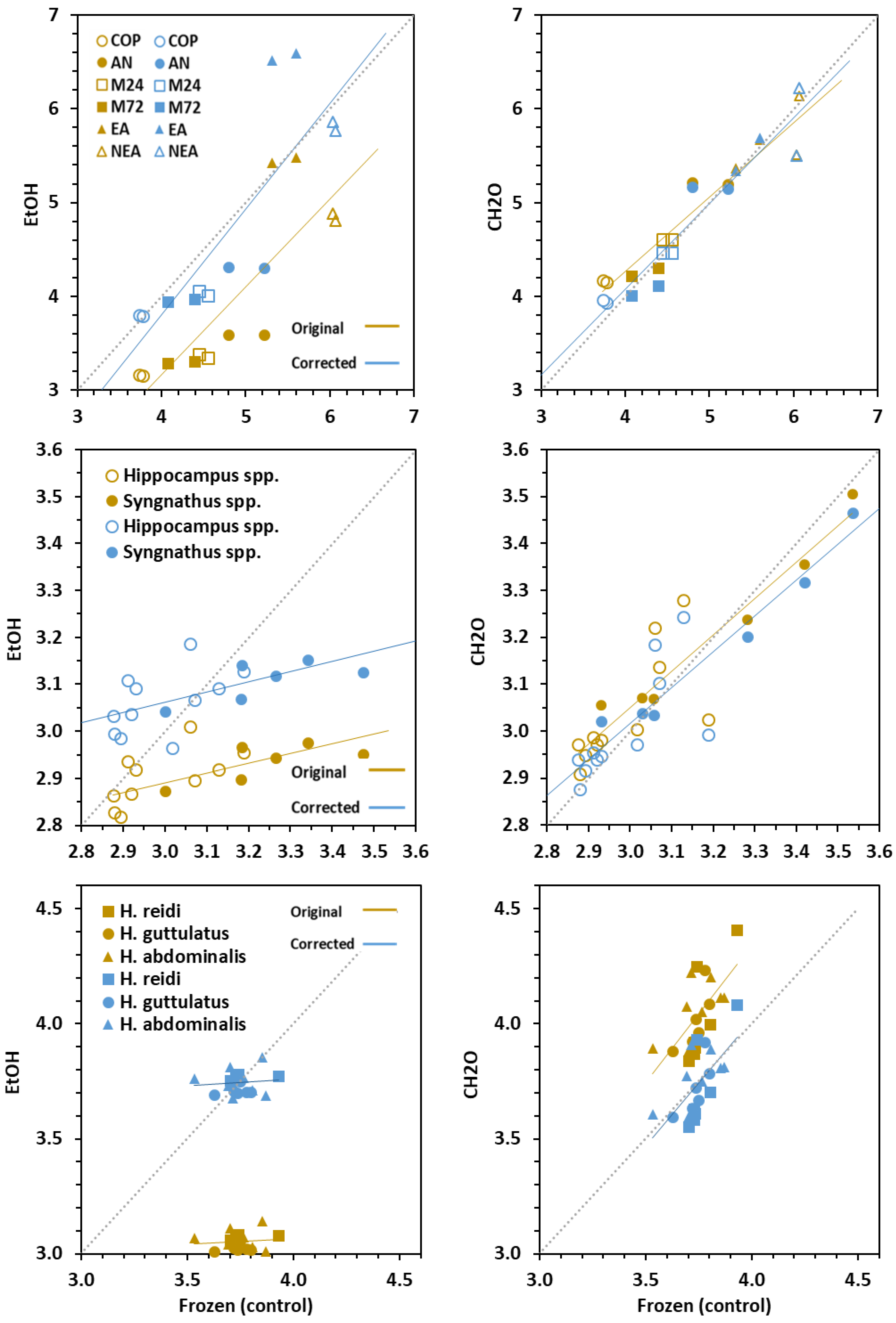
3.1. Effect on Carbon and Nitrogen Ratios

Preservation treatments (freezing, EtOH and CH2O) of experimental samples (prey, fins and newborns) differed significantly (ANCOVA, p < 0.001) in their effect on C:N values (p < 0.081) (Table 1). When comparing to control groups (freezing), C:N values decreased significantly (p < 0.001) in EtOH-treated samples (Δ = −0.88 in prey; −0.15 in fins and −0.69 in newborn), whereas the effect of CH2O was minimal (Δ = 0.10 in prey and, 0.03 in fins) (p > 0.05), except in newborn seahorses (Δ = 0.29) (p < 0.001) (Table 1; Figure 2).
Considering dorsal fins, adjusted means for C:N values in Hippocampus (2.99) and Syngnathus (3.14) differed significantly (p = 0.001), even though the difference was small. In seahorse newborn, mean C:N values were similar (p = 0.506), ranging from 3.59 in H. guttulatus to 3.64 in H. abdominalis.

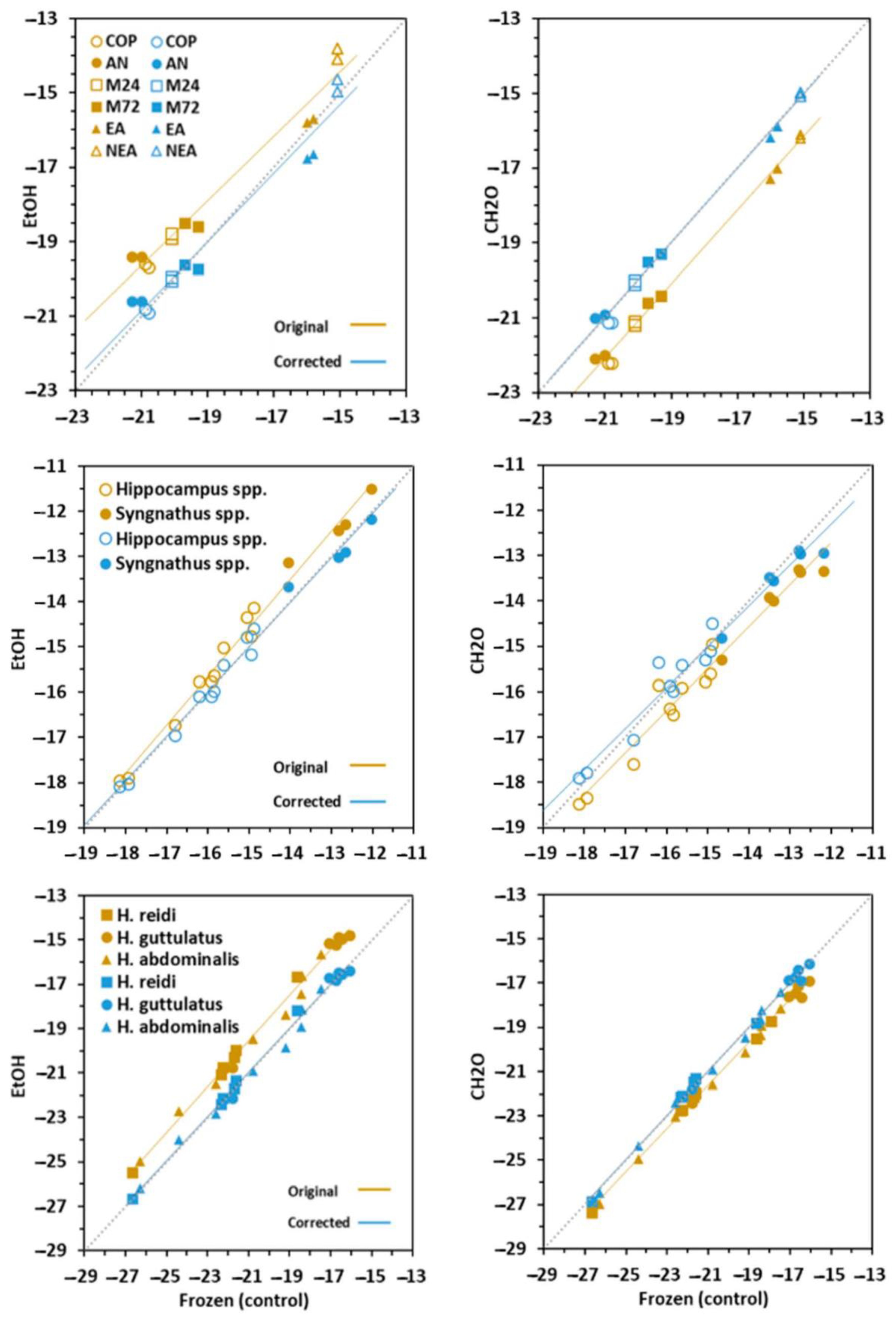
3.2. Effect on δ13C Signatures

The signatures for δ13C in prey, fins and newborns was highly affected (p < 0.005) by chemical preservation (Table 1, Figure 3). The effects were directly related to C:N values in bulk frozen tissues. Accordingly, prey and fins were more (p < 0.001) and less (p = 0.031) affected, respectively. The isotopic signal increased (enriched) significantly (p < 0.001) in EtOH-treated samples (Δ = 1.1‰ in prey; 1.0‰ in newborn), except in clipped fins (Δ = 0.2‰) (p = 0.192). The treatment with CH2O led to decreased δ13C signals in prey (Δ = 1.1‰) (p < 0.001) but not in fish tissues (Δ = 0.0‰ in fins and 0.7‰ in newborns) (p = 0.365 and 0.391, respectively). Broadly, the effects of preservation methods in newborn seahorses were similar to those in fins.
Considering prey, adjusted means for δ13C were −20.9‰ in copepods and Artemia nauplii, −20‰ to −19.5‰ in Artemia metanauplii and −15.1‰ in non-enriched adult Artemia, and −16.3‰ in adult enriched Artemia. Isotopic signals for 13C in fish fins were −16.3‰ in Hippocampus and −13.3‰ in Syngnathus. Adjusted means in seahorse newborns were −17.2‰ in H. guttulatus, −19.5‰ in H. abdominalis and −21.0‰ in H. reidi.

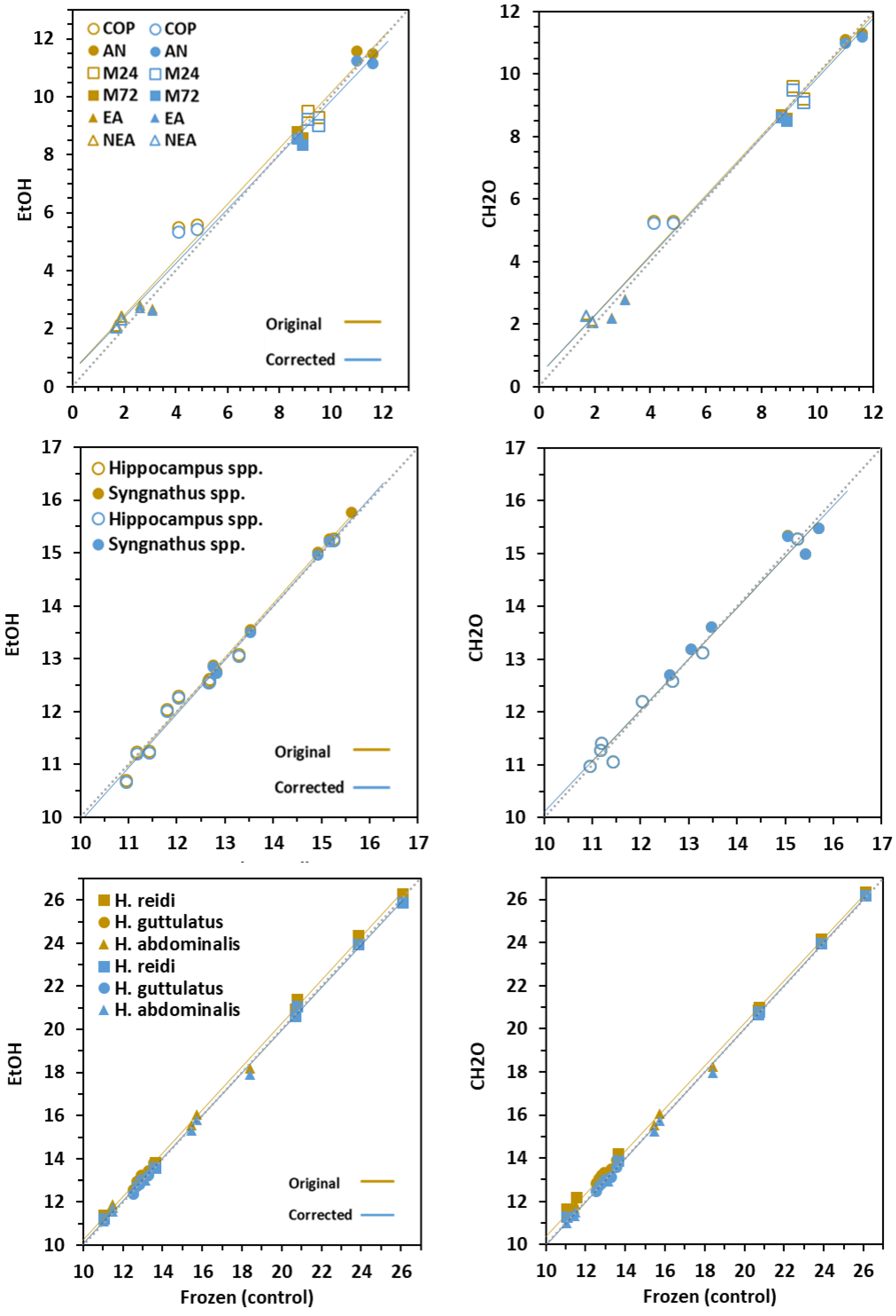
3.3. Effect on δ15N Signatures

Chemical treatments performed similarly to frozen samples, and the effects on δ15N values were negligible, especially in fins (Δ < 0.2‰) and newborns (Δ < 0.3‰) (p < 0.001) (Table 1, Figure 4). The highest differences were achieved in prey (Δ = 0.1‰ for EtOH; Δ = 0.3‰ for CH2O).
Adjusted means for δ15N in prey ranged from 2.7‰ in copepods and 20‰ to −11.3‰ in Artemia nauplii. Dorsal fins in Hippocampus and Syngnathus fishes differed in isotopic adjusted means (12.4 and 14.2‰, respectively). Adjusted means for δ15N in newborns were 12.5‰ in H. abdominalis, 13.2‰ in H. guttulatus and 20.7‰ in H. reidi.

3.4. Conversion Models

The correction equations to account for changes of C:N, δ15N and δ13C values in tissues treated with chemical preservatives relative to freezing treatment are provided in Table 2 and visualized in Figure 2, Figure 3 and Figure 4. Corrected values in prey revealed worse adjustment and predictability compared to fish tissues. Except for C:N in CH2O-preserved newborns (Adj R2 > 0.384), the models were highly significant (Adj R2 > 0.9; range = 0.894–0.999). The main discrepancies between the original and corrected models occurred in C:N values, especially in EtOH–treated samples.
Simple arithmetic corrections could be applied to δ15N and δ13C values in fins and newborns for treatment correction. Overall, isotopic corrected lines for both chemical preservatives did not differ from the 1:1 line. Besides this, the slopes in most uncorrected and corrected linear models did not differ significantly, except for δ13C in prey and fins. On the contrary, regression intercepts were significantly different in many cases. However, the differences were highly reduced (<0.01) in δ15N models, and corrected and control values were statistically undistinguishable.

4. Discussion

The present study demonstrates for the first time the effects of freezing and two chemical preservation methods (ethanol—EtOH and formaldehyde—CH2O) on isotopic signals (δ13C and δ15N) and C:N ratios in syngnathid fishes, including adults (dorsal fin), and newborn juveniles, and in prey commonly used to feed marine fishes in rearing systems. Considering the preservation methods tested, EtOH and CH2O are polar and non-polar solvents frequently used as preservation chemicals. Formaldehyde hydrolyzes proteins and systematically affects δ13C signatures [44,45,46]. Even though EtOH does not remove lipids completely, the solvent is capable of extracting many fats, including phospholipids and free fatty acids. The global effects of each preservation method tested implied important differences across treatments depending on the type of tissue considered. CH2O-treated materials were more similar to control samples than those preserved in EtOH, especially for C:N values. Correcting C:N ratios and isotope signatures in chemically preserved tissues with derived correction equations revealed significant differences for C:N and δ13C values between treated and control (freezing) samples. The easiest, fastest and most economical procedure for SIA in adult syngnathids in laboratory experiments would be freezing of clipped-fin tissue, whereas EtOH (does not requires defatting) or CH2O-treatments would be recommended in the field to ensure better sample maintenance during sampling.
Ethanol led to a sharp but calculable decrease in C:N values. The magnitude of observed drop across tissues was directly correlated (Pearson’s R = 0.84) with C:N values (i.e., lipid content) in controls. Hence, lower C:N ratios in bulk tissues (fins) led to lower shifts in EtOH-treated samples (−0.2, −0.7 and −0.9 in fins, newborn and prey, respectively). Corrections on C:N ratios were reliable in prey but ineffective in fish tissues due to the low ratio variability derived from chemical treatments.
As compared to ethanol, formaldehyde-treated materials provided small shifts and low variability, affecting especially C:N values (all tissues) and δ13C signals. The depletion in 13C varied depending on the type of tissue, but prey and newborn were mostly affected (−1.1, −0.7 and −0.2‰ in prey, newborn and fin, respectively). Those changes would be concordant with tissue lipid content [25] and/or tissue formalin uptake [35,44].
Considering the average shifts achieved in δ13C signatures in fins (Δ < 0.2‰) and newborns (Δ < 1.0‰), we advise application of further corrections in chemical preserved tissues. On the contrary, δ15N signatures in solvent preserved materials were small enough (Δ < 0.2‰ in fins and <0.3‰ in newborns) to correct the values. Interestingly, average δ15N-shifts in both isotopes were much lower than the estimated trophic enrichment factor of 3.4‰ in aquatic animals [3] and 3.9–4.2‰ in adult syngnathids [23]. Hence, differences in δ15N between frozen and solvent preserved tissues will not affect data interpretation in food web studies for syngnathids.
The diverse tissues of an animal differ in turnover rates due to inter-tissue differences in isotopic fractionation [10,11,12,47,48,49]. Muscle tissue is the focal material in many isotopic studies due to its intermediate and less variable turnover rates [12,49]. The disadvantage of sampling fish muscle is that small specimens must be sacrificed [50]. However, there are alternative tissues that do not entail fish killing, providing isotopic signatures highly correlated to those in muscle [15,45,51]. Compared to many teleost, syngnathids are small fishes and their fins are difficult to sample, especially in small specimens. Consequently, the most convenient tissue for sampling is dorsal fin. However, differences might arise comparing dorsal fins with other fin tissues.
Fish fins are excellent tissues for isotopic assessment, especially in studies involving threatened or endangered species [17,22]. Fin clipping has been successfully used for SIA in syngnathids, both in field collected samples [23] and ex situ experimental studies [24]. Partial fin clipping results are advantageous when compared to other not detrimentally sampled tissues such as the dorsal fleshy filaments present in some seahorse species (e.g., H. guttulatus and H. kuda). Clipped filaments provide enough biomass for DNA analysis [39,52,53] but not for SIA. Fin and filament clipping does not impair fish behaviour [52,54] and both tissues are able to regenerate [53]. However, clipped filaments regenerate more slowly, and can recover histologically but not completely in size [52,53].
There is a high availability of isotopic studies on the effects of preservation methods and lipid normalization procedures in fish tissues [31,46,55,56]. Lipids are depleted in 13C when compared to proteins and carbohydrates [32]. Consequently, the main effect of EtOH preservation on δ13C of tissues is the loss of lipids and/or proteins, resulting in 13C enrichment [55]. In agreement with those findings, our EtOH-treated tissues were enriched in 13C, especially in materials with high C:N values (prey and newborn, in increasing order). However, other studies reported different results [25,44].
Even though lipids mainly affect the heavier 13C isotope, lipid extraction might also result in deviations in the δ15N of the lipid-free tissue [12,57,58]. Although there is a large variability, lipid extracts may be more depleted in 15N than bulk tissues. Commonly, δ15N signals in bulk tissues increase with trophic levels of organisms [33], as reflected in the present study when comparing prey with fish tissues (δ15Nprey < δ15Nfin > δ15Nnewborn). However, the preservation in EtOH did not affect δ15N signals, suggesting that lipids in all tissues analysed were highly 15N-depleted, and that storage in those solvents will not impair the reliability of the analyses.
Impaired δ15N values might also result due to artifacts in the extraction of lipids [5]. In the present study, we did not assess the effects of complete lipid extraction usually applied to samples with C:N ratios above 3.5 [34,56]. In this regard, it is likely that EtOH samples be isotopically equivalent to lipid-extracted bulk samples. A previous study carried out in seahorses comparing dorsal fin, muscle, and liver tissues reported similarities between δ13C and δ15N values in dorsal fin and muscle tissue, and significant effects of lipid extraction on δ13C values in muscle and liver [22]. The study concluded that lipid removal was not necessary in dorsal fin tissues due to a lipid content (2.6% dry weight) lower than in muscle tissue (7.1%). The low lipid content in fin tissues of syngnathids was confirmed by C:N values in the present study (2.88–3.19 in Hippocampus spp.; 2.93–3.53 in Syngnathus spp.). Accordingly, fin clipped samples could be submitted to SIA without the need of previous lipid extraction nor further mathematical lipid corrections for δ15N, whereas the regression models given in Table 2 should be applied for δ13C.
The main aim of the present study was to provide practical mathematical models for the conversion of isotopic signals in preserved live prey and syngnathids. The regression equations provided would be useful in field studies when samples cannot be properly stored, requiring preservation until further analysis. In addition, in spite of further assessment on the effect of long-term storage of sampled tissues on isotopic signals [25], the models might be applied to syngnathid-preserved specimens in natural history or museum collections. With regard to field studies in syngnathids, our study on adults was performed with samples collected both in the field and in cultivated fishes (seahorses vs. pipefishes) and both types responded similarly to solvents. An important feature in syngnathids is that fin tissues are really thin (with a low lipid content) compared to other large fishes, and it is likely that the results would be the same, whatever the scenario considered.

5. Conclusions

The results achieved revealed different effects of chemical preservation on isotopic signatures and C:N ratios both in syngnathid fishes (dorsal fin in adults and bulk newborns) and in prey commonly used to feed those fishes in the laboratory. Considering that dorsal fins are valid isotopic subrogates of muscle tissue in syngnathids, our results would be comparable to those in muscle tissue samples. However, the effects of feeding activity on potential differences between both tissue types deserve further consideration [59]. The impacts of ethanol were higher than those of formaldehyde, especially for δ13C signals and C:N ratios, but the effects of the former were consistent and predictable, and can be corrected. Conversely, the shifts in δ15N of chemically preserved tissues were small enough to be ignored. Hence, both solvents provided consistent and reliable results. The first conversion models for the mathematical correction of data across the tested preservatives were constructed specifically for syngnathids. Those taxa-specific models may be applied to field collected samples as well as to historical collections. Further work should be conducted to determine the isotopic effect of lipid extraction and duration of preservation, except in dorsal fins.

Author Contributions

Conceptualization, M.P.; formal analysis, M.P., A.P., M.D.D.C. and J.H.-U.; investigation, M.P., A.P., M.D.D.C. and J.H.-U.; methodology, M.P., A.P., M.D.D.C. and J.H.-U.; project administration, M.P.; resources, M.P.; supervision, M.P.; validation, M.P.; writing—original draft preparation, M.P.; writing—review and editing of the manuscript, M.P., A.P., M.D.D.C. and J.H.-U. All authors have read and agreed to the published version of the manuscript.

Funding

This study was financially supported by the Spanish Government with projects Hippoeco (Ref. CGL2015-68110-R, Ministerio de Ciencia, Innovación y Universidades and Fondos FEDER) and Hippoparques (Ref. 1541S/2015, Organismo Autónomo de Parques Nacionales de España, Ministerio para la Transición Ecológica). J.H.U. was supported by a Juan de la Cierva’s post-doc research grant (FJCI-2016-30990; Ministerio de Ciencia, Innovación y Universidades, Spain), A.P. was supported by Erasmus+ funding programme (Università Politecnica delle Marche, Italy) and M.D.D.C. was granted by Fundação Coordenação de Aperfeiçoamento de Pessoal de Nível Superior—CAPES (PDSE 88881.187275/2018-01; Ministério da Educação—MEC, Brazil).

Acknowledgments

The authors are grateful to Cofradía de Pescadores de Arcade (Ría de Vigo, NW Spain) for providing Syngnathus spp. specimens, Alexandro Chamorro for fish rearing technical support and Antía González Barreiro for sampling assistance. Special thanks to Loïc N. Michel (IFREMER) for a critical review of the manuscript.

Conflicts of Interest

The authors declare no conflict of interest. The funders had no role in the design of the study; in the collection, analyses, or interpretation of data; in the writing of the manuscript, or in the decision to publish the results.

References

  1. Post, D.M. Using stable isotopes to estimate trophic position: Models, methods, and assumptions. Ecology 2002, 83, 703–718. [Google Scholar] [CrossRef]
  2. Peterson, B.J.; Fry, B. Stable isotopes in ecosystem studies. Annu. Rev. Ecol. Syst. 1987, 18, 293–320. [Google Scholar] [CrossRef]
  3. Vander Zanden, M.J.; Rasmussen, J.B. Variation in δ15N and δ13C trophic fractionation: Implications for aquatic food web studies. Limnol. Oceanogr. 2001, 46, 2061–2066. [Google Scholar] [CrossRef]
  4. Gannes, L.Z.; O’Brien, D.M.; Martínez del Rio, C. Stable isotopes in animal ecology: Assumptions, caveats, and a call for more laboratory experiments. Ecology 1997, 78, 1271–1276. [Google Scholar] [CrossRef]
  5. Boecklen, W.J.; Yarnes, C.T.; Cook, B.A.; James, A.A. On the use of stable isotopes in trophic ecology. Annu. Rev. Ecol. Evol. Syst. 2011, 42, 411–440. [Google Scholar] [CrossRef] [Green Version]
  6. Herzka, S.Z.; Holt, G.J. Changes in isotopic composition of red drum (Sciaenops ocellatus) larvae in response to dietary shifts: Potential applications to settlement studies. Can. J. Fish. Aquat. Sci. 2000, 57, 137–147. [Google Scholar] [CrossRef]
  7. Le Vay, L.; Gamboa-Delgado, J. Naturally-occurring stable isotopes as direct measures of larval feeding efficiency, nutrient incorporation and turnover. Aquaculture 2011, 315, 95–103. [Google Scholar] [CrossRef] [Green Version]
  8. Hansson, S.; Hobbie, J.E.; Elmgren, R.; Larsson, U.; Fry, B.; Johansson, S. The stable nitrogen isotope ratio as a marker of food-web interactions and fish migration. Ecology 1997, 78, 2249–2257. [Google Scholar] [CrossRef]
  9. Hobson, K.A. Tracing origins and migration of wildlife using stable isotopes: A review. Oecologia 1999, 120, 314–326. [Google Scholar] [CrossRef]
  10. DeNiro, M.J.; Epstein, S. Influence of diet on the distribution of carbon isotopes in animals. Geochim. Cosmochim. Acta 1978, 42, 495–506. [Google Scholar] [CrossRef]
  11. DeNiro, M.J.; Epstein, S. Influence of diet on the distribution of nitrogen isotopes in animals. Geochim. Cosmochim. Acta 1981, 45, 341–351. [Google Scholar] [CrossRef]
  12. Pinnegar, J.K.; Polunin, N.V.C. Differential fractionation of δ13C and δ15N among fish tissues: Implications for the study of trophic interactions. Funct. Ecol. 1999, 13, 225–231. [Google Scholar] [CrossRef]
  13. Wolf, N.; Carleton, S.A.; Martínez del Rio, C. Ten years of experimental animal isotopic ecology. Funct. Ecol. 2009, 23, 17–26. [Google Scholar] [CrossRef]
  14. Minagawa, M.; Wada, E. Stepwise enrichment of 15N along food chains: Further evidence and the relation between δ15N and animal age. Geochim. Cosmochim. Acta 1984, 48, 1135–1140. [Google Scholar] [CrossRef]
  15. Jardine, T.D.; Gray, M.A.; McWilliam, S.M.; Cunjak, R.A. Stable isotope variability in tissues of temperate stream fishes. Trans. Am. Fish. Soc. 2005, 134, 1103–1110. [Google Scholar] [CrossRef]
  16. Caut, S.; Angulo, E.; Courchamp, F. Variation in discrimination factors (∆15N and ∆13C): The effect of diet isotopic values and applications for diet reconstruction. J. Appl. Ecol. 2009, 46, 443–453. [Google Scholar] [CrossRef]
  17. Jardine, T.D.; Hunt, R.J.; Pusey, J.B.; Bunn, S.E. A non-lethal sampling method for stable carbon and nitrogen isotope studies of tropical fishes. Mar. Freshw. Res. 2011, 62, 83–90. [Google Scholar] [CrossRef] [Green Version]
  18. Blanco, A.; Deudero, S.; Box, A. Muscle and scale isotopic offset of three fish species in the Mediterranean Sea: Dentex dentex, Argyrosomus regius and Xyrichtys novacula. Rapid Commun. Mass Spectrom. 2009, 23, 2321–2328. [Google Scholar] [CrossRef]
  19. Cano-Rocabayera, O.; Maceda-Veiga, A.; de Sostoa, A. Fish fins and scales as non-lethally sampled tissues for stable isotope analysis in five fish species of north—Eastern Spain. Environ. Biol. Fishes 2015, 98, 925–932. [Google Scholar] [CrossRef]
  20. Winter, E.R.; Nyqvist, M.; Britton, J.R. Non-lethal sampling for stable isotope analysis of pike Esox lucius: How mucus, scale and fin tissue compare to muscle. J. Fish Biol. 2019, 95, 956–958. [Google Scholar] [CrossRef]
  21. Sanderson, B.L.; Tran, C.D.; Coe, H.J.; Pelekis, V.; Steel, E.A.; Reichert, W.L. Nonlethal sampling of fish caudal fins yields valuable stable isotope data for threatened and endangered fishes. Trans. Am. Fish. Soc. 2009, 138, 1166–1177. [Google Scholar] [CrossRef]
  22. Valladares, S.; Planas, M. Non-lethal dorsal fin sampling for stable isotope analysis in seahorses. Aquat. Ecol. 2012, 46, 363–370. [Google Scholar] [CrossRef] [Green Version]
  23. Planas, M.; Chamorro, A.; Paltrinieri, A.; Campos, S.; Jiménez, A.; Nedelec, K.; Hernández-Urcera, J. Effect of diet on breeders and inheritance in syngnathids: Application of isotopic experimentally derived data to field studies. Mar. Ecol. Prog. Ser. 2020, 650, 107–123. [Google Scholar] [CrossRef]
  24. Planas, M.; Olivotto, I.; González, M.J.; Laurà, R.; Zarantoniello, M. A multidisciplinary experimental study of the effects of breeders diet on newborn seahorses (Hippocampus guttulatus). Front. Mar. Sci. 2020, 7, 638. [Google Scholar] [CrossRef]
  25. Bosley, K.L.; Wainright, S.C. Effects of preservatives and acidification on the stable isotope ratios (15N:14N, 13C:12C) of two species of marine animals. Can. J. Fish. Aquat. Sci. 1999, 56, 2181–2185. [Google Scholar] [CrossRef]
  26. Kelly, B.; Dempson, J.B.; Power, M. The effects of preservation on fish tissue stable isotope signatures. J. Fish Biol. 2006, 69, 1595–1611. [Google Scholar] [CrossRef]
  27. Vizza, C.; Sanderson, B.L.; Burrows, D.G.; Coe, H.J. The effects of ethanol preservation on fish fin stable isotopes: Does variation in C:N ratio and body size matter? Trans. Am. Fish. Soc. 2013, 142, 1469–1476. [Google Scholar] [CrossRef]
  28. Stallings, C.D.; Nelson, J.A.; Rozar, K.L.; Adams, C.S.; Wall, K.R.; Switzer, T.S.; Winner, B.L.; Hollander, D.J. Effects of preservation methods of muscle tissue from upper-trophic level reef fishes on stable isotope values (δ(13)C and δ(15)N). PeerJ 2015, 3, e874. [Google Scholar] [CrossRef] [Green Version]
  29. Kishe-Machumu, M.A.; van Rijssel, J.C.; Poste, A.; Hecky, R.E.; Witte, F. Stable isotope evidence from formalin–ethanol-preserved specimens indicates dietary shifts and increasing diet overlap in Lake Victoria cichlids. Hydrobiologia 2017, 791, 155–173. [Google Scholar] [CrossRef]
  30. Focken, U.; Becker, K. Metabolic fractionation of stable carbon isotopes: Implications of different proximate compositions for studies of the aquatic food webs using δ13C data. Oecologia 1998, 115, 337–343. [Google Scholar] [CrossRef]
  31. Logan, J.M.; Jardine, T.D.; Miller, T.J.; Bunn, S.E.; Cunjak, R.A.; Lutcavage, M.E. Lipid corrections in carbon and nitrogen stable isotope analyses: Comparison of chemical extraction and modelling methods. J. Anim. Ecol. 2008, 77, 838–846. [Google Scholar] [CrossRef] [PubMed]
  32. Logan, J.M.; Lutcavage, M.E. A comparison of carbon and nitrogen stable isotope ratios of fish tissues following lipid extractions with non-polar and traditional chloroform/methanol solvent systems. Rapid Commun. Mass Spectrom. 2008, 22, 1081–1086. [Google Scholar] [CrossRef] [PubMed]
  33. Svensson, E.; Schouten, S.; Hopmans, E.C.; Middelburg, J.J.; Sinninghe Damsté, J.S. Factors controlling the stable nitrogen isotopic composition (δ15N) of lipids in marine animals. PLoS ONE 2016, 11, e0146321. [Google Scholar] [CrossRef] [PubMed] [Green Version]
  34. Kiljunen, M.; Grey, J.; Sinisalo, T.; Harrod, C.; Immonen, H.; Jones, R. A revised model for lipid-normalizing δ13C values from aquatic organisms, with implications for isotope mixing models. J. Appl. Ecol. 2006, 43, 1213–1222. [Google Scholar] [CrossRef]
  35. Edwards, M.S.; Turner, T.F.; Sharp, Z.D. Short and long-term effects of fixation and preservation on stable isotope values (δ13C, δ15N, δ34S) of fluid-preserved museum specimens. Copeia 2002, 4, 1106–1112. [Google Scholar] [CrossRef]
  36. Ahnesjö, I.; Craig, J.F. The biology of Syngnathidae: Pipefishes, seadragons and seahorses. J. Fish Biol. 2011, 78, 1597–1602. [Google Scholar] [CrossRef]
  37. Vincent, A.C.J.; Foster, S.J.; Koldewey, H.J. Conservation and management of seahorses and other Syngnathidae. J. Fish Biol. 2011, 78, 1681–1724. [Google Scholar] [CrossRef]
  38. Planas, M.; Silva, C.; Quintas, P.; Chamorro, A.; Piñero, S. Ongrowing and enhancement of n-3 HUFA profile in adult Artemia: Short vs. long-time enrichment. J. Appl. Phycol. 2017, 29, 1409–1420. [Google Scholar] [CrossRef]
  39. Planas, M.; Chamorro, A.; Quintas, P.; Vilar, A. Establishment and maintenance of threatened long-snouted seahorse, Hippocampus guttulatus, broodstock in captivity. Aquaculture 2008, 283, 19–28. [Google Scholar] [CrossRef] [Green Version]
  40. Randazzo, B.; Rolla, L.; Ofelio, C.; Planas, M.; Gioacchini, G.; Vargas, A.; Giorgini, E.; Olivotto, I. The influence of diet on the early development of two seahorse species (H. guttulatus and H. reidi): Traditional and innovative approaches. Aquaculture 2018, 490, 75–90. [Google Scholar] [CrossRef]
  41. R Core Team. R: A Language and Environment for Statistical Computing. 2014. Available online: http://www.R-project.org (accessed on 23 February 2018).
  42. Howell, D.C. Statistical Methods for Psychology; Wadsworth Publishing Company: Belmont, CA, USA, 2013; p. 770. [Google Scholar]
  43. Hothorn, T.; Bretz, F.; Westfall, P. Simultaneous inference in general parametric models. Biom. J. 2008, 50, 346–363. [Google Scholar] [CrossRef] [PubMed] [Green Version]
  44. Hobson, K.A.; Gibbs, H.L.; Gloutney, M.L. Preservation of blood and tissue samples for stable-carbon and stable-nitrogen isotope analysis. Can. J. Zool. 1997, 75, 1720–1723. [Google Scholar] [CrossRef]
  45. Kelly, M.H.; Hagar, W.G.; Jardine, T.D.; Cunjak, R.A. Nonlethal sampling of sunfish and slimy sculpin for stable isotope analysis: How scale and fin tissue compare with muscle tissue. N. Am. J. Fish Manag. 2006, 26, 921–925. [Google Scholar] [CrossRef]
  46. Sarakinos, H.C.; Johnson, M.L.; Zanden, M.J.V. A synthesis of tissue-preservation effects on carbon and nitrogen stable isotope signatures. Can. J. Zool. 2002, 80, 381–387. [Google Scholar] [CrossRef] [Green Version]
  47. Reich, K.J.; Bjorndal, K.A.; Martinez Del Rio, C. Effects of growth and tissue type on the kinetics of 13C and 15N incorporation in a rapidly growing ectotherm. Oecologia 2008, 155, 651–663. [Google Scholar] [CrossRef]
  48. Britton, J.R.; Busst, G.M.A. Stable isotope discrimination factors of omnivorous fishes: Influence of tissue type, temperature, diet composition and formulated feeds. Hydrobiologia 2018, 808, 219–234. [Google Scholar] [CrossRef] [Green Version]
  49. Tieszen, L.L.; Boutton, T.W.; Tesdahl, K.G.; Slade, N.A. Fractionation and turnover of stable carbon isotopes in animal tissues: Implications for δ13C analysis of diet. Oecologia 1983, 57, 32–37. [Google Scholar] [CrossRef]
  50. Vizzini, S.; Mazzola, A. Stable isotopes and trophic positions of littoral fishes from a Mediterranean marine protected area. Environ. Biol. Fishes 2009, 48, 13–25. [Google Scholar] [CrossRef]
  51. McCarthy, I.D.; Waldron, S. Identifying migratory Salmo trutta using carbon and nitrogen stable isotope ratios. Rapid Commun. Mass Spectrom. 2000, 14, 1325–1331. [Google Scholar] [CrossRef]
  52. Correia, M.; Campoy, A.; Madeira, C.; Andrade, J.P. Is filament clipping an effective tool for tissue sampling in Hippocampus guttulatus? Environ. Biol. Fish 2018, 101, 1517–1523. [Google Scholar] [CrossRef]
  53. Gristina, M.; Bertrandino, S.; Cardone, F.; Mentino, D.; Corriero, G.; Scillitani, G. Skin filament recovery after clipping in Hippocampus guttulatus: Behavioural and histological aspects. Aquat. Biol. 2017, 26, 149–157. [Google Scholar] [CrossRef]
  54. Woodall, L.C.; Jones, R.; Zimmerman, B.; Guillaume, S.; Stubbington, T.; Shaw, P.; Koldewey, H.J. Partial fin-clipping as an effective tool for tissue sampling seahorses, Hippocampus spp. J. Mar. Biol. Assoc. 2012, 92, 1427–1432. [Google Scholar] [CrossRef]
  55. DeNiro, M.J.; Epstein, S. Mechanism of carbon isotope fractionation associated with lipid synthesis. Science 1977, 197, 261–263. [Google Scholar] [CrossRef]
  56. Post, D.M.; Craig, A.; Layman, D.; Arrington, D.A.; Takimoto, G.; Quatrocchi, J.; Montaña, C.G. Getting to the fat of the matter: Models, methods and assumptions for dealing with lipids in stable isotope analyses. Oecologia 2007, 152, 179–189. [Google Scholar] [CrossRef] [PubMed]
  57. Sotiropoulos, M.A.; Tonn, W.M.; Wassenaar, L.I. Effects of lipid extraction on stable carbon and nitrogen isotope analyses of fish tissues: Potential consequences for food web studies. Ecol. Freshw. Fish 2004, 13, 155–160. [Google Scholar] [CrossRef]
  58. Sweeting, C.J.; Polunin, N.V.C.; Jennings, S. Effects of chemical lipid extraction and arithmetic lipid correction on stable isotope ratios of fish tissues. Rapid Commun. Mass Spectrom. 2006, 20, 595–601. [Google Scholar] [CrossRef]
  59. Perga, M.E.; Gerdeaux, D. Are fish what they eat’ all year round? Oecologia 2005, 144, 598–606. [Google Scholar] [CrossRef]
Figure 1. Stable isotope biplot of means (±SD) for δ13C and δ15N in prey, dorsal fins in adult syngnathids and bulk newborn seahorses. Values correspond to frozen samples. Ha—Hippocampus abdominalis; Hg—H. guttulatus; Hh—H. hippocampus; Hr—H. reidi; Sa—Syngnathus acus; St—S. typhle.
Animals 10 02301 g001
Figure 2. Original (brown symbols) and corrected (blue symbols) values for C:N in prey, fins and newborns. Regression lines are given for frozen, EtOH (ethanol) and CH2O (formaldehyde) relationships. A 1:1 dashed line (slope = 1, intercept = 0) is given for clarity. Prey: COP—copepods, AN—Artemia nauplii, M24 and M72—Artemia metanauplii, and; EA and NEA—enriched and non-enriched adult Artemia. Fin: genera Syngnathus and Hippocampus; Seahorse newborns: H. guttulatus, H. abdominalis and H. reidi.
Animals 10 02301 g002
Figure 3. Original (brown symbols) and corrected (blue symbols) values for δ13C in prey, fins and newborns. Regression lines are given for frozen, EtOH (ethanol) and CH2O (formaldehyde) relationships. A 1:1 dashed line (slope = 1, intercept = 0) is given for clarity. Prey: COP—copepods, AN—Artemia nauplii, M24 and M72—Artemia metanauplii, and; EA and NEA—enriched and non-enriched adult Artemia. Fin: genera Syngnathus and Hippocampus; Seahorse newborns: H. guttulatus, H. abdominalis and H. reidi.
Animals 10 02301 g003
Figure 4. Original (brown symbols) and corrected (blue symbols) values for δ15N in prey, fins and newborns. Regression lines are given for frozen, EtOH (ethanol) and CH2O (formaldehyde) relationships. A 1:1 dashed line (slope = 1, intercept = 0) is given for clarity. Prey: COP—copepods, AN—Artemia nauplii, M24 and M72—Artemia metanauplii, and; EA and NEA—enriched and non-enriched adult Artemia. Fin: genera Syngnathus and Hippocampus; Seahorse newborns: H. guttulatus, H. abdominalis and H. reidi.
Animals 10 02301 g004
Table 1. Summary of ANCOVA and pairwise comparisons for C:N, δ15N and δ13C values in organisms/tissues submitted to three preservation treatments (FR—frozen; ET—ethanol; FO—formaldehyde). Significant p-values are given in bold. Prey: copepods and Artemia (several developmental stages); Fin: genera Syngnathus and Hippocampus; Seahorse newborns: H. guttulatus, H. abdominalis and H. reidi.
ANCOVAGroup Comparisons (p)
EffectSSd.f.MSFAdj. pFR–ETFR–FOET–FO
Prey
C:NTreatment6.9023.4541.43<0.001<0.0010.403<0.001
Prey18.8353.7745.19<0.001
Residuals2.33280.08
δ13CTreatment28.39214.19199.00<0.001<0.001<0.001<0.001
Prey186.41537.28522.80<0.001
Residuals2.00280.07
δ15NTreatment0.5020.242.750.0810.0270.3110.201
Prey435.30587.05990.40<0.001
Residuals2.50280.09
Fins
C:NTreatment0.57020.2813.01<0.001<0.0010.384<0.001
Genus0.31010.3114.15<0.001
Residuals1.206550.02
δ13CTreatment9.2624.633.700.0310.1920.3650.037
Genus127.921127.92102.11<0.001
Residuals68.90551.25
δ15NTreatment0.3820.190.100.9010.7120.7180.998
Genus42.74142.7423.67<0.001
Residuals92.09551.81
Newborns
C:NTreatment7.3723.68332.51<0.001<0.001<0.001<0.001
Species0.0120.010.690.506
Residuals0.443400.010
δ13CTreatment36.75218.384.740.0140.0400.3910.005
Species118.48259.2415.29<0.001
Residuals154.99403.87
δ15NTreatment1.0020.490.050.9550.8010.7870.986
Species613.902306.9628.38<0.001
Residuals427.304010.68
Table 2. Summary of least-square linear regression of C:N, δ15N and δ13C data and preservation methods (FR—frozen; ET—ethanol; FO—formaldehyde) across organisms (dorsal fin in adult syngnathids and bulk newborn seahorses). S, I—Original and corrected regression models: Significant differences in slopes (S) and intercepts (I), respectively. S.E.—Standard error.
Conversion Model (Linear Regression)
TreatmentsModel S.E.S.E.
y-xnFpy = ax + b abAdj R2β
Prey
C:NFR-ET12736<0.017y = 1.201xI0.044 0.8940.993
FR-FO12104<0.001y = 1.154x − 0.854I0.1130.5620.9030.955
δ13CFR-ET1219,070<0.001y = 1.061S,I0.008 0.9080.999
FR-FO122009<0.001y = 1.006x + 1.222S,I0.0220.4490.9950.998
δ15NFR-ET122138<0.001y = 0.971xI0.021 0.9030.997
FR-FO122519<0.001y = 0.990xI0.020 0.9040.998
Dorsal fins
C:NFR-ET177510<0.001y = 1.059xI0.012 0.9350.999
FR-FO1723,903<0.001y = 0.989xI0.006 0.9370.908
δ13CFR-ET14865.0<0.001y = 0.918x − 1.617S,I0.0310.4660.9850.993
FR-FO1630,169<0.001y = 0.969xI0.006 0.9990.999
δ15NFR-ET15107,259<0.001y = 0.997xI0.003 0.9280.999
FR-FO1650,677<0.001y = 1.000x 0.004 0.9230.999
Newborn
C:NFR-ET2032,140<0.001y = 1.226xI0.007 0.9990.999
FR-FO2122,370<0.001y = 0.349x + 2.338I0.0960.3840.3840.999
δ13CFR-ET202045<0.001y = 0.961x − 2.178I0.0210.4070.9910.996
FR-FO214622<0.001y = 1.024x + 1.166I0.0150.3170.9960.998
δ15NFR-ET20146,531<0.001y = 0.984x 0.003 0.9990.999
FR-FO2112,356<0.001y = 0.982x − 0.528I0.0030.3150.9990.999
Publisher’s Note: MDPI stays neutral with regard to jurisdictional claims in published maps and institutional affiliations.

