Article

Molecular Identification of Phytoplasmas Infecting Diseased Pine Trees in the UNESCO-Protected Curonian Spit of Lithuania

by
Deividas Valiunas
1,*,†,
Rasa Jomantiene
1,†,
Algirdas Ivanauskas
1,†,
Indre Urbonaite
1,
Donatas Sneideris
1 and
Robert E. Davis
2
1
Nature Research Centre, Institute of Botany, Phytovirus Laboratory, Akademijos g. 2, Vilnius LT-08412, Lithuania
2
USDA-Agricultural Research Service, Molecular Plant Pathology Laboratory, 10300 Baltimore Avenue Bldg. 004, BARC-WEST, Beltsville, MD 20705, USA
*
Author to whom correspondence should be addressed.
These authors contributed equally to this work.
Forests 2015, 6(7), 2469-2483; https://doi.org/10.3390/f6072469
Submission received: 16 June 2015 / Revised: 9 July 2015 / Accepted: 10 July 2015 / Published: 17 July 2015

Abstract

:
Although mainly known as pathogens that affect angiosperms, phytoplasmas have recently been detected in diseased coniferous plants. In 2008–2014, we observed, in the Curonian Spit of Western Lithuania and in forests of Southern Lithuania (Varena district), diseased trees of Scots pine (Pinus sylvestris) and mountain pine (Pinus mugo) with unusual symptoms similar to those caused by phytoplasmas. Diseased trees exhibited excessive branching, dwarfed reddish or yellow needles, dried shoots and ball-like structures. restriction fragment length polymorphism (RFLP) and nucleotide sequence analysis of 16S rRNA gene fragments revealed that individual trees were infected by Candidatus (Ca.) Phytoplasma pini-related strains (members of phytoplasma subgroup 16SrXXI-A) or by Ca. Phytoplasma asteris-related strains (subgroup 16SrI-A). Of the nearly 300 trees that were sampled, 80% were infected by phytoplasma. Ninety-eight percent of the positive samples were identified as Ca. Phytoplasma pini-related strains. Strains belonging to subgroup 16SrI-A were identified from only few trees. Use of an additional molecular marker, secA, supported the findings. This study provides evidence of large-scale infection of Pinus by Ca. Phytoplasma pini in Lithuania, and it reveals that this phytoplasma is more widespread geographically than previously appreciated. This is also the first report of phytoplasma subgroup 16SrI-A in pine trees.

1. Introduction

In Lithuania, the timber industry generates about 2% of industrial production and engages 13% of the nation’s workforce. Timber, including wood from pine trees, is an economically important Lithuanian export commodity, and Scots pine (Pinus sylvestris L.) is highly valued in the wood industry for supplying raw materials for products including furniture, paper, firewood, cork, and wood-pulp [1]. Diseases caused by phytoplasmas in forest trees, including alder, willow, and oak, have already been reported in Lithuania [2,3,4]. Our recent experimental results have revealed that the timber industry in Lithuania may be threatened by yet another disease caused by a phytoplasma; in this case, the disease is affecting gymnosperms, Scots (Pinus sylvestris L.) and mountain (Pinus mugo Turra) pines. Mountain pine is native to Alpine mountains and was introduced from Denmark to dunes of the United Nations Educational, Scientific, and Cultural Organization (UNESCO) protected territory of the Curonian Spit in Lithuania. Mountain pines were introduced since the mid-1800s for dune protection from deflation [5].
Phytoplasmas are plant pathogenic, wall-less unculturable bacteria that belong to class Mollicutes, and cause diseases that result in harvest losses and affect natural ecosystems. Phloem-feeding insects, mainly leafhoppers, planthoppers, and psyllids, transmit phytoplasmas from plant to plant [6]. Phytoplasmas are classified in a series of groups and subgroups based on restriction fragment length polymorphism (RFLP) analysis of 16S rRNA gene sequences [7]. More than 30 groups (16Sr groups), and over 90 subgroups have been delineated on the basis of results from RFLP analysis of 16S ribosomal (r) DNA [8,9,10,11,12,13,14,15,16]. More than 30 Candidatus (Ca.) Phytoplasma species have been described [17]. The high economic and societal impacts of phytoplasmal diseases can be attributed to their worldwide distribution, combined with the extremely wide range of plant families and species that are susceptible to phytoplasma infection.
In 2008, on the bank of the Nemunas river in southern Lithuania, we observed diseased Scots pine (P. sylvestris) exhibiting symptoms of excessive branching, dwarfed needles, and dried shoots (devoid of needles). The symptom syndrome of the disease (named pine bunchy top, PineBT) did not resemble that of any previously reported disease of pine trees in the country. In 2011, we observed diseased pine trees (P. sylvestris and P. mugo) exhibiting similar (red short needles, yellow dwarfed needles, dried shoots and ball-like structures) symptoms in the unique pine forest ecosystem of the Neringa peninsula, UNESCO protected Curonian Spit of western Lithuania (latitude/longitude: 55.137219/20.803179). Recent reports of phytoplasmas in diseased coniferous plants elsewhere [18,19,20,21,22] prompted us to question whether the disease of pine trees in Curonian Spit and southern Lithuania may be due to infection by a phytoplasma. A preliminary synoptic report of a phytoplasma infecting Scots pine trees in Southern Lithuania and pine trees in Curonian Spit have been published in abstract form [23,24]. Here we report that diseased Scots and mountain pine trees in Lithuania are infected by Ca. Phytoplasma pini-related phytoplasma strains and by Ca. Phytoplasma asteris-related strains (subgroup 16SrI-A). We communicate results and interpretations from analyses of 16S rRNA and secA (preprotein translocase subunit protein) gene sequences, enabling classification of the Ca. Phytoplasma pini-related phytoplasma strains in group 16SrXXI (subgroup 16SrXXI-A) and providing previously unavailable secA molecular markers for finer classification and characterization of Ca. Phytoplasma pini in Lithuania, Poland and other regions where it may be found.

2. Materials and Methods

2.1. Plant Samples and DNA Extraction

Samples of needles were collected from symptomless trees and from 300 symptomatic pine trees in Smiltyne (latitude/longitude: 55.682573/ 21.120496), Preila (55.379487, 21.061432), Pervalka (55.420652, 21.084098), Juodkrante (55.563706, 21.116726), and Nida (55.336083, 21.021618) regions of the Neringa peninsula (Curonian Spit). Samples of needles were also collected from three apparently healthy trees and from seven symptomatic Scots pines in the Varena region of Southern Lithuania (54.081189/24.054947). Samples of needles were collected in August and September 2008–2014. Samples of needles from Pinus sylvestris var. lapponica (infected by Ca. Phytoplasma pini isolate PsylLap2) [20] were kindly provided by Dr. M. Kaminska (Institute of Horticulture, Pomologiczna, Skierniewice, Poland). Nucleic acid for use as template in the polymerase chain reaction (PCR) was extracted from plant tissue using Genomic DNA Purification Kit (Thermo Fisher Scientific Baltics, Vilnius, Lithuania) according to manufacturer’s instructions. Fifty milligrams of needles was used in the extraction. Extracted DNA was disolved in 100 µL of sterile deionized water and 1 µL was used in 50 µL of PCR mixture.

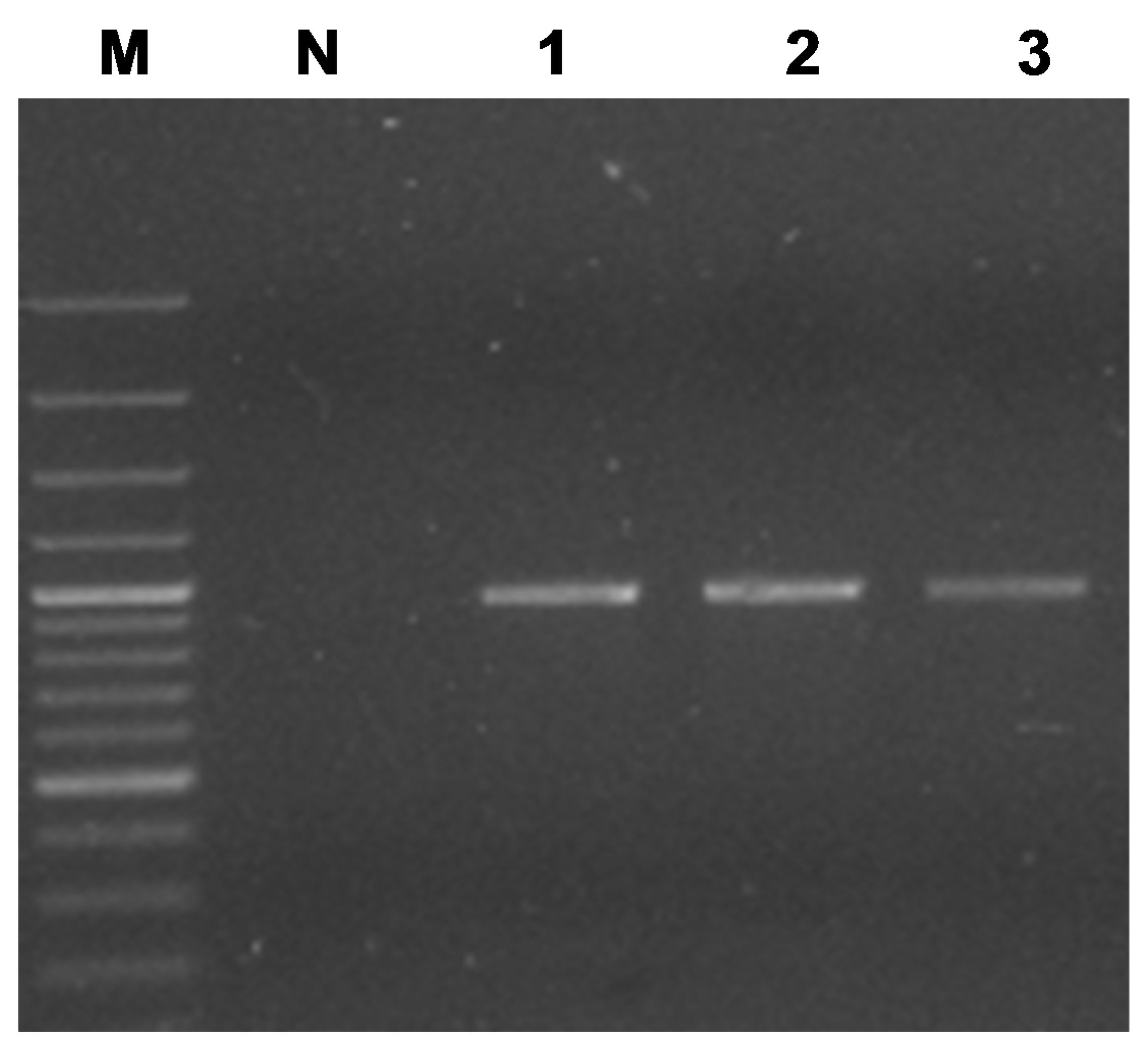
2.2. Amplification of 16S rRNA and secA Gene Sequences

Nested PCRs using extracted DNAs and primer pairs P1A/R16-SR [25,26] and P1/P7 [27,28] and R16F2n/R16R2n [29,30] were catalyzed by AmpliTaq Gold polymerase (Applied Biosystems, Waltham, MA, USA); reaction conditions and analysis of PCR products were as previously described [8,30].
A partial secA gene sequence was amplified from DNA of infected Scots pine trees by using the degenerate primer pair secAFdg: 5′-ATG AAA ACT GGR GAA GGW AAA AC-3′ and secARdg: 5′-ATG AAA GAA TYT TGT TGW CC-3′ (constructed by authors of this communication). Amplification was conducted as described by Lee et al. [8] for the amplification of 16S rDNA, except that the annealing temperature was 50 °C. PCR products were analyzed as previously described [8]. We also designed a primer pair for use in nested PCR for amplification of a 965 bp segment of the secA gene sequence from ‘Ca. Phytoplasma pini’. The nucleotide sequences of the primers were as follows: PiniSecAF2; 5′-tcg atg aag caa gaa atc ctt tg-3′ and PiniSecARA; 5′-aaa acg aga aaa tcc agg-3′. For use as template in the nested PCR, the first round PCR product was diluted 1:30 with sterile water. Amplification was conducted as described for PCRs using degenerate primer pair secAFdg/secARdg. Alternatively, direct PCR was carried out using primer pair PiniSecAF2/PiniSecARA.

2.3. Cloning and Nucleotide Sequences

16S rRNA gene (16S rDNA) products of PCR primed by R16F2n/R16R2n and secA gene sequences amplified in PCR primed by secAFdg/secARdg and PiniSecAF2 PiniSecARA were cloned in Esherichia coli using the TOPO-TA Cloning Kit (Invitrogen, Carlsbad, USA); plasmids were purified using QIAquick extraction kit (Qiagen, Hilden, Germany) according to manufacturer’s instructions, and sequenced using an automated DNA sequencer (Applied Biosystems (ABI) Prism model 3730, Netherlands), to achieve a minimum of 3 × coverage per base position. Sequences of the same gene determined from different plant samples were mutually identical.
SecA and 16S rRNA gene sequences were deposited in the GenBank database under accession numbers given in Table 1. Other nucleotide sequences used in this study were obtained from the GenBank database (Accession numbers are given in figures). For calculations of sequence similarities, sequences were aligned using DNASTAR software MegAlign option (DNASTAR, Inc., Madison, USA).
Table 1. Phytoplasma strains and their gene fragments sequenced and analysed in this study.
Phytoplasma strain and RFLP based classificationAbbreviationPlant hostGenBank accession no.
Pine dwarf yellow needle
16SrXXI-A
PineDYN27Pinus mugo TurraKR051474 *
Pine dwarf red needle
16SeXXI-A
PineDRN12Pinus mugoKR051470 *
Pine red short needle
16SrXXI-A
PineRShNPinus mugoKF801674 *
KF791911 **
Pine bunchy top
16SrXXI-A
PineBTPinus sylvestris L.GU289676 *
KF791913 **
KF791912 ***
Pine dwarf yellow needle
16SrXXI-A
PineDYN12Pinus sylvestrisKR051471 *
Pine ball-like
16SrXXI-A
PineBLPinus mugoKR051475 *
KR051469 **
Pine proliferation decline
16SrXXI-A
PinePD13Pinus mugoKR051473 *
Pine sessile needle
16SrXXI-A
PineSN13Pinus mugoKR051472 *
Pine dwarf necrotic needle
16SrI-A
PineDNN14Pinus sylvestrisKR054620 *
Pine decline
16SrI-A
PineD14Pinus mugoKR054619 *
Pine yellow short needle
16SrXXI-A
PineYShNPinus sylvestrisKF791914 **
16SrXXI-APsylLap2Pinus sylvestris var. lapponicaKF791910 **
* 16Sr DNA fragmnent; ** secA gene fragment; *** secA gene fragment (clone161) primed by primers pair secAFdg/secARdg; RFLP, restriction fragment length polymorphism.

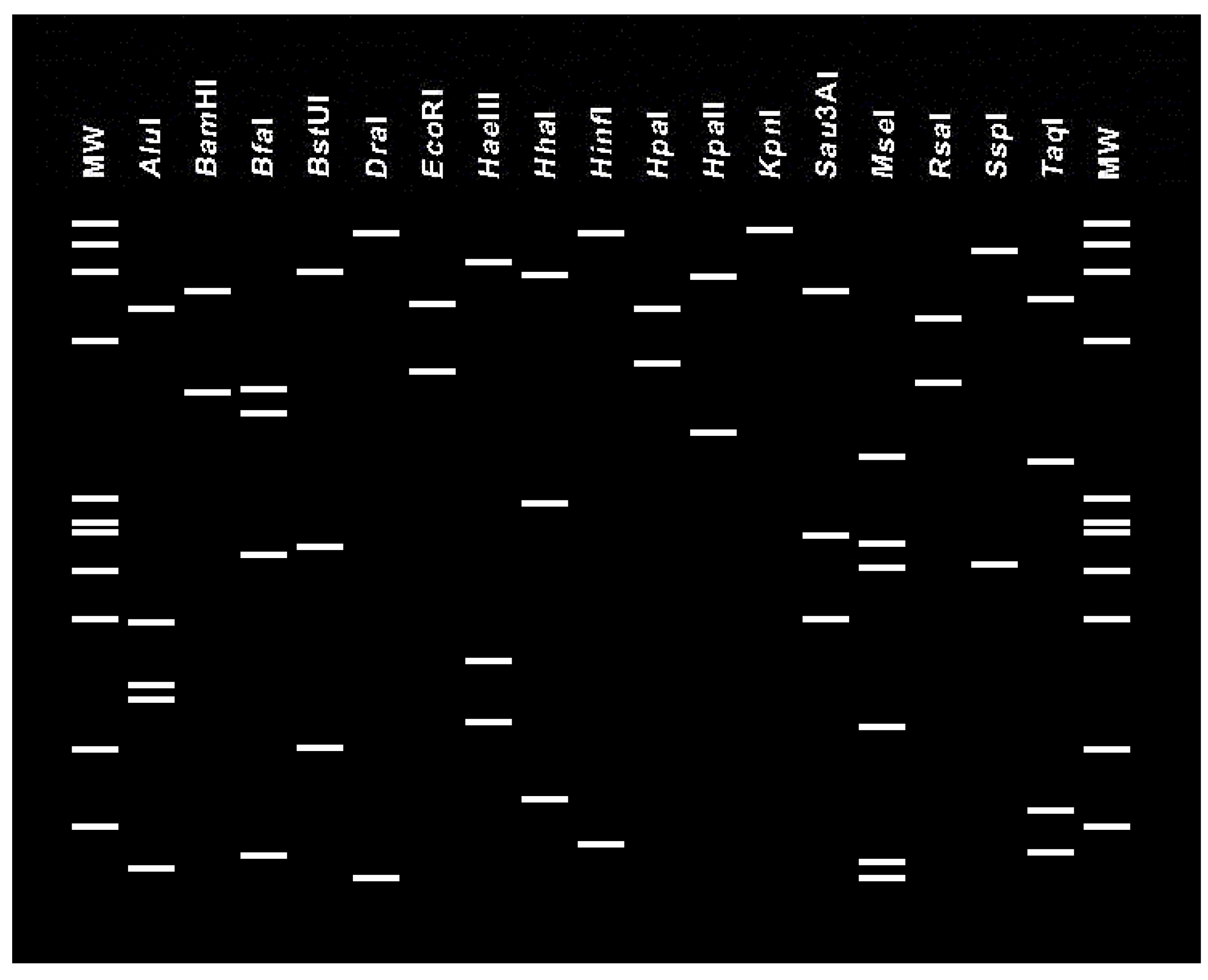
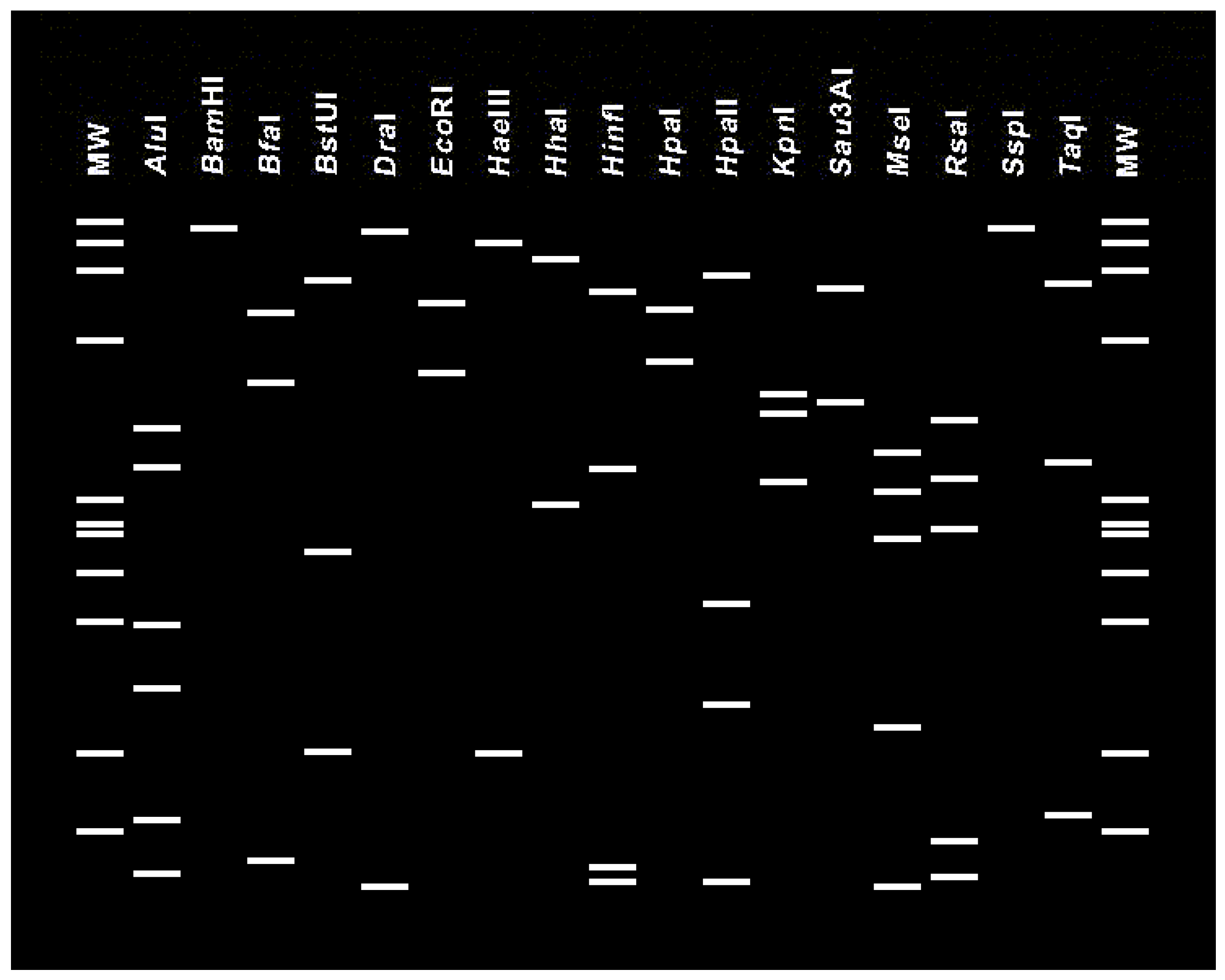
2.4. RFLP Analysis and 16Sr Group/Subgroup Classification

Products from nested PCR primed by R16F2n/R16R2n were analyzed by single enzyme digestion, according to manufacturer’s instructions, with AluI, BfaI, HaeIII, HhaI, HinfI, HpaII, KpnI, MseI, RsaI, and TaqI (Thermo Fisher Scientific Baltics, Vilnius, Lithuania). The restriction fragment length polymorphism (RFLP) profiles of digested rDNA were analyzed by electrophoresis through 5% polyacrylamide gel; DNA fragment size standard was ØX174 DNA/BsuRI (HaeIII) digest (Thermo Fisher Scientific Baltics). RFLP profiles of 16S rDNA were also observed as virtual patterns of nucleotide sequences, and 16Sr group/subgroup affiliations were assessed using iPhyClassifier [31].

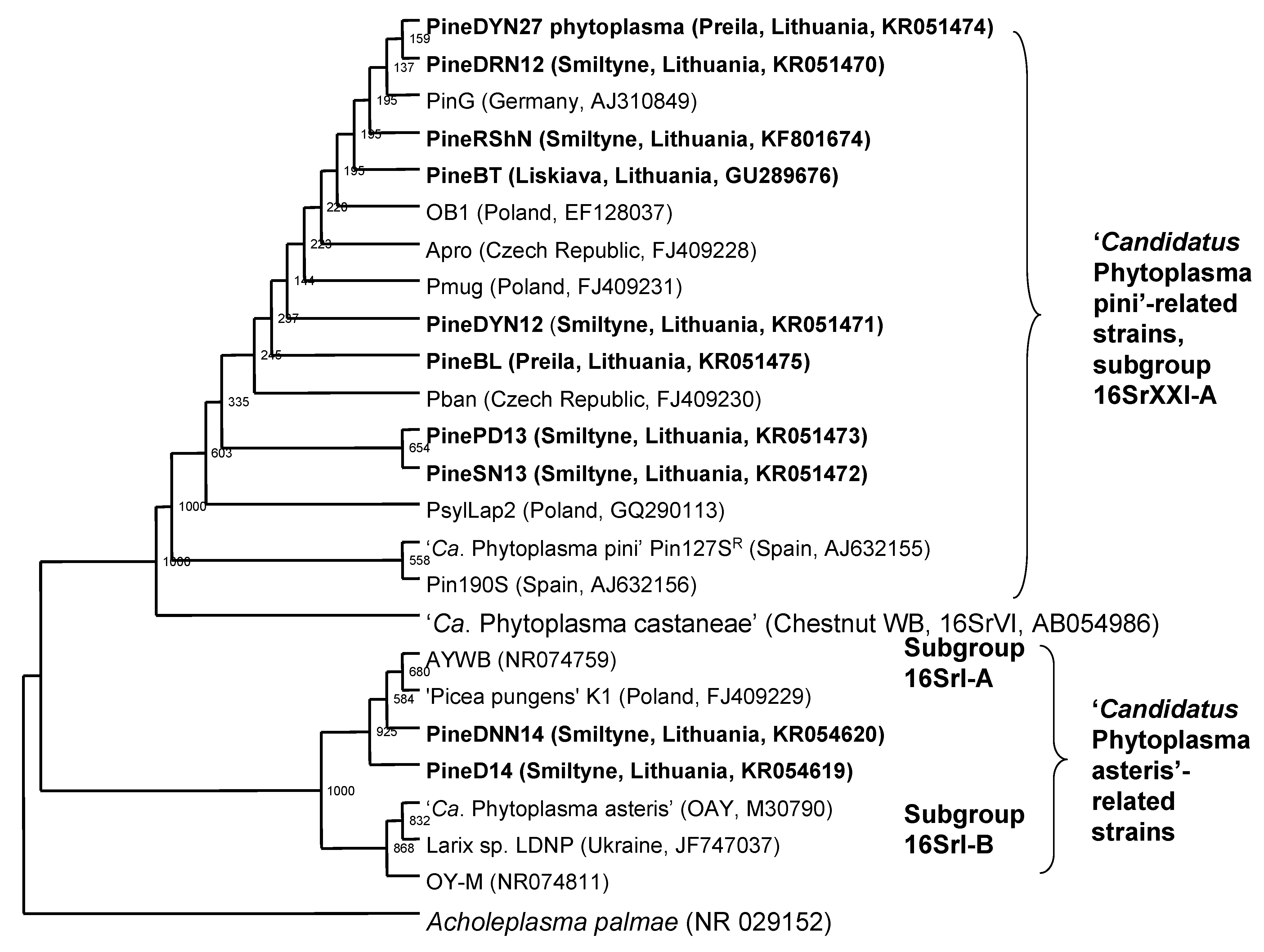
2.5. Phylogenetic Analysis

16S rRNA gene sequences (1.2 kbp in size, representing the sequence between annealing sites of primer pair R16F2n/R16R2n) from Ca. Phytoplasma pini-related phytoplasmas, Ca. Phytoplasma castaneae, and Ca. Phytoplasma asteris-related phytoplasmas were aligned, for phylogenetic analysis, using Clustal X version 1.63b [32]. A phylogenetic tree was constructed by the neighbor-joining method, the tree was bootstrapped 1000 times, and the cladogram was viewed by using TreeViewPPC [33]. Acholeplasma palmae was selected as the out-group to root the tree. Phylogenetic analysis of secA gene (968 bp in size), representing the sequence between annealing sites of primer pair PiniSecAF2/PiniSecARA from PineBT, PineRShN, PineYShN, PineBL, and PsylLap2 phytoplasma strains was accomplished using the same methods and software as that used to analyze rDNA in this study.

3. Results

3.1. Detection and Classification of the Ca. Phytoplasma pini- and Ca. Phytoplasma asteris-Related Phytoplasma Strains

All seven naturally infected Scots pines in the Varena district in southern Lithuania exhibited the same symptoms of excessive branching, dwarfed needles and dried shoots (Figure 1a). Mountain (220 plants) and Scots (80 plants) pine trees in the Smiltyne, Juodkrate, Pervalka, Preila, and Nida of the Neringa region of Curonian Spit in western Lithuania exhibited symptoms of excessive branching, dwarfed reddish or yellow needles, dried shoots and ball-like structures (Figure 1b and data not shown). DNA was extracted from the diseased and several symptomless pine tree samples and used as template in separate PCRs. In each PCR, a phytoplasma-characteristic 1.2 kb 16S rDNA product was amplified from DNA of diseased, but not from healthy pine, using phytoplasma universal primer pairs P1/P7 and/or P1A/R16-SR and R16F2n/R16R2n in nested PCRs (data not shown). These results indicated that the diseased plants were infected by phytoplasma. The phytoplasmas detected in the diseased pines were termed according to major symptoms observed, and those for which 16S rDNA and/or secA gene fragments were sequenced are listed in Table 1.
Figure 1. (a) Naturally infected Scots pine tree, exhibiting symptoms of excessive branching, dwarfed needles and dry shoots, is infected by pine bunchy top (The symptom syndrome of the diseased Scots pine (P. sylvestris) exhibiting excessive branching, dwarfed needles, and dried shoots (devoid of needles) (PineBT)) phytoplasma, a Candidatus Phytoplasma pini-related strain. On the right is an asymptomatic branch on an asymptomatic plant (indicated with arrow). (b) Naturally infected Scot pine tree exhibiting symptoms of excessive branching, dwarfed reddish needles and dry shoots infected by pine red short needle (PineRShN) phytoplasma, a Ca. Phytoplasma pini-related strain.
Forests 06 02469 g001
The 16S rDNA PCR products from all 245 positive PCRs were separately analyzed by single enzyme digestion (data not shown). Two hundred forty products yielded the same collective RFLP patterns. Comparison of these RFLP patterns, with patterns previously published for 16S rDNA from other phytoplasmas [8,15,22], revealed that the phytoplasmas belong to group 16SrXXI (pine shoot proliferation phytoplasma group) subgroup 16SrXXI-A and are related to Ca. Phytoplasma pini. The other five phytoplasmas were identified as relataed to Ca. Phytoplasma asteris, subgroup 16SrI-A. Results from virtual RFLP analysis of the strains 16S rDNA nucleotide sequences (Figure 2 and Figure 3) were in excellent agreement with results from the enzymatic RFLP analysis.
Figure 2. Virtual restriction fragment length polymorphism (RFLP), RFLP patterns of 16S rRNA gene sequences corresponding to fragments amplified, in the polymerase chain reaction (PCR) primed by primer pair R16F2/R16R2n, from PineBT phytoplasma (GU289676) classified in group 16SrXXI, subgroup 16SrXXI-A. RFLP patterns observed as virtual patterns using iPhyClassifier [31]. MW, virtual ØX174 HaeIII digest size standard.
Forests 06 02469 g002
Figure 3. Virtual restriction fragment length polymorphism (RFLP) patterns of 16S rRNA gene sequences corresponding to fragments amplified, in the polymerase chain reaction (PCR) primed by primer pair R16F2/R16R2n, from PineD14 phytoplasma (KR054619) classified in group 16SrI, subgroup 16SrI-A. RFLP patterns observed as virtual patterns using iPhyClassifier [31]. MW, virtual ØX174 HaeIII digest size standard.
Forests 06 02469 g003

3.2. SecA Gene from the PineBT Phytoplasma

A 2 kbp fragment of the secA gene was amplified, from PineBT phytoplasma, in PCR primed by the degenerate primer pair secAFdg and secARdg (this study). A negative control devoid of DNA template, and PCRs containing template DNA extracted from symptomless tree, yielded no detectable product. The secA gene fragment was cloned and sequenced, and the 1998-base nucleotide sequence was deposited in the GenBank database under acc. no. KF791912.

3.3. Primers for Nested or Direct PCR for secA-Based Detection of Ca. Phytoplasma pini

Primer pair PiniSecAF2/PiniSecARA was used to prime PCR amplification of a 965 bp DNA fragment (Figure 4, three PCR products are shown) of the secA gene from 17 samples of Ca. Phytoplasma pini and Ca. Phytoplasma pini from isolate PsylLap2 from Poland [20] (16S rDNA acc. no. GQ290113). The five cloned PCR products were sequenced (Table 1). Alignments of the nucleotide sequences of primers PiniSecAF2 and PiniSecARA with all available phytoplasmal secA sequences indicated that these primers may offer some specificity for rapid detection and identification of Ca. Phytoplasma pini strains.
Figure 4. Evaluation of primers: DNA products amplified from Ca. Phytoplasma pini-related strains PineBT (1), PineRShN (2), and PineYShN (3) in PCRs primed by PiniSecAF2/PiniSecARA primer pair. M, GeneRuler 100 bp DNA Ladder Plus, fragment sizes: 3000, 2000, 1500, 1200, 1031, 900, 800, 700, 600, 500, 400, 300, 200, and 100 bp. N, negative control.
Forests 06 02469 g004

3.4. Phylogenetic Analysis and Alignments of 16S rDNA and secA Gene Sequences

The 16S rDNA phylogenetic tree (cladogram) indicated that PineDYN27, PineDRN12, PineRShN, PineBT, PineDYN12, PineBL, PinePD13, and PineSN13 phytoplasmas from Lithuania are closely related to Ca. Phytoplasma pini or Ca. Phytoplasma pini-related strains from other countries. In the 16S rDNA cladogram, the group 16SrXXI phytoplasmas clustered with Ca. Phytoplasma castanea as described by Schneider et al. [21], consistent with the concept that these phytoplasmas shared a common ancestor (Figure 5). In the secA phylogenetic tree, PineBT, PineRShN, PineYShN, PineBL, and PsylLap2 phytoplasmas were separated according to geographical region where found (Figure 6). The 16S rDNA cladogram indicated that PineDNN14 and PineD14 phytoplasmas are related to Ca. Phytoplasma asteris and belong to subgroup 16SrI-A (Figure 5).
Alignment of the 968 bp fragments of the secA gene (amplified in PCRs primed by PiniSecAF2/PiniSecARA) from phytoplasma strains PineBT, PineRShN, PineBL and PineYShN revealed that they shared 100% nucleotide sequence identity. The 968 bp fragments secA from isolate (strain) PsylLap2 (from Poland) shared 99.8% sequence identity with secA from the Lithuanian strains, suggesting differences related to geographical location, although the nucleotide sequence differences reflected silent mutations, because the deduced amino acid were identical between all the strains from Lithuania and Poland.
Figure 5. Cladogram constructed by the Neighbor-Joining method of 16S rRNA gene sequences from phytoplasma strains from Lithuania (in bold), other Ca. Phytoplasma pini strains, Ca. Phytoplasma castaneae, and Ca. Phytoplasma asteris, employing Acholeplasma palmae as the outgroup. Accession numbers of phytoplasma strains are indicated in brackets. Numbers on the nodes are bootstrap (confidence) values.
Forests 06 02469 g005
Figure 6. Cladogram constructed by the Neighbor-Joining method of secA gene nucleotide sequences (corresponding to the region amplified in polymerase chain reaction (PCRs) primed by primer pair PiniSecAF2/PiniSecARA) from phytoplasma strains from Lithuania (in bold), and from isolate (strain) PsylLap2 phytoplasma from Poland. GenBank accession numbers are indicated in brackets. Numbers on the nodes are bootstrap (confidence) values.
Forests 06 02469 g006
Ca. Phytoplasma pini-related strains PineDYN27, PineDRN12, PineRShN, PineBT, PineDYN12, PineBL, PinePD13, and PineSN13 (from Lithuania), and strain PsylLap2 (from Poland) shared 99.7%–99.9% rDNA sequence identity; all Lithuanian strains shared 99.7%–100% rDNA sequence identity with one another.
The 1.2 kb 16S rDNAs from Ca. Phytoplasma asteris-related strains PineDNN14 and PineD14 shared 99.7% nucleotide sequence identity with one another and 99.6%–99.8% identity of 16S rDNA with strain aster yellows withes’-broom (AY-WB), previously classified in subgroup 16SrI-A.

4. Discussion

Comparative 16S rDNA RFLP and nucleotide sequence analyses revealed that symptomatic Scots pine and mountain pine trees in Lithuania are infected by Ca. Phytoplasma pini-related strains and that some diseased trees are infected by Ca. Phytoplasma asteris-related strains belonging to phytoplasma subgroup 16SrI-A.
Ball-like structures resembling those reported on Ca. Phytoplasma pini-infected Scots pine trees in Germany and Poland [20,21] were observed rarely on phytoplasma-infected Scots and mountain pine trees in Lithuania. The most common symptoms were dwarfed needles that ranged in color from yellow to reddish, and dried branches. Symptomatic diseased pine trees were found throughout most areas of Curonian Spit. Of 300 symptomatic trees that were sampled, 80% were infected by phytoplasma resulting in serious and widespread damage to the landscape of the UNESCO-protected Curonian Spit.
In other parts of the world, phytoplasmas have been reported in association with several diseases of gymnosperms. A group 16SrIII phytoplasma was identified from Cypress sp. in Italy [34]. In other regions of Europe, phytoplasmal diseases of gymnosperms (Pinus spp., Picea spp., Abies spp., and Larix sp.) have been reported in Germany, Spain, Poland, Czech Republic, Croatia, and Ukraine [19,20,21,22,35]. Taxonomically diverse phytoplasmas, including Ca. Phytoplasma pini-, Ca. Phytoplasma asteris-, and Ca. Phytoplasma pruni-related strains have been found associated with diseases of gymnosperms in Europe [36]. In India, a phytoplasma of group 16SrVI was identified from Norfolk Island pine [37]. In North America, a new subgroup 16SrIX-E, Ca. Phytoplasma phoenicium-related, phytoplasma was found in juniper exhibiting symptoms of a witches’ broom disease [18]. A Ca. Phytoplasma pini-related strain was identified in bald-cypress in China [38]. Our communication presents the first report of Ca. Phytoplasma asteris-related strains (phytoplasmas belonging to the subgroup 16SrI-A) in pine trees. The present communication and prior literature illustrate that diverse gymnosperms are susceptible to infection by at least five phytoplasma species level lineages, emphasizing the need for adequate quarantine surveillance of regional and intercontinental movement of gymnospermae germplasm and underscoring the need to learn the identities of insect vectors responsible for the spread of phytoplasmas among gymnosperms.
Quarantine surveillance and studies of insect vectors of Ca. Phytoplasma pini-related phytoplasma strains may be facilitated by the availability of secA gene sequence data herein, which provide new molecular markers for finer characterization and future epidemiological studies of the phytoplasma in conifers. Primer pair PiniSecAF2/PiniSecARA, designed in the present work, rendered possible direct amplification of secA sequences from Ca. Phytoplasma pini, whereas, use of primers previously published [39] yielded no observable DNA amplification or non-specific DNA amplification in the present work (data not shown). As noted by Hodgetts et al. [39], a relatively short (480 bp) secA gene fragment may provide an informative alternative molecular marker for pathogen identification and diagnosis of phytoplasma diseases, and for monitoring phytoplasma movement between geographic regions [39,40]. The 968 bp secA fragment amplified using primers described in the present study should be even more highly informative.
Multiple gene-based classification could provide molecular criteria for improved delineation of Ca. Phytoplasma’ species and related phytoplasma strains. SecA could serve as a useful alternative to 16S rDNA for phytoplasma classification [39]. Analysis of secA gene from 12 major 16Sr phytoplasma groups supported the previous classification systems and provided improved resolution of those groups and constituent subgroups [39]. In the present communication, we expand this capability by providing secA gene sequence data for a representative of a 13th major phytoplasma group (16SrXXI), which previously had been characterized based on 16S rDNA alone. Analysis of secA gene fragments from Ca. Phytoplasma pini distinguished between strains from Lithuania and Poland, reinforcing the concept that multiple gene-based classification improves phytoplasma strain differentiation and can support epidemiological studies.

5. Conclusions

Scots pine (Pinus sylvestris) and mountain pine (Pinus mugo) in Lithuania are suffering from a serious disease characterized by dwarfed needles ranging in color from yellow to reddish, branches devoid of needles, dying and dead branches, and death of trees. The disease is associated with infection of the trees by phytoplasma, a very small bacterium that lacks a rigid wall, lives in plant phloem tissue, and is spread by phloem sap-feeding insects. Diseased trees were infected by either Candidatus Phytoplasma pini or Ca. Phytoplasma asteris. In the UNESCO-protected Curonian Spit of western Lithuania, Ca. Phytoplasma pini accounted for 80% of the infections in symptomatic trees. The wide occurrence of the disease and the extensive damage to forests underscore the serious threat of this disease to industry and ecology. Continued development and use of multiple molecular genetic markers, as in the present work, will be needed to diagnose the disease, identify insect vector(s), and devise strategies for reducing its damage and stopping its spread.

Acknowledgments

This research was funded by a grant (No. MIP-51/2013) from the Research Council of Lithuania. We thank M. Kaminska (Department of Plant Protection, Institute of Horticulture, Pomologiczna 18, 96-100 Skierniewice, Poland) for providing diseased pine tree needles.

Author Contributions

All authors generated the data. Deividas Valiunas, Rasa Jomantiene, Algirdas Ivanauskas, and Robert E. Davis analyzed and discussed the data. The manuscript was written by Deividas Valiunas and Robert E. Davis.

Conflicts of Interest

The authors declare no conflict of interest.

References

  1. Mizaras, S.; Mizaraite, D.; Lebedys, A.; Pivoriunas, A.; Belova, O. Lithuania. Acta Silv. Lign. Hung. 2005, 1, 437–466, Special Edition. [Google Scholar]
  2. Jomantiene, R.; Davis, R.E.; Alminaite, A.; Staniulis, J.; Valiunas, D. Ribosomal RNA interoperon sequence heterogeneity in new phytoplasma lineages infecting oak, campion, thistle, and dandelion. In Proceedings of the American Phytopathological Society, Potomac Division Meeting Abstracts, Williamsburg, VA, USA, 4–6 March 2002; Available online: http://eurekamag.com/research/035/670/035670482.php (accessed on 4 March 2002).
  3. Valiunas, D.; Alminaite, A.; Staniulis, J.; Jomantiene, R.; Davis, R.E. First report of alder yellows phytoplasma in the Eastern Baltic Region. Plant Dis. 2001, 85, 1120. [Google Scholar] [CrossRef]
  4. Valiunas, D. Identification of Phytoplasmas in Lithuania and Estimation of Their Biodiversity and Molecular Evolutionary Relationships. Ph.D. Thesis, Institute of Botany and Vilnius University, Vilnius, Lithuania, 2003. [Google Scholar]
  5. Aučina, A.; Rudawska, M.; Leski, T.; Ryliškis, D.; Pietras, M.; Riepšas, E. Ectomycorrhizal fungal communities on seedlings and conspecific trees of Pinus mugo grown on the coastal dunes of the Curonian Spit in Lithuania. Mycorrhiza 2011, 21, 237–245. [Google Scholar] [CrossRef] [PubMed]
  6. Weintraub, P.G.; Beanland, L. Insect vectors of phytoplasma. Annu. Rev. Entomol. 2006, 51, 91–111. [Google Scholar] [CrossRef] [PubMed]
  7. Davis, R.E.; Lee, I.M. Phytoplasma. In Encyclopedia of Microbiology, 2nd ed.; Lederberg, J., Ed.; Academic Press Inc.: San Diego, CA, USA, 2000; pp. 640–646. [Google Scholar]
  8. Lee, I.M.; Gundersen-Rindal, D.E.; Davis, R.E.; Bartoszyk, I. Revised classification scheme of phytoplasmas based on RFLP analyses of 16S rRNA and ribosomal protein gene sequences. Int. J. Syst. Bacteriol. 1998, 48, 1153–1169. [Google Scholar] [CrossRef]
  9. Marcone, C.; Lee, I.M.; Davis, R.E.; Ragozzino, A.; Seemüller, E. Classification of aster yellows-group phytoplasmas based on combined analyses of rRNA and tuf gene sequences. Int. J. Syst. Evol. Microbiol. 2000, 50, 1703–1713. [Google Scholar] [PubMed]
  10. Davis, R.E.; Dally, E.L. Revised subgroup classification of group 16SrV phytoplasmas and placement of flavescence dorée-associated phytoplasmas in two distinct subgroups. Plant Dis. 2001, 85, 790–797. [Google Scholar] [CrossRef]
  11. Jomantiene, R.; Davis, R.E.; Valiunas, D.; Alminaite, A. New group 16SrIII phytoplasma lineages in Lithuania exhibit rRNA interoperon sequence heterogeneity. Eur. J. Plant Pathol. 2002, 108, 507–517. [Google Scholar] [CrossRef]
  12. Jomantiene, R.; Zhao, Y.; Lee, I.M.; Davis, R.E. Phytoplasmas infecting sour cherry and lilac represent two distinct lineages having close evolutionary affinities with clover phyllody phytoplasma. Eur. J. Plant Pathol. 2011, 130, 97–107. [Google Scholar] [CrossRef]
  13. Valiunas, D.; Jomantiene, R.; Davis, R.E. Establishment of a new phytoplasma subgroup, 16SrI-Q, to accomodate a previously undescribed phytoplasma found in diseased cherry in Lithuania. J. Plant Pathol. 2009, 91, 71–75. [Google Scholar]
  14. Valiunas, D.; Jomantiene, R.; Ivanauskas, A.; Abraitis, R.; Staniene, G.; Zhao, Y.; Davis, R.E. First report of a new phytoplasma subgroup, 16SrIII-T, associated with decline disease affecting sweet and sour cherry trees in Lithuania. Plant Dis. 2009, 93, 550. [Google Scholar] [CrossRef]
  15. Wei, W.; Davis, R.E.; Lee, I.M.; Zhao, Y. Computer-simulated RFLP analysis of 16S rRNA genes: Identification of ten new phytoplasma groups. Int. J. Syst. Evol. Microbiol. 2007, 57, 1855–1867. [Google Scholar] [CrossRef] [PubMed]
  16. Zhao, Y.; Sun, Q.; Wei, W.; Davis, R.E.; Wu, Q.; Liu, Q. Candidatus Phytoplasma tamaricis, a novel taxon discovered in witches’-broom-diseased salt cedar (Tamarix chinensis Lour.). Int. J. Syst. Evol. Microbiol. 2009, 59, 2496–2504. [Google Scholar] [CrossRef] [PubMed]
  17. Davis, R.E.; Zhao, Y.; Dally, E.L.; Lee, I.M.; Jomantiene, R.; Douglas, S.M. Candidatus Phytoplasma pruni, a novel taxon associated with X-disease of stone fruits, Prunus spp.: Multilocus characterization based on 16S rRNA, secY, and ribosomal protein genes. Int. J. Syst. Evol. Microbiol. 2013, 63, 766–776. [Google Scholar] [CrossRef] [PubMed]
  18. Davis, R.E.; Dally, E.; Zhao, Y.; Lee, I.M.; Jomantiene, R.; Detweiler, A.J.; Putnam, M.L. First report of a new subgroup 16SrIX-E (Candidatus Phytoplasma phoenicium-related) phytoplasma associated with juniper witches’ broom disease in Oregon, USA. Plant Pathol. 2010, 59, 1161. [Google Scholar] [CrossRef]
  19. Jomantiene, R.; Valiunas, D.; Ivanauskas, A.; Urbanaviciene, L.; Staniulis, J.; Davis, R.E. Larch is a new host for a group 16SrI, subgroup B phytoplasma in Ukraine. B. Insectol. 2011, 64, S101–S102. [Google Scholar]
  20. Kaminska, M.; Berniak, H.; Obdrzalek, J. New natural host plants of Candidatus Phytoplasma pini in Poland and the Czech Republic. Plant Pathol. 2011, 60, 1023–1029. [Google Scholar] [CrossRef]
  21. Schneider, B.; Torres, E.; Martin, M.P.; Schröder, M.; Behnke, H.D.; Seemüller, E. Candidatus Phytoplasma pini, a novel taxon from Pinus silvestris and Pinus halepensis. Int. J. Syst. Evol. Microbiol. 2005, 55, 303–307. [Google Scholar] [CrossRef] [PubMed]
  22. Śliwa, H.; Kaminska, M.; Korszun, S.; Adler, P. Detection of Candidatus Phytoplasma pini in Pinus sylvestris trees in Poland. J. Phytopathol. 2008, 156, 88–92. [Google Scholar] [CrossRef]
  23. Valiunas, D.; Jomantiene, R.; Ivanauskas, A.; Sneideris, D.; Staniulis, J.; Davis, R.E. A possible threat to the timber industry: Candidatus Phytoplasma pini in Scots pine (Pinus sylvestris L.) in Lithuania. In Current Status and Perspectives of Phytoplasma Disease Research and Management, Proceedings of the Abstract book of the combined meeting of Work Groups 1–4, COST Action FA0807, Sitges, Spain, 1–2 February 2010; Bertaccini, A., Lavifia, A., Torres, E., Eds.; p. 38.
  24. Valiunas, D.; Jomantiene, R.; Ivanauskas, A.; Urbonaite, I.; Davis, R.E. Molecular identification of pine tree-infecting phytoplasmas from the UNESCO-protected Curonian Spit in Lithuania. In Proceedings of the Joint IUFRO Working Party Meetings, Uppsala, Sweden, 7–12 June 2015; p. 89.
  25. Lee, I.M.; Martini, M.; Bottner, K.D.; Dane, R.A.; Black, M.C.; Troxclair, N. Ecological implications from a molecular analysis of phytoplasmas involved in an aster yellows epidemic in various crops in Texas. Phytopathology 2003, 93, 1368–1377. [Google Scholar] [CrossRef] [PubMed]
  26. Lee, I.M.; Martini, M.; Marcone, C.; Zhu, S.F. Classification of phytoplasma strains in the elm yellows group (16SrV) and proposal of Candidatus Phytoplasma ulmi for the phytoplasma associated with elm yellows. Int. J. Syst. Evol. Microbiol. 2004, 54, 337–347. [Google Scholar] [CrossRef] [PubMed]
  27. Deng, S.; Hiruki, C. Amplification of 16S rRNA genes from culturable and non-culturable mollicutes. J. Microbiol. Meth. 1991, 14, 53–61. [Google Scholar] [CrossRef]
  28. Schneider, B.; Seemüller, E.; Smart, C.D.; Kirkpatrick, B.C. Phylogenetic classification of plant pathogenic mycoplasma-like organisms or phytoplasmas. In Molecular and Diagnostic Procedures in Mycoplasmology; Razin, S., Tully, J.G., Eds.; Academic Press: San Diego, CA, USA, 1995; Volume 1, pp. 369–380. [Google Scholar]
  29. Gundersen, D.E.; Lee, I.M. Ultrasensitive detection of phytoplasmas by nested-PCR assays using two universal primer pairs. Phytopathol. Mediterr. 1996, 35, 144–151. [Google Scholar]
  30. Lee, I.-M.; Bottner, K.D.; Munyaneza, J.E.; Davis, R.E.; Crosslin, J.M.; du Toit, L.J. Carrot purple leaf: A new spiroplasma disease associated with carrots in Washington state. Plant Dis. 2006, 90, 989–993. [Google Scholar] [CrossRef]
  31. Zhao, Y.; Wei, W.; Lee, I.M.; Shao, J.; Suo, X.; Davis, R.E. Construction of an interactive online phytoplasma classification tool, iPhyClassifier, and its application in analysis of the peach X-disease phytoplasma group (16SrIII). Int. J. Syst. Evol. Microbiol. 2009, 59, 2582–2593. [Google Scholar] [CrossRef] [PubMed]
  32. Thompson, J.D.; Gibson, T.J.; Plewniak, F.; Jeanmougin, F.; Higgins, D.G. The Clustal X windows interface: Flexible strategies for multiple sequence alignment aided by quality analysis tools. Nucleic Acids Res. 1997, 25, 4876–4882. [Google Scholar] [CrossRef] [PubMed]
  33. Page, R.D. TreeView: An application to display phylogenetic trees on personal computers. Comput. Appl. Biosci. 1996, 12, 357–358. [Google Scholar] [PubMed]
  34. Paltrinieri, S.; Martini, M.; Pondrelli, M.; Bertaccini, A. X-disease related phytoplasmas in ornamental trees and shrubs with witches’-broom and malformation symptoms. J. Plant Pathol. 1998, 80, 261. [Google Scholar]
  35. Ježić, M.; Poljak, I.; Šafarić, B.; Idžojtić, M.; Ćurković-Perica, M. Candidatus Phytoplasma pini in pine species in Croatia. J. Plant Dis. Prot. 2013, 120, 160–163. [Google Scholar]
  36. Kaminska, M.; Berniak, H. Detection and identification of three Candidatus Phytoplasma species in Picea spp. trees in Poland. J. Phytopathol. 2011, 159, 796–798. [Google Scholar] [CrossRef]
  37. Gupta, M.K.; Samad, A.; Shasany, A.K.; Ajayakumar, P.V.; Alam, M. First report of a 16SrVI Candidatus Phytoplasma trifolii isolate infecting Norfolk Island pine (Araucaria heterophylla) in India. Plant Pathol. 2009, 59, 399. [Google Scholar] [CrossRef]
  38. Huang, S.; Tiwari, A.K.; Rao, G.P. Candidatus Phytoplasma pini affecting Taxodium distichum var. imbricarium in China. Phytopathogenic Mollicutes 2011, 1, 91–94. [Google Scholar] [CrossRef]
  39. Hodgetts, J.; Boonham, N.; Mumford, R.; Harrison, N.; Dickinson, M. Phytoplasma phylogenetics based on analysis of secA and and 23S rRNA gene sequences for improved resolution of candidate species of Candidatus Phytoplasma. Int. J. Syst. Evol. Microbiol. 2008, 58, 1826–1837. [Google Scholar] [CrossRef] [PubMed]
  40. Makarova, O.; Contaldo, N.; Paltrinieri, S.; Bertaccini, A.; Nyskjold, H.; Nicolaisen, M. DNA bar-coding for phytoplasma identification. Methods Mol. Biol. 2013, 938, 301–317. [Google Scholar] [PubMed]

