Article

Distribution of Duffy Phenotypes among Plasmodium vivax Infections in Sudan

1
Institute of Endemic Diseases, University of Khartoum, Khartoum 11111, Sudan
2
Faculty of Laboratory Sciences, University of Sinnar, Khartoum 11111, Sudan
3
Department of Biological Sciences, University of North Carolina at Charlotte, Charlotte, NC 28223, USA
4
Faculty of Medicine, Sudan University of Science and Technology, Khartoum 11111, Sudan
*
Authors to whom correspondence should be addressed.
These authors contributed equally to this work.
Submission received: 24 April 2019 / Revised: 5 June 2019 / Accepted: 5 June 2019 / Published: 8 June 2019
(This article belongs to the Special Issue Genetics and Genomics of Malaria Parasites)

Abstract

:
Negative Duffy expression on the surface of human red blood cells was believed to be a barrier for Plasmodium vivax infection in most Africans. However, P. vivax has been demonstrated to infect Duffy-negative individuals in several Central and East African countries. In this study, we investigated the distribution of Duffy blood group phenotypes with regard to P. vivax infection and parasitemia in Sudan. Out of 992 microscopic-positive malaria samples, 190 were identified as P. vivax positive infections. Among them, 186 were P. vivax mono-infections and 4 were mixed P. vivax and Plasmodium falciparum infections. A subset of 77 samples was estimated with parasitemia by quantitative real-time PCR. Duffy codons were sequenced from the 190 P. vivax positive samples. We found that the Duffy Fy(a-b+) phenotype was the most prevalent, accounting for 67.9% of all P. vivax infections, while homozygous Duffy-negative Fy(a-b-) accounted for 17.9% of the P. vivax infections. The prevalence of infection in Fy(a-b+) and Fy(a+b-)were significantly higher than Fy(a-b-) phenotypes (p = 0.01 and p < 0.01, respectively). A significantly low proportion of P. vivax infection was observed in Duffy negative individuals Fy(a-b-). This study highlights the prevalence of P. vivax in Duffy-negatives in Sudan and indicates low parasitemia among the Duffy-negative individuals.

1. Introduction

Malaria is a serious health problem, especially in developing countries. According to the latest estimates from the World Health Organization, there were 214 million new cases and 435,000 deaths, mostly occurring in sub-Saharan Africa [1].The predominant malaria parasite species is Plasmodium falciparum. However, Plasmodium vivax is widely spread and it has been increasingly recognized as a cause of severe malaria and even death [1,2]. Although the prevalence of P. vivax was believed to be much lower in Africa, using a compiled database, a recent study has highlighted the widespread presence of P. vivax across all malaria-endemic regions of Africa [3].
Based on the WHO World Malaria Report 2018, malaria constitutes a major public health problem with 720,819 reported confirmed cases and 3885 deaths in Sudan [1]. Most cases are caused by P. falciparum. However, in recent years the prevalence of P. vivax malaria has witnessed a sharp increase and accounted for up to one fifth of the total malaria cases [1] and disease is becoming widespread in areas where it has not been documented before and some severe cases due P. vivax have been reported [4,5].
The human Duffy antigen also known as Duffy antigen receptor for chemokines (DARC) or the FY gene is the key molecule required for P. vivax and Plasmodium knowlesi parasites to invade red cells through interaction with P. vivax Duffy binding protein (PvDBP) [6,7,8]. The FY gene is located on chromosome 1 and possesses four main allelic forms, including FYA, FYB, FYAES, and FYBES. This gene encodes a glycosylated membrane protein (FY glycoprotein) [9].
Polymorphisms in the DARC gene alter the expression of Duffy antigen protein and determine the Duffy blood group system [10]. The theoretical expectation is that Duffy-negative individuals lack Duffy antigen expression on their red blood cells and thus are protected against P. vivax infection [7]. The FY gene encodes red blood group antigens and has two co-dominant alleles (FYA and FYB), which differ by a single nucleotide polymorphism, 125 G→A, leading to an amino acid change from Gly to Asp at position 42. The composition of these two alleles gives rise to the Duffy phenotypes, Fy(a+b+), Fy(a+b−), and Fy(a−b+). Individuals with these phenotypes exhibit Duffy antigen expression and are considered as Duffy positive. The silent FYBES allele results from a mutation in the GATA box motif of the FYB promoter (−33T→C) which is known to disrupt the binding of erythroid transcription factor to GATA-1, leading to a reduced Duffy antigen expression [11]. The Duffy-negative homozygote with phenotype Fy(a-b-) has been shown to be most prevalent among Africans and individuals of African ancestry, who were thought to be protected from P. vivax infection [12]. However, recently, P. vivax infections in Duffy-negative individuals have been documented in several African countries [12], including Madagascar [13], Mauritania [14], Ethiopia [15], Cameroon [16], and Sudan [4], and thus Duffy negativity alone does not prevent infection with P. vivax. There is yet no documented evidence for the associations between Duffy phenotypes and the prevalence of P. vivax infection in malaria endemic regions in East Africa. The aim of this study was to determine the distribution of Duffy phenotypes and to estimate P. vivax parasitemia in relation to the different Duffy phenotypes.

2. Materials and Methods

2.1. Ethical Statement

The study was approved by the Research Ethical Review Committee, Institute of Endemic Diseases University of Khartoum, Sudan (certificate reference number 9/2016). Prior to enrollment in the study, written informed consent was obtained from all adult patients and parents or legal guardians of the children.

2.2. Study Sites and Subjects

Malaria transmission in Sudan is seasonal with two peaks of transmission: One during the rainy season between August and October and the other one during the winter season between December to February. Anopheles arabiensis is the principal malaria vector in Sudan [17]. Sample collection was conducted in the Central, Northern, and Eastern part of Sudan including Khartoum (15°53’11.2″N 32°31’39.9″E), New Halfa (15°20’00.0″N, 35°35’00″E), and River Nile (16°59’58.5″N 35°29’04.4″E), between August 2013 and 2017. Symptomatic malaria patients were recruited from Gerri and Gezira Slanj primary health centers in Northern Khartoum, New Halfa teaching hospital in New Halfa, as well as Seedon, Al-Zaidab, and Atabra primary health centers in River Nile. Thick and thin blood films were collected and stained with Giemsa using standard procedures. Slides were examined twice at the study centers and re-examined at the Department of Parasitology and Medical Entomology, Institute of Endemic Diseases, University of Khartoum. Microscopy positive samples were confirmed by 18S rRNA nested PCR assay. In this study, a total of 190 PCR-confirmed P. vivax blood samples were collected from symptomatic malaria patients (101 from Khartoum, 40 from New Halfa, and 49 from River Nile), 186 samples were confirmed P. vivax mono-infection and four samples were mixed infection (P. vivax and P. falciparum) based on nested PCR assay. The male to female ratio was 1.73 and the mean age was 25 years. Non-malaria subjects attending the same health facility who tested negative for Plasmodium by microscopy and PCR were included as controls. Matching cases were selected according to age, gender, and ethnicity.

2.3. Sample Collection and DNA Extraction

Approximately 3 to 4 spots of finger-prick blood samples (each 50 microliters) were collected on Whatman filter paper (Whatman International Ltd, Maidstone, England) from microscopy positive malaria patients (N = 190). In addition, 2 to 3 mL of whole blood was collected in EDTA tubes from 67 healthy subjects with no prior history of malaria and were tested as negative for malaria by microscopy and nested PCR at the time of blood collection. Genomic DNA was extracted from 2 to 3 dried blood spots using a Qiagen mini DNA kit (Qiagen, Germany) with minor modification, while DNA from blood samples was extracted using a DNeasy Blood kit (Qiagen, Germany). DNA was eluted in total of 40µL of warm elution buffer (AE) (Qiagen, Germany) and kept at −20 °C until used.

2.4. Molecular Identification of Plasmodium Species

Identification of Plasmodium species was performed using nested PCR. Species-specific primers that amplify the 18S rRNA gene for P. falciparum, P. vivax, Plasmodium malariae, and Plasmodium ovale as a single or mixed infection were used as described by previous studies [18]. The 18S rRNA amplicon was sequenced in both the 5’ and 3’ directions to confirm P. vivax infection (Macrogen Inc., Amsterdam, The Netherlands).

2.5. Estimation of P. vivax Parasitemia

The estimation of parasitemia was performed using the 18S rRNA SYBR green quantitative real-time PCR method. The P.vivax 18S rRNA specific primers (forward: 5′-GAATTTTCTCTTCGGAGTTTATTCTTAGATTGC-3′ and reverse: 5′-GCCGCAAGCTCCACGCCTGGTGGTGC-3′) were used for amplifications. Amplification was performed in 20 μL reactions which contained 10 μL of 2× SYBR Green Master Mix (Thermo Scientific, Waltham, MA), 0.5 μM of the forward and reverse primers, 2 μL of genomic DNA, and 7 μL of water. The initial denaturation was run at 95 °C for 3 minutes, followed by 45 cycles of 94 °C for 30 seconds, 55 °C for 30 seconds, and 68 °C for 1 minute which was followed by a hold step of 95 °C for 10 seconds. This was followed by a melting curve stage from 65 to 95 °C with 0.5 increments for 5 seconds [19]. Each assay included positive controls of P. vivax Pakchong (MRA-342G) and Nicaragua (MRA-340G) isolates, in addition to negative controls, including uninfected samples and water. A standard curve was produced from a 10-fold dilution series of the control plasmids to determine the amplification efficiency. A cut-off threshold of 0.02 fluorescence units that robustly represented the threshold cycle at the log-linear phase of the amplification and above the background noise was set to determine the Ct value for each assay. The mean Ct value was calculated from three independent assays of each sample. Samples yielding Ct values higher than 40 (as indicated in the negative controls) were considered negative for Plasmodium species. The amount of parasite DNA in a sample was quantified using the following equation: Parasite DNA (per/μL) = [2 E×(40−Ctsample)/10]; where Ct is the threshold cycle of the sample and E is the amplification efficiency.

2.6. Duffy Blood Group Genotyping

A 1100-bp fragment of the human DARC (Duffy antigen receptor for chemokines; FY) gene that encompasses the GATA-1 transcription factor-binding site of the gene promoter was initially amplified using published primers [13]. In addition, another set of primers for the human DARC were designed using Primer 3plus software (Macrogen In., Seoul, Korea). The first PCR includes an upstream 5’ GTGGGGTAAGGCTTCCTGAT 3’ and downstream 5’CAGAGCTGCGAGTGCTACCT 3’ primers which amplify 998 bp of exon 2 of the DARC gene (125 G→A). The second PCR amplified a 638 bp fragment which includes the GATA-1 transcription factor binding site of the FY gene (−33T→C) using an upstream primer 5’ GGATGGAGGAGCAGTGAGAG 3’ and downstream primer 5’CAAAGGGAGGGACACAAGAG 3’. The PCR reaction contained 20 μL DreamTaq PCR Mastermix, 1 μL of DNA template, and 0.5 μL of each primer. PCR conditions were 94 °C for 2 minutes, followed by 35 cycles of 94 °C for 20 seconds, 58 °C for 30 seconds, and 68 °C for 60 seconds, followed by a 4 minute extension. PCR products were sequenced and the chromatograms were visually inspected to determine the Fy genotypes (see the Duffy blood group nomenclature in [14]. The PCR conditions were as follows: Initial denaturation at 95 °C for 3 minutes, followed by 35 cycles of 94 °C for 30 seconds, 55 °C for 1 min and 72 °C for 1 min, with a final extension at 72°C for 10 minutes. PCR products were purified and sequenced using both 5’ and 3’ primers on a 3730 DNA Analyzer (Macrogen Inc., Amsterdam, The Netherlands)

2.7. Statistical Analyses

Descriptive and inferential statistics were performed using IBM SPSS version 21. Fisher’s exact test was used to compare proportions of Duffy genotypes in relation to infection. Odds ratios were performed to measure the strength of association between P. vivax infection and Duffy phenotypes. Gene sequences were visualized using FinchTV software version 14.0 [20] nucleotide BLAST. Multiple sequence alignment was done using BioEdit Sequence Alignment Editor software version 7.2. All reference sequences were retrieved from the NCBI web site [21] using the Basic Local Alignment Search Tool (BLAST).

3. Results

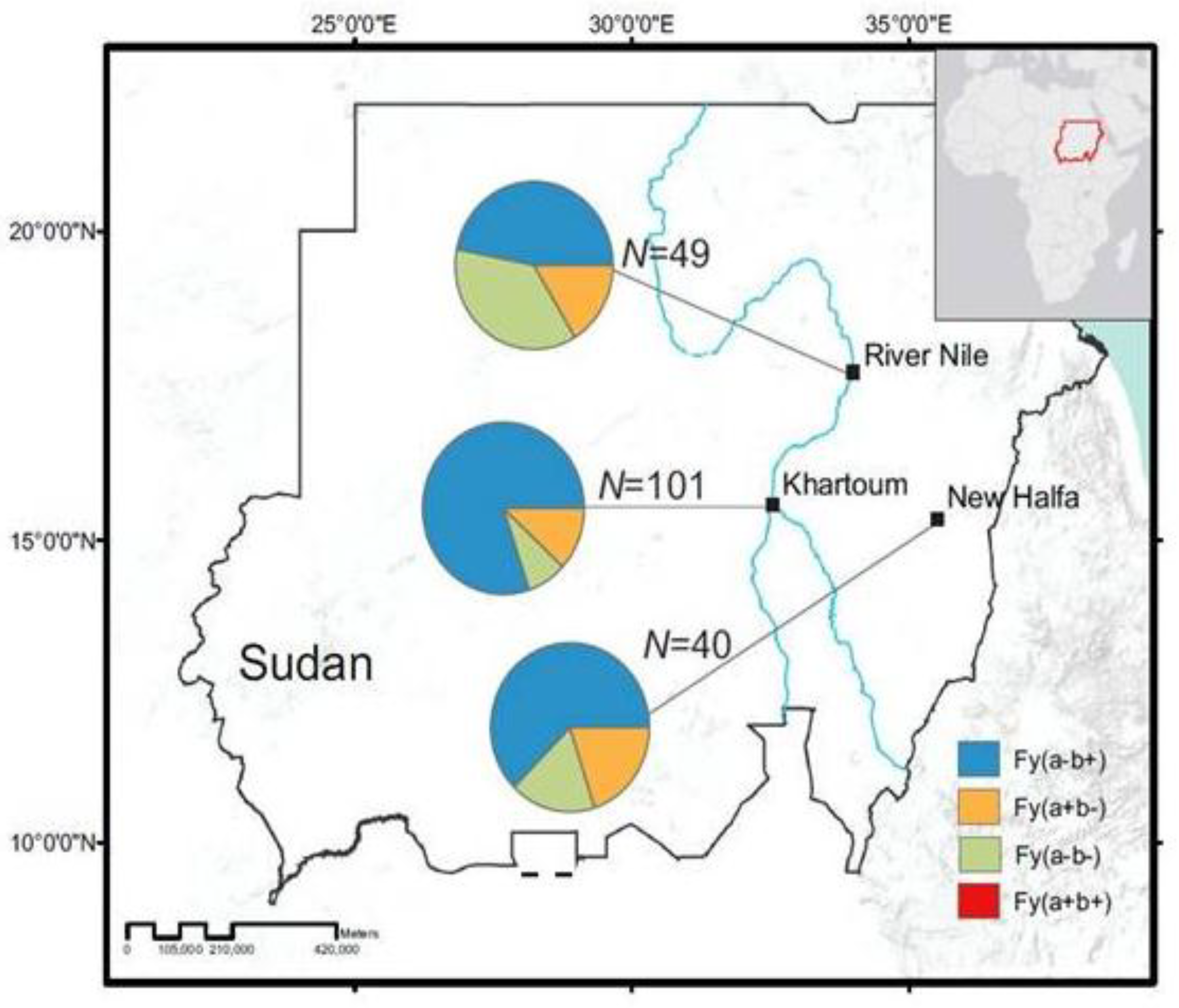
3.1. Distribution of Duffy Phenotypes among P. vivax Infections

Out of the 992 microscopy positive samples, 186 were confirmed as P. vivax mono-infection and an additional four were identified as mixed infections of P. vivax and P. falciparum based on nested PCR. We further confirmed P. vivax infections by DNA sequencing the P. vivax 18S rRNA gene in six monoclonal infected samples (accession number MF540769-MF540774) when compared with the reference P. vivax Sal strain (accession number U07367.1). The mean parasite count in the positive samples was 5453.7 parasite/µL (95% confidence interval 2896 to 8010).
Duffy genotyping was performed in 190 P. vivax positive patients (as confirmed by both microscopy and PCR), and in 67 healthy non-malaria infected individuals with matched age and gender. Among the 190 PCR-confirmed P. vivax samples (186 mono-infections and 4 mixed infections), 129 samples (67.9%) were Fy(a-b+), whereas Fy(a+b-) and Fy(a-b-) only accounted for 14.2% and 17.9%, respectively, of the samples (Table 1). No Fy(a+b+) was detected among the samples. The proportion of the three Duffy phenotypes in healthy non-malaria individuals was significantly different from that in P. vivax infected patients. Among the 67 healthy non-malaria infected individuals, 45 (67.1%) were Fy(a-b-), followed by Fy(a-b+) (29.9%). Fy(a+b-) was the least prevalent with only 3% of the samples (Table 1). The Fy(a-b+) and Fy(a+b-) phenotypes were shown to be significantly higher in the P. vivax-infected patients than the healthy individuals (p = 0.01 and p < 0.01, respectively). By contrast, a significantly lower proportion of P. vivax infection was observed in Duffy-negative Fy(a-b-) individuals (p < 0.01; Table 1).
For all three sites (New Halfa, Khartoum, and River Nile), Fy(a-b+) was the highest prevalent phenotype. In New Halfa, 25 out of 40 (62.5%) P. vivax samples were Fy(a-b+) though other phenotypes, including (Fy(a+b-) and Fy(a-b-), were also detected with a smaller proportion (Figure 1). The prevalence of infection in Khartoum and River Nile showed a similar trend, Fy(a-b+) was 80.2% and 46.9%, respectively (Figure 1, Table 1, File S1).

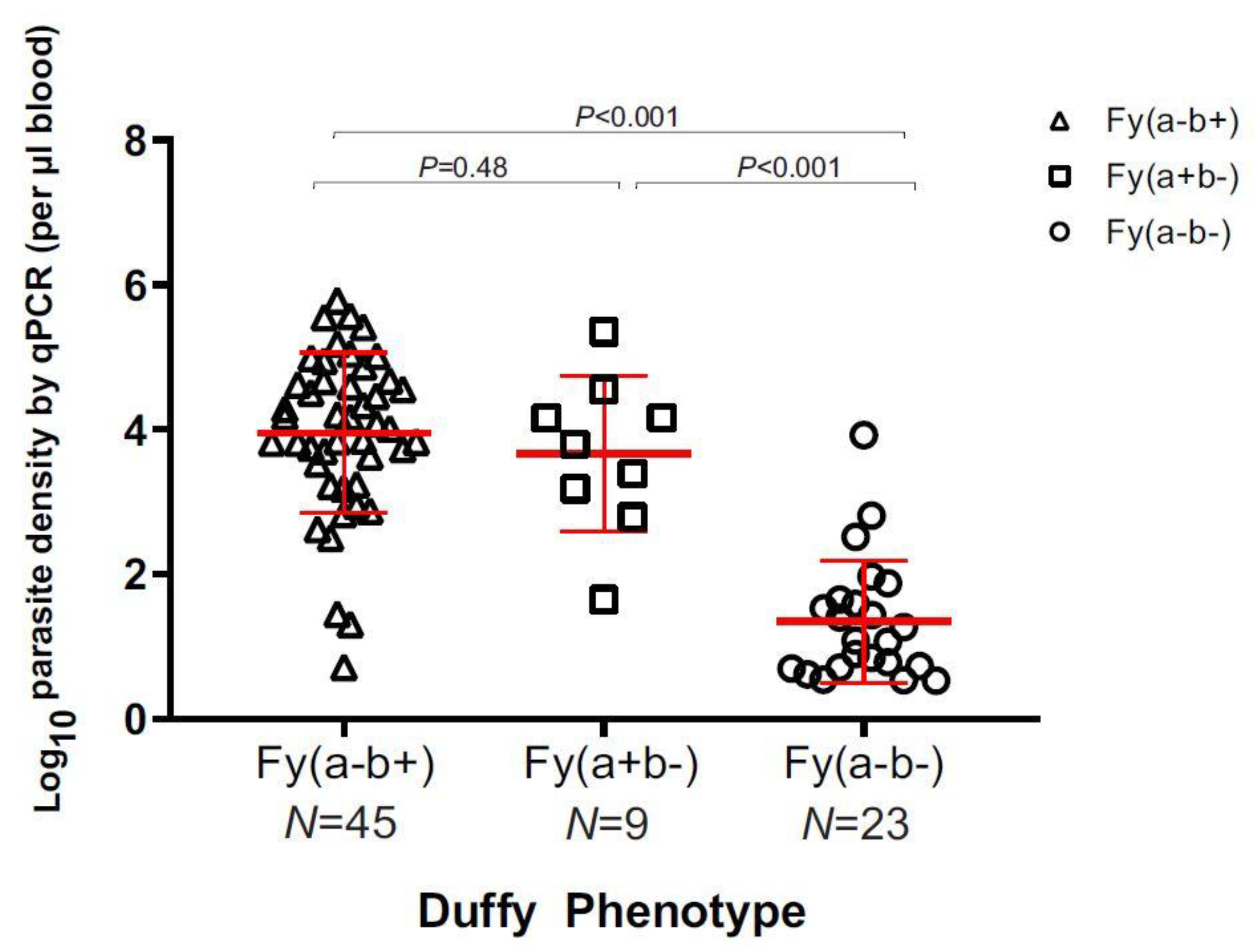
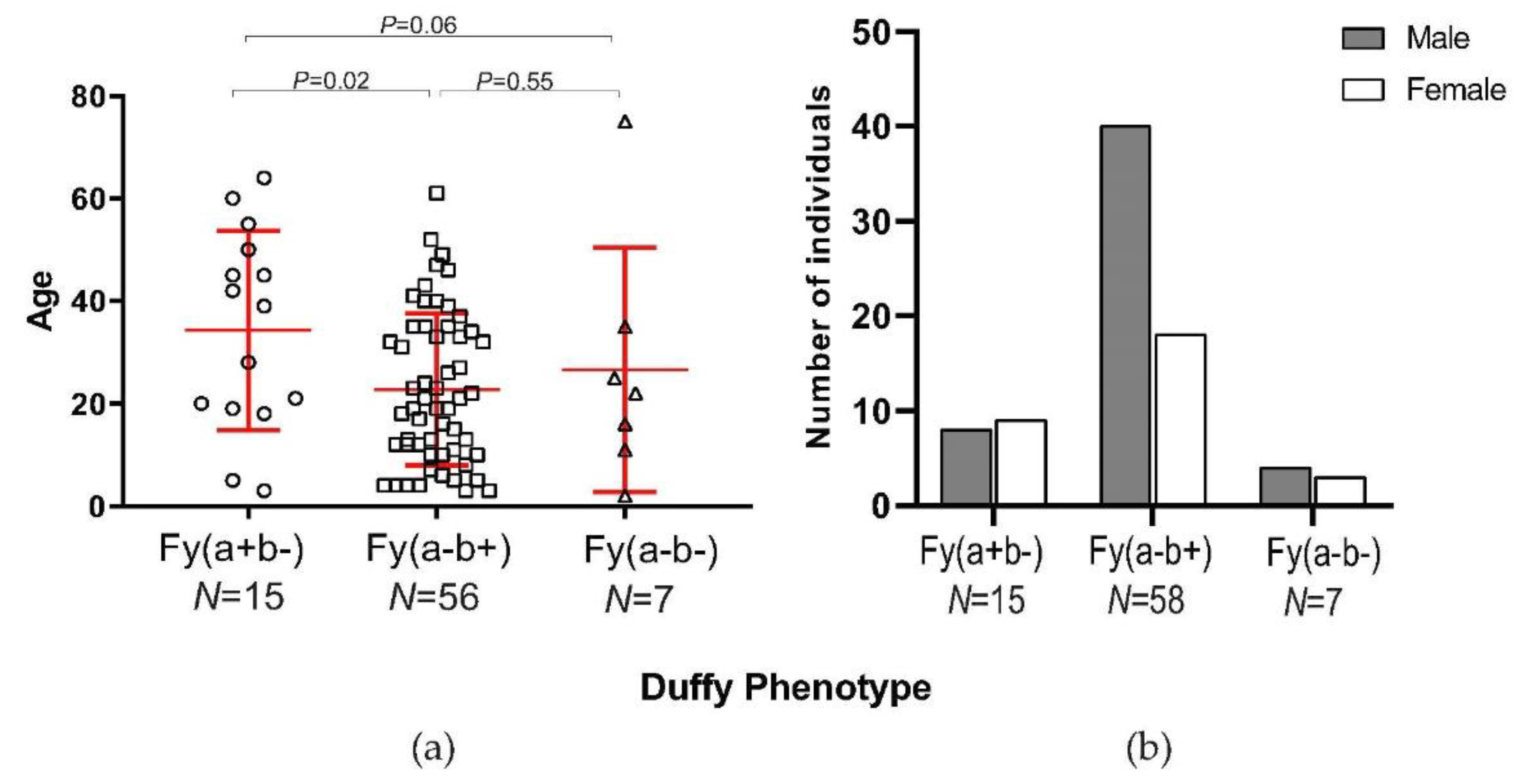
3.2. Parasite Density and Demographic Features in Duffy-Negatives

Parasite density of a subset of 77 P. vivax samples representing the three Duffy phenotypes was estimated by qPCR assay. A significant difference was observed in the parasite density among the Duffy phenotypes (Figure 2). Individuals of Fy(a-b-) had the lowest amount of P. vivax parasites (mean log parasite density = 1.35 ± 0.63 µL−1) compared to individuals of Fy(a+b-) (mean log parasite density = 3.67 ± 1.02 µL−1) and Fy(a-b+) (mean log parasite density = 3.96 ± 0.83 µL−1) phenotypes (p < 0.001; Figure 2). No significant difference was detected between the Fy(a+b-) and Fy(a-b+) individuals (p = 0.48), despite a relatively small number of Fy(a+b-) individuals.
For a subset of 77 P. vivax samples, the average age of Fy(a-b-) individuals was similar to those of Fy(a-b+), but significantly younger than those of the Fy(a+b-) phenotype (Figure 3A, File S2). Despite a small sample size, the numbers of males and females were almost similar among the Fy(a-b-) and Fy(a+b-) individuals, whereas a larger number of males than females was observed among the Fy(a-b+) individuals (Figure 3B, File S2).

4. Discussion

This study was carried out in three P. vivax malaria endemic areas (Khartoum, New Halfa, and River Nile) in Sudan where Duffy positive and Duffy-negative individuals live side-by-side [22]. While a large number of Fy(a-b+) individuals were detected among the P. vivax infections, approximately 17% of these infections were Fy(a-b-) Duffy-negatives in our study sites. In the past, individuals with the Duffy-negative phenotype (Fy(a-b-)) were thought to confer resistance to P. vivax infection [9]. However, recently, in many parts of Africa, P. vivax infection was reported in individuals with the Duffy-negative phenotype [12,15,23,24,25]. In this study, we found a relatively high percentage (17.4%; 34/190) of P. vivax-infected patients who were Duffy-negative, much higher than that reported in Madagascar, where 8.8% of Duffy-negative hosts were P. vivax positive [13]. This finding confirms our previous study in central Sudan [4] and supports the hypothesis that Duffy-negativity is not fully protective against P. vivax infection [13].
Among the three study sites, River Nile had a higher proportion of Duffy-negative individuals (38.8%; 19/49) in comparison to Khartoum (7.9%; 8/101) and New Halfa (17.5%; 7/40). It is unclear if this contrasting pattern is related to the ethnicity difference among Sudanese in the three study sites. In this study, we did not detect any unique demographic features among these the Duffy-negatives who were infected with P. vivax. The epidemiological factors related to P. vivax infections in Duffy-negatives merit further investigation.
Compared to the P. vivax infected individuals, the majority of the healthy uninfected individuals were Duffy-negative Fy(a-b-) whereas a majority of the P. vivax-infected individuals were Duffy positive Fy(a-b+). This pattern agrees with the expectation that homozygous (Fy(a-b-)) individuals are less susceptible to P. vivax infection and corroborates with the previous reports from Ethiopia [26]. The observation of a small proportion of Fy(a+b-) individuals infected with P. vivax compared to Fy(a-b+) individuals suggest that these individuals could have a relatively low susceptibility to P. vivax infection, consistent with previous observations in Brazil and Papua New Guinea [27,28], or it may be due to the low frequency of this phenotype in our population. Future investigation is needed with a larger number of Fy(a+b-) individuals to test this hypothesis.
The observation of a high P. vivax infection among homozygote Duffy-negative (Fy(a-b-)) in this study suggests that the association between P. vivax and the Duffy antigens is more complex than habitually described. Further, the observation of lower parasitemia in Duffy-negatives corroborates previous findings in Ethiopia [24], supporting the hypothesis that the infectivity of the parasite to human red blood cells is reduced in the absence of or with reduced Duffy antigen expression. The parasite could have evolved an alternative red cell invasion pathway that is independent of Duffy antigen. This hypothesis merits further experimental studies to elucidate the molecular pathogenesis of P. vivax infection in the absence of the Duffy receptor and in-depth analyses of the level of Duffy antigen expression in different Duffy phenotypes.
In summary, this study indicates a relatively high rate of P. vivax malaria in Duffy-negative (Fy(a-b-)) individuals, particularly at the River Nile site in northeastern Sudan. While our data lends support to low parasitemia in the Duffy-negative Fy(a-b-) individuals in Sudan, the high prevalence may imply a Duffy-independent invasion pathway of the parasite or change in the host susceptibility, which can in turn drastically influence P. vivax distribution. P. vivax malaria could become a new epidemic or severe disease in East Africa given that the current antimalarial vaccine is developed based on the Duffy binding protein.

Supplementary Materials

The following are available online at https://0-www-mdpi-com.brum.beds.ac.uk/2073-4425/10/6/437/s1, File S1: Duffy genotypes and phenotypes of P. vivax positive samples at each of the studied sites. File S2: Age and gender information of Duffy positive and negative samples.

Author Contributions

Conceptualization, M.M.A.H., E.L., M.M.A.A., and M.E.I.; methodology, M.M.A.A., K.P., S.A., M.E., and E.G.; software, K.P., M.M.A.A. and E.G.; validation, M.M.A.H., K.P., E.L., S.M.A., and M.E.I.; formal analysis, K.P., M.M.A.A.; investigation, M.M.A.A., M.M.A.H.; resources, M.M.A.H.; data curation, K.P., M.M.A.A., M.M.A.H.; writing—original draft preparation, M.M.A.H., S.M.A., E.L., K.P.; writing—review and editing, M.M.A.H., E.L., K.P.; visualization, M.M.A.A., K.P.; supervision, M.M.A.H., E.L., A.M.M.; project administration, M.M.A.H., E.L.; funding acquisition, M.M.A.H.

Funding

This research is partially funded by The World Academy of Sciences (TWAS) RGA No: 13-145 RG/BIO/AF/AC_G and EMRO/TDR 2014 small grant scheme No: TDR 5/4 T5/72/6.

Acknowledgments

We thank all participants of the study and health workers at respective study sites for facilitating this study, in particular Alsamuel Omer. We also thank Mohamed Salih and Mutaz Amin for their contribution in genetics analysis. Our thanks are extended to Rosalind Howes and Kyakonye Yasin who kindly accepted to proofread the manuscript.

Conflicts of Interest

The authors declare that they have no conflicts of interests

References

  1. World Health Organization. World Malaria Report 2018. Available online: https://www.who.int/malaria/publications/world_malaria_report/en/ (accessed on 19 April 2019).
  2. Douglas, N.M.; Lampah, D.A.; Kenangalem, E.; Simpson, J.A.; Poespoprodjo, J.R.; Sugiarto, P.; Anstey, N.M.; Price, R.N. Major burden of severe anemia from non-falciiparum malaria species in Southern Papua: a hospital-based surveillance study. PLoS Med. 2013, 10, e1001575. [Google Scholar] [CrossRef] [PubMed]
  3. Twohig, K.A.; Pfeffer, D.A.; Baird, J.K.; Price, R.N.; Zimmerman, P.A.; Hay, S.I.; Gething, P.W.; Battle, K.E.; Howes, R.E. Growing evidence of Plasmodium vivax across malaria-endemic Africa. PLoS Negl. Trop. Dis. 2019, 13. [Google Scholar] [CrossRef] [PubMed]
  4. Abdelraheem, M.H.; Albsheer, M.M.A.; Mohamed, H.S.; Amin, M.; Abdel, H.M.M. Transmission of Plasmodium vivax in Duffy-negative individuals in central Sudan. Trans.R.Soc.Trop.Med.Hyg. 2016, 110, 258–260. [Google Scholar] [CrossRef] [PubMed]
  5. Suliman, M.M.A.; Hamad, B.M.; Albasheer, M.M.A.; Elhadi, M.; Mustafa, M.A.; Elobied, M.; Hamid, M.M.A. Molecular evidence of high proportion of Plasmodium vivax malaria infection in White Nile area in Sudan. J. Parasitol. Res. 2016, 2016. [Google Scholar] [CrossRef] [PubMed]
  6. Chaudhuri, A.; Polyakova, J.; Zbrzezna, V.; Williams, K.; Gulati, S.; Pogo, A.O. Cloning of glycoprotein D cDNA, which encodes the major subunit of the Duffy blood group system and the receptor for the Plasmodium vivax malaria parasite. Proc. Natl. Acad. Sci. USA 1993, 90, 10793–10797. [Google Scholar] [CrossRef] [PubMed]
  7. Miller, L.H.; Mason, S.J.; Clyde, D.F.; McGinniss, M.H. The Resistance Factor to Plasmodium vivax in Blacks. N. Engl. J. Med. 1976, 295, 302–304. [Google Scholar] [CrossRef]
  8. Chitnis, C.E.; Sharma, A. Targeting the Plasmodium vivax Duffy-binding protein. Trends Parasitol. 2008, 24, 29–34. [Google Scholar] [CrossRef]
  9. King, C.L.; Adams, J.H.; Zianli, J.; Grimberg, B.T.; McHenry, A.M.; Greenberg, L.J.; Siddiqui, A.; Howes, R.E.; da Silva-Nunes, M.; Ferreira, M.U.; et al. Fya/Fyb antigen polymorphism in human erythrocyte Duffy antigen affects susceptibility to Plasmodium vivax malaria. Proc. Natl. Acad. Sci. USA 2011, 108. [Google Scholar] [CrossRef]
  10. De Silva, J.R.; Lau, Y.L.; Fong, M.Y. Genotyping of the Duffy Blood Group among Plasmodium knowlesi-Infected Patients in Malaysia. PLoS ONE 2014, 9, e108951. [Google Scholar] [CrossRef]
  11. Tournamille, C.; Colin, Y.; Cartron, J.P.; Le Van Kim, C. Disruption of a GATA motif in the Duffy gene promoter abolishes erythroid gene expression in Duffy–negative individuals. Nat. Genet. 1995, 10, 224–228. [Google Scholar] [CrossRef]
  12. Zimmerman, P.A. Plasmodium vivax infection in Duffy-Negative People in Africa. Am. J. Trop. Med. Hyg. 2017, 97. [Google Scholar] [CrossRef]
  13. Ménard, D.; Barnadas, C.; Bouchier, C.; Henry-Halldin, C.; Gray, L.R.; Ratsimbasoa, A.; Thonier, V.; Carod, J.F.; Domarle, O.; Colin, Y.; et al. Plasmodium vivax clinical malaria is commonly observed in Duffy-negative Malagasy people. Proc. Natl. Acad. Sci. USA 2010, 107, 5967–5971. [Google Scholar] [CrossRef] [PubMed]
  14. Wurtz, N.; Lekweiry, K.M.; Bogreau, H.; Pradines, B.; Rogier, C.; Boukhary, A.O.M.S.; Hafid, J.E.; Salem, M.S.O.A.; Trape, J.F.; Basco, L.K.; et al. Vivax malaria in Mauritania includes infection of a Duffy-negative individual. Malar. J. 2011, 10, 336. [Google Scholar] [CrossRef] [PubMed]
  15. Woldearegai, T.G.; Kremsner, P.G.; Kun, J.F.; Mordmüller, B. Plasmodium vivax malaria in duffy-negative individuals from Ethiopia. Trans. R. Soc. Trop. Med. Hyg. 2013, 107, 328–331. [Google Scholar] [CrossRef] [PubMed]
  16. Russo, G.; Faggioni, G.; Paganotti, G.M.; Dongho, G.B.D.; Pomponi, A.; De Santis, R.; Tebano, G.; Mbida, M.; Sobze, M.S.; Vullo, V.; et al. Molecular evidence of Plasmodium vivax infection in Duffy negative symptomatic individuals from Dschang, West Cameroon. Malar. J. 2017, 16, 74. [Google Scholar] [CrossRef] [PubMed]
  17. Aboud, M.; Makhawi, A.; Verardi, A.; El Raba’a, F.; Elnaiem, D.-E.; Townson, H. A genotypically distinct, melanic variant of Anopheles arabiensis in Sudan is associated with arid environments. Malar. J. 2014, 13, 492. [Google Scholar] [CrossRef] [PubMed]
  18. Snounou, G.; Viriyakosol, S.; Zhu, X.P.; Jarra, W.; Pinheiro, L.; do Rosario, V.E.; Thaithong, S.; Brown, K.N. High sensitivity of detection of human malaria parasites by the use of nested polymerase chain reaction. Mol.Biochem.Parasitol. 1993, 61, 315–320. [Google Scholar] [CrossRef]
  19. Baum, E.; Sattabongkot, J.; Sirichaisinthop, J.; Kiattibutr, K.; Davies, D.H.; Jain, A.; Lo, E.; Lee, M.C.; Randall, A.Z.; Molina, D.M.; et al. Submicroscopic and asymptomatic Plasmodium falciparum and Plasmodium vivax infections are common in western Thailand—Molecular and serological evidence. Malar. J. 2015, 14, 95. [Google Scholar] [CrossRef]
  20. FinchTV 1.4. Available online: https://finchtv.software.informer.com/1.4/ (accessed on 7 June 2019).
  21. NCBI Basic Local Alignment Search Tool. Available online: https://blast.ncbi.nlm.nih.gov/ (accessed on 7 June 2019).
  22. Kempinska-Podhorodecka, A.; Knap, O.; Drozd, A.; Kaczmarczyk, M.; Parafiniuk, M.; Parczewski, M.; Ciechanowicz, A. Analysis for genotyping Duffy blood group in inhabitants of Sudan, the Fourth Cataract of the Nile. Malar. J. 2012, 11, 115. [Google Scholar] [CrossRef]
  23. Cavasini, C.E.; de Mattos, L.C.; Couto, Á.A.D.; Bonini-Domingos, C.R.; Valencia, S.H.; Neiras, W.C.; Alves, R.T.; Rossit, A.R.; Castilho, L.; Machado, R.L. Plasmodium vivax infection among Duffy antigen-negative individuals from the Brazilian Amazon region: An exception? Trans. R. Soc. Trop. Med. Hyg. 2007, 101, 1042–1044. [Google Scholar] [CrossRef]
  24. Mendes, C.; Dias, F.; Figueiredo, J.; Mora, V.G.; Cano, J.; de Sousa, B.; do Rosário, V.E.; Benito, A.; Berzosa, P.; Arez, A.P. Duffy negative antigen is no longer a barrier to Plasmodium vivax—Molecular evidences from the African West Coast (Angola and Equatorial Guinea). PLoS Negl. Trop. Dis. 2011, 5. [Google Scholar] [CrossRef] [PubMed]
  25. Ryan, J.R.; Stoute, J.A.; Amon, J.; Dunton, R.F.; Mtalib, R.; Koros, J.; Owour, B.; Luckhart, S.; Wirtz, R.A.; Barnwell, J.W.; et al. Evidence for transmission of Plasmodium vivax among a Duffy antigen negative population in Western Kenya. Am. J. Trop. Med. Hyg. 2006, 27, 575–581. [Google Scholar] [CrossRef]
  26. Lo, E.; Yewhalaw, D.; Zhong, D.; Zemene, E.; Degefa, T.; Tushune, K.; Ha, M.; Lee, M.C.; James, A.A.; Yan, G. Molecular epidemiology of Plasmodium vivax and Plasmodium falciparum malaria among Duffy-positive and Duffy-negative populations in Ethiopia. Malar. J. 2015, 14, 84. [Google Scholar] [CrossRef] [PubMed]
  27. Kasehagen, L.J.; Mueller, I.; Kiniboro, B.; Bockarie, M.J.; Reeder, J.C.; Kazura, J.W.; Kastens, W.; McNamara, D.T.; King, C.H.; Whalen, C.C.; et al. Reduced plasmodium vivax Erythrocyte infection in PNG Duffy-negative heterozygotes. PloS ONE 2007, 2. [Google Scholar] [CrossRef] [PubMed]
  28. Weber, S.S.; Tadei, W.P.; Martins, A.S. Association between Duffy blood group system variants and susceptibility and resistance to malaria in the Brazilian Amazon. Rev. Biol. Neotrop. 2011, 7, 39–44. [Google Scholar] [CrossRef]
Figure 1. Proportion of Duffy phenotypes in Plasmodium positive patients among the three sites in River Nile, Khartoum, and New Halfa. The proportion of Fy(a-b-) was significantly higher in River Nile than the other two sites. In addition, Fy(a+b+) was not recorded in any of the healthy or Plasmodium-positive patients.
Genes 10 00437 g001
Figure 2. Estimated log parasite density per μL of blood based on Duffy phenotype. There was no significant difference between Fy(a-b+) and Fy(a+b-) phenotypes, but there was a significant difference between the Fy(a-b-) phenotype and the Fy(a-b+) and the Fy(a+b-) phenotypes (p < 0.001).
Genes 10 00437 g002
Figure 3. (a) Distribution of age of patient in years among the different phenotypes in Plasmodium positive patients; (b) proportion of male and female Plasmodium-positive patients between the different Duffy phenotypes. It is noted that there were two patient samples that we did not have their age data for. Thus, the sample size in Fy(a-b+) was different.
Genes 10 00437 g003
Table 1. Proportion of the three Duffy phenotypes in P. vivax positive and negative healthy patients
Duffy PhenotypeNPositive
(N = 190)
Negative
(N = 67)
p1OR
(95%CI)2
Fy(a+b-)2927 (14.2%)2 (3%)0.01220.1858
Fy(a-b+)149129 (67.9%)20 (29.9%)0.00010.2012
Fy(a-b-)7734 (17.9%)45 (67.1%)0.00011.9567
1p-values were calculated by Fisher’s exact test at a significance level of 0.05.* Significant difference between the two groups after correction for multiple testing.2 Odd’s ratio with a 95% confidence interval.

