Article

Diversity of Bacterial Communities of Fitness Center Surfaces in a U.S. Metropolitan Area

1
Division of Epidemiology, Biostatistics, and Environmental Health, School of Public Health, The University of Memphis, 338 Robison Hall, 3825 Desoto Avenue, Memphis, TN 38152, USA
2
Molecular Research LP (MR DNA), 503 Clovis Road, Shallowater, TX 79363, USA
3
WMC TV Action News 5, NBC Memphis, 1960 Union Ave, Memphis, TN 38104, USA
*
Author to whom correspondence should be addressed.
Int. J. Environ. Res. Public Health 2014, 11(12), 12544-12561; https://0-doi-org.brum.beds.ac.uk/10.3390/ijerph111212544
Submission received: 16 October 2014 / Revised: 26 November 2014 / Accepted: 26 November 2014 / Published: 3 December 2014
(This article belongs to the Special Issue Environmental Determinants of Infectious Disease Transmission)

Abstract

:
Public fitness centers and exercise facilities have been implicated as possible sources for transmitting community-acquired bacterial infections. However, the overall diversity of the bacterial community residing on the surfaces in these indoor environments is still unknown. In this study, we investigated the overall bacterial ecology of selected fitness centers in a metropolitan area (Memphis, TN, USA) utilizing culture-independent pyrosequencing of the 16S rRNA genes. Samples were collected from the skin-contact surfaces (e.g., exercise instruments, floor mats, handrails, etc.) within fitness centers. Taxonomical composition revealed the abundance of Firmicutes phyla, followed by Proteobacter and Actinobacteria, with a total of 17 bacterial families and 25 bacterial genera. Most of these bacterial genera are of human and environmental origin (including, air, dust, soil, and water). Additionally, we found the presence of some pathogenic or potential pathogenic bacterial genera including Salmonella, Staphylococcus, Klebsiella, and Micrococcus. Staphylococcus was found to be the most prevalent genus. Presence of viable forms of these pathogens elevates risk of exposure of any susceptible individuals. Several factors (including personal hygiene, surface cleaning and disinfection schedules of the facilities) may be the reasons for the rich bacterial diversity found in this study. The current finding underscores the need to increase public awareness on the importance of personal hygiene and sanitation for public gym users.

Graphical Abstract

1. Introduction

As we aspire to stay fit and healthy, many of us regularly visit fitness centers or “gyms”. In fact, the data from the International Health, Racquet & Sportsclub Association (IHRSA) indicates a surge in the number of people visiting fitness centers in the last five years [1]. In the year 2012, more than 58 million persons used health clubs in the U.S., in other words, one out of five (20%) Americans of ages 6 years and older are using health club facilities [2]. However, there is a lack of knowledge about the diversity of microbial communities at fitness centers. It is important to note that the overall microbial load and diversity of the environment are often implicated as a critical indicator of hygiene and cleanliness [3,4,5]. Several previous studies focusing on environmental hygiene and sanitation (e.g., in food production or health care settings) had found a direct relationship between microbial load in the surrounding environment and the risk of pathogen transmission [4,6,7]. Therefore, an understanding of overall bacterial population and diversity in gymnasiums and athletic facilities would obviously shed light on the risk of the pathogen propagation from these facilities. Most of the studies reported to date from gymnasiums, playgrounds, athletic facilities, or venues where individuals come in contact with others are focused on the transmission of staphylococci, in particular on methicillin-resistant Staphylococcus aureus (MRSA) [8,9,10,11,12,13,14]. It is also important to note that most of these studies relied on culture-based techniques [15]. Because a large number of microorganisms are difficult to culture [16], thus, the overall microbial diversity associated with fitness center environments remains largely unknown. The recent advancement of high-throughput sequencing techniques and related bioinformatics tools made it possible to envisage the microbial communities that inhabit humans and surroundings in great detail. By enabling identification of both “cultivable” and “non-cultivable” microbial populations, this culture independent method provides a vivid realization of the relationship among humans, microbes, and the environment. For example, microbial communities of several indoor environments, including hospitals [17], office buildings [18], kitchen [19], public restrooms [20], showers [21], have been documented. In most of the previous findings, human skin was found to be the main source of bacteria in each of the above-mentioned locations. However, other attributes such as soil and outdoor air (by air conditioning), dust from human shoes, etc. could also serve as potential sources of harboring and transmission of various microorganisms in indoor environments. Pathogenic microorganisms can survive on inanimate surfaces for prolonged periods of time as reported in several previous studies [22,23,24,25]. These pathogens can readily be transferred from surfaces to the human body through the touch of hands or other body parts. Carpets, yoga mat, clothes, equipment handles, etc. may serve as excellent living places for bacteria. The bacterial communities found on different surfaces are reported to be distinct [19]. Moreover, some studies delineated the role of environmental surfaces in the transmission of bacteria [26,27,28]. For instance, surfaces in public places namely, computers, telephones, telephone mouthpieces, headsets, desks, automated teller machines (ATM), cash machines, elevator buttons, are all reported as potential sources for transmitting infectious microorganisms [26,29,30,31,32].
Compared to other indoor environments, it is interesting to note that fitness centers offer a unique setting to explore microbial diversity. This can be attributed to the physical activities with high frequency of surface touch by individuals with different personal hygienic practices. Such factors are likely to have strong influences on the types of bacteria observed on fitness center surfaces. However, there is a lack of information on the microbial ecology of fitness centers in terms of the potential danger to the environment and public health. In the current study, we explored the overall bacterial ecology of selected fitness centers in a metropolitan area (Memphis, TN, USA) utilizing culture-independent pyrosequencing of the 16S rRNA genes. Our goal was to assess and comprehensively understand the microbial diversity associated with fitness center surfaces; and to determine if different surfaces of fitness centers (e.g., exercise instruments, floor mats, handrails, etc.) serve as potential reservoirs for different bacterial communities.

2. Experimental Section

2.1. Sample Collection

For this study, surface swab samples were collected from four membership-based fitness centers around the Memphis metropolitan area in Tennessee. Two out of the four fitness centers are open 24 h/7-days a week. Samples were collected by trained volunteers from the skin-contact surfaces on exercise equipment (nautilus machine, treadmill, stationary bike, power strider, elliptical machine, and leg press), dumbbell, toilet handles, and handrails on stairs of the fitness centers during October 2013. The samples were obtained from certain places that had not been sanitized before sample collection. Cotton-tipped swabs (SanicultTM, Thermo Remel/ Starplex Scientific Inc., Etobicoke, ON, Canada) were used to wipe the surface (approximately 10 cm2) by rotating over 4–5 times (~10 s). While swabbing on equipment surfaces with different shapes, appropriate care was taken to cover approximately the same surface area. After swabbing, the swab sticks were immediately placed back into the tube containing sterile diluent solution and the samples were transported in a refrigerated container to the laboratory within four hours for analysis.

2.2. DNA Extraction and Pyrosequencing

Genomic DNA was extracted from the swab samples using the DNA extraction kit (MO BIO Laboratories, Carlsbad, CA, USA), following the manufacturer’s protocol for isolation of DNA from microbial cultures. The extracted genomic DNA samples were quantified spectrophotometrically using a NanoDrop spectrophotometer (Thermo Scientific, Wilmington, DE, USA). The DNA samples were pooled based on the types of equipment and surfaces and subjected for pyrosequencing. The details of pooling swab samples is described in Table A1 (Appendix Material).
In this study, we performed bTEFAP® (MR DNA www.mrdnalab.com) which was originally described by Dowd et al. [33,34] and that has been utilized to describe a wide range of environmental- and health-related microbiomes [33,34,35]. To evaluate the microbial ecology of samples, 16S universal Eubacterial primers 27Fmod (AGRGTTTGATCMTGGCTCAG) and 519Rmod (GTNTTACNGCGG CKGCTG) were utilized on the Illumina MiSeq v3 2 × 300 bp sequencing platform (Illumina, San Diego, CA, USA). A single-step 30 cycle PCR using HotStarTaq Plus Master Mix Kit (Qiagen, Valencia, CA, USA) were used under the following conditions: 94 °C for 3 min, followed by 28 cycles of 94 °C for 30 s; 53 °C for 40 s and 72 °C for 1 min; after which a final elongation step at 72 °C for 5 min was performed. After the completion of PCR, all amplicon products from different samples were mixed in equal concentrations and purified using Agencourt Ampure beads (Agencourt Bioscience Corporation, Beverly, MA, USA).

2.3. Computational and Statistical Analyses

The Q25 sequence data derived from the sequencing process was processed using a proprietary analysis pipeline (www.mrdnalab.com, MR DNA, Shallowater, TX, USA). Sequences were depleted of barcodes and primers with short sequences (<200 bp), sequences with ambiguous base calls, and sequences with homopolymer runs exceeding 6 bp were removed. Sequences are then denoised and chimeras removed. Operational Taxonomic Units (OTUs) were defined after the removal of singleton sequences, clustering at 3% divergence (97% similarity). [33,34,36,37,38]. OTUs were then taxonomically classified using BLASTn against a curated GreenGenes/RDP/NCBI derived database [39] and compiled into each taxonomic level into both “counts” and “percentage” files. Counts files contain the actual number of sequences while the percent files contain the relative (proportion) percentage of sequences within each sample that map to the designated taxonomic classification. For example, if there are 1000 sequences and 100 of the sequences are classified as Staphylococcus then it has been represented as Staphylococcus being 10% of the total population. Statistical analysis in this study was performed using a variety of computer packages including XLstat, NCSS 2007, “R” and NCSS 2010. Alpha and beta diversity analysis was conducted as described previously [33,34,36,37,38] using QIIME (Quantitative Insights Into Microbial Ecology). Significance reported for any analysis is defined as p < 0.05.

3. Results

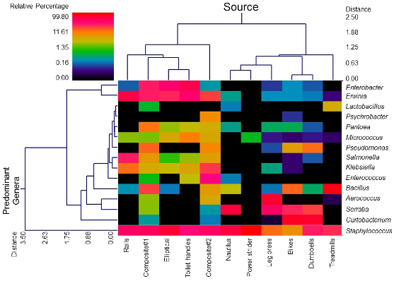
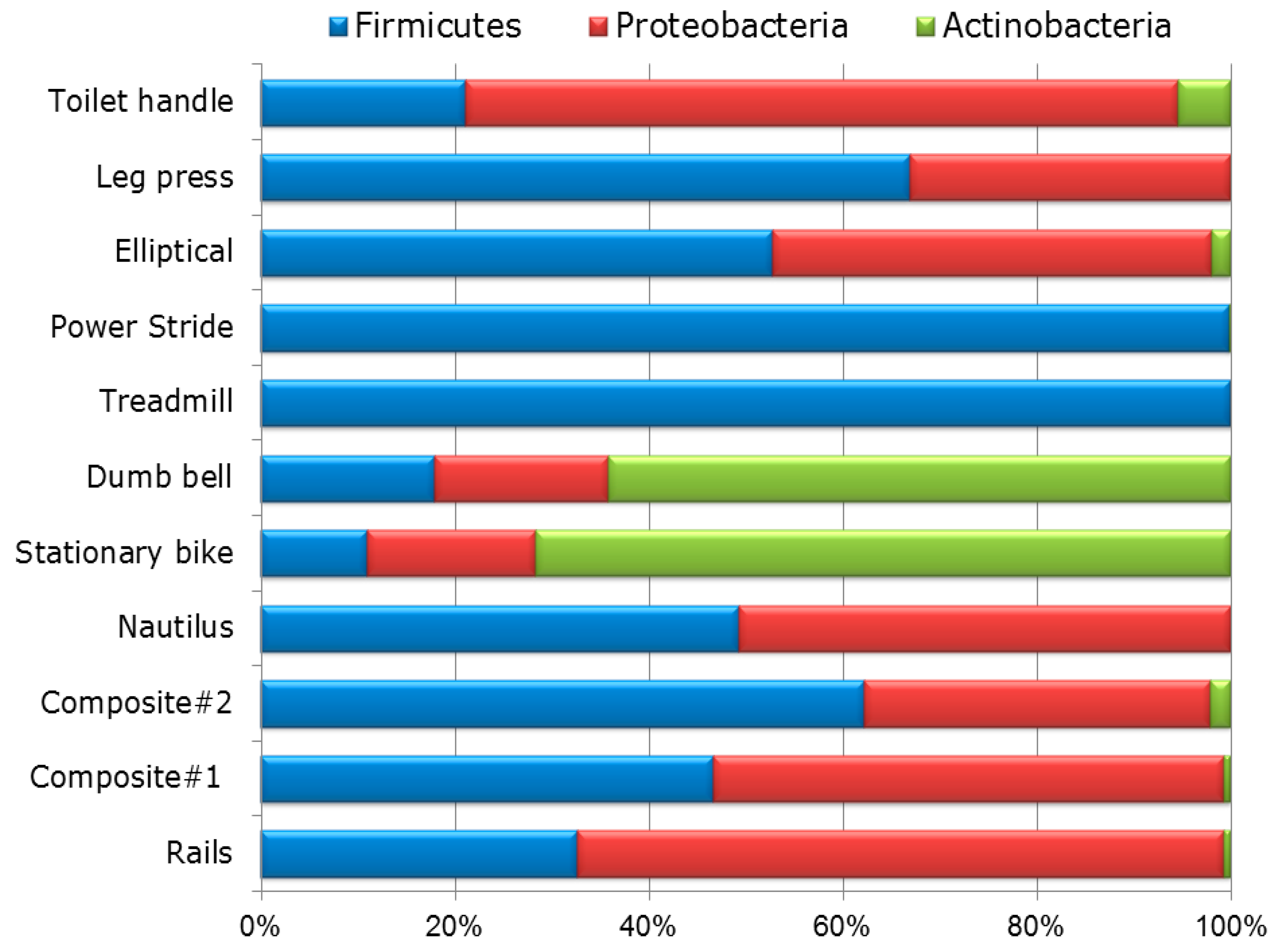
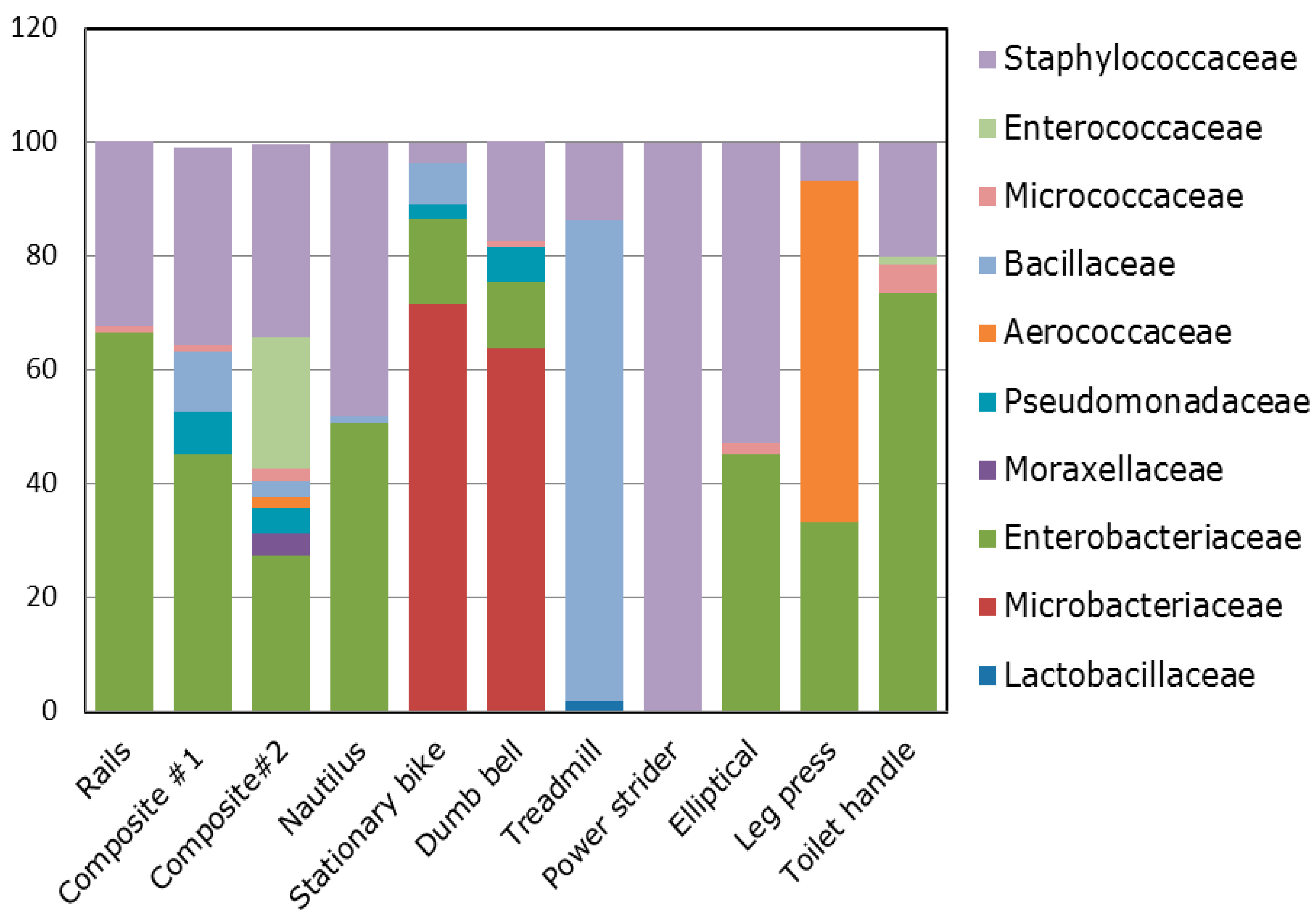
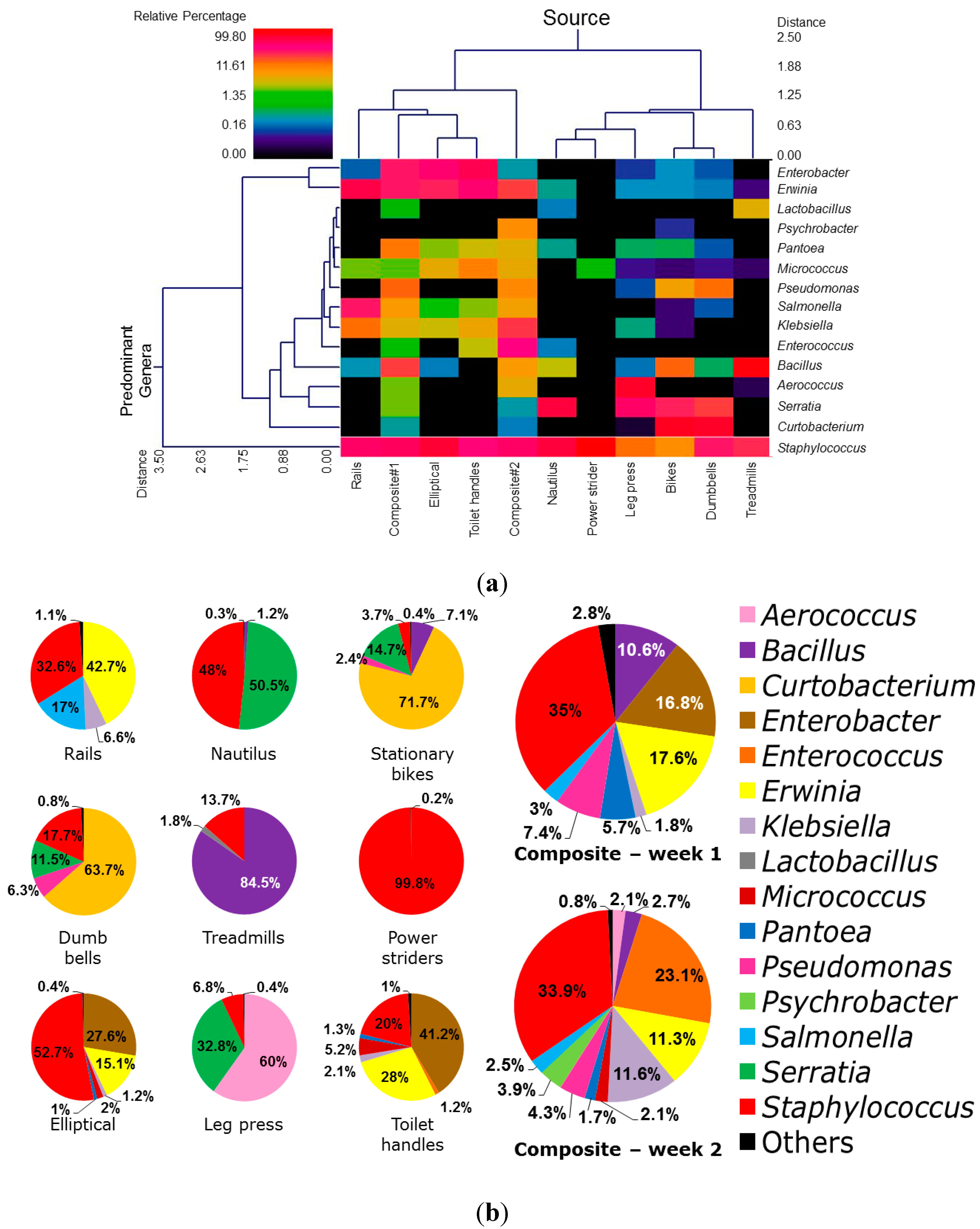
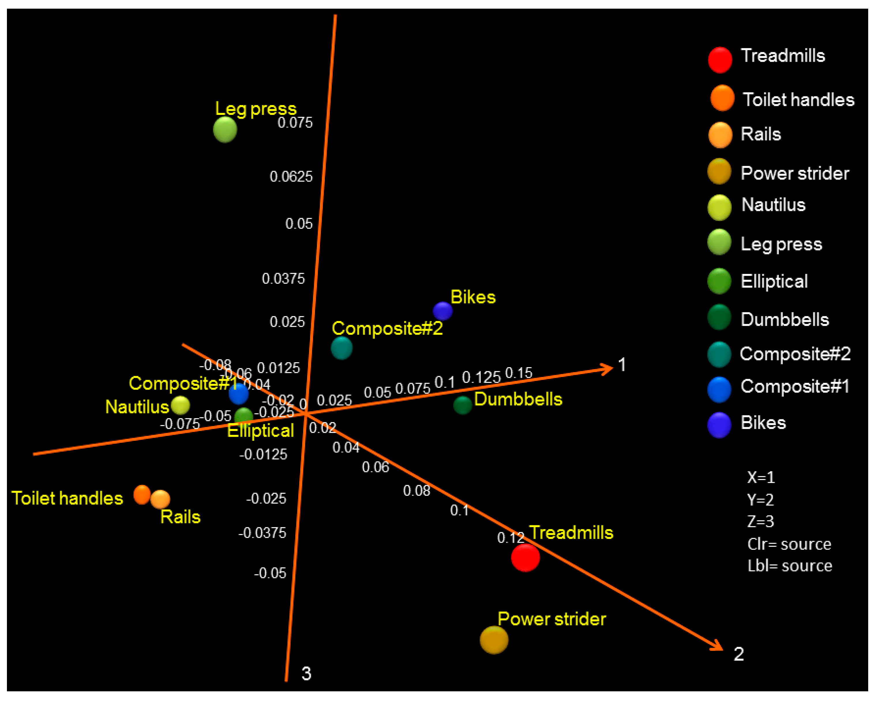
3.1. Diversity and Relative Abundance of Bacterial Genera in Surface Swab Sample of Fitness Center

DNA extraction from all surface swab samples contained measurable quantities of microbial DNA (48–152 ng/µL); negative controls (no cells) had no quantifiable DNA. After stringent quality sequence curation, a total of 122,454 sequences were generated by the bTEFAP® of DNAs from thirty two surface samples (swabs). A total of 111,663 sequences were utilized for the analyses of the eleven sample groups (which were based on the equipment types sampled, including the pooled samples from week 1 and 2). The details of pooling samples and clustering into groups have been described in Table A1.
Taxonomical composition indicated that the Firmicutes phyla was most common, followed by Proteobacteria and Actinobacteria phyla (Figure 1) with a total of 25 identified genera and 63 species across all surface swab samples. Within these dominant phyla, bacterial family associated with mostly human normal flora and environmental bacteria such as Staphylococcaceae, Enterobacteriaceae, Microbacteriaceae, Bacillaceae, Aerococcaceae, and Pseudomonadaceae have been found abundantly in all surface swab samples (Figure 2).
Figure 1. Relative abundance of bacterial diversity of different surfaces of gym equipment at phylum level as determined by bTEFAP®. Bacterial diversity varied between equipment or surface types. Firmicutes are found to the predominant phyla, followed by Proteobacteria and Actinobacteria phyla The least bacterial diversity at phylum level was found in surfaces of power striders and treadmills which consisted mostly of Firmicutes (>99%).
Ijerph 11 12544 g001
The relative abundance of bacterial diversity in genus level is presented in Figure 3. A high abundance of Staphylococcus spp. was observed in most of the samples. Some other bacterial species such as, Bacillus, Serratia, Aerococcus, Erwinia, and Enterobacter spp. were observed predominantly on treadmills, nautilus machines, leg press, rails (handrail on stairs), elliptical, and toilet handles, respectively. Some previously unreported bacterial genera associated with the surfaces of equipment in the fitness center have been identified in this study, namely Klebsiella, Lactobacillus, Salmonella, Curtobacterium, Pantoea, Psychrobacter, Serratia, Bacillus, Pseudomonas, Micrococcus, Enterococcus, Erwinia, and Aerococcus. The most prevalent bacterial species identified across all the samples observed were staphylococci, including S. aureus, S. epidermidis, and S. saprophyticus. In this study, Staphylococcus were found predominantly in power striders (99.8%), followed by elliptical machines (52.7%), and nautilus machines (48%), rails (32.6%), toilet handles (20%), dumb bells (17.7%), treadmills (13.6%), leg presses (6.8%), stationary bikes (3.7%), composite samples of week 1 (34.7%) and composite samples of week 2 (33.9%). Interestingly, S. aureus and S. epidermidis were found to be present on all the surfaces tested.
A total of 25 bacterial genera were detected in all swabs sampled in this study. The predominant bacterial genera in the surface swab samples analyzed based on the relative abundance cutoff of 1.0% are shown in Figure 3b. The major bacterial genera include Pseudomonas, Pantoea, Micrococcus, Staphylococcus, Enterobacter, Klebsiella, and Bacillus (Figure 3). In addition, our results indicate that other prevalent bacterial genus such as Erwinia spp. were found on rails (42.7%) and in the composite samples from week 1 (17.6%). Enterococcus was prevalently found in the composite samples from week 2 (23.1%), whereas Serratia spp. was abundant on nautilus machines (50.5%). Curtobacterium spp. were observed on stationary bikes and dumb bells with an abundance of 71.7% and 63.7%, respectively. The air-borne bacteria Aerococcus spp. was prevalent on leg press machines (60%). Among the swab samples from treadmills, Bacillus spp. was the most abundant (84.5%) genus, whereas Enterobacter spp. was observed in elliptical machines and toilet handles with the relative abundance of 27.6% and 41.2%, respectively.
Figure 2. Relative abundance of bacterial families of different surfaces of gym equipment as determined by bTEFAP®. Multi-colored stack bar graphs represent the relative abundance of bacterial family in each sample.
Ijerph 11 12544 g002
Figure 3. (a) Dual hierarchical clustering dendogram of the bacterial genera profiles for fitness center equipment and items. Samples with more similar microbial populations are clustered closer together. The top ~15 most abundant genera (average across all samples) are used for clustering. The heatmap represents the relative percentages of each bacterial genus. The predominant genera are represented along the right Y-axis, while each sample (surface swabs from equipment and items) is clustered on the X-axis. The legend for the heatmap is provided in the upper left corner. (b) Relative abundance of bacterial diversity at the genus level in the surface swabs of gym equipment. Bacterial genus abundance less than 1% were grouped as “Others”.
Ijerph 11 12544 g003
Samples were rarefied to 2000 sequences for alpha and beta diversity and bootstrapping analyses. The indices of bacterial diversity were estimated using a Rarefaction Curve (Figure 4) based on OTUs. Rarefaction Curve modeling indicated 97% similarity of OTUs at the 3% divergence was attained for each sample [33,34,36,37,38] suggesting adequate depth of coverage. By rarefaction analysis estimates (Figure 4), the trend for species richness in different equipment surfaces was found to be (high to low): stationary bikes > toilet handles > dumb bells > elliptical machines > leg presses > treadmills > nautilus machines > stair rails > power striders. As expected, the composite samples showed the highest bacterial species diversity since they were pooled DNA of all samples for a specific sampling week.
Figure 4. Observed taxonomic units analysis (Rarefaction Curve) of bacterial species diversity of surface swab samples in fitness centers. This curves show that as might be expected the composite samples from weeks 1 and 2 have the highest observed species while power Striders had the lowest observed species values.
Ijerph 11 12544 g004
Using weighted Principal Coordinates Analysis (PCoA) of the microbiome of each sample based upon UniFrac method, we see that the samples most distant from the central cluster are leg press, treadmills, and power striders (Figure 5). Toilet handles and rails to the stairs cluster together, bikes and dumbbells cluster near to each other and the nautilus and elliptical machines group together.

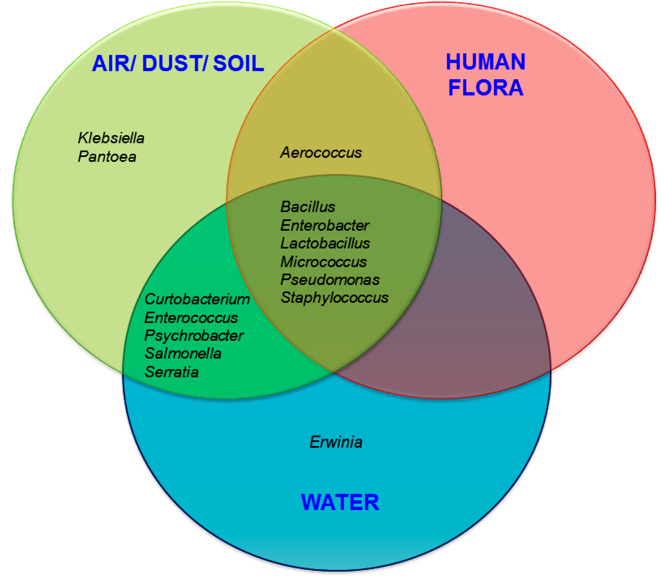
3.2. Probable Source of Bacteria in Surface Swab Sample of Fitness Center

The tentative source/habitat of the bacteria has been shown in Figure 6. The probable environmental sources/habitats of bacteria were classified based on previous reports/literature [20,40,41]. It is evident from our results that bacterial genera commonly associated with air/soil/dust-borne transmissions contribute to a relatively high proportion of the bacterial community in the fitness centers. They include Aerococcus, Bacillus, Curtobacterium, Paenibacillus, Pantoea, Pseudomonas, Psychrobacter, and Serratia.
Figure 5. PCoA analysis of the microbiome of each equipment and items swabbed based upon UniFrac method. Different colored symbols are indicative of the various gym equipment and items. Symbols that are closer together represent similar surface bacterial communities.
Ijerph 11 12544 g005
Human associated bacteria (human flora) were found in this study are Aerococcus, Bacillus, Enterobacter, Lactobacillus, Micrococcus, Pseudomonas, and Staphylococcus [42]. Bacterial genera such as Aerococcus, Bacillus, Enterobacter, Lactobacillus, Micrococcus, Pseudomonas, and Staphylococcus share their habitat in three clusters as depicted in Figure 6. Additionally, many of these genera, namely, Aerococcus, Bacillus, Curtobacterium, Enterobacter, Enterococcus, Lactobacillus, Micrococcus, Pseudomonas, Psychrobacter, Salmonella, Serratia, and Staphylococcus have been reported previously from both air/dust/soil and water [20,40,41].
Notably, the presence of some pathogenic (or potentially pathogenic) bacterial genera were observed in relatively low abundance, including Bacillus in composite samples from week 2 (2.7%), nautilus machine (1.2%), stationary bike (7.1%); Enterococcus (1.2%) on the toilet handle; Klebsiella on rails (6.6%), composite samples from week 1 (1.8%), elliptical machines (1.2%), toilet handle (2.1%); Pantoea in composite samples from week 1 (5.7%), composite samples from week 2 (1.7%), elliptical machines (1%), toilet handle (1.3%); Pseudomonas in composite samples from week 1 (7.4%), composite samples from week 2 (4.3%), stationary bike (2.4%), dumb bell (6.3%). Moreover, it is interesting to note that pathogenic Salmonella spp. were found on rails (17%), composite samples from week 1 (2.6%), and in composite samples from week 2 (2.5%).
Figure 6. Results showing the potential contributions of different sources to the fitness center surface associated bacterial communities.
Ijerph 11 12544 g006

4. Discussion and Conclusions

This study explored the bacterial loads and diversity associated with different equipment surfaces in fitness centers using culture independent sequencing based methods. Taxonomical composition revealed that the predominant phyla (in terms of percentages and read) were Firmicutes, Proteobacteria and Actinobacteria. Prevalence of these phyla have also been reported previously from various indoor environments utilizing both culture-dependent and independent techniques [43,44,45]. Within these dominant phyla, the bacterial families with the highest relative abundances across all the samples were Bacillaceae, Staphylococcaceae, Enterobacteriaceae, Aerococcaceae, and Microbacteriaceae. In general, the most common bacterial genus observed in this study was found to be Staphylococcus. The presence of Staphylococcus has been frequently reported from gymnasium, playground, beach, college, daycare centers, athletic facilities, where person to person contact may occur [8,9,10,11,12,13,14]. In the present study, we identified the presence of several Staphylococcus spp. (tentatively, S. aureus, S. cohnii, S. epidermidis, S. haemolyticus, S. hominis, S. pasteuri, and S. saprophyticus) in all surface swab samples. Among them, S. saprophyticus is the most predominant bacterial species, followed by S. epidermidis, and S. aureus. Pathogenic S. saprophyticus, commonly present in the human urogenital and gastrointestinal tract, in food products such as cheese, meat, and vegetables, and in the environment, has been associated with urinary tract infections (UTI), particularly in young women [46,47,48,49,50,51]. Human behaviors such as outdoor swimming, sexual intercourse, and work in meat production may be associated with UTI caused by S. saprophyticus [52]. S. epidermidis, a human skin flora [53] also been considered as an opportunistic pathogen, can cause several health problems, including bacteremia, surgical wound, dialysis-associated and prosthetic-joint infections [54,55]. Production of biofilms, implicated as a major virulence factor, protects S. epidermidis from antibiotics and host immune defenses [55,56,57]. S. epidermidis exhibits resistance to multiple antibiotics, including methicillin and many additional antibiotics [58]. One of the most infamous antibiotic resistant strains among staphylococci, MRSA, has been known to transmit from people to people by skin contact, fomite to people contact, or through touching of contaminated surfaces [59,60]. The transmission of S. aureus has been reported from the public places such as gymnasiums, playgrounds, beaches, schools, daycare centers, and athletic facilities [8,9,10,11,12,13,14]. Moreover, MRSA was also identified in indoor environments such as kitchen and bathroom surfaces [61]. Interestingly, S. aureus can survive on inanimate surfaces for a long time [23,24,25]. The human infection related to community-associated MRSA (CA-MRSA) is distributed widely throughout the world [62]. Moreover, S. aureus is implicated to skin and soft-tissue infections (SSTI). In USA, the prevalence of SSTI is increasing. In 2005, there were more than 14 million outpatient clinic visits for SSTI (~50 visits/1,000 in the US) compared with only 8.6 million visits (~32 visits/1000) in 1997 [63]. Due to this substantial burden, MRSA is a top priority for the Institute of Medicine’s Comparative Effectiveness Research Program [64]. The presence of S. aureus in this study is an obvious public health concern. Future study is needed to evaluate the prevalence of antibiotic resistance of S. aureus isolates obtained in the present study. However, the high prevalence of Staphylococcus spp. in these samples is not surprising as most of these species are part of human normal flora.
Most of the bacteria found in this study belong to environments such as, soil, dust, air and water and human flora. The prevalence of human flora and environmental bacteria on the swab samples is not surprising as most gym equipment surfaces frequently come into contact with human skin. In addition, many other human-associated bacteria, including several lineages associated with the gut, mouth, and urine, (e.g., Klebsiella pneumoniae, Enterobacter faecalis, Staphylococcus saprophyticus, Staphylococcus aureus, Staphylococcus epidermidis, etc.) were observed on the surfaces of toilet handles, which is also not surprising.
Some pathogenic or potentially pathogenic bacteria such as tentatively Salmonella enterica, Klebsiella pneumoniae, Enterococcus faecalis, Baciluus cereus, Pantoea agglomerans have beendetected in swab samples. The presence of food-borne pathogenic bacteria Salmonella enterica (belonging to the bacterial family Enterobacteriaceae), associated with cattle and poultry [65], have been observed on stair rails, and in the composite samples from week 1 and 2. The probable reasons of the presence of Salmonella enterica in our study may be attributed to gym users who are either exposed to or come in contact with livestock or work in a veterinary clinic or having prior exposures to the infection source. Another pathogenic bacteria, Klebsiella pneumoniae (belonging to the bacterial family Enterobacteriaceae), associated with urinary tract infections [66] and bacteremic liver abscess [67], have been identified in our study. The presence of these bacteria may also be a public health concern.
Previous studies established that Staphylococcus and Micrococcus spp. are the most common bacteria found in indoor air environments, although Aeromonas spp. and some other bacteria belonging to the family Pseudomonadaceae are often present in indoor air environments [68,69]. Human normal flora M. luteus and S. saprophyticus have also been reported from the indoor environment [70]. Some dust-borne potential/opportunistic pathogens which are previously reported from indoor environments [71] were also identified in the current study including Pseudomonas, Pantoea, Micrococcus, Staphylococcus, Enterobacter, Klebsiella, and Bacillus. In addition, the air-borne bacterium Aerococcus viridans, which has previously been identified from the air of occupied rooms [45] was identified in high abundance on leg press equipment in this study.
The survival of microorganisms and the microbial diversity in indoor environments depends on indoor ventilation design, air circulation, and relative humidity (RH) [72]. The bacteria capable of transmission through aerosols such as, Pseudomonas, Enterobacter, Erwinia, and Klebsiella species can survive in high RH and in low temperature [73,74]. Serratia marcescens, identified in high abundance from nautilus machines, stationary bikes, dumb bells, leg press, treadmills, and composite samples from weeks 1 and 2 in this study, cannot survive in high RH (70%–90% RH) environments [75]. While another pathogenic bacteria, Klebsiella pneumoniae, identified from rails and stationary bikes in this study, has been known to survive at 60% RH [76]. The probable reason of the presence of these RH sensitive bacteria may be due to the transport from a different place to fitness centers by several routes of entry, and these may reflect both climatic and personal hygiene influences.
Although the bacteria communities identified in this study can be transferred by surface touch, it is difficult to estimate the risk of acquiring the disease through surface touch as there are no reports of any associated diseases. Such reporting is rare, unless associated with a large epidemic outbreak. Here, the identification of bacterial communities was performed using DNA-based pyrosequencing methods, which reveal bacterial presence regardless of their viability status. These methodologies report non-viable or non-culturable cells along with culturable cells. Future study employing RNA-based methods, such as RNAsec, is required to confirm the presence of viable bacterial communities in the fitness centers. Other functional information such as presence of pathogenic determinants can be obtained by conducting real-time PCR-based assays or bioassays. Nevertheless, the current study provides a comprehensive assessment on the diversity in bacterial communities in the fitness center along with the knowledge of the potential presence of pathogenic organisms. Overall, our study represents the microbiome of selected fitness centers from metropolitan Memphis area (representing approximately 1.2 million populations) in Tennessee, USA, which can be deemed as a representative model of a large metropolitan setting. As revealed by our study, a high degree of microbial diversity originating from inanimate surfaces of fitness centers may be alarmingly implicated to poor personnel hygiene of facility users as well as to the inadequate cleanliness of the facilities. To conclude, it is critical to underscore the need of proper hygienic practices in fitness centers and gyms for minimizing the spread of disease-causing organisms.

Acknowledgments

This research was partly supported by a grant from the U.S. Food & Drug Administration to Pratik Banerjee, under award number 1U54FD004330-01 and by start-up funds from University of Memphis. The authors thank Dharma Teja Ravi for help in sample analyses.

Author Contributions

Pratik Banerjee conceived the project idea. Andy Wise and Pratik Banerjee designed the sample collection protocols. Nabanita Mukherjee conducted and coordinated experimental protocols. Sapna Kedia and Varun Vohra performed laboratory experiments. Scot E. Dowd conducted the sequencing experiments. Data analyses were done by Scot E. Dowd and Nabanita Mukherjee. Pratik Banerjee and Nabanita Mukherjee wrote the paper. All authors have read and approved the final version.

Conflicts of Interest

The authors declare no conflict of interest.

References

  1. Walsh, K. Global Health Club Industry Proves Resilient. Available online: http://download.ihrsa.org/press/2013_IHRSA_Global_Report_Feature.pdf (accessed on 23 August 2014).
  2. 58.5 Million Americans Utilize Health Clubs. Available online: http://www.ihrsa.org/media-center/2013/5/8/585-million-americans-utilize-health-clubs.html (accessed on 23 August 2014).
  3. Ayliffe, G.A.; Collins, B.J.; Lowbury, E.J.; Babb, J.R.; Lilly, H.A. Ward floors and other surfaces as reservoirs of hospital infection. J. Hyg. 1967, 65, 515–536. [Google Scholar] [CrossRef] [PubMed]
  4. De Oliveira, A.B.A.; da Cunha, D.T.; Stedefeldt, E.; Capalonga, R.; Tondo, E.C.; Cardoso, M.R.I. Hygiene and good practices in school meal services: Organic matter on surfaces, microorganisms and health risks. Food Control 2014, 40, 120–126. [Google Scholar] [CrossRef]
  5. Centers for Disease Control and Prevention (CDC). Microbes in pool filter backwash as evidence of the need for improved swimmer hygiene-metro-Atlanta, Georgia, 2012. MMWR Morb. Mortal. Wkly. Rep. 2013, 62, 385–388. [Google Scholar]
  6. Cooper, R.A. Surgical site infections: Epidemiology and microbiological aspects in trauma and orthopaedic surgery. Int. Wound J. 2013, 10, 3–8. [Google Scholar] [CrossRef] [PubMed]
  7. Oliver, S.P.; Jayarao, B.M.; Almeida, R.A. Foodborne pathogens in milk and the dairy farm environment: Food safety and public health implications. Foodborne Pathog. Dis. 2005, 2, 115–129. [Google Scholar] [CrossRef] [PubMed]
  8. David, M.Z.; Mennella, C.; Mansour, M.; Boyle-Vavra, S.; Daum, R.S. Predominance of methicillin-resistant Staphylococcus aureus among pathogens causing skin and soft tissue infections in a large urban jail: Risk factors and recurrence rates. J. Clin. Microbiol. 2008, 46, 3222–3227. [Google Scholar] [CrossRef] [PubMed]
  9. Gorwitz, R.J. A review of community-associated methicillin-resistant Staphylococcus aureus skin and soft tissue infections. Pediatr. Infect. Dis. J. 2008, 27, 1–7. [Google Scholar] [CrossRef] [PubMed]
  10. Montgomery, K.; Ryan, T.J.; Krause, A.; Starkey, C. Assessment of athletic health care facility surfaces for MRSA in the secondary school setting. J. Environ. Health 2010, 72, 8–11. [Google Scholar] [PubMed]
  11. Newsome, A.; DuBois, J.; Tenney, J. Disinfection of football protective equipment using chlorine dioxide produced by the ICA TriNova system. BMC Public Health 2009, 9. [Google Scholar] [CrossRef]
  12. Rackham, D.M.; Ray, S.M.; Franks, A.S.; Bielak, K.M.; Pinn, T.M. Community-associated methicillin-resistant Staphylococcus aureus nasal carriage in a college student athlete population. Clin. J. Sport Med. 2010, 20, 185–188. [Google Scholar] [CrossRef] [PubMed]
  13. Stanforth, B.; Krause, A.; Starkey, C.; Ryan, T.J. Prevalence of community-associated methicillin-resistant Staphylococcus aureus in high school wrestling environments. J. Environ. Health 2010, 72, 12–16. [Google Scholar] [PubMed]
  14. Ryan, K.A.; Ifantides, C.; Bucciarelli, C.; Saliba, H.; Tuli, S.; Black, E.; Thompson, L.A. Are gymnasium equipment surfaces a source of staphylococcal infections in the community? Am. J. Infect. Control. 2011, 39, 148–150. [Google Scholar] [CrossRef] [PubMed]
  15. Goldhammer, K.A.; Dooley, D.P.; Ayala, E.; Zera, W.; Hill, B.L. Prospective study of bacterial and viral contamination of exercise equipment. Clin. J. Sport Med. 2006, 16, 34–38. [Google Scholar] [CrossRef] [PubMed]
  16. Pace, N.R. A molecular view of microbial diversity and the biosphere. Science 1997, 276, 734–740. [Google Scholar] [CrossRef] [PubMed]
  17. Kembel, S.W.; Jones, E.; Kline, J.; Northcutt, D.; Stenson, J.; Womack, A.M.; Bohannan, B.J.; Brown, G.Z.; Green, J.L. Architectural design influences the diversity and structure of the built environment microbiome. ISME J. 2012, 6, 1469–1479. [Google Scholar] [CrossRef] [PubMed]
  18. Hewitt, K.M.; Gerba, C.P.; Maxwell, S.L.; Kelley, S.T. Office space bacterial abundance and diversity in three metropolitan areas. PLoS One 2012, 7. [Google Scholar] [CrossRef] [PubMed]
  19. Flores, G.E.; Bates, S.T.; Caporaso, J.G.; Lauber, C.L.; Leff, J.W.; Knight, R.; Fierer, N. Diversity, distribution and sources of bacteria in residential kitchens. Environ. Microbiol. 2013, 15, 588–596. [Google Scholar] [CrossRef] [PubMed]
  20. Flores, G.E.; Bates, S.T.; Knights, D.; Lauber, C.L.; Stombaugh, J.; Knight, R.; Fierer, N. Microbial biogeography of public restroom surfaces. PLoS One 2011, 6. [Google Scholar] [CrossRef] [PubMed]
  21. Feazel, L.M.; Baumgartner, L.K.; Peterson, K.L.; Frank, D.N.; Harris, J.K.; Pace, N.R. Opportunistic pathogens enriched in showerhead biofilms. Proc. Natl. Acad. Sci. 2009, 106, 16393–16399. [Google Scholar] [CrossRef]
  22. Barker, J.; Bloomfield, S.F. Survival of Salmonella in bathrooms and toilets in domestic homes following salmonellosis. J. Appl. Microbiol. 2000, 89, 137–144. [Google Scholar] [CrossRef] [PubMed]
  23. Desai, R.; Pannaraj, P.S.; Agopian, J.; Sugar, C.A.; Liu, G.Y.; Miller, L.G. Survival and transmission of community-associated methicillin-resistant Staphylococcus aureus from fomites. Am. J. Infect. Control. 2011, 39, 219–225. [Google Scholar] [CrossRef] [PubMed]
  24. Huang, R.; Mehta, S.; Weed, D.; Price, C.S. Methicillin-resistant Staphylococcus aureus survival on hospital fomites. Infect. Control. Hosp. Epidemiol. 2006, 27, 1267–1269. [Google Scholar] [CrossRef] [PubMed]
  25. Sanford, M.D.; Widmer, A.F.; Bale, M.J.; Jones, R.N.; Wenzel, R.P. Efficient detection and long-term persistence of the carriage of methicillin-resistant Staphylococcus aureus. Clin. Infect. Dis. 1994, 19, 1123–1128. [Google Scholar] [CrossRef] [PubMed]
  26. Bures, S.; Fishbain, J.T.; Uyehara, C.F.; Parker, J.M.; Berg, B.W. Computer keyboards and faucet handles as reservoirs of nosocomial pathogens in the intensive care unit. Am. J. Infect. Control. 2000, 28, 465–471. [Google Scholar] [CrossRef]
  27. Ekanem, E.E.; DuPont, H.L.; Pickering, L.K.; Selwyn, B.J.; Hawkins, C.M. Transmission dynamics of enteric bacteria in day-care centers. Am. J. Epidemiol. 1983, 118, 562–572. [Google Scholar] [PubMed]
  28. Manning, M.L.; Archibald, L.K.; Bell, L.M.; Banerjee, S.N.; Jarvis, W.R. Serratia marcescens transmission in a pediatric intensive care unit: A multifactorial occurrence. Am. J. Infect. Control. 2001, 29, 115–119. [Google Scholar] [CrossRef] [PubMed]
  29. Abad, F.X.; Pinto, R.M.; Bosch, A. Survival of enteric viruses on environmental fomites. Appl. Environ. Microbiol. 1994, 60, 3704–3710. [Google Scholar] [PubMed]
  30. Brooke, J.S.; Annand, J.W.; Hammer, A.; Dembkowski, K.; Shulman, S.T. Investigation of bacterial pathogens on 70 frequently used environmental surfaces in a large urban U.S. university. J. Environ. Health 2009, 71, 17–22. [Google Scholar] [PubMed]
  31. Reynolds, K.A.; Watt, P.M.; Boone, S.A.; Gerba, C.P. Occurrence of bacteria and biochemical markers on public surfaces. Int. J. Environ. Health Res. 2005, 15, 225–234. [Google Scholar] [CrossRef] [PubMed]
  32. Zhang, M.; O’dononghue, M.; Boost, M.V. Characterization of staphylococci contaminating automated teller machines in Hong Kong. Epidemiol. Infect. 2012, 140, 1366–1371. [Google Scholar] [CrossRef] [PubMed]
  33. Dowd, S.E.; Callaway, T.R.; Wolcott, R.D.; Sun, Y.; McKeehan, T.; Hagevoort, R.G.; Edrington, T.S. Evaluation of the bacterial diversity in the feces of cattle using 16S rDNA bacterial tag-encoded FLX amplicon pyrosequencing (bTEFAP). BMC Microbiol. 2008, 8. [Google Scholar] [CrossRef] [PubMed]
  34. Dowd, S.E.; Sun, Y.; Wolcott, R.D.; Domingo, A.; Carroll, J.A. Bacterial tag-encoded FLX amplicon pyrosequencing (bTEFAP) for microbiome studies: bacterial diversity in the ileum of newly weaned Salmonella-infected pigs. Foodborne Pathog. Dis. 2008, 5, 459–472. [Google Scholar] [CrossRef] [PubMed]
  35. Callaway, T.R.; Dowd, S.E.; Edrington, T.S.; Anderson, R.C.; Krueger, N.; Bauer, N.; Kononoff, P.J.; Nisbet, D.J. Evaluation of bacterial diversity in the rumen and feces of cattle fed different levels of dried distillers grains plus solubles using bacterial tag-encoded FLX amplicon pyrosequencing. J. Anim. Sci. 2010, 88, 3977–3983. [Google Scholar] [CrossRef] [PubMed]
  36. Edgar, R.C. Search and clustering orders of magnitude faster than BLAST. Bioinformatics 2010, 26, 2460–2461. [Google Scholar] [CrossRef] [PubMed]
  37. Eren, A.M.; Zozaya, M.; Taylor, C.M.; Dowd, S.E.; Martin, D.H.; Ferris, M.J. Exploring the diversity of Gardnerella vaginalis in the genitourinary tract microbiota of monogamous couples through subtle nucleotide variation. PLoS One 2011, 6. [Google Scholar] [CrossRef] [PubMed]
  38. Swanson, K.S.; Dowd, S.E.; Suchodolski, J.S.; Middelbos, I.S.; Vester, B.M.; Barry, K.A.; Nelson, K.E.; Torralba, M.; Henrissat, B.; Coutinho, P.M.; et al. Phylogenetic and gene-centric metagenomics of the canine intestinal microbiome reveals similarities with humans and mice. ISME J. 2011, 5, 639–649. [Google Scholar] [CrossRef] [PubMed]
  39. DeSantis, T.Z.; Hugenholtz, P.; Larsen, N.; Rojas, M.; Brodie, E.L.; Keller, K.; Huber, T.; Dalevi, D.; Hu, P.; Andersen, G.L. Greengenes, a chimera-checked 16S rRNA gene database and workbench compatible with ARB. Appl. Environ. Microbiol. 2006, 72, 5069–5072. [Google Scholar] [CrossRef] [PubMed] [Green Version]
  40. Knights, D.; Kuczynski, J.; Charlson, E.S.; Zaneveld, J.; Mozer, M.C.; Collman, R.G.; Bushman, F.D.; Knight, R.; Kelley, S.T. Bayesian community-wide culture-independent microbial source tracking. Nat. Methods 2011, 8, 761–763. [Google Scholar] [CrossRef] [PubMed]
  41. Costello, E.K.; Lauber, C.L.; Hamady, M.; Fierer, N.; Gordon, J.I.; Knight, R. Bacterial community variation in human body habitats across space and time. Science 2009, 326, 1694–1697. [Google Scholar] [CrossRef] [PubMed]
  42. Capone, K.A.; Dowd, S.E.; Stamatas, G.N.; Nikolovski, J. Diversity of the human skin microbiome early in life. J. Invest. Dermatol. 2011, 131, 2026–2032. [Google Scholar] [CrossRef] [PubMed]
  43. Lee, L.; Tin, S.; Kelley, S. Culture-independent analysis of bacterial diversity in a child-care facility. BMC Microbiol. 2007, 7, 27. [Google Scholar] [CrossRef] [PubMed]
  44. McManus, C.J.; Kelley, S.T. Molecular survey of aeroplane bacterial contamination. J. Appl. Microbiol. 2005, 99, 502–508. [Google Scholar] [CrossRef] [PubMed]
  45. Rintala, H.; Pitkaranta, M.; Toivola, M.; Paulin, L.; Nevalainen, A. Diversity and seasonal dynamics of bacterial community in indoor environment. BMC Microbiol. 2008, 8. [Google Scholar] [CrossRef] [PubMed]
  46. Coton, E.; Desmonts, M.H.; Leroy, S.; Coton, M.; Jamet, E.; Christieans, S.; Donnio, P.Y.; Lebert, I.; Talon, R. Biodiversity of coagulase-negative staphylococci in French cheeses, dry fermented sausages, processing environments and clinical samples. Int. J. Food Microbiol. 2010, 137, 221–229. [Google Scholar] [CrossRef] [PubMed]
  47. Gillespie, B.E.; Headrick, S.I.; Boonyayatra, S.; Oliver, S.P. Prevalence and persistence of coagulase-negative Staphylococcus species in three dairy research herds. Vet. Microbiol. 2009, 134, 65–72. [Google Scholar] [CrossRef] [PubMed]
  48. Hedman, P.; Ringertz, O.; Eriksson, B.; Kvarnfors, P.; Andersson, M.; Bengtsson, L.; Olsson, K. Staphylococcus saprophyticus found to be a common contaminant of food. J. Infect. 1990, 21, 11–19. [Google Scholar] [CrossRef] [PubMed]
  49. Leroy, S.; Giammarinaro, P.; Chacornac, J.P.; Lebert, I.; Talon, R. Biodiversity of indigenous staphylococci of naturally fermented dry sausages and manufacturing environments of small-scale processing units. Food Microbiol. 2010, 27, 294–301. [Google Scholar] [CrossRef] [PubMed]
  50. Raz, R.; Colodner, R.; Kunin, C.M. Who are you—Staphylococcus saprophyticus? Clin. Infect. Dis. 2005, 40, 896–898. [Google Scholar] [CrossRef] [PubMed]
  51. Soge, O.O.; Meschke, J.S.; No, D.B.; Roberts, M.C. Characterization of methicillin-resistant Staphylococcus aureus and methicillin-resistant coagulase-negative Staphylococcus spp. isolated from US West Coast public marine beaches. J. Antimicrob. Chemother. 2009, 64, 1148–1155. [Google Scholar] [CrossRef] [PubMed]
  52. Hedman, P.; Ringertz, O. Urinary tract infections caused by Staphylococcus saprophyticus. A matched case control study. J. Infect. 1991, 23, 145–153. [Google Scholar] [CrossRef] [PubMed]
  53. Huebner, J.; Goldmann, D.A. Coagulase-negative staphylococci: Role as pathogens. Annu. Rev. Med. 1999, 50, 223–236. [Google Scholar] [CrossRef] [PubMed]
  54. Heikens, E.; Fleer, A.; Paauw, A.; Florijn, A.; Fluit, A.C. Comparison of genotypic and phenotypic methods for species-level identification of clinical isolates of coagulase-negative staphylococci. J. Clin. Microbiol. 2005, 43, 2286–2290. [Google Scholar] [CrossRef] [PubMed]
  55. Vuong, C.; Otto, M. Staphylococcus epidermidis infections. Microbes Infect. 2002, 4, 481–489. [Google Scholar] [CrossRef] [PubMed]
  56. Raad, I.; Alrahwan, A.; Rolston, K. Staphylococcus epidermidis: Emerging resistance and need for alternative agents. Clin. Infect. Dis. 1998, 26, 1182–1187. [Google Scholar] [CrossRef] [PubMed]
  57. von Eiff, C.; Peters, G.; Heilmann, C. Pathogenesis of infections due to coagulasenegative staphylococci. Lancet Infect. Dis. 2002, 2, 677–685. [Google Scholar]
  58. Rupp, M.E.; Archer, G.L. Coagulase-negative staphylococci: Pathogens associated with medical progress. Clin. Infect. Dis. 1994, 19, 231–245. [Google Scholar] [CrossRef] [PubMed]
  59. Eady, E.A.; Cove, J.H. Staphylococcal resistance revisited: Community-acquired methicillin resistant Staphylococcus aureus—An emerging problem for the management of skin and soft tissue infections. Curr. Opin. Infect. Dis. 2003, 16, 103–124. [Google Scholar] [CrossRef] [PubMed]
  60. Miller, L.G.; Diep, B.A. Colonization, fomites, and virulence: Rethinking the pathogenesis of community-associated methicillin-resistant Staphylococcus aureus infection. Clin. Infect. Dis. 2008, 46, 752–760. [Google Scholar] [CrossRef] [PubMed]
  61. Scott, E.; Duty, S.; McCue, K. A critical evaluation of methicillin-resistant Staphylococcus aureus and other bacteria of medical interest on commonly touched household surfaces in relation to household demographics. Am. J. Infect. Control. 2009, 37, 447–453. [Google Scholar] [CrossRef] [PubMed]
  62. Zetola, N.; Francis, J.S.; Nuermberger, E.L.; Bishai, W.R. Community-acquired meticillin-resistant Staphylococcus aureus: An emerging threat. Lancet Infect. Dis. 2005, 5, 275–286. [Google Scholar] [CrossRef] [PubMed]
  63. Hersh, A.L.; Chambers, H.F.; Maselli, J.H.; Gonzales, R. NAtional trends in ambulatory visits and antibiotic prescribing for skin and soft-tissue infections. Arch. Int. Med. 2008, 168, 1585–1591. [Google Scholar] [CrossRef]
  64. Initial National Priorities for Comparative Effectiveness Research. Available online: http://www.iom.edu/Reports/2009/ComparativeEffectivenessResearchPriorities.aspx (accessed on 23 August 2014).
  65. Scallan, E.; Hoekstra, R.M.; Angulo, F.J.; Tauxe, R.V.; Widdowson, M.A.; Roy, S.L.; Jones, J.L.; Griffin, P.M. Foodborne illness acquired in the United States—Major pathogens. Emerg. Infect. Dis. 2011, 17, 7–15. [Google Scholar] [CrossRef] [PubMed]
  66. Lin, W.H.; Kao, C.Y.; Yang, D.C.; Tseng, C.C.; Wu, A.B.; Teng, C.H.; Wang, M.C.; Wu, J.J. Clinical and microbiological characteristics of Klebsiella pneumoniae from community-acquired recurrent urinary tract infections. Eur. J. Clin. Microbiol. Infect. Dis. 2014, 33, 1533–1539. [Google Scholar] [CrossRef] [PubMed]
  67. Wang, J.H.; Liu, Y.C.; Lee, S.S.; Yen, M.Y.; Chen, Y.S.; Wang, J.H.; Wann, S.R.; Lin, H.H. Primary liver abscess due to Klebsiella pneumoniae in Taiwan. Clin. Infect. Dis. 1998, 26, 1434–1438. [Google Scholar] [CrossRef] [PubMed]
  68. Gorny, R.L.; Dutkiewicz, J. Bacterial and fungal aerosols in indoor environment in Central and eastern European countries. Ann. Agric. Environ. Med. 2002, 9, 17–23. [Google Scholar] [PubMed]
  69. Gorny, R.L.; Dutkiewicz, J.; Krysińska-Traczyk, E. Size distribution of bacterial and fungal bioaerosols in indoor air. Ann. Agric. Environ. Med. 1999, 6, 105–113. [Google Scholar] [PubMed]
  70. Bonetta, S.; Bonetta, S.; Mosso, S.; Sampo, S.; Carraro, E. Assessment of microbiological indoor air quality in an Italian office building equipped with an HVAC system. Environ. Monit. Assess. 2010, 161, 473–483. [Google Scholar] [CrossRef] [PubMed]
  71. Andersson, A.M.; Weiss, N.; Rainey, F.; Salkinoja-Salonen, M.S. Dust-borne bacteria in animal sheds, schools and children’s day care centres. J. Appl. Microbiol. 1999, 86, 622–634. [Google Scholar] [CrossRef] [PubMed]
  72. Aliabadi, A.A.; Rogak, S.N.; Bartlett, K.H.; Green, S.I. Preventing airborne disease transmission: Review of methods for ventilation design in health care facilities. Adv. Prev. Med. 2011, 2011. [Google Scholar] [CrossRef] [PubMed]
  73. Marthi, B.; Fieland, V.P.; Walter, M.; Seidler, R.J. Survival of bacteria during aerosolization. Appl. Environ. Microbiol. 1990, 56, 3463–3467. [Google Scholar] [PubMed]
  74. Walter, M.V.; Marthi, B.; Fieland, V.P.; Ganio, L.M. Effect of aerosolization on subsequent bacterial survival. Appl. Environ. Microbiol. 1990, 56, 3468–3472. [Google Scholar] [PubMed]
  75. Won, W.D.; Ross, H. Effect of diluent and relative humidity on apparent viability of airborne Pasteurella pestis. Appl. Microbiol. 1966, 14, 742–745. [Google Scholar]
  76. Bolister, N.J.; Johnson, H.E.; Wathes, C.M. The ability of airborne Klebsiella pneumoniae to colonize mouse lungs. Epidemiol. Infect. 1992, 109, 121–131. [Google Scholar] [PubMed]

Appendix

Table A1. The detail of pooling swab samples.
GroupDescriptionNumber of Samples (n) *
1Stair rail (Rails)2
2Nautilus machine4
3Stationary bike4
4Dumb bell4
5Treadmill4
6Power Stride4
7Elliptical4
8Leg press4
9Toilet handle2
10Composite sample, week 1 **
11Composite sample, week 2 **
Total32
* Samples were collected from four fitness centers with repeat sampling for two consecutive weeks. For bTEFAP®, DNA samples were pooled based on the types of equipment and surfaces resulting in nine (9) equipment/item groups. ** Two additional groups of samples (Composite samples) were created by pooling all samples from week 1 and week 2 (total 11 sample groups).

