Review

Transport and Use of Bicarbonate in Plants: Current Knowledge and Challenges Ahead

1
Plant Physiology Lab., Bioscience Faculty, Universidad Autónoma de Barcelona, 08193 Barcelona, Spain
2
Department Biologia. Vegetal, Campus Teatinos, Universidad de Málaga, 29071 Málaga, Spain
*
Author to whom correspondence should be addressed.
Int. J. Mol. Sci. 2018, 19(5), 1352; https://0-doi-org.brum.beds.ac.uk/10.3390/ijms19051352
Submission received: 13 April 2018 / Revised: 27 April 2018 / Accepted: 28 April 2018 / Published: 3 May 2018
(This article belongs to the Special Issue Plasma-Membrane Transport)

Abstract

:
Bicarbonate plays a fundamental role in the cell pH status in all organisms. In autotrophs, HCO3 may further contribute to carbon concentration mechanisms (CCM). This is especially relevant in the CO2-poor habitats of cyanobacteria, aquatic microalgae, and macrophytes. Photosynthesis of terrestrial plants can also benefit from CCM as evidenced by the evolution of C4 and Crassulacean Acid Metabolism (CAM). The presence of HCO3 in all organisms leads to more questions regarding the mechanisms of uptake and membrane transport in these different biological systems. This review aims to provide an overview of the transport and metabolic processes related to HCO3 in microalgae, macroalgae, seagrasses, and terrestrial plants. HCO3 transport in cyanobacteria and human cells is much better documented and is included for comparison. We further comment on the metabolic roles of HCO3 in plants by focusing on the diversity and functions of carbonic anhydrases and PEP carboxylases as well as on the signaling role of CO2/HCO3 in stomatal guard cells. Plant responses to excess soil HCO3 is briefly addressed. In conclusion, there are still considerable gaps in our knowledge of HCO3 uptake and transport in plants that hamper the development of breeding strategies for both more efficient CCM and better HCO3 tolerance in crop plants.

Graphical Abstract

1. Introduction

Life on Earth is based on the photosynthetic transformation of inorganic carbon (Cinorg) and water into energy-rich organic carbon (Corg) compounds. In turn, these are oxidized by heterotrophs to obtain cellular energy, releasing again Cinorg in the form of CO2 into the atmosphere. Atmospheric CO2 is the main form of Cinorg assimilated by the terrestrial photosynthetic organisms. Dissolution of CO2 in water provides carbonic acid, which dissociates into bicarbonate (HCO3) and carbonate (CO32−). Ocean water contains about 90% of Cinorg in the form of HCO3. It is calculated that, at preindustrial concentrations of atmospheric CO2, the seawater concentration of HCO3 was 1757 µmol/kg. Even higher HCO3 concentrations are currently observed due to increasing atmospheric CO2, which leads to acidification of the ocean and higher solubility of carbonate under these lower pH conditions. Photosynthetic marine organisms as well as submerged freshwater plants can use this abundant HCO3 as a source for the biosynthesis of Corg [1,2,3].
Due to the weathering of limestone and dolomite, bicarbonate enters into the soil solution. The HCO3 concentration in a solution phase should be controlled by the solubility of CaCO3. Calcite is the main carbonate mineral with an ion concentration product (Ksp) at 25 °C of 10−8.35 [4]. However, considerably higher HCO3 levels than those predicted based on carbonate solubility constants may occur in soil solutions [5]. Biological activity contributes to HCO3 build-up in soil solutions by hydrating CO2 from the atmosphere and from the respiratory activity of plant roots, microorganisms, and soil fauna. The CO2 hydration process catalyzed by soil carbonic anhydrase activity provided mostly by soil cyanobacteria and microalgae [6] can be considerably higher than the un-catalyzed process [7].
Terrestrial cyanobacteria can use the HCO3 dissolved in the surrounding aqueous film for photosynthesis [8]. In contrast, high soil HCO3 concentrations can injure the performance of higher land plants especially of the carbonate sensitive calcifuge species. In these calcareous soils with high pH, the availability of Fe and other essential micronutrients like Zn, Mn, and Cu is usually low due to precipitation as oxides or carbonates. This leads to the so-called lime-induced chlorosis and low yield in sensitive varieties of crops. Dicots such as citrus, deciduous fruit trees, vineyard, and legumes are the most sensitive mainly because of the interference of bicarbonate with their Fe acquisition mechanisms (strategy I). Grasses are less affected. Their Fe acquisition is based on phytosiderophore production (strategy II) [9]. Nonetheless, monocots like rice, maize barley, or wheat can be affected by severe Zn deficiency on carbonate-rich soils [10]. To what extent terrestrial higher plants are able to assimilate HCO3—either soil-derived or of respiratory origin produced by soil microorganism and plant roots—is still under debate [11] and will be discussed below.
Heterotrophs, which are animals and humans, are net producers of CO2 by respiration. Bicarbonate is the main transport form of Cinorg from cells to the lungs where it is exhaled in the form of CO2. The carbonic acid/bicarbonate buffer is considered the most important system for cell pH homeostasis. Kidneys filter and reabsorb HCO3. These processes are essential for the acid-base balance of the body [12]. Furthermore, HCO3 transport plays an essential role in pH regulation during amelogenesis, which is the formation of enamel during tooth development [13], and in other biological calcification processes such as the development of reef cnidarians [14].
The presence of HCO3 in all organisms opens the question of how this molecule is taken up, membrane-transported, and compartmentalized in these different biological systems. This review aims to give a comprehensive overview of the transport and metabolic processes related to HCO3 in plants. Bicarbonate transport in cyanobacteria and human cells is much better documented and will be briefly presented for comparison.

2. Bicarbonate Transport

As an anion, HCO3 is not freely permeable to the lipid bilayer of biological membranes. The presence of HCO3 inside cells is either due to HCO3 transport mediated by membrane transporter proteins or transmembrane diffusion of CO2 followed by fast transformation into HCO3 using carbonic anhydrase (CA).
CO2 crosses biological membranes by diffusion either through the lipid bilayer or through pores. A subset of aquaporins and related proteins [15,16] can behave as CO2 channels [17]. Models based on artificial lipid bilayers indicated that the resistance for CO2 diffusion is small and mostly limited by unstirred layers. According to the authors, an estimated permeability of 3.6 cm s−1 makes it unlikely that CO2 is transported through aquaporins or other transporter proteins [18]. Contrastingly, studies on real bio-membranes provided clear evidence that membranes can offer resistance to CO2 diffusion and that this resistance depends on the membrane’s protein composition [19]. There is now functional evidence that some, but not all, plant aquaporins can enhance CO2 diffusion into both stomatal guard cells and mesophyll cells [20,21]. However, the relative contribution of both CO2 diffusion pathways to the downhill, non-energized movement of CO2 through the membranes of cyanobacteria, eukaryotic algae, and embryophytes has to be further clarified [22]. Transport of HCO3 through aquaporins has not been shown. As a charged chemical species, the HCO3 ion is submitted to electrochemical gradients that govern plasma membrane ion transport. Since cyanobacteria, algae, and plant cells have an inside negative membrane potential (Em), the uphill HCO3 uptake must be energized. It takes place through transporters in cyanobacteria [23], microalgae [24,25], macro algae [26], and seagrasses [27]. The plasma membrane downhill HCO3 efflux takes place through anion channels. The only available evidence for an HCO3 permeable anion channel is the R-type, which has been found in the hypocotyls of Arabidopsis thaliana with a selectivity sequence for anions. NO3 (2.6) > SO42– (2.0) > Cl (1.0) > HCO3 (0.8) >> malate2− (0.03) [28,29]. In addition, the inner chloroplast envelope protein LCIA of Chlamydomonas reinhardtii has been proposed to be an HCO3 permeable channel [30,31,32]. Furthermore, Raven et al. [33] have proposed the existence of HCO3 permeable anion channels in the thylakoid membrane as an element of the carbon concentration mechanisms (CCM) in microalgae.

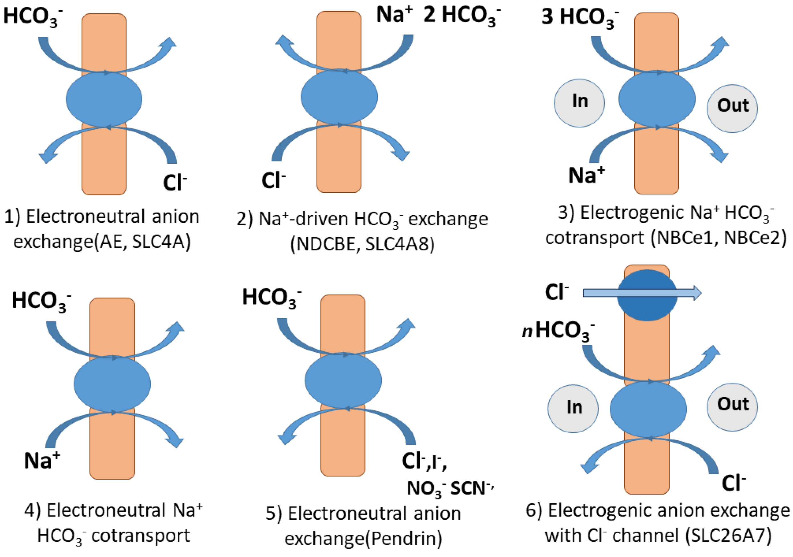
2.1. Bicarbonate Transport by Solute Carriers (SLC) in Humans and Mammals

Most bicarbonate transporters belong to the solute carriers (SLC), which is a large group of secondary active membrane transporters for relatively small molecules. The best studied bicarbonate transporters are SLC in humans due to severe diseases related to the malfunctioning of these transporters [34,35]. In humans, 430 members organized in 52 families have been identified [36]. Proteins transporting HCO3 belong to the families SLC26 (Sulfate permease SulP) and SLC4. The phylogenetically ancient gene family SLC26 encodes for multiple anion exchangers and channels. Some are relatively ion specific, but others have a broad substrate range. Besides transporting inorganic anions like HCO3, Cl, SO42−, and I, oxalate and formate may be transported. Structural models indicate that these polypeptides have 10 to 14 membrane-spanning domains flanked by a cytoplasmic N-terminal and a cytoplasmic C-terminal bound to a STAS (sulphate transporter anti sigma factor-like) domain [37]. The gene family SLC4 contains genes that code for proteins transporting HCO3 or the closely related CO32− along with a monovalent anion (Cl) or cation (Na+) [38]. These proteins have 14 transmembrane spanning domains grouped into a 7 + 7 inverted repeat topology.
Different ways for HCO3 transport through the membranes in mammalian and human cells can be distinguished (see Figure 1). The first includes electroneutral, Na+- independent anion exchange between HCO3 and Cl using anion exchange transporters (AE) encoded by genes of the SLC4A family. The second includes sodium-driven Cl/HCO3 exchanger (NDCBE) encoded by SLC4A8. This transporter is thought to exchange 1 Cl for 2 HCO3 and 1 Na+. The next transport mechanism comprises electrogenic Na+/HCO3-cotransport performed by NCBT transporter proteins NBCe1 and NBCe2 encoded by SLC4A4 and SLC4A5. The fourth way is the electroneutral Na+- HCO3 cotransport or Na+-driven Cl/HCO3 exchange through the transporter protein encoded by SLC4A10 [39,40,41]. The fifth mechanisms involves electroneutral Cl/HCO3 exchangers that also can exchange either I, NO3, SCN, or formate encoded by SLC26A (Pendrin) or NO3, OH, SO42−, oxalate, and formate (SLC26A6). (Electrogenic Cl/HCO3 exchange, with channel activity for Cl, SO42−, and oxalate (SLC26A7) and Electrogenic Cl/HCO3 exchange with Cl- channel activity, NaCl cotransport or Cl -independent HCO3 transport (SLC26A9) are also HCO3 transport mechanisms [35] (see Figure 1).

2.2. Cinorg Transporters in Cyanobacteria

Five modes of Cinorg transport have been described in cyanobacteria. (i) BCT1 is an inducible high affinity (K0.5 for HCO3 ≈ 15 µM) transporter located in the plasma membrane that belongs to the ATP binding cassette (ABC) transporter family [42] although transport energization by ATP consumption has not been proven [43]. BCT1 is a multi-meric complex composed by four subunits. CmpA is located in the periplasmic space and binds HCO3 with a very low K0.5 of 5 µM [44] and also binds Ca2+ as a cofactor [45]. CmpB is a dimer within the plasma membrane and CmpC and CmpD are extrinsic proteins that share binding sites for ATP. CmpC has an extra domain involved in the allosteric regulation of BCT1 similar to the NrtC protein of NRT1 transporter. In this later case, the domain of the NrtC protein is involved in the inhibition of transport in the presence of NH4+ [46]. BCT1 is found in ß-cyanobacteria but absent in marine cyanobacteria. However, it is present in the α-cyanobacteria Synechococcus WH5701, which can live in a wide range of Cinorg concentrations and salinities [43].
(ii) SbtA is a low Cinorg-inducible, high affinity (K0.5 2–5 µM), plasma membrane HCO3 transporter that uses Na+ as a driving ion with a half saturation constant around 1 mM for this ion [47]. Although initially considered a single unit-type transporter, it has a bigger complex size, which suggests that, in its functional form in the plasma membrane, this transporter is a tetramer [48]. It has been suggested that SbtA is activated by a serine-threonine protein kinase [49] that also depends on Na+ [50]. SbtA homologs seem to be present in many ß-cyanobacteria although this has only been confirmed in Synechocystis PCC6803 [47] and Synechococcus PCC7002 [50].
(iii) BicA HCO3 transporters are also dependent on Na+. Their affinity for HCO3 transport with a Km ranging from 74 µM to 353 µM (1.7 mM for Na+) is lower than that of SbtA. Nonetheless, BicA is able to maintain a high flow of Cinorg for photosynthesis. BicA transporters are expressed at low levels under conditions of high CO2 but they are highly inducible under low CO2. They have been discovered in the coastal marine cyanobacterium Synechococcus PCC7002 [51] and they are present in both α-cyanobacteria and ß-cyanobacteria. BicA transporters belong to the large family of prokaryotic and eukaryotic transporters often described as sulphate, SulP family transporters. The C-terminus includes a hydrophilic STAS domain (see also Section 2.1) involved in the allosteric regulation that has also been found in A. thaliana sulfate transporters [52].
(iv) NDH-I4 is a constitutive protein complex located in the plasma membrane that accelerates CO2 uptake. The passive entry of CO2 is followed by the conversion (NDH-I mediated) to HCO3 [53,54].
(v) NDH-I3 is a second, complex, low CO2 -inducible system involved in CO2 uptake located in the thylakoid membrane. It works in a similar manner to NDH-I4 [53,54].

2.3. Cinorg Transport in Microalgae

C. reindhardtii uptake of Cinorg has been associated with the activity of an ATP-binding cassette transporter, HLA3, and the homolog of a formate-nitrite transporter LCIA that is also called NAR1.2 [30]. HLA3 is located in the plasma membrane and LCIA in the chloroplast envelope. The absence of LCIA decreases the amount of HLA3 mRNA, which indicates a regulation by the chloroplast-encoded LCIA of the expression of HL3 encoded in the nuclear genome [31]. While the HCO3 transport mechanism of HL3 seems to be clear, LCIA has been proposed to be an HCO3 channel [22,30,31]. If so, HCO3 ions would be transported through such a channel downhill and could not accumulate HCO3 over the equilibrium prediction. However, the addition of mM concentrations of HCO3 to Xenopus laevis oocytes expressing NAR1.2 evokes a membrane depolarization as does the addition of mM concentrations of NO2, which suggests an HCO3 transport into the chloroplast by H+ symport instead of the transport through a channel. This mechanism would also be consistent with the need to overcome the electrochemical gradient for HCO3 in the stroma relative to the cytosol [55]. The ycf10 is also related to Cinorg transport. Disruption of the plastid ycf10 inhibits the Cinorg accumulation in the chloroplast. Its gene product known as the protein CemA was originally proposed as a Cinorg transporter, but its similarities with the cyanobacterial PxcA involved in Na+-dependent H+ extrusion suggest that CemA may play a similar role in the energization of the chloroplast envelope [55]. The HCO3 uphill transport through the plasma membrane and the chloroplast envelope agrees with the early observation of a vanadate sensitive Cinorg transport at both levels. A second Cinorg transporter proposed for the plasma membrane in C. reindhardtii is LCI1 [56,57]. The overexpression of this protein increases the affinity for Cinorg and enhances HCO3 uptake. The protein is encoded by an orphan gene [55] and does not have any known functional motif. The proteins CCP1/2 have also been proposed to take part in Cinorg uptake by the chloroplasts. They show similarities with the mitochondrial carrier proteins superfamily, but knock-outs of CCP1/2 do not show defects in photosynthesis [58]. Thus the specific role of CCP1/2 proteins in Cinorg transport has yet to be clarified.
The active uptake of HCO3 was described for natural populations of marine phytoplankton dominated by large diatoms [59]. However, the HCO3 transport mechanisms at the molecular level have been studied in the model diatom species Phaeodactylum tricornutum and Thalassiosira pseudonana [60,61]. In P. tricornutum, ten putative HCO3 transporters have been identified. They are similar to the unrelated SLC4 and SLC26 mammalian protein families (see Section 2.1). SLC4 has been characterized as a HCO3 transporter in the plasma membrane of P. tricornutum and SLC4 homologs have also been found in T. pseudonana [25]. Photosynthesis in the diatom species is sensitive to 4,4′-diisothiocyanatostilbene-2, 2′-disulfonic acid (DIDS), which is an inhibitor of anion exchange, and depends on the presence of Na+ in the medium (K0.5 28 mM, saturation at 100 mM Na+). This suggests the existence of an HCO3 uptake mechanism based on Na+ symport or on a Na+ dependent Cl/HCO3 anti-port [25]. A different group of SLC4 transporters located in the chloroplast envelope have been proposed for transporting HCO3 to the chloroplast stroma [25,60,61]. The active transport rate of dissolved inorganic carbon through the chloroplast envelope is ten-fold that of HCO3 transport across the plasmalemma [54]. However, further investigations are required to elucidate the molecular identity of the protein and the transport mechanism in the context of the complex four-layer chloroplast envelope of diatoms [62].
In micro-algal species, genetic tools are still not available and HCO3 uptake has been revealed by physiological methods that include the photosynthetic sensitivity to inhibitors of external CA, pH buffers to dissipate electrochemical H+ gradients, and inhibitors of anion exchangers. Therefore, a direct entry of HCO3 has been proposed for the marine eustigmatophycean Nannochloropsis gaditana [63,64,65]. The absence of external CA and the sensitivity to DIDS suggest an anion exchange mechanism for HCO3 transport. A DIDS and 4-acetamido-4′-isothiocyanato-stilbene-2, 2′-disulfonic acid (SITS) sensitive photosynthesis has been described in Eminliania huxleyi [66]. SITS is the putative inhibitor of the anion exchanger 1 (AE1), which works as a Cl/HCO3 antiporter in red blood cells [67] (see Figure 1). In contrast, a DIDS/SITS insensitive HCO3 transport has been described for Dunaliella tertiolecta [68].

2.4. Cinorg Transport in Macroalgae

One of the first examples for the use of HCO3 in macro-algae was described in the giant inter-nodal cells of Characeae living in alkaline media [69]. The active efflux of H+ through the putative H+-ATPase causes a local acidification of the apoplast in about two pH units [69]. The presence of CA activity in the acidic zones accelerates the conversion of HCO3 to CO2 that diffuses across the plasmalemma [70]. The cytosolic pH homeostasis requires the presence of alkaline areas between the acid zones, which produces the spectacular banding observed in these organisms under the light [71]. An alternative mechanism for HCO3 use and hence for banding was given by Lucas et al. [72]. These authors, by using quasi apoplastic pH measurements in flow- through experiments, provide evidence for an H+/HCO3 symport in the acid bands in which the electrochemical proton gradient generated by the H+-ATPases is secondarily used for HCO3 transport. According to the model by Walker et al. [69], the alkaline zones are needed for compensating cytosolic pH through OH efflux, which originated via the catalyzed cytosolic dehydration of HCO3. A similar model to the one proposed by Walker et al. [69] has been described for freshwater flowering plants where the acid zone is the abaxial (lower) leaf surface and the alkaline zone is the adaxial (upper) leaf surface [22,73].
The use of HCO3 as a source of inorganic carbon for photosynthesis has been described for the majority of marine macro-algae and seagrasses [74,75,76]. The most common mechanism of HCO3 use is the apoplastic conversion to CO2, which is shown in Condrus chrispus [77], Porphyra leucosticta [78], a series of red macroalgae [79], and Phyllariopsis puspurascens [80]. More information is available in References [33,75,81,82]. Alternatively, other algal species have been described as direct HCO3 users. Most of the evidence for a direct uptake of HCO3 ions comes from experiments in which the inhibitors of anion exchanger, mainly DIDS and SITS, are used to inhibit HCO3 transport and, therefore, photosynthesis [82]. Larsson and Axelson [83] examined 11 green, 5 red, and 11 brown macro algae. Photosynthesis was DIDS-sensitive only in Chaetomorpha, Monostroma, and ulvaceans (Ulva and Enteromorpha), but not in the rest of green, red, or brown algae tested. More information is available in Reference [84]. Fernández et al. [26] show a DIDS-sensitive anion exchanger as the main mechanism for HCO3 uptake in the giant kelp Macrocystis pyrifera. DIDS-sensitivity has also been reported in the red algae Eucheuma denticulatum [85] while a residual DIDS-sensitive photosynthetic activity was found in Gracilaria gaditana [86]. Calculations made from photosynthetic conductance were used to suggest direct HCO3 uptake in Laurencia pinnatifida [87].

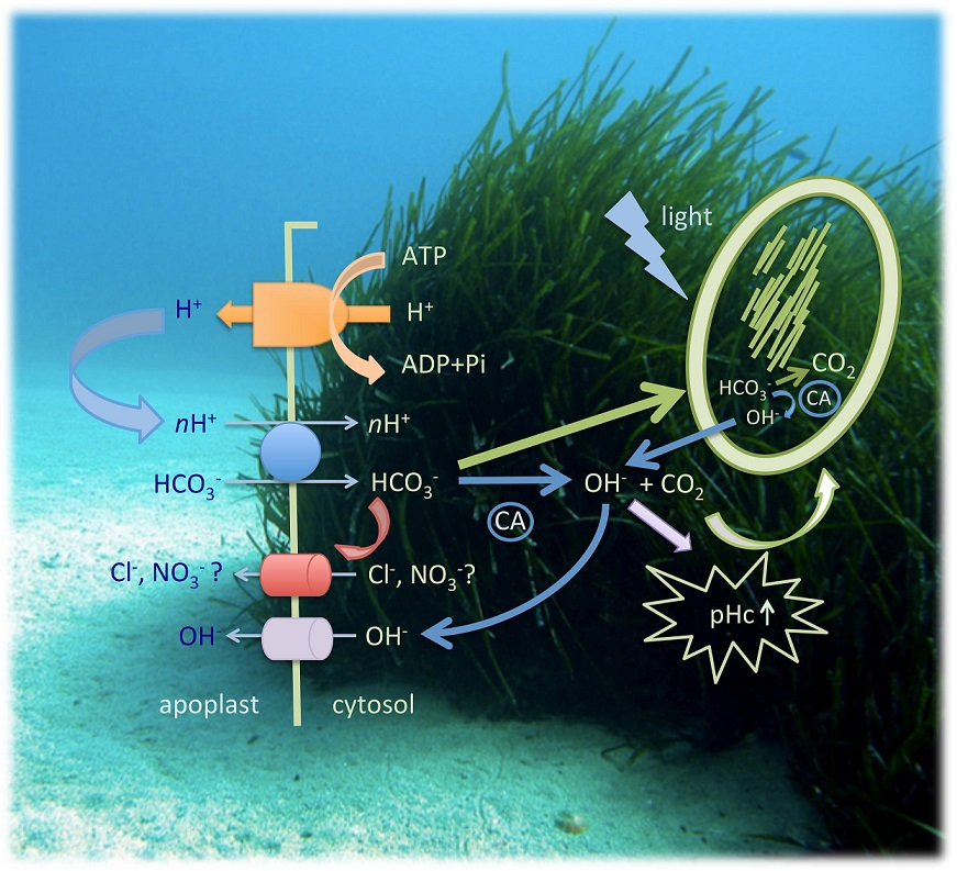
2.5. Cinorg Transport in Seagrasses

Seagrasses have been described as HCO3 users [82,88,89,90,91]. Based on the lack of photosynthesis inhibition in seagrasses by DIDS and SITS, Larkum et al. [91] hold that HCO3 influx through anion exchangers does not take place in the leaves of seagrasses. These substances inhibit AE1 that are present in algae but in angiosperms (including marine) DIDS and SITS have been described as inhibitors of anion channels [29,92] that may have a distinct role in plasma membrane anion transport [87]. As an alternative, Larkum et al. [91] suggest a proton symport as the mechanism for direct uptake of HCO3. Such a mechanism has been proposed for Zostera marina [88,93,94], Zostera noltii [95], Posidonia oceanica and Cymodocea nodosa [96], Halophila stipulacea and Ruppia maritima [96], Ruppia cirrhosa [97], and Halophila ovalis [98]. Based on the response of seagrasses to acetazolamide (AZ) and TRIS buffers, Beer et al. [88] suggest three mechanisms for carbon acquisition. First, an apoplastic dehydration of HCO3 catalyzed by CA and the subsequent diffusion of CO2 across the plasmalemma. This mechanism is proposed for plants that show AZ-sensitive TRIS-insensitive photosynthesis, but the ubiquitous presence of plasmalemma H+-ATPase cannot be ignored [99]. Second, the catalyzed apoplastic dehydration of HCO3 to CO2 in acid regions generated by the activity of the H+-ATPases. This mechanism would be sensitive to AZ and TRIS. Third, the direct uptake of HCO3 ions by symport with H+. In this case, the electrochemical gradient for H+ generated by the activity of the plasmalemma H+-ATPases drives the direct HCO3 transport. This mechanism would be AZ-insensitive and TRIS-sensitive. The two first mechanisms involve apoplastic accumulation of OH and CO2 diffusion across the plasmalemma and the third one implies accumulation of HCO3 and likely OH in the cytosol (see Table 1). In contrast to humans (see Figure 1) and cyanobacteria (Section 2.2), no Na+ -dependent HCO3 uptake system has been reported in plants. The only example for a Na+-driven ion transport system is the high affinity transporter for NO3 and Pi in the seagrass Zostera marina [100]. In that case the electrochemical gradient for Na+ is maintained because of very low membrane permeability for Na+ and the action of a Na+/H+ antiporter, which is similar to the SOS1 present in terrestrial vascular plants [101].
The availability of the genome of Zostera marina [103] allows the in silico search for genes potentially involved in Cinorg transport. Using the web application Phytozome (http://www.phytozome.net), which is a comparative platform for green plant genomics [104], we searched for genes with homologies with the HLA3 transporter of C. reinhardtii and SLC4 transporter of P. tricornutum. The search for homologies in the genome of Z. marina with ChHLA3 sequence results in six genes with high homology, all of them listed as ABC transporters. In contrast, the search for homologies with PhSLC4 results in five sequences of medium-to-low homologies with genes encoding boron transporters and anion exchangers. The public availability of the genome of seagrasses will be a valuable tool for the future investigation of the exact molecular identities of Cinorg transporters, cellular location, mechanism, kinetic properties, and regulation.

2.6. Cinorg Transport in Higher Land Plants

While HCO3 transporters are already quite well-characterized in cyanobacteria, algae, and mammals, the information on higher vascular land plants is scarce. Seven loci of genes coding for transporters of the HCO3 family are listed in the gene databases of the genetically well-characterized A. thaliana. The best studied protein is BOR1. This protein belongs to the solute carrier family type SLC4 and presents homology to SLC4A1, which is the band 3 transporter highly abundant in erythrocytes. As SLC4A1, BOR1 has a gate and a core domain and acts with an elevator mechanism. However, BOR1 has an inward rotated core domain providing an occluded state, which suggests that it may undergo structural transitions allowing access from either side of the membrane [105]. Bicarbonate transport by Band 3 is a unidirectional pathway out of the erythrocyte. A further substantial difference is that BOR1 is an efflux-type borate transporter responsible for root-to-shoot transport of this essential plant nutrient. BOR1 is located in the xylem parenchyma cells and loads borate into the xylem, which is then transported to shoots by the transpiration stream [106]. The other six genes code for BOR2 to BOR7 [107]. All seem to be involved in the transport of borate or boric acid rather than in HCO3 transport.
Although no selective HCO3 transporters or channels have so far been characterized in higher land plants, the possibility of membrane transport by specific or unspecific anion transporting proteins cannot be excluded. Several studies provide indirect support for HCO3 uptake by plant roots. Under exposure to high HCO3 (5 mM to 20 mM), a strong inhibition of nitrate, sulphate, and phosphate uptake by roots has been observed [108]. Such inhibition could be caused, at least in part, by competition between HCO3 and other anions for transport mechanisms with low anion specificity. An electrophysiological approach to ion selectivity of a voltage-dependent anion channel in A. thaliana hypocotyls revealed low but reproducible HCO3 currents. A permeability ranking of NO3 ≥ SO24− > Cl > HCO3 >> mal2− could be established [28]. More recently, such a channel with permeability for several anions has been identified as QUAC1/ALMT12, which is a channel that releases anions from guard cells [109]. In fact, anion channel currents in plants have mainly been studied in guard cells where they contribute to the mechanisms for controlling stomatal resistance (see Section 3.3). Slow Anion Channels (SLACs) and Quick Anion Channels (QUAC) are involved in the transport of NO3 and Cl (SLAC1), NO3 (SLAH3), or malate (QUAC1/ALMT1) [110]. QUAC1/ALMT12 is activated by xylem derived extracellular SO42− [111].
Anion channels in roots are less characterized. In A. thaliana roots, a slah3-1 mediated Cl and possibly NO3 efflux in response to ABA has been shown [112]. Recently, Canales et al. [113] reported comparative root expression profiles at a cell resolution level for anion channels in A. thaliana. Nitrate channel SLAH3 was strongly expressed in the mature root zone while the Voltage Dependent Anion Channel (VDAC1) was localized to the meristem zone. VDACs 2 and 4 have been reported expressed in all plant organs [114]. At the subcellular level, VDACs are localized at the outer mitochondrial membrane and in small vesicles located in the cell periphery [115]. The ion selectivity of VDACs depends on ionic strength. Higher selectivity for Cl is achieved with lower ionic strength [116].
It has been stated that, on limestone soils, HCO3 can passively enter into plant roots. Then it is long–distant transported via xylem vessels to the leaves where, after transformation by CA anhydrase, the resulting CO2 can be assimilated along with the atmospheric CO2 [11]. The apoplastic, passive radial transport pathway in the roots is disrupted at the endodermal level due to the hydrophobic Casparian strip. Therefore, to reach the vascular cylinder, a substance has to first pass through the plasma membrane into the symplasm. This implies either a still unidentified HCO3 membrane transport system or the conversion of HCO3 into CO2, which may easily diffuse into the stele. Apoplastic by flow, either through the young root tips where the Casparian strip has still not fully developed or at sites where lateral root emergence from the pericycle disrupts this hydrophobic barrier, may be another way HCO3 enters the stele. Contribution of this apoplastic bypass is relatively small in the case of NaCl [117] or Cd [118]. We could not find specific data for HCO3.
Early investigations using 11C or 14C isotopes as markers for HCO3 provided evidence for uptake of HCO3 by roots and transport to the shoots [119,120,121,122]. However, the contribution of Cinorg taken up by roots may be less than 1% taken up by leaves [123]. The 14C from labelled H14CO3 supplied through the roots was found to be incorporated into sugar, starch, and proteins of leaves [124]. As plants can acquire Cinorg from different sources including atmospheric CO2 and respiratory CO2, the experimental design is critical. Solution pH used for supplying labelled HCO3 to the plants deserves special attention. At pH 8, most of the labelled Cinorg is in the form of HCO3 but a small percentage of labelled CO2 can be present and CO2 diffusion into the root cells may occur, which will be followed by transformation of this CO2 into labelled HCO3 by CA. This transformation can be even more relevant considering that the pH of cell walls and xylem sap are usually in the acid range. The pH of the leaf apoplast of sunflowers remained stable around 6.4 to 6.5 even if roots were exposed to 10 mM HCO3 [125]. However, it has to be taken into account that apoplast alkalinization is a general response to stress in plants [126]. Enhanced Cl supply under stress causes alkalization of the root apoplast due to the symport of 2 H+ per 1 Cl [127]. Increasing the external pH of the root bathing solution also increases pH of both A. thaliana root cell walls [128] and xylem sap [129]. This favors HCO3 over CO2 formation. Nonetheless, even under severe stress conditions, such as drought or fungal infection with a strong alkalinization effect in the apoplast, the increased pH values remain nearly neutral [130]. Therefore, in plants with their roots exposed to HCO3, the proportion of HCO3 over CO2 during radial transport of Cinorg from soil to the stele and within the xylem sap up to the leaves may be considerably lower than in the soil solution surrounding the plant roots. The direct use of root-derived HCO3 by CA in chloroplasts to supply CO2 for Rubisco is unlikely when taking into account the low chloroplast permeability of HCO3 (1 × 10⁻⁸ m s−1) in comparison to CO2 (range from 2.3 × 10⁻⁴ to 8 × 10⁻⁴ m s−1), which was recently shown by mass inlet mass spectrometry (MIMS) using 18O labelled Cinorg [131].

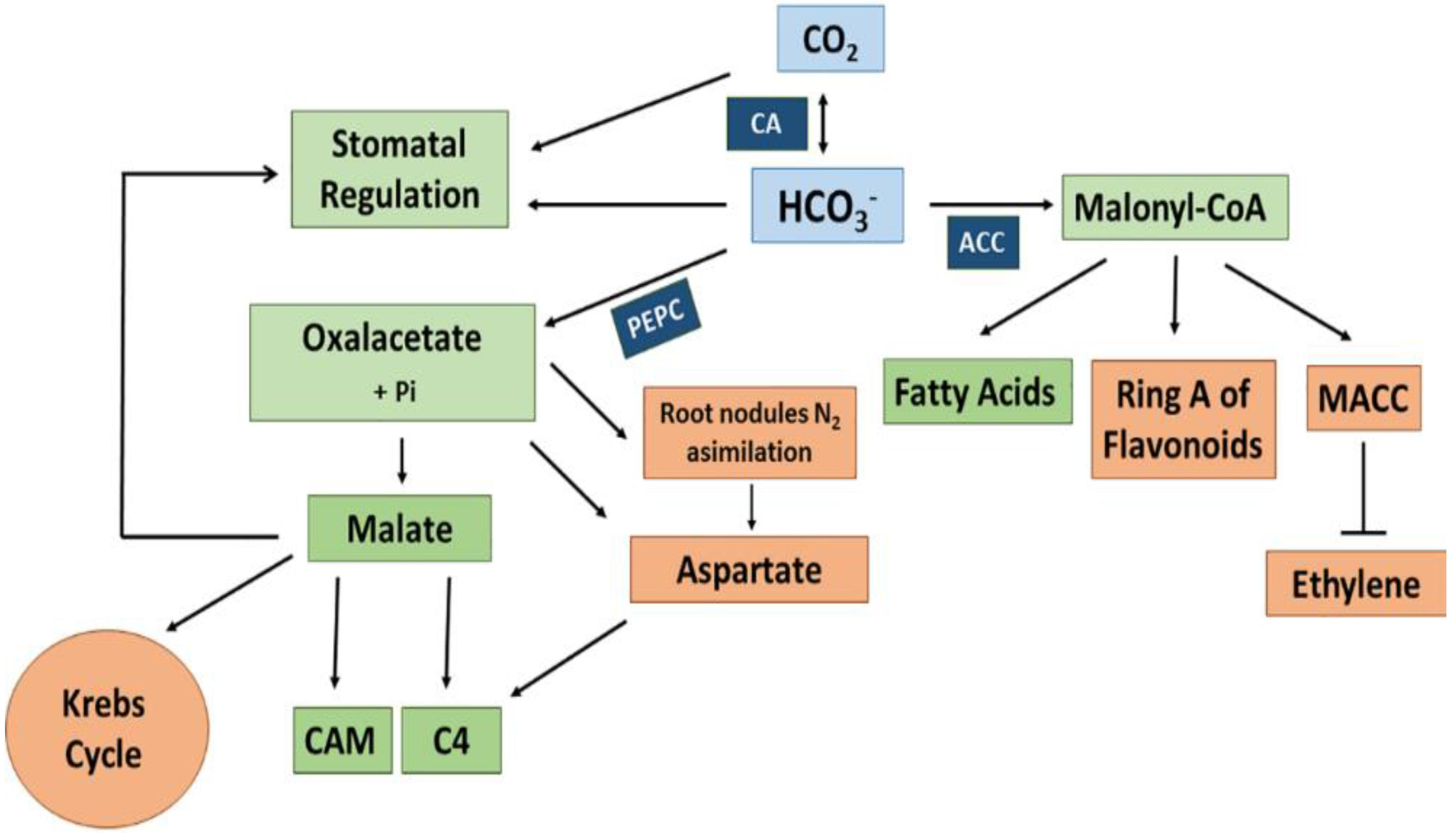
3. Formation and Use of Bicarbonate in Plants

As seen in higher plants, no selective HCO3 transporter or channel has been characterized at the molecular level. Membrane transport of HCO3 in these organisms is still unclear. Contrastingly, the contribution of HCO3 to essential metabolic pathways and the total assimilation of Cinorg in plants is reliably documented.
The CA-generated HCO3 serves as a substrate for different carboxylases among others acetyl-CoA carboxylase (ACCase, EC.6.4.1.2) and phosphoenolpyruvate carboxylase (PEPC, EC 4.1.1.31). ACCase contains a biotin carboxylase, a biotin carboxyl carrier protein, and a carboxyl transferase. It catalyzes the carboxylation of acetyl-CoA to malonyl-CoA in the chloroplast and the cytosol [132]. Malonyl-CoA is the precursor for fatty acid formation and elongation. Moreover, it participates in the biosynthesis of ring A of flavonoids through the polycetic pathway and in the biosynthesis of malonylated aminocyclopropane-1-carboxylic acid (MACC), which is involved in the down-regulation of ethylene production in plants (see Figure 2). Other biotin-containing carboxylases operating with HCO3 are 3-methylcrotonyl-CoA carboxylase, which is involved in the mitochondrial pathway of leucine catabolism. Geranyl-CoA carboxylase likely works in the metabolism of cyclic terpenes [133,134].
PEPC plays a major role in the carbon assimilation processes in plants. This enzyme in the presence of Mg2+ or Mn2+ ions catalyzes the β-carboxylation of phosphoenolpyruvate (PEP) yielding oxalacetate (OAA) and inorganic phosphate (Pi) in an irreversible reaction (see Figure 2).
The relative importance of the contribution of HCO3 to the total plant Corg as well as the assimilation mechanisms and their consequences for plant adaptation to different environmental conditions depend on the plant species and the characteristics of the habitat (see Section 3.2 and Section 4). In all cases, the cooperation of the two enzymes, CA and, in higher plants, PEPC, is essential.

3.1. Plant Carbonic Anhydrase and Phosphoenolpyruvate Carboxylase

Carbonic anhydrases (CAs, EC 4.2.1.1) are metallo-enzymes that catalyze the reversible hydration of CO2 forming HCO3. Zinc is the required metal at the catalytic site for CA activity. Some exceptions are several coastal diatoms with cadmium–containing CA (CDCA). The Cd2+ at the catalytic site is fully exchangeable for Zn2+ [135]. The natural use of Cd2+ in this ζ-CA class enzyme is considered an evolutionary adaptation to low Zn2+ availability in marine habitats [136].
CA enzymes are ubiquitous in nature (animals, plants, archaebacteria, and eubacteria) and are an example of convergent evolution. Based on sequence comparison, CA proteins are grouped into seven distinct classes: α, β, γ, δ, ζ, η, and θ-CAs [137,138,139,140]. In higher land plants, only α, β, γ CAs are found. The δ and ζ classes are restricted to marine diatoms and η-CA so far has only been reported in Plasmodium falsiparum [141]. θ-CA seems more widely distributed in algae and cyanobacteria [142] and it has been reported critical for photosynthesis in the diatom Phaedactylum tricornutum [143]. The ubiquity of the distribution of CAs implies that they play diverse and essential roles in many biological processes. They have been related to respiration and transport of CO2/HCO3 between tissues, pH and CO2 homeostasis, electrolyte secretion in a variety of tissues/organs, various biosynthetic reactions, and CO2 fixation [142,144]. In addition, CA is a plausible source of hydrogen sulphide (H2S) within plant leaves by catalyzing the conversion of carbonyl sulphide (COS) to CO2 and H2S [145].
Higher plants contain three evolutionarily distinct CA families including αCAs, βCAs, and γ CAs where each family is represented by multiple isoforms in all species [142,146,147]. Alternative splicing of CA transcripts is common. Consequently, the number of functional CA isoforms in a species may exceed the number of genes [147]. CAs are expressed in numerous plant tissues and in different cellular locations. The most prevalent CAs are those in the chloroplast, cytosol, and mitochondria. CAs have been found in the thylakoid lumen of Chlamydomonas and Phaeodactylum. They are an important component of the CCM in these species and, therefore, essential for photosynthesis and growth [143,148]. This diversity in location is paralleled in the many physiological and biochemical roles that CAs play in plants [142,147,149,150]. As in humans and animals, many of these roles are related to the CA-driven regulation of cell pH, which, in turn, can participate in multiple regulatory processes through electrical signals, changes in cytosolic Ca2+ concentrations, and plant hormones [150,151,152] among others.

3.1.1. Plant α-Carbonic Anhydrases (αCA)

Arabidopsis thaliana contain eight αCA (AtαCA1-8) [153]. Genes for αCa1, αCA2, and αCA3 are expressed in green and reproductive tissue (stems, rosette leaves, caulinar leaves, and flowers). Only αCA2 presents root expression. While expression of αCA1 is independent of the level of CO2, the expressions of αCA2 and αCA3 are induced under conditions of low CO2 concentrations [149]. αCa1 is expressed in chloroplasts and αCA2 is expressed in the plasma membrane. α-CA4 is implicated in the processes leading to energy dissipation in the PSII antenna [154]. Arabidopsis αCA8 is clearly a pseudogene since it encodes in-frame stop codons [147]. Tissue-specific expression has also been reported for other species. In sorghum, the αCA Sb5G039000 is expressed specifically in anthers while, in the legume species Medicago truncatula αCAs Mt1g059900 and Mt1g059940, are expressed in root nodules [147]. There is increasing evidence that αCAs can play an important role in photosynthesis [150]. Under conditions of increasing light intensity, the expression of αCA2 decreases while the expression of αCA4 increases. Knock-out mutants of these chloroplast-located αCAs exhibit contrasting responses in comparison of the wild type. Both the quantum yield at photosystem 2 (PS2) and the electron transfer to O2 decreased while non-photochemical quenching (NPQ) and CO2 assimilation were enhanced in plants lacking αCA2. The opposite was observed in αCA4 knock-outs [155]. The authors hypothesize that these αCAs may participate in the regulation of H+ flux into the PS2 protein PsbS, which regulates qE-type NPQ.

3.1.2. Plant β-Carbonic Anhydrases

βCAs are most abundant in land plants where they participate in photosynthesis [147]. Arabidopsis thaliana has six βCAs [147]. βCAs genes are highly expressed in leaf tissue. Expressed sequence tag experiments revealed that βCA1 to βCA6 are expressed in rosette leaves, caulinar leaves, and flowers. βCA3 is also strongly expressed in reproductive tissue while βCA4, βCA5, and βCA6 are expressed in all tissues including roots. βCAs have been found in chloroplasts, mitochondria, the cytosol, and the plasma membrane [144,149]. Targeting analysis using green fluorescent protein fusion proteins confirmed the subcellular localization of plant βCAs: βCA1 and βCA5 are expressed in chloroplasts while βCA2 and βCA3 are cytosolic. Isoforms βCA4, βCA4.1, are localized in the plasma membrane while the short form, βCA4.2, is cytosolic. βCA5 and βCA6 are localized in the chloroplast and mitochondria, respectively [149].
The role of βCAs in photosynthesis of land plants seems especially relevant in grasses with C4-type photosynthesis [156] or for plants under limited Cinorg supply (see Section 3.2). Carbonic anhydrases could be versatile. They may be involved not only in photosynthesis and responses to CO2 and light but also in seed germination, morphogenesis, nodule development, and responses to abiotic stress [157,158]. The tobacco salicylic acid-binding protein 3 (SABP3) is a chloroplast βCA that exhibits antioxidant activity and plays a role in the hypersensitive defense response [159]. Furthermore, βCA1 is related to ethylene signaling responses, photosynthetic performance of cotyledons, and Arabidopsis seedling survival [160].

3.1.3. Plant γ-Carbonic Anhydrases

Plant γCAs are codified in the nucleus but localized in mitochondria [139]. So far, no higher plant γCA with CA activity has been identified. Nonetheless, plant proteins with the active-site residues found in γCAs from archaebacteria and cyanobacteria have been found. In A. thaliana, five γCA- related genes have been reported including three γCA genes and two genes encoding γCA-like proteins. In contrast to γCA proteins, the γCA-like proteins do not have the required Zn-coordinating amino acid residues. Plant γCA genes encode for a part of the mitochondrial Complex I (NADH-ubiquinone oxidoreductase). Complex I knock-out lines present adverse effects: non-viable seeds, high levels of mitochondrial Complexes II and IV, and the alternative oxidase. However, this is in contrast with reduced levels of photosynthetic proteins [161]. A proteomic approach has recently found enhanced γCA root levels during the induction phase of Al-tolerance in the hyper-resistant grass Urochloa decumbens. This increase occurred along with higher adenylate kinase activity and supports a role for γCA in the maintenance of ATP-production during the Al tolerance response [162].

3.1.4. Plant PEP Carboxylases

Phosphoenolpyruvate carboxylases (PEPC) are located in the cytosol and catalyze the β-carboxylation of PEP to oxaloacetate using HCO3 in an irreversible process. The OAA can then be reduced through NADH or NADPH-dependent malate dehydrogenase to malate in a reversible process. PEPCs are present in bacteria, algae, and plants. The typical plant PEPC (class 1 PEPC) has four identical subunits of 107 kDa. Multiple isoforms have been identified in leaves of C3, C4, and CAM plants [163,164,165]. In Sorghum bicolor, which is a plant with C4-type photosynthesis, five PEPC genes (PEPC1-5) have been identified. The plant PEPC is highly regulated. Phosphorylation through PEP carboxykinase (PEPCK) at the N-terminal phosphorylation domain [166] and allosteric regulation by glycine and glucose-6-P enhances the activity. Inhibition is achieved by both allosteric regulation especially by malate and by ubiquitination [167]. In addition to the typical plant-type PEPC, plants also contain a bacterial-type PEPC (BPEPC) of 118 kDa [153]. The BPEPC is highly expressed in floral tissues as well as in seeds and fruits. It has recently been shown that high BPEPC occurs in tissues that accumulate high malate concentrations [168]. There is a tight interaction between PTPC and BTPC, which forms the class 2 PEP. This is an enzyme complex that, in contrast to class 1 PEPC, is mostly insensitive to allosteric inhibition by high malate concentrations [169,170]. While class 1 PEPCs are constitutively expressed in the cytosol, the BPEPC is associated with the outer mitochondrial surface. This location is in line with a central role of this enzyme in collaboration with CA in the efficient fixation of respiratory CO2 and the anaplerotic supply of organic acids to the Krebs cycle [171]. This is especially relevant in developing seeds that store fatty acids such as castor bean seeds [172]. PEPC activity also plays a central role in symbiotic N2 fixation in root nodules (see Figure 2) where it provides OAA for nitrogen assimilation and malate for the bacteroids [173].

3.2. Carbon Concentration Mechanisms (CCM) in Terrestrial Plants

Under certain environmental conditions, CO2 may become a limiting factor for photosynthesis not only in cyanobacteria, algae, and aquatic macrophytes where CCMs have been intensively studied but also in terrestrial higher plants. High-temperature favoring photorespiration and drought imposes an increase of stomatal resistance. These are the main factors limiting CO2 availability for RuBisCo in land plants [174].
Long distance transport of HCO3 from roots to leaves usually makes only a small contribution to Cinorg for photosynthesis (see Section 2.6). Exceptions are aquatic plants in the Lycophyta genus Isoetes and the non-stomatous land plant Stylites. They acquire all Cinorg for photosynthesis from the soil through the roots and recycle carbon by CAM [175]. Other terrestrial plants take up most of the Cinorg in the form of CO2 through stomata of the leaves. This CO2 diffuses into the chloroplast where it is assimilated by RuBisCO, which forms phopshoglycerate (PGA) as the first stable product of Cinorg assimilation. After activation with ATP and reduction by NADPH provided by the light–driven chloroplastic electron transport, PGA forms phosphoglyceraldehyde, which is the first sugar molecule of the photosynthetic carbon metabolism. Most terrestrial plants fix CO2 directly onto ribuslose-bis-phosphate. In contrast to plants with this so-called C3-type photosynthesis, plants with C4-type photosynthesis convert CO2 entering through the stomata into the mesophyll cells to HCO3 using a cytosolic βCA. This Cinorg in the form of HCO3 is initially fixed by PEP carboxylase in the cytosol of the outer mesophyll cells of the leaves. In this case, OAA is a first stable organic compound. Oxalacetate is either reduced to malate or transformed by transamination to aspartate. Malate or aspartate are the molecules that transfer the newly fixed carbon to the inner bundle sheet cells of the leaves where decarboxylation provides CO2, which is the substrate for Rubisco [176]. While CA activity is high in C3 chloroplasts where it facilitates the availability of CO2 for RuBisCo, the absence of CA activity from bundle sheet cells seems essential for the C4 mechanism [177].
This CCM around RuBisCo in C4 plants is considered an evolutionary adaptation to reduce the oxygenase activity of RuBisCo, which inhibits photorespiration and is especially enhanced under high temperature in tropical or subtropical areas [178]. However, C4-type photosynthesis can also be induced in certain amphibious plant species such as Eleocharius vivipara [179] under conditions of leaf emergence under dry conditions. Extreme adaptation to drought is observed in many CAM plants, which capture CO2 during the night when a lower temperature and a higher relative humidity in the atmosphere reduces transpiratory water loss. During the dark period, this Cinorg is fixed in the form of HCO3 by PEP carboxylase and stored in large vacuoles mostly in the form of malate. The CO2 for fixation with RuBisCo is obtained by decarboxylation of malate during the following day-light period [180].
Limitations of CCMs in higher plants, especially of the C3- type of photosynthesis, and advances in our understanding of CCMs in cyanobacteria and microalgae like C. rheinhardii have promoted genetic engineering approaches to introduce efficient CCM into crop plants for increasing yield. Different approaches include manipulation of photorespiration, C3 to C4 engineering, and introduction of CCMs of cyanobacteria of C. rheinharddii into C3 crops. This well-known topic has recently been reviewed in detail by Mackinder [181] who identified gaps in our knowledge on bicarbonate transporter structure, functioning, and localization as important constraints that need priority attention for successful development of CCM engineered plants.

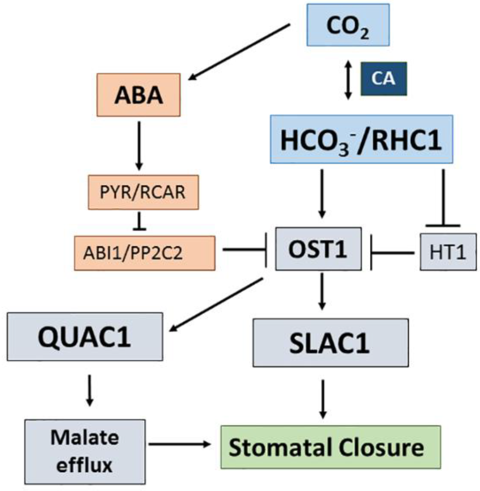
3.3. CO2/Bicarbonate Signalling in Stomatal Guard Cells

Regardless the type of photosynthesis, C3, C4, or CAM, the CO2 flux from the atmosphere into the plants is regulated by the stomatal opening and closure due to turgor changes in the stomatal guard cells. These changes are strictly controlled by multiple external and internal factors. Among those, the binomial CO2/HCO3 plays a central role (see Figure 3). High CO2 promotes stomatal closure, which is brought about by the activation of efflux anion channels: SLAC1 (S-type) facilitating Cl or NO3 efflux and R-type (AtALMT12/QUAC1 in A. thaliana) for malate efflux (see Section 2.6). The signal for stomatal closing in response to high CO2 seems to be a combination of alkaline pH, high Ca2+, and high HCO3 in the cytosol [182]. The carbonic anhydrase double mutant ca1ca4 does not show any effect on stomatal conductance when CO2 concentration is changed from 100 ppm to 80 ppm [183]. This points to HCO3 being the key signal. Abscisic acid (ABA) dependent and ABA-independent mechanisms seem to operate in stomatal closure under a high amount of CO2 (see Figure 3). OST1 (Open Stomata 1) is a positive regulator of the anion efflux channels. In the ABA-independent signaling pathway, a high amount of HCO3 activates a MATE–like transporter protein (RHC1, Resistance to High CO2), which acts as a positive regulator of OST 1 by inhibiting HT1 (High Leaf Temperature) known as a protein kinase that inactivates OST1 [183,184].

4. Plant Response to Bicarbonate-Rich Soils

It is common knowledge that limestone soils containing high carbonate/bicarbonate concentrations restrict the performance of calcifuge plant species and limit yield especially in iron-inefficient crops such as certain varieties of citrus, peach, pear, or soybeans suffering from lime-induced chlorosis [185,186]. Low pH leads to low availability of essential nutrients (especially Fe, Zn, and P) and high Ca soil concentrations are considered the main constraining factors. However, HCO3 at concentrations occurring in the solution of limestone soils can inhibit root growth in sensitive plant species like the calcifuge grass Deschampsia caespitosa [187]. However, dicots like peas, beans, or sunflowers suffer more intense root growth inhibition due to CO2 and/or HCO3 than the monocots barley and oats [123]. Recently soil carbonate has been identified as a main selection factor that drives local adaptation in natural populations of A. thaliana, which is a calcifuge species able to colonize soils with moderate carbonate contents [188].
Currently, no HCO3 transporter has been characterized in higher plants (see Section 2.6). Nonetheless, HCO3-induced root growth inhibition is paralleled by enhanced root production of organic acids especially malate, succinate, and citrate [189]. This suggests that excess HCO3 enters the root and is metabolized by CA and PEP, which yields enhanced organic acid levels. However, CO2 diffusing from the soil’s atmosphere into the root can also be transformed inside the root into HCO3 by CA. Bicarbonate can be released again into the soil rhizosphere, which contributes to the plant’s cation-anion balance. Especially under conditions of high nitrate uptake, enhanced HCO3 efflux from the roots has been claimed to contribute to the characteristic alkalinization of the rhizosphere when nitrate is the main N source for the plants [190,191]. In fact, in maize and a tomato, the sum of K+ and NO3 uptake and the HCO3 efflux have been reported to be in electrical equilibrium [192]. However, no selective bicarbonate efflux transporters in plant roots have been reported and alkalinization can also be a consequence of either or both OH release or H+ uptake in cotransport with nitrate [193]. Actually, root supply of low HCO3 concentrations tended to increase rather than decrease root nitrate uptake in Populus canescens. Exposure to 1 mM external NaHCO3 enhanced both nitrate reduction and assimilation as well as exported nitrogen to the shoots of poplar plants [194]. Higher HCO3 concentrations cause net K+ and NO3 efflux as well as accumulation of organic acids, mainly malate, in the roots [195]. To what extent dark fixation of Cinorg entering the roots plays a role in the carbon budget of terrestrial plants has been considered mainly in relation to lime-induced chlorosis in calcifuge plants. This type of chlorosis affects sensitive plant species when growing on carbonate-rich soil and may reflect an interference of HCO3 in the mechanisms of Fe acquisition and transport [196]. Key processes potentially impaired by HCO3 include the dicots’ strategy 1 such as the acidification of the rhizosphere due to the strong buffer ability of HCO3 and the reduction of FeIII to FeII by ferric reductase, which operates optimally at acid pH [197]. The induction of root exudation of phenolic substances is not affected and is even stimulated by HCO3. Induction of root accumulation and exudation of coumarin-type phenolics with high affinity for Fe has been reported as a response to Fe-deficiency under high pH conditions in A. thaliana [198]. An A. thaliana population which is naturally adapted to moderate soil carbonate had higher rates of coumarin root exudation than a sensitive population [188]. Furthermore, prevention of the imbalance of organic acid concentrations caused by dark fixation of HCO3 and shifting of Corg into the shikimate pathway for the production of phenolic compounds has been reported as a mechanism of the extreme HCO3 tolerance in Parietaria difusa [199]. In the view of the multiple implications of HCO3 in plants’ metabolism, breeding programs for better crop yield on carbonate-rich soil would greatly benefit from the characterization at the genetic and molecular level of the of bicarbonate uptake and efflux mechanisms in higher land plants.

5. Conclusions

During the last decade, there has been significant progress of our knowledge on the mechanisms of HCO3 transport and CCM in cyanobacteria, algae, and seagrass species due to improved genetic and molecular tools and electrophysiological approaches. In contrast, in higher land plants, no HCO3 transporter has been characterized so far. Advanced knowledge of the metabolic use of HCO3 in terrestrial plants has mainly been made in relation to C4 and CAM metabolism including the genetic and molecular characterization of CAs and PEPC involved. However, there are still important gaps in our knowledge about the mechanisms of compartmentation and regulation especially regarding the complex interactions between light and dark fixation of Cinorg, the recycling of respiratory and photorespiratory CO2, and the importance of anaplerotic supply of organic acids to the Krebs cycle. Filling these gaps is essential for progress in both genetic engineering approaches for transferring CCMs from cyanobacteria or microalgae to higher plants and breeding for bicarbonate tolerance in crops sensitive to lime-induced chlorosis.

Author Contributions

All co-authors contributed to the design of the review’s outline and to the critical discussion of the relevant information. C.P. and J.A.F. wrote the article with continuous inputs from L.R., J.B., L.P., and J.T. Figures were designed and drawn by C.P. and J.B. The graphical abstract was provided by J.A.F.

Acknowledgments

Open access costs of this publication are supported by the Spanish MICINN project Sal-CAL-MED (BFU2016-75176-R) participated by C.P., J.B., L.P., and J.T. Work by J.A.F. and L.R. is financed by the Spanish MICINN, project BFU2017-85117-R, and both participate in BIO2016-81957-REDT.

Conflicts of Interest

The authors declare no conflict of interest. The founding sponsors had no role in the design of the study, in the collection, analyses, or interpretation of data, in the writing of the manuscript, or in the decision to publish the results.

Abbreviations

ABAAbscisic acid
CCMCarbon concentration mechanism
CACarbonic anhydrase
PEPCPhosphoenolpyruvate carboxylase
SLACSlow anion channel
SLCSolute carriers
QACQuick anion channel

References

  1. Madsen, T.V.; San-Jensen, K. Photosynthetic carbon assimilation in aquatic macrophytes. Aquat. Bot. 1991, 41, 5–40. [Google Scholar] [CrossRef]
  2. Keeley, J.E.; Sandquist, D.R. Carbon: Freshwater plants. Plant Cell Environ. 1992, 15, 1021–1035. [Google Scholar] [CrossRef]
  3. Falkowsky, P.G.; Raven, J.A. Aquatic Photosynthesis, 2nd ed.; Princeton University Press: Princeton, NJ, USA, 2007; Chapter 1; pp. 1–43. ISBN 9780691115511. [Google Scholar]
  4. Krauskopf, K.B.; Bird, D.K. Introduction to Geochemistry, 3rd ed.; McGraw Hill: New York, NY, USA, 1995; Chapter 3; pp. 61–85. ISBN 0-07-o35820-6. [Google Scholar]
  5. Bloom, P.R.; Inskeep, W.P. Factors affecting bicarbonate chemistry and iron chlorosis in soils. J. Plant Nutr. 1986, 9, 215–228. [Google Scholar] [CrossRef]
  6. Sauze, J.; Jones, S.P.; Wingate, L.; Wohl, S.; Ogée, J. The role of soil pH on soil carbonic anhydrase activity. Biogeosciences 2018, 597–612. [Google Scholar] [CrossRef]
  7. Jones, S.P.; Ogée, J.; Sauze, J.; Wohl, S.; Saavedra, N.; Fernández-Pardo, N.; Maire, J.; Launois, T.; Bosc, A.; Wingate, L. Non-destructive estimates of soil carbonic anhydrase activity and associated soil water oxygen isotope composition. Hydrol. Earth Syst. Sci. 2017, 21, 6363–6377. [Google Scholar] [CrossRef]
  8. Gao, K.S.; Zou, D.G. Photosynthetic bicarbonate utilization by a terrestrial cyanobacterium Nostoc flagelliforme (Cyanophyceae). J. Appl. Phycol. 2001, 37, 768–771. [Google Scholar] [CrossRef]
  9. Römheld, V. Different strategies for iron acquisition in higher plants. Physiol. Plant. 1987, 70, 231–234. [Google Scholar] [CrossRef]
  10. Rashid, A.; Ryan, J. Micronutrient constraints to crop production in soils with Mediterranean-type characteristics: A review. J. Plant Nutr. 2004, 27, 959–975. [Google Scholar] [CrossRef]
  11. Rao, S.; Wu, Y. Root derived bicarbonate assimilation in response to variable water deficit in Camptotheca acuminate seedlings. Photosynth. Res. 2017, 134, 59–70. [Google Scholar] [CrossRef] [PubMed]
  12. Brown, D.; Bouley, R.; Păunecu, T.G.; Breton, S.; Lu, H.A.J. New insights into the dynamic regulation of water and acid-base balance by renal epithelial cells. Am. J. Physiol. Cell Physiol. 2012, 302, C1421–C1433. [Google Scholar] [CrossRef] [PubMed]
  13. Yin, K.; Paine, M.L. Bicarbonate transport during enamel maturation. Calcif. Tissue Int. 2017, 101, 457–464. [Google Scholar] [CrossRef] [PubMed]
  14. Zoccola, D.; Ganot, P.; Bertucci, A.; Caminiti-Segonds, N.; Techer, N.; Voolstra, C.R.; Aranda, M.; Tambutté, E.; Allemand, D.; Casey, J.R.; Tambutté, S. Bicarbonate transporters in corals point towards a key step in the evolution of cnidarian calcification. Sci. Rep. 2015, 5, 9983. [Google Scholar] [CrossRef] [PubMed]
  15. Boron, W.F.W.F.; Endeward, V.; Gros, G.; Musa-Aziz, R.; Pohl, P. Intrinsic CO2 permeability of cell membranes and potential biological relevance of CO2 channels. ChemPhysChem 2011, 12, 1017–1019. [Google Scholar] [CrossRef] [PubMed]
  16. Talbot, K.; Kwong, R.W.M.; Gilmour, K.M.; Perry, S.F. The water channel aquaporin-1a1 facilitates movement of CO2 and ammonia in zebrafish (Danio rerio) larvae. J. Exp. Biol. 2015, 218, 3931–3940. [Google Scholar] [CrossRef] [PubMed]
  17. Endeward, V.; Arias-Hidalgo, M.; Al-Samir, S.; Gros, G. CO2 permeability of biological membranes and role of CO2 channels. Membranes 2017, 7, 61. [Google Scholar] [CrossRef] [PubMed]
  18. Missner, A.; Kügler, P.; Saparov, S.M.; Sommer, K.; Mathai, J.C.; Zeidel, M.L. Carbon dioxide transport through membranes. J. Biol. Chem. 2008, 283, 25340–25347. [Google Scholar] [CrossRef] [PubMed]
  19. Uehlein, N.; Kai, L.; Kaldenhoff, R. Plant aquaporins and CO2. In Plant Aquaporins; Signaling and Communications in Plants Series; Chaumont, F., Tyerman, S.D., Eds.; Springer Int. Publ.: Cham, Switzerland, 2017; pp. 255–265. [Google Scholar] [CrossRef]
  20. Flexas, J.; Ribas-Carbó, M.; Hanson, D.T.; Bota, J.; Otto, B.; Cifre, J.; McDowell, M.C.; Medrano, H.; Kaldenhoff, R. Tobacco aquaporin NTAQP1 is involved in mesophyll conductance to CO2 in vivo. Plant J. 2006, 48, 427–439. [Google Scholar] [CrossRef] [PubMed]
  21. Groszmann, M.; Osborn, H.L.; Evans, J.R. Carbon dioxide and water transport through aquaporins. Plant Cell Environ. 2017, 40, 938–961. [Google Scholar] [CrossRef] [PubMed]
  22. Raven, J.A.; Beardall, J.; Sánchez-Baracaldo, P. The possible evolution and future of CO2-concentrating mechanisms. J. Exp. Bot. 2017, 68, 3701–3716. [Google Scholar] [CrossRef] [PubMed]
  23. Price, G.D.; Shelden, M.C.; Howitt, S.M. Membrane topology of the cyanobacterial bicarbonate transporter, SbtA, and identification of potential regulatory loops. Mol. Membr. Biol. 2011, 28, 265–275. [Google Scholar] [CrossRef] [PubMed]
  24. Duanmu, D.; Miller, A.R.; Horken, K.M.; Weeks, D.P.; Spalding, M.H. Knockdown of limiting-CO2-induced gene HLA3 decreases HCO3 transport and photosynthetic Ci affinity in Chlamydomonas reinhardtii. Proc. Nat. Acad. Sci. USA 2009, 106, 5990–5995. [Google Scholar] [CrossRef] [PubMed]
  25. Nakajima, K.; Tanaka, A.; Matsuda, Y. SLC4 family transporters in a marine diatom directly pump bicarbonate from seawater. Proc. Natl. Acad. Sci. USA 2013, 110, 1767–1772. [Google Scholar] [CrossRef] [PubMed]
  26. Fernández, P.A.; Hurd, C.L.; Roleda, M.Y. Bicarbonate uptake via an anion exchange protein is the main mechanism of inorganic carbon acquisition by the giant kelp Macrocystis pyrifera (Laminariales, Phaeophyceae) under variable pH. J. Phycol. 2014, 50, 998–1008. [Google Scholar] [CrossRef] [PubMed]
  27. Rubio, L.; García, D.; García-Sánchez, M.J.; Niell, F.X.; Felle, H.H.; Fernández, J.A. Direct uptake of HCO3 in the marine angiosperm Posidonia oceanica (L.) Delile driven by a plasma membrane H+ economy. Plant Cell Environ. 2017, 40, 2820–2830. [Google Scholar] [CrossRef] [PubMed]
  28. Frachisse, J.M.; Thomine, S.; Colcombet, J.; Guern, J.; Barbier-Brygoo, H. Sulfate is both a substrate and an activator of the voltage-dependent anion channel of Arabidopsis hypocotyl cells. Plant Physiol. 1999, 121, 253–261. [Google Scholar] [CrossRef] [PubMed]
  29. Roberts, S.K. Plasma membrane anion channels in higher plants and their putative functions in roots. New Phytol. 2006, 169, 647–666. [Google Scholar] [CrossRef] [PubMed]
  30. Wang, Y.; Spalding, M.J. Acclimation to very low CO2: Contribution of limiting CO2 inducible proteins, LCIb and LCIA, to inorganic carbon uptake in Chlamydomonas reinhardtii. Plant Physiol. 2014, 166, 2040–2050. [Google Scholar] [CrossRef] [PubMed]
  31. Yamano, T.; Sato, E.; Iguchi, H.; Fukuda, Y.; Fukuzawa, H. Characterization of cooperative bicarbonate uptake into chloroplast stroma in the green alga Chlamydomonas reinhardtii. Proc. Natl. Acad. Sci. USA 2015, 112, 7315–7320. [Google Scholar] [CrossRef] [PubMed] [Green Version]
  32. Jungnick, N.; Ma, Y.; Mukherjee, B.; Cronan, J.C.; Speed, D.J.; Laborde, S.M.; Longstreth, D.J.; Moroney, J.V. The carbon concentrating mechanism in Chlamydomonas reinhardtii: Finding the missing pieces. Photosynth. Res. 2014, 121, 159–173. [Google Scholar] [CrossRef] [PubMed]
  33. Raven, J.A.; Beardall, J.; Giordano, M. Energy costs of carbon dioxide concentrating mechanisms in aquatic organisms. Photosynth. Res. 2014, 121, 111–124. [Google Scholar] [CrossRef] [PubMed]
  34. Parker, M.D.; Boron, W.F. The divergence, actions, roles, and relatives of sodium-coupled bicarbonate transporters. Physiol. Rev. 2013, 93, 803–959. [Google Scholar] [CrossRef] [PubMed]
  35. Alka, K.; Casey, J.R. Bicarbonate transport in health and disease. Int. Union Biochem. Mol. Biol. 2014, 66, 596–615. [Google Scholar] [CrossRef] [PubMed]
  36. Perland, E.; Bagchi, S.; Klaesson, A.; Fredriksson, R. Characteristics of 29 novel atypical solute carriers of major facilitator superfamily type: Evolutionary conservation, predicted structure and neuronal co-expression. Open Biol. 2017, 7, 170142. [Google Scholar] [CrossRef] [PubMed]
  37. Alper, S.L.; Sharmer, A.K. The SLC26 gene family of anion transporters and channels. Mol. Aspects Med. 2013, 34, 494–515. [Google Scholar] [CrossRef] [PubMed]
  38. Romero, M.F.; Chen, A.-P.; Parler, D.; Boron, W.F. The SLC4 family of bicarbonate (HCO3) transporters. Mol. Aspects Med. 2013, 34, 159–182. [Google Scholar] [CrossRef] [PubMed]
  39. Bonar, P.T.; Casey, J.R. Plasma membrane Cl/HCO3 exchangers: Structure, mechanism and physiology. Channels 2008, 2, 337–345. [Google Scholar] [CrossRef] [PubMed]
  40. Aalkjaer, C.; Boedtkjer, E.; Choi, I.; Lee, S. Cation-coupled bicarbonate transporters. Compr. Physiol. 2014, 4, 1605–1637. [Google Scholar] [CrossRef] [PubMed]
  41. Alvadia, C.M.; Sommer, T.; Bjerregaard-Andersen, K.; Damkier, H.H.; Montrasio, M.; Aalkjaer, C.; Morth, J.P. The crystal structure of the regulatory domain of the human sodium-driven chloride/bicarbonate exchanger. Sci. Rep. 2017, 7, 12131. [Google Scholar] [CrossRef] [PubMed]
  42. Omata, T.; Price, G.D.; Badger, M.R.; Okamura, M.; Gohta, S.; Ogawa, T. Identification of an ATP-binding cassette transporter involved in bicarbonate uptake in the cyanobacterium Synechococcus sp. Strain PCC7942. Proc. Natl. Acad. Sci. USA 1999, 23, 13571–13576. [Google Scholar] [CrossRef]
  43. Price, G.D.; Badger, M.R.; Woodger, F.J.; Long, B.M. Advances in understanding the cyanobacterial CO2-concentrating mechanism (CCM): Functional components, Ci transporters, diversity, genetic regulation and prospects for engineering into plants. J. Exp. Bot. 2008, 59, 1441–1461. [Google Scholar] [CrossRef] [PubMed]
  44. Maeda, S.; Price, G.D.; Badger, M.R.; Enomoto, C.; Omata, T. Bicarbonate binding activity of the CmpA protein of the cyanobacterium Synechococcus sp. strain PCC7942 involved in active transport of bicarbonate. J. Biol. Chem. 2000, 275, 20551–20555. [Google Scholar] [CrossRef] [PubMed]
  45. Koropatkin, N.M.; Koppenaal, D.W.; Pakrasi, H.B.; Smith, T.J. The structure of a cyanobacterial bicarbonate transport protein, CmpA. J. Biol. Chem. 2007, 282, 2606–2614. [Google Scholar] [CrossRef] [PubMed]
  46. Maeda, S.I.; Omata, T. Substrate-binding lipoprotein of the cyanobacterium Synechococcus sp. strain PCC7942 involved in the transport of nitrate and nitrite. J. Biol. Chem. 1997, 272, 3036–3041. [Google Scholar] [CrossRef] [PubMed]
  47. Shibata, M.; Katoh, H.; Sonoda, M.; Ohkawa, H.; Shimoyama, M.; Fukuzawa, H.; Kaplan, A.; Ogawa, T. Genes essential to sodium-dependent bicarbonate transport in cyanobacteria: Function and phylogenetic analysis. J. Biol. Chem. 2002, 277, 18658–18664. [Google Scholar] [CrossRef] [PubMed]
  48. Zhang, P.P.; Battchikova, N.; Jansen, T.; Appel, J.; Ogawa, T.; Aro, E.M. Expression and functional roles of the two distinct NDH-1 complexes and the carbon acquisition complex NdhD3/NdhF3/CupA/Sll1735 in Synechocystis sp. PCC 6803. Plant Cell 2004, 16, 3326–3340. [Google Scholar] [CrossRef] [PubMed]
  49. Sültemeyer, D.; Klughammer, B.; Badger, M.R.; Price, G.D. Fast induction of high-affinity HCO3 transport in cyanobacteria. Plant Physiol. 1998, 116, 183–192. [Google Scholar] [CrossRef]
  50. Amoroso, G.; Seimetz, N.; Sultemeyer, D. The dc13 gene upstream of ictB is involved in rapid induction of the high affinity Na+ dependent HCO3 transporter in cyanobacteria. Photosynth. Res. 2003, 77, 127–138. [Google Scholar] [CrossRef] [PubMed]
  51. Price, G.D.; Woodger, F.J.; Badger, M.R.; Howitt, S.M.; Tucker, L. Identification of a SulP-type bicarbonate transporter in marine cyanobacteria. Proc. Natl. Acad. Sci. USA 2004, 101, 18228–18233. [Google Scholar] [CrossRef] [PubMed]
  52. Shibagaki, N.; Grossman, A.R. Probing the function of STAS domains of the Arabidopsis sulfate transporters. J. Biol. Chem. 2004, 279, 30791–30799. [Google Scholar] [CrossRef] [PubMed]
  53. Shibata, M.; Ohkawa, H.; Kaneko, T.; Fukuzawa, H.; Tabata, S.; Kaplan, A.; Ogawa, T. Distinct constitutive and low CO2 induced CO2 uptake systems in cyanobacteria: Genes involved and their phylogenetic relationship with homologous genes in other organisms. Proc. Natl. Acad. Sci. USA 2001, 98, 11789–11794. [Google Scholar] [CrossRef] [PubMed]
  54. Maeda, S.; Badger, M.R.; Price, G.D. Novel gene products associated with NdhD3/D4-containing NDH-1 complexes are involved in photosynthetic CO2 hydration in the cyanobacterium, Synechococcus sp. PCC7942. Mol. Microbiol. 2002, 43, 425–435. [Google Scholar] [CrossRef] [PubMed]
  55. Wang, Y.; Stessman, D.J.; Spalding, M.H. The CO2 concentrating mechanism and photosynthetic carbon assimilation in limiting CO2: How Chlamydomonas works against the gradient. Plant J. 2015, 82, 429–448. [Google Scholar] [CrossRef] [PubMed]
  56. Burow, M.D.; Chen, Z.Y.; Mouton, T.M.; Moroney, J.V. Isolation of cDNA clones of genes induced upon transfer of Chlamydomonas reinhardtii cells to low CO2. Plant Mol. Biol. 1996, 31, 443–448. [Google Scholar] [CrossRef] [PubMed]
  57. Ohnishi, N.; Mukherjee, B.; Tsujikawa, T.; Yanase, M.; Nakano, H.; Moroney, J.V.; Fukuzawa, H. Expression of a low CO2-inducible protein, LCI1, increases inorganic carbon uptake in the green alga Chlamydomonas reinhardtii. Plant Cell 2010, 22, 3105–3117. [Google Scholar] [CrossRef] [PubMed]
  58. Pollock, S.V.; Prout, D.L.; Godfrey, A.C.; Lemaire, S.D.; Moroney, J.V. The Chlamydomonas reinhardtii proteins Ccp1 and Ccp2 are required for long-term growth, but are not necessary for efficient photosynthesis, in a low CO2 environment. Plant Mol. Biol. 2004, 56, 125–132. [Google Scholar] [CrossRef] [PubMed]
  59. Tortell, P.D.; Reinfelder, J.R.; Morel, F.MM. Active uptake of bicarbonate by diatoms. Nature 1997, 390, 243–244. [Google Scholar] [CrossRef]
  60. Hopkinson, B.M.; Dupont, C.L.; Matsuda, Y. The physiology and genetics of CO2 concentrating mechanisms in model diatoms. Curr. Opin. Plant Biol. 2016, 31, 51–57. [Google Scholar] [CrossRef] [PubMed]
  61. Shen, C.; Dupont, C.L.; Hopkins, B.M. The diversity of CO2-concetrationg mechanisms in marine diatoms as inferred from their genetic content. J. Exp. Bot. 2017, 68, 3937–3948. [Google Scholar] [CrossRef] [PubMed]
  62. Gibbs, S.P. The chloroplast endoplasmic reticulum: Structure, function and evolutionary significance. Int. Rev. Cytol. 1981, 72, 49–99. [Google Scholar] [CrossRef]
  63. Huertas, I.E.; Lubián, L.M. Comparative study of dissolved inorganic carbon and photosynthetic responses in Nannochloris (Chlorophyceae) and Nannochloropsis (Eustigmatophyceae) species. Can. J. Bot. 1997, 76, 1104–1108. [Google Scholar] [CrossRef]
  64. Huertas, I.E.; Espie, G.S.; Colman, B.; Lubián, L.M. Light-dependent bicarbonate uptake and CO2 efflux in the microalga Nannochloropsis gaditana. Planta 2000, 211, 43–49. [Google Scholar] [CrossRef] [PubMed]
  65. Huertas, I.E.; Lubián, L.M.; Espie, G.S. Mitochondrial-driven bicarbonate transport supports photosynthesis in a marine microalga. Plant Physiol. 2002, 130, 284–291. [Google Scholar] [CrossRef] [PubMed]
  66. Herfort, L.; Thake, B.; Roberts, J. Acquisition and use of bicarbonate by Emiliania huxleyi. New Phytol. 2002, 156, 427–436. [Google Scholar] [CrossRef]
  67. Bruce, L.J.; Robinson, H.C.; Guizouarn, H.; Borgese, F.; Harrison, P.; King, M.J.; Goede, J.S.; Coles, S.E.; Gore, D.M.; Lutz, H.U.; et al. Monovalent cation leaks in human red cells caused by single amino-acid substitutions in the transport domain of the band 3 chloride-bicarbonate exchanger, AE1. Nat. Genet. 2005, 37, 1258–1263. [Google Scholar] [CrossRef] [PubMed]
  68. Young, E.B.; Beardall, J.; Giordano, M. Investigation of inorganic carbon acquisition by Dunaliella tertiolecta (Chlorophyta) using inhibitors of putative HCO3 utilization pathways. Eur. J. Phycol 2001, 36, 81–88. [Google Scholar] [CrossRef]
  69. Walker, N.A.; Smith, F.A.; Cathers, I.R. Bicarbonate assimilation by freshwater charophytes and higher plants. I. Membrane transport of bicarbonate is not proven. J. Membr. Biol. 1980, 12, 241–256. [Google Scholar]
  70. Price, G.D.; Badger, M.R.; Bassatt, M.E.; Whitecross, M.I. Involvement of plasmalemmasomes and carbonic anhydrase in photosynthetic utilization of bicarbonate in Chara corallina. Aust. J. Plant Physiol. 1985, 12, 241–256. [Google Scholar] [CrossRef]
  71. Beilby, M.J.; Bisson, M.A. pH banding in charophyte algae. In Plant Electrophysiological Methods and Cell Electrophysiology; Volkov, A.G., Ed.; Springer: Berlin, Germany, 2012; pp. 247–271. ISBN 978-3-642-29118-0. [Google Scholar]
  72. Lucas, W.J.; Nuccitelli, R. HCO3 and OH transport across the plasmalemma of Chara. Planta 1980, 150, 120–131. [Google Scholar] [CrossRef] [PubMed]
  73. Maberly, S.C.; Madsen, T.V. Freshwater agiosperm carbon concentration mechanisms: Processes and patterns. Funct. Plant Biol. 2002, 29, 393–405. [Google Scholar] [CrossRef]
  74. Maberly, S.C. Exogenous sources of inorganic carbon for photosynthesis by marine macroalgae. J. Phycol. 1990, 26, 439–449. [Google Scholar] [CrossRef]
  75. Koch, M.; Bowes, G.; Ross, C.; Zhang, X.-H. Climate change and ocean acidification effects on seagrasses and marine microalgae. Glob. Chang. Biol. 2013, 19, 103–132. [Google Scholar] [CrossRef] [PubMed]
  76. Cornwall, C.E.; Revill, A.T.; Hall-Spencer, J.M.; Milazzo, M.; Raven, J.A.; Hurd, C.L. Inorganic carbon physiology underpins macroalgal responses to elevated CO2. Sci. Rep. 2017, 7, 46297. [Google Scholar] [CrossRef] [PubMed]
  77. Smith, R.G.; Bidwell, R.G.S. Mechanism of photosynthetic carbon dioxide uptake by the red macroalga, Chondrus crispus. Plant Physiol. 1989, 89, 93–99. [Google Scholar] [CrossRef] [PubMed]
  78. Mercado, J.M.; Niell, F.X.; Figueroa, F.L. Regulation of the mechanism for HCO3 use by the inorganic carbon level in Porphyra leucosticta in Le Jolis (Rhotophyta). Planta 1997, 201, 319–325. [Google Scholar] [PubMed]
  79. Moulin, P.; Andría, J.R.; Axelsson, L.; Mercado, J.M. Different mechanisms of inorganic carbon acquisition in red macroalgae (Rhodophyta) revealed by the use of TRIS buffer. Aquat. Bot. 2011, 95, 31–38. [Google Scholar] [CrossRef]
  80. Flores-Moya, A.; Fernández, J.A. The role of external carbonic anhydrase in the photosynthetic use of inorganic carbon in the deep-water alga Phyllariopsis purpurascens (Laminariales, Phaeophyta). Planta 1998, 207, 115–119. [Google Scholar] [CrossRef]
  81. Giordano, M.; Beardall, J.; Raven, J.A. CO2 concentrating mechanisms in algae: Mechanisms, environmental modulation, and evolution. Ann. Rev. Plant Biol. 2005, 6, 99–131. [Google Scholar] [CrossRef] [PubMed]
  82. Raven, J.A.; Hurd, C.J. Ecophysiology of photosynthesis in macroalgae. Photosynth. Res. 2012, 113, 105–125. [Google Scholar] [CrossRef] [PubMed]
  83. Larsson, C.; Axelsson, L. Bicarbonate uptake and utilization in marine macroalgae. Eur. J. Phycol. 2010, 34, 79–86. [Google Scholar] [CrossRef]
  84. Larsson, C.; Axelsson, L.; Ryberg, H.; Beer, S. Photosynthetic carbon utilization by Enteromorpha intestinalis (Chlorophyta) from a Swedish rockpool. Eur. J. Phycol. 1997, 32, 49–54. [Google Scholar] [CrossRef]
  85. Granbon, M.; Pedersen, M. Carbon acquisition strategies of the red alga Eucheuma denticulatum. Hydrobiologia 1999, 399, 349–354. [Google Scholar] [CrossRef]
  86. Andría, J.R.; Peérez-Lloréns, J.L.; Vergara, J.J. Mechanisms of inorganic carbon acquisition in Gracilaria gaditana nom. prov. (Rhodophyta). Planta 1999, 208, 564–573. [Google Scholar] [CrossRef]
  87. Johnston, A.M.; Maberly, S.C.; Raven, J.A. The acquisition of inorganic carbon by four red algae. Oecologia 1992, 92, 317–326. [Google Scholar] [CrossRef] [PubMed]
  88. Beer, S.; Bjork, M.; Hellblom, F.; Axelsson, L. Inorganic carbon utilization by marine angiosperms (seagrasses). Funct. Plant Biol. 2002, 29, 349–354. [Google Scholar] [CrossRef]
  89. Borum, J.; Pedersen, O.; Kotula, L.; Fraser, M.W.; Statton, J.; Colmer, T.D.; Kendrick, G.A. Photosynthetic response to globally increasing CO2 of co-occurring temperate seagrass species. Plant Cell Environ. 2016, 39, 1240–1250. [Google Scholar] [CrossRef] [PubMed]
  90. Larkum, A.W.D.; Drew, E.A.; Ralph, P.J. Photosynthesis and metabolism in seagrasses at the cellular level. In Seagrasses: Biology, Ecology and Conservation; Larkum, A.W.D., Orth, J.J., Duarte, C.A., Eds.; Springer: Berlin, Germany, 2006; pp. 323–345. ISBN 978-1-4020-2983-7. [Google Scholar]
  91. Larkum, A.W.D.; Davey, P.A.; Kuo, J.; Ralph, P.J.; Raven, J.A. Carbon-concentrating mechanisms in seagrasses. J. Exp. Bot. 2017, 68, 3773–3784. [Google Scholar] [CrossRef] [PubMed]
  92. Barbier-Brygoo, H.; Vinauger, M.; Colcombet, J.; Ephritikhine, G.; Frachisse, J.M.; Maurel, C. Anion channels in higher plants: Functional characterization, molecular structure and physiological role. Biochim. Biophy. Acta Biomembr. 2000, 1465, 199–218. [Google Scholar] [CrossRef]
  93. Beer, S.; Rehnberg, J. The acquisition of inorganic carbon by the seagrass Zostera marina. Aquat. Bot. 1997, 56, 277–283. [Google Scholar] [CrossRef]
  94. Hellblom, F.; Beer, S.; Björk, M.; Axelsson, L. A buffer sensitive inorganic carbon utilisation system in Zostera marina. Aquat. Bot. 2001, 69, 55–62. [Google Scholar] [CrossRef]
  95. Mercado, J.M.; Niell, F.X.; Silva, J.; Santos, R. Use of light and inorganic carbon acquisition by two morphotypes of Zostera noltii Hornem. J. Exp. Mar. Biol. Ecol. 2003, 297, 71–81. [Google Scholar] [CrossRef]
  96. Invers, O.; Perez, M.; Romero, J. Bicarbonate utilization in seagrass photosynthesis: Role of carbonic anhydrase in Posidonia oceanica (L.) Delile and Cymodocea nodosa (Ucria) Ascherson. J. Exp. Mar. Biol. Ecol. 1999, 235, 125–133. [Google Scholar] [CrossRef]
  97. Hellblom, F.; Axelsson, L. External HCO3 dehydration maintained by acid zones in the plasma membrane is an important component of the photosynthetic carbon uptake in Ruppia cirrhosa. Photosynth. Res. 2003, 77, 173–181. [Google Scholar] [CrossRef] [PubMed]
  98. Uku, J.; Beer, S.; Björk, S. Buffer sensitivity of photosynthetic carbon utilization in eight tropical seagrasses. Mar. Biol. 2005, 147, 1085–1090. [Google Scholar] [CrossRef]
  99. Fernández, J.A.; García-Sández, M.J.; Felle, H.H. Physiological evidence for a proton pump and sodium exclusion mechanisms at the plasma membrane of the marine angiosperm Zostera marina L. J. Exp. Bot. 1999, 50, 1763–1768. [Google Scholar] [CrossRef]
  100. Rubio, L.; Belver, A.; Venema, K.; García-Sánchez, M.J.; Fernández, J.A. Evidence for a sodium efflux mechanism in the leaf cells of the seagrass Zostera marina L. J. Exp. Mar. Biol. Ecol. 2011, 402, 56–64. [Google Scholar] [CrossRef]
  101. Rubio, L.; Linares-Rueda, A.; García-Sánchez, M.J.; Fernández, J.A. Physiological evidence for a sodium-dependent high-affinity phosphate and nitrate transport at the plasma membrane of leaf and root cells of Zostera marina L. J. Exp. Bot. 2005, 56, 613–622. [Google Scholar] [CrossRef] [PubMed]
  102. James, P.L.; Larkum, A.W.D. Photosynthetic Ci acquisition of Posidonia australis. Aq. Bot. 1996, 55, 149–157. [Google Scholar]
  103. Olsen, J.L.; Rouzé, P.; Verhelst, B.; Lin, Y.-C.; Bayer, T.; Collen, J.; Dattolo, E.; de Paoli, E.; Dittami, S.; Maumus, F.; et al. The genome of the seagrass Zostera marina reveals angiosperm adaptation to the sea. Nature 2016, 530, 331–335. [Google Scholar] [CrossRef] [PubMed] [Green Version]
  104. Goodstein, D.M.; Shu, S.; Howson, R.; Neupane, R.; Hayes, R.D.; Fazo, J.; Mitros, T.; Dirks, W.; Hellsten, U.; Putnam, N.; et al. Phytozome: A comparative platform for green plant genomics. Nucleic Acids Res. 2012, 40, 1178–1186. [Google Scholar] [CrossRef] [PubMed]
  105. Thurtle Schmidt, B.; Stroud, R.M. Structure of Bor1 supports an elevator transport mechanism for SLC4 anion exchangers. Proc. Natl. Acad. Sci. USA 2016, 113, 10542–10546. [Google Scholar] [CrossRef] [PubMed]
  106. Takano, J.; Tanaka, M.; Toyoda, A.; Miwa, K.; Kasai, K.; Fuji, K.; Onouchi, H.; Naito, S.; Fujiwara, T. Polar localization and degradation of Arabidopsis boron transporters through distinct trafficking pathways. Proc. Natl. Acad. Sci. USA 2010, 107, 5220–5225. [Google Scholar] [CrossRef] [PubMed]
  107. Takano, J.; Miwa, K.; Fujiwara, T. Boron transport mechanisms: Collaboration of channels and transporters. Trends Plant Sci. 2008, 13, 451–457. [Google Scholar] [CrossRef] [PubMed]
  108. Al Mansouri, H.M.; Alhendawi, R.A.M. Effect of increasing concentration of bicarbonate on plant growth and nutrient uptake by maize plants. Am.-Eur. J. Agric. Environ. Sci. 2014, 14, 1–6. [Google Scholar] [CrossRef]
  109. Meyer, S.; Mumm, P.; Imes, D.; Endler, A.; Weder, B.; Al-Rasheid, K.A.; Geiger, D.; Marten, I.; Martinoia, E.; Hedrich, R. AtALMT12 represents an R-type anion channel required for stomatal movement in Arabidopsis guard cells. Plant J. 2010, 63, 1054–1062. [Google Scholar] [CrossRef] [PubMed] [Green Version]
  110. Dryer, I.; Gomez-Porras, J.L.; Riaño-Pachón, D.M.; Hedrich, R.; Geiger, D. Molecular evolution of slow and quick anion channels (SLACs and QUACs/ALMTs). Front. Plant Sci. 2012, 3, 263. [Google Scholar] [CrossRef] [PubMed]
  111. Machelska, F.; Ahmad, A.; Batool, S.; Müller, H.M.; Ludwig-Müller, J.; Kreuzwieser, J.; Randewig, D.; Hänsch, R.; Mendel, R.R.; Hell, R.; et al. Drought enhanced xylem sap sulfate closes stomata affecting ALMT12 and guard cell ABA synthesis. Plant Physiol. 2017, 174, 798–814. [Google Scholar] [CrossRef]
  112. Planes, M.D.; Niñoles, R.; Rubio, L.; Bissoli, G.; Bueso, E.; García-Sánchez, M.J.; Alejandro, S.; Gonzalez-Guzmán, M.; Hedrich, R.; Rodriguez, P.L.; et al. A mechanism of growth inhibition by abscisic acid in germinating seeds of Arabidopsis thaliana based on inhibition of plasma membrane H+-ATPase and decreased cytosolic pH, K+, and anions. J. Exp. Bot. 2015, 66, 813–825. [Google Scholar] [CrossRef] [PubMed]
  113. Canales, J.; Henriquez-Valencia, C.; Brauchi, S. The integration of electrical signals originating in the root of vascular plants. Front. Plant Sci. 2017, 8, 2173. [Google Scholar] [CrossRef] [PubMed]
  114. Tateda, C.; Watanabe, K.; Kusano, T.; Takahashi, Y. Molecular and genetic characterization of the gene family encoding the voltage-dependent anion channel in Arabidopsis. J. Exp. Bot. 2011, 4773–4785. [Google Scholar] [CrossRef] [PubMed]
  115. Wandrey, M.; Trevaski, B.; Brewin, N.; Udvardi, M.K. Molecular and cell biology of a family of voltage dependent anion channel porins in Lotus japonicus. Plant Physiol. 2004, 134, 182–193. [Google Scholar] [CrossRef] [PubMed]
  116. Krammer, E.-M.; Homblé, F.; Prévost, M. Concentration dependent ion selectivity in VDAC: A molecular dynamics simulation study. PLoS ONE 2011, 6, e27994. [Google Scholar] [CrossRef] [PubMed]
  117. Foster, K.J.; Miklavcic, S.J. A comprehensive biophysical model of ion and water transport in plant roots. I. Clarifying the roles of endodermal barriers in the salt stress response. Front. Plant Sci. 2017, 8, 1326. [Google Scholar] [CrossRef] [PubMed]
  118. Van der Vliet, L.; Peterson, C.; Hale, B. Cd accumulation in roots and shoots of durum wheat: The roles of transpiration rate and apoplastic bypass. J. Exp. Bot. 2007, 58, 2939–2947. [Google Scholar] [CrossRef] [PubMed]
  119. Wallace, A.; Mueller, R.T.; Wood, R.A.; Soufi, S.M. Plant uptake of bicarbonate as measured with the 11C isotope. Plant Soil 1979, 51, 431–435. [Google Scholar] [CrossRef]
  120. Vapaabuori, E.M.; Pelkonen, P. HCO3 uptake through the roots and its effect on the productivity of willow cuttings. Plant Cell Environ. 1985, 8, 531–534. [Google Scholar] [CrossRef]
  121. Vourinen, A.H.; Vapaavuori, E.M.; Lapinjoki, S. Time course of uptake of dissolved inorganic carbon through willow roots in light and in darkness. Physiol. Plant. 1989, 77, 33–38. [Google Scholar] [CrossRef]
  122. Overstreet, R.; Ruben, S.; Broyer, T.C. The absorption of bicarbonate ion by barley plants as indicated by studies with radioactive carbon. Proc. Natl. Acad. Sci. USA 1940, 26, 688–695. [Google Scholar] [CrossRef] [PubMed]
  123. Stolwijk, J.A.J.; Thimann, K.V. On the utake of carbon dioxide and bicarbonate by roots, and its influence on growth. Plant Physiol. 1957, 32, 513–520. [Google Scholar] [CrossRef] [PubMed]
  124. Stringer, J.W.; Kimmerer, T.W. Refixation of xylem sap CO2 in Populus deltoides. Physiol. Plant. 1993, 89, 243–251. [Google Scholar] [CrossRef]
  125. Nikolic, M.; Römheld, V. The role of leaf apoplast in iron nutrition of plants. In Plant Nutrition—Food Security and Sustainability of Agro-Ecosystems; Horst, W.J., Schenk, M.K., Bürkert, A., Claassen, N., Flessa, H., Frommer, W.B., Goldbach, H., Olfs, H.-W., Römheld, V., Sattelmacher, B., et al., Eds.; Kluwer Academic Publ.: Dordrecht, The Netherlands, 2001; pp. 274–275. [Google Scholar] [CrossRef]
  126. Geilfus, C.-M. The pH of the apoplast: Dynamic factor with functional impact under stress. Mol. Plant 2017, 10, 1371–1386. [Google Scholar] [CrossRef] [PubMed]
  127. Felle, H.H. The H+/Cl symporter in root hair cells of Sinapis alba (an electrophysiological study using ion-selective microelectrodes). Plant Physiol. 1994, 106, 1131–1136. [Google Scholar] [CrossRef] [PubMed]
  128. Barbez, E.; Dünser, K.; GAidora, A.; Lendl, T.; Busch, W. Auxin sterrs root cell expansion via apoplastic pH regulation in Arabidopsis thaliana. Proc. Natl. Acad. Sci. USA 2017, 114, E4884–E4893. [Google Scholar] [CrossRef] [PubMed]
  129. Wegner, L.H.; Zimmermann, U. Bicarbonate-induced alkalinization of the xylem sap in intact maize seedlings as measured in situ with a novel xylem pH probe. Plant Physiol. 2004, 136, 3469–3477. [Google Scholar] [CrossRef] [PubMed]
  130. Felle, H.H. pH regulation in anoxic plants. Ann. Bot. 2005, 96, 519–532. [Google Scholar] [CrossRef] [PubMed]
  131. Tolleter, D.; Chochois, V.; Poiré, R.; Price, G.D.; Badger, M.R. Measuring CO2 and HCO3 permeabilities of isolated chloroplasts using MIMS-18O approach. J. Exp. Bot. 2017, 68, 3915–3924. [Google Scholar] [CrossRef] [PubMed]
  132. Sasaki, Y.; Nagano, Y. Plant acetyl-CoA carboxylase: Structure, biosynthesis, regulation, and gene manipulation for plant breeding. Biosci. Biotechnol. Biochem. 2004, 68, 1175–1184. [Google Scholar] [CrossRef] [PubMed]
  133. Nikolau, B.J.; Ohlrogge, J.B.; Wurtele, E.S. Plant biotin-containing carboxylases. Arch. Biochem. Biophys. 2003, 414, 211–222. [Google Scholar] [CrossRef]
  134. Tong, L. Structure and function of biotin-dependent carboxylases. Cell Mol. Life Sci. 2013, 70, 863–891. [Google Scholar] [CrossRef] [PubMed]
  135. Xu, Y.; Feng, L.; Jeffrey, P.D.; Shi, Y.; Morel, F.M.M. Structure and metal exchange in the cadmium carbonic anhydrase of marine diatoms. Nature 2008, 452, 56–62. [Google Scholar] [CrossRef] [PubMed]
  136. Alterio, V.; Langella, E.; De Dimone, G.; Monti, S.S. Cadmium-containing carbonic anhydrase CDCA1 in marine diatom Thalassiosira weissflogii. Mar. Drugs 2015, 13, 1688–1697. [Google Scholar] [CrossRef] [PubMed]
  137. Lapointe, M.; MacKenzie, T.D.B.; Morse, D. An external carbonic anhydrase in a free-living marine dinoflagellate may circumvent diffusion-limited carbon acquisition. Plant Physiol. 2008, 147, 1427–1436. [Google Scholar] [CrossRef] [PubMed]
  138. Wang, M.; Zhang, Q.; Liu, F.C.; Xie, W.F.; Wang, G.D.; Gao, Q.H.; Duan, K. Family-wide expression characterization of Arabidopsis beta-carbonic anhydrase genes using qRT-PCR and Promoter::GUS fusions. Biochimie 2014, 97, 219–227. [Google Scholar] [CrossRef] [PubMed]
  139. Parisi, G.; Perales, M.; Fornasari, M.S.; Colaneri, A.; González-Schain, N.; Gómez-Casati, D.; Zimmermann, S.; Brennike, A.; Araya, A.; Ferry, J.; et al. Gamma carbonic anhydrases in plant mitochondria. Plant Mol. Biol. 2004, 55, 193–207. [Google Scholar] [CrossRef] [PubMed]
  140. De Simeone, G.; Du Fiore, A.; Capasso, C.; Supuran, C.T. The zinc coordination pattern in η-carbonic anhydrase from Plasmodium falciparum is different from all other carbonic anhydrase genetic families. Bioorg. Med. Chem Lett. 2015, 25, 1385–1389. [Google Scholar] [CrossRef] [PubMed]
  141. Del Prete, S.; Vullo, D.; Fisher, G.M.; Andrews, K.T.; Poulsen, S.A.; Capasso, C.; Supuran, C.T. Discovery of a new family of carbonic anhydrases in the malaria pathogen Plasmodium falciparum—The η-carbonic anhydrases. Bioorg. Med. Chem. Lett. 2014, 24, 4389–4396. [Google Scholar] [CrossRef] [PubMed]
  142. DiMario, R.J.; Machingura, M.C.; Waldrop, G.L.; Moroney, J.V. The many types of carbonic anhydrases in photosynthetic organisms. Plant Sci. 2018, 268, 11–17. [Google Scholar] [CrossRef] [PubMed]
  143. Kikutani, S.; Nakajima, K.; Nagasato, C.; Tsuji, Y.; Miyatake, A.; Matsuda, Y. Thylakoid luminal θ–carbonic anhydrase critical for growth and photosynthesis in the marine diatom Phaedactylum tricornutum. Proc. Natl. Acad. Sci. USA 2016, 113, 9828–9833. [Google Scholar] [CrossRef] [PubMed]
  144. Badger, M.R.; Price, G.D. The Role of Carbonic Anhydrase in Photosynthesis. Annu. Rev. Plant. Physiol. Plant Mol. Biol. 1994, 45, 369–392. [Google Scholar] [CrossRef]
  145. Protoschill-Krebs, G.; Wilhelm, C.; Kesselmeier, J. Consumption of carbonyl sulphide (COS) by higher plant carbonic anhydrase (CA). Atmos. Environ. 1996, 30, 3151–3156. [Google Scholar] [CrossRef]
  146. Banerjee, S.; Deshpande, P.A. On origin and evolution of carbonic anhydrase isozymes: A phylogenetic analysis from whole-enzyme to active site. Comput. Biol. Chem. 2016, 61, 121–129. [Google Scholar] [CrossRef] [PubMed]
  147. DiMario, R.J.; Clayton, H.; Mukherjee, A.; Ludwig, M.; Moroney, J.V. Plant carbonic anhydrases: Structures, locations, evolution, and physiological roles. Mol. Plant 2017, 10, 30–46. [Google Scholar] [CrossRef] [PubMed]
  148. Karlsson, J.; Clarke, A.K.; Chen, Z.-Y.; Hugghins, Y.; Moroney, J.V.; Samuelsson, G. A novel α-type carbonic anhydrase associated with the thylakoid membrane in Chlamydomonas reinhardtii is required for growth at ambient CO2. EMBO J. 1998, 17, 1208–1216. [Google Scholar] [CrossRef] [PubMed]
  149. Fabre, N.; Reiter, I.M.; Becuwe-Linka, N.; Genty, B.; Rumeau, D. Characterization and expression analysis of genes encoding carbonic anhydrases in Arabidopsis. Plant Cell Environ. 2007, 30, 617–629. [Google Scholar] [CrossRef] [PubMed]
  150. Rudenko, N.N.; Ignatova, L.K.; Fedorchuk, T.P.; Ivanov, B.N. Carbonic anhydrases in photosynthetic cells of higher plants. Biochemistry 2015, 80, 674–687. [Google Scholar] [CrossRef] [PubMed]
  151. Sherstneva, O.N.; Vodeneev, V.A.; Katicheva, L.A.; Surova, L.M.; Sukhov, V.S. Participation if intracellular and extracellular pH changes in photosynthetic response development induced by variation potential in pumpkin seedlings. Biochemistry 2015, 80, 776–784. [Google Scholar] [CrossRef] [PubMed]
  152. Kader, M.A.; Lindberg, S. Cytosolic calcium and pH signaling in plants under stress. Plant Sign. Behav. 2010, 5, 233–238. [Google Scholar] [CrossRef]
  153. Hewett-Emmert, D.; Tashian, R.E. Functional diversity, conservation, and convergence in the evolution of the α-, β-, and γ-carbonic anhydrase gene families. Mol. Phylogenet. Evol. 1996, 5, 50–77. [Google Scholar] [CrossRef] [PubMed]
  154. Rudenko, N.N.; Fedorchik, T.P.; Vetoshinka, D.V.; Zhurikova, E.M.; Ignatova, L.K.; Ivanov, B.N. Influence of knockout of At4g20990 gene encoding α-CA4 on photosystem II light-harvesting antenna in plants grown under different light intensities and day lengths. Protoplasma. 2018, 255, 69–78. [Google Scholar] [CrossRef] [PubMed]
  155. Zhurikova, E.M.; Ignatova, L.K.; Rudenko, N.N.; Mudrik, V.A.; Vetoshinka, D.V.; Ivanov, B.N. Participation of two carbonic anhydrases of the lpha family in photosynthetic reactions in Arabidopsis thaliana. Biochemistry 2016, 81, 1182–1187. [Google Scholar] [PubMed]
  156. Ludwig, M. Carbonic anhydrase and the molecular evolution of C4 photosynthesis. Plant Cell Environ. 2012, 35, 22–37. [Google Scholar] [CrossRef] [PubMed]
  157. Flemetakis, F.J.; Dimou, M.; Cotzur, D.; Aivalakis, G.; Efrose, R.C.; Kenoutis, C.; Udvardi, M.; Katinakis, P. A Lotus japonicus β-type carbonic anhydrase gene expression pattern suggests distinct physiological roles during nodule development. Biochim. Biophys. Acta Gene Struct. Expr. 2003, 1628, 186–194. [Google Scholar] [CrossRef]
  158. Floryszak-Wieczorek, J.; Arasimowicz-Jelonek, M. The multifunctional face of plant carbonic anhydrase. Plant Physiol. Biochem. 2017, 112, 362–368. [Google Scholar] [CrossRef] [PubMed]
  159. Slaymaker, D.H.; Navarre, D.A.; Clark, A.; del Pozo, O.; Martin, G.B.; Klessig, D.F. The tobacco salicylic acid-binding protein 3 (SABP3) is the chloroplast carbonic anhydrase, which exhibits antioxidant activity and plays a role in the hypersensitive response. Proc. Natl. Acad. Sci. USA 2002, 99, 11640–11645. [Google Scholar] [CrossRef] [PubMed]
  160. Ferreira, F.J.; Guo, C.; Coleman, J.R. Reduction of plastid localized carbonic anhydrase activity results in reduced Arabidopsis seedling survivorship. Plant Physiol. 2018, 147, 585–594. [Google Scholar] [CrossRef] [PubMed]
  161. Fromm, S.; Senkler, J.; Eubel, H.; Peterhänsel, C.; Braun, H.P. Life without complex I: Proteome analysis of an Arabidopsis mutant lacking the mitochondrial NADH dehydrogenase complex. J. Exp. Bot. 2016, 67, 3079–3093. [Google Scholar] [CrossRef] [PubMed]
  162. Arroyave, C.; Tolrà, R.; Chaves, L.; de Souza, M.; Barceló, J.; Poschenrieder, C. A proteomic approach to the mechanisms underlying activation of aluminium resistance in roots of Urochloa decumbens. J. Inorg Biochem. 2018, 181, 145–151. [Google Scholar] [CrossRef] [PubMed]
  163. Lepiniec, L.; Vidal, J.; Chollet, R.; Gadal, P.; Cretin, C. Phosphoenolpyruvate carboxylase: Structure, regulation and volution. Plant Sci. 1994, 99, 11–124. [Google Scholar] [CrossRef]
  164. Rao, S.K.; Noël, C.; Magnin, C.; Reiskind, J.B.; Bowes, G. Photosynthetic and other phosphoenolpyruvate carboxylase isoforms in the single-cell, facultative C4 system of Hydrilla verticillata. Plant Physiol. 2002, 130, 876–886. [Google Scholar] [CrossRef] [PubMed]
  165. Silvera, K.; Winter, K.; Rodriguez, B.L.; Albion, R.L.; Cushman, J.C. Multiple isoforms of phosphoenolpyruvate carboxylase in the Orchidaceae (subtribe Oncidiinae): Implications for the evolution of crassulacean acid metabolism. J. Exp. Bot. 2014, 65, 3623–3636. [Google Scholar] [CrossRef] [PubMed]
  166. Nimmo, H.G. Control of the phosphorylation of phosphoenolpyruvate carboxylase in higher plants. Arch. Biochem. Biophys. 2003, 414, 189–196. [Google Scholar] [CrossRef]
  167. Ruiz-Ballesta, I.; feria, A.B.; She, Y.M.; Plaxton, W.C.; Echevarría, C. In vivo monoubiquitination of anaplerotic phosphoenolpyruvate carboxylase at Lys624 in germinating sorghum seeds. J. Exp. Bot. 2014, 65, 443–451. [Google Scholar] [CrossRef] [PubMed]
  168. Sánchez, R.; Cejudo, F.J. Identification and expression analysis of a gene encoding a bacterial-type phosphoenolpyruvate carboxylase from Arabidopsis in rice. Plant Physiol. 2003, 132, 949–957. [Google Scholar] [CrossRef] [PubMed]
  169. Gennidakis, S.; Rao, S.; Greenham, K.; Uhrig, R.G.; O’Leary, B.; Snedden, W.A.; Lu, C.; Plaxton, W.C. Bacterial- and plant-type phosphoenolpyruvate carboxylase polypeptides interact in the heterooligomeric Class-2 PEPC complex of developing castor oil seeds. Plant J. 2007, 52, 839–849. [Google Scholar] [CrossRef] [PubMed]
  170. Ying, S.; Hill, A.T.; Pyc, M.; Anderson, E.M.; Snedden, W.A.; Mullen, R.T.; She, Y.-M.; Plaxton, W.C. Regulatory phosphorylation of bacterial-type PEP carboxylase by the Ca2+- dependent protein kinase RcCDPK1 in developing castor oil seeds. Plant Physiol. 2017, 174, 1012–1027. [Google Scholar] [CrossRef] [PubMed]
  171. Park, J.; Khuu, N.; Howard, A.S.M.; Mullen, R.T.; Plaxton, W.C. Bacterial and plant-type phosphoenolpyruvate carboxylase isozymes from developing castor oil seeds interact in vivo and associate with the surface of mitochondria. Plant J. 2012, 71, 251–262. [Google Scholar] [CrossRef] [PubMed]
  172. Plaxton, W.C.; O’Leary, B. The central role of phosphoenolpyruvate metabolism in developing castor oil seeds. In Seed Development. Omics. Technologies Toward Improvement of Seed Quality and Crop Yield; Agrawal, G.K., Rakwal, R., Eds.; Springer: Berlin, Germany, 2012; pp. 279–301. [Google Scholar] [CrossRef]
  173. Fischinger, S.A.; Schulze, J. The importance of nodule CO2 fixation for the efficiency of symbiotic nitrogen fixation in pea at vegetative growth and during pot formation. J. Exp. Bot. 2010, 61, 2281–2291. [Google Scholar] [CrossRef] [PubMed]
  174. Morales, A.; Kaiser, E.; Yin, X.; Harbinson, J.; Molenaar, J.; Driever, S.; Struik, P.C. Dynamic modelling of limitations on improving leaf CO2 assimilation under fluctuating irradiance. Plant Cell Environ. 2018, 41, 589–604. [Google Scholar] [CrossRef] [PubMed]
  175. Keeley, J. Aquatic CAM photosynthesis: A brief history of its discovery. Aquat. Bot. 2014, 118, 38–44. [Google Scholar] [CrossRef]
  176. Niyogi, K.K.; Wolosiuk, R.A.; Malkin, R. Photosynthesis. In Biochemistry & Molecular Biology of Plants, 2nd ed.; Buchanan, B.B., Gruissem, W., Jones, R.L., Eds.; Wiley & Sons: Oxford, UK, 2015; pp. 508–566. ISBN 9780470714218. [Google Scholar]
  177. DiMario, R.J.; Quebedeaux, J.C.; Longstreth, D.J.; Dassanayake, M.; Hartman, M.M.; Moroney, J.V. The cytoplasmic carbonic anhydrases βCA2 and βCA4 are required for optimal plant growth at low CO2. Plant Physiol. 2016, 171, 280–293. [Google Scholar] [CrossRef] [PubMed]
  178. Sage, R.F.; Sage, T.L.; Kocacinar, F. Photorespiration and the evolution of C4 photosynthesis. Annu. Rev. Plant Biol. 2012, 63, 19–47. [Google Scholar] [CrossRef] [PubMed]
  179. Ueno, U. Environmental regulation of C3 and C4 differentiation in the amphibious sedge Eleocharis vivipara. Plant Phsyiol. 2001, 127, 1524–1532. [Google Scholar] [CrossRef]
  180. Lüttge, U. Ecophysiology of Crassulacean acid metabolism (CAM). Ann. Bot. 2004, 93, 629–652. [Google Scholar] [CrossRef] [PubMed]
  181. Mackinder, L.C.M. The Chlamydomonas CO2-concentrating mechanism and its potential for engineering photosynthesis in plants. New Phytol. 2018, 217, 54–61. [Google Scholar] [CrossRef] [PubMed]
  182. Xue, S.; Hu, H.; Ries, A.; Merilo, E.; Kollist, H.; Schroeder, J.I. Central functions of bicarbonate in S-type anion channel activation and OST1 protein kinase in CO2 signal transduction guard cell. EMBO J. 2011, 30, 1645–1658. [Google Scholar] [CrossRef] [PubMed]
  183. Engineer, C.B.; Hashimoto-Sugimoto, M.; Negi, J.; Israelsson-Nordsröm, M.; Azoulay-Shemer, T.; Rappel, W.-J.; Iba, K.; Schroeder, J.I. CO2 sensing and CO2 regulation of stomatal conductance: Advances and open questions. Trends Plant Sci. 2016, 21, 16–30. [Google Scholar] [CrossRef] [PubMed]
  184. Tian, W.; Hou, C.; Ren, Z.; Pan, J.J.; Zhang, H.; Bai, F.; Zhang, P.; Zhu, H.; He, Y.; Luo, S.; et al. A molecular pathway for CO2 response in Arabidopsis guard cells. Nat. Commun. 2015, 6, 6057. [Google Scholar] [CrossRef] [PubMed]
  185. Kinzel, H. Influence of limestone, silicates and soil pH on vegetation. In Encyclopedia of Plant Physiology, New Series; Lange, O.L., Nobel, P.S., Osmond, C.B., Ziegler, H., Eds.; Springer: Heidelberg, Germany, 1983; Volume 12, pp. 201–244. ISBN 3-540-10907-2. [Google Scholar]
  186. Kruckeberg, A.R. Geology and Plant Life. In The Effects of Landforms and Rock Types on Plants; University of Washington Press: Seattle, WA, USA, 2002; Chapter 5; pp. 103–228. ISBN 0-295-98203-9. [Google Scholar]
  187. Lee, J.A.; Woolhouse, H.W. A comparative study of bicarbonate inhibition of root growth of certain grasses. New Phytol. 1969, 68, 1–11. [Google Scholar] [CrossRef]
  188. Terés, J.; Busoms, S.; Pérez, L.; Villaroya, A.; Álvarez-Fernández, A.; Tolrà, R.; Salt, D.; Poschenrieder, C. Soil carbonate drives local adaptation in Arabidopsis thaliana. New Phytol. 2018. under review. [Google Scholar]
  189. Yang, X.; Römheld, V.; Marschner, H. Effect of bicarbonate on root growth and accumulation of organic acids in Zn-inefficient and Zn-efficient rice cultivars (Oryza sativa L.). Plant Soil 1994, 164, 1–7. [Google Scholar] [CrossRef]
  190. Raven, L.A.; Smith, F.A. Nitrogen assimilation and transport in vascular land plants in relation to intracellular pH regulation. New Phytol. 1976, 76, 415–431. [Google Scholar] [CrossRef]
  191. Marschner, H.; Römheld, V. In vivo measurements of root induced pH changes at the soil-root interface: Effect of plant species and nitrogen source. Z. Pflanzenphysiol. 1983, 111, 241–251. [Google Scholar] [CrossRef]
  192. Veen, B.W. Influence of oxygen deficiency on growth and function of plant roots. Plant Soil 1988, 111, 259–266. [Google Scholar] [CrossRef]
  193. Lucena, J.J. Effects of bicarbonate, nitrate and other environmental factors on iron deficiency chlorosis: A review. J. Plant Nutr. 2000, 23, 1561–1606. [Google Scholar] [CrossRef]
  194. Wanek, W.; Popp, M. Effects of rhizospheric bicarbonate on net nitrate uptake and partitioning between the main nitrate utilising processes in Populus canescens and Sambucus nigra. Plant Soil 2000, 221, 13–24. [Google Scholar] [CrossRef]
  195. Alhendawi, R.A.; Römheld, V.; Kirkby, E.A.; Marschner, H. Influence of increasing bicarbonate concentrations on plant growth, organic acid accumulation in roots and iron uptake by barley, sorghum, and maize. J. Plant Nutr. 2008, 20, 1731–1753. [Google Scholar] [CrossRef]
  196. Martínez-Cuenca, M.R.; Legaz, F.; Forner Giner, M.A.; Iglesias, D.J. Bicarbonate blocks iron translocation from cotyledons inducing iron stress responses in Citrus roots. J. Plant Physiol. 2013, 170, 899–905. [Google Scholar] [CrossRef] [PubMed]
  197. Lucena, C.; Rolera, F.J.; Rojas, C.L.; García, M.J.; Alcántara, E.; Pérez-Vicente, R. Bicarbonate blocks the expression of several genes involved in the physiological responses to Fe deficiency of strategy 1 plants. Funct. Plant Biol. 2007, 34, 1002–1009. [Google Scholar] [CrossRef]
  198. Sisó-Terraza, P.; Luis-Villaroya, A.; Fourcroy, P.; Briat, J.-F.; Abadía, A.; Gaymard, F.; Abadía, J.; Álvarez-Fernández, A. Accumulation and secretion of coumarinolignans and other coumarins in Arabidopsis thaliana roots in response to iron deficiency at high pH. Front. Plant Sci. 2016, 23, 1711. [Google Scholar] [CrossRef] [PubMed]
  199. Donnini, S.; De Nisi, P.; Gabotti, D.; Tato, L.; Zocchi, G. Adaptive strategies of Parietaria diffusa (M.& K.) to calcareous habitat with limited iron availability. Plant Cell Environ. 2012, 35, 1171–1184. [Google Scholar] [CrossRef] [PubMed]
Figure 1. Mechanisms of HCO3 transport by solute carriers (SLC) in humans and mammals drawn with information from [37,40].
Ijms 19 01352 g001
Figure 2. Metabolic pathways related to three major plant carboxylases using HCO3 as substrate. CA, carbonic anhydrase; PEPC, phosphoenolpyruvate carboxylase; ACC, acetyl-CoA carboxylase; MCC, malonyl-1-aminocyclopropane-1-carboxylic acid.
Ijms 19 01352 g002
Figure 3. Mechanisms of stomatal closure induced by high CO2 or HCO3 concentrations according to [183,184]. CA, carbonic anhydrase; ABA, abscisic acid; ABA receptor, PYR/RCAR, Pyrabactin Resistance (PYR) Regulator Component of ABA Receptor (RCAR); ABI1/PP2C2, Abscisic acid Insensitive Protein Phosphates C2; RHC1 Resistant to High CO2, MATE-type transporter specific activated by HCO3; HT1, High Leaf Temperature kinase; OST1, Open Stomata1 protein kinase; SLAC1, Slow Anion Channel1; QUAC1, Quick Anion Channel1.
Ijms 19 01352 g003
Table 1. Cinorg uptake mechanisms proposed for several seagrass species based on their photosynthetic sensitivity to TRIS and AZ. Question mark (?) denotes that the mechanism is partially supported by available evidences.
Cinorg Uptake MechanismAZTRISSeagrass SpeciesReferences
Apoplastic dehydration of HCO3 catalysed by CA+Zostera marina[93]
Cymodocea nodosa[96]
Halophyla ovalis[98]
Cymodocea serrulata[98]
Cymodocea rotundata[98]
Synringodium isoetifolium[98]
Halodule wrightii[98]
Thalassia hemprichii[98]
Thalassodendron ciliatum[98]
Enhalus acoroides[98]
Posidonia australis[102]
Apoplastic dehydration of HCO3 in acid regions++Halophila stipulacea[88]
Rupia maritima[88]
Cymodocea nodosa (?)[96]
Cymodocea rotundata[98]
Synringodium isoetifolium[98]
Halodule wrightii[98]
Thalassia hemprichii[98]
Thalassodendron ciliatum[98]
Enhalus acoroides[98]
Plasma membrane HCO3/H+ symport+Posidonia oceanica[27] 1
Zostera marina[94]
Halophyla stipulacea[88]
Rupia maritima[88]
Cymodocea nodosa (?)[96]
Halophila ovalis[98]
+ sensitive; − insensitive; 1 Direct evidences for a plasma membrane HCO3/H+ symport mechanism.

