Review

Actions of Brain-Derived Neurotrophin Factor in the Neurogenesis and Neuronal Function, and Its Involvement in the Pathophysiology of Brain Diseases

1
Department of Cell Modulation, Institute of Molecular Embryology and Genetics, Kumamoto University, Kumamoto 860-0811, Japan
2
Department of Mental Disorder Research, National Institute of Neuroscience, National Center of Neurology and Psychiatry (NCNP), Tokyo 187-8551, Japan
3
Department of Biomedical Chemistry, School of Science and Technology, Kwansei Gakuin University, Sanda city, Hyogo 669-1337, Japan
*
Author to whom correspondence should be addressed.
Int. J. Mol. Sci. 2018, 19(11), 3650; https://0-doi-org.brum.beds.ac.uk/10.3390/ijms19113650
Submission received: 11 October 2018 / Revised: 13 November 2018 / Accepted: 15 November 2018 / Published: 19 November 2018
(This article belongs to the Special Issue Brain-Derived Neurotrophic Factor 2018)

Abstract

:
It is well known that brain-derived neurotrophic factor, BDNF, has an important role in a variety of neuronal aspects, such as differentiation, maturation, and synaptic function in the central nervous system (CNS). BDNF stimulates mitogen-activated protein kinase/extracellular signal-regulated kinase (MAPK/ERK), phosphoinositide-3kinase (PI3K), and phospholipase C (PLC)-gamma pathways via activation of tropomyosin receptor kinase B (TrkB), a high affinity receptor for BDNF. Evidence has shown significant contributions of these signaling pathways in neurogenesis and synaptic plasticity in in vivo and in vitro experiments. Importantly, it has been demonstrated that dysfunction of the BDNF/TrkB system is involved in the onset of brain diseases, including neurodegenerative and psychiatric disorders. In this review, we discuss actions of BDNF and related signaling molecules on CNS neurons, and their contributions to the pathophysiology of brain diseases.

Graphical Abstract

1. Introduction

BDNF, one of the neurotrophic factors, is expressed in a variety of brain regions including the cortex and hippocampus, and exerts beneficial effects in the development, survival, and maintenance of neurons in the central nervous system (CNS). It is well known that MAPK/ERK-, PI3K/Akt, and PLC-gamma signaling pathways are stimulated after activation of TrkB receptor by BDNF, and intensive investigations on these signaling have been performed because the BDNF/TrkB-mediated intracellular signals are involved in many neuronal aspects, including neuronal survival and synaptic plasticity [1,2,3,4]. Increasing evidence demonstrates its contributions in the recovery from depressive symptoms by stimulating neurogenesis in hippocampal region [4,5,6]. Several lines of studies suggest that the BDNF/TrkB system is involved in hippocampal neurogenesis with distinct effects in dentate gyrus (DG) and subventricular zone (SVZ) in the hippocampus [7]. The amount of BDNF in brain tissues is regulated via several cellular process (e.g., transcription, translation, and BDNF mRNA and protein stability). Importantly, BDNF protein is synthetized as a pre-pro form that consists of signal peptide, the pro-region, and the mature BDNF [8,9,10]. The pro-region is removed by proteases intra-/extra-cellularly and secreted mature BDNF binds to TrkB receptor. TrkB receptor possesses tyrosine residues in its kinase domain, and phosphorylated-TrkB triggers to stimulate signaling pathways, including MAPK, PI3K and PLC-gamma cascades, to exert beneficial effects such as neuronal survival promotion. On the other hand, truncated form of TrkB that does not have intracellular kinase domain functions as dominant negative inhibitor against mature BDNF signaling via activation of full length TrkB. Further, it is well-known that pro-BDNF preferentially binds to pan-neurotrophin receptor p75 (p75NTR), which causes negative effects such as neuronal death while mature BDNF binds to p75NTR with low affinity [8,9,10]. In this review, we mainly focus on the role of mature BDNF through TrkB receptor in neurogenesis and neurodegeneration.
Concerning neurodegenerative disorders, such as Alzheimer’s disease (AD) that exhibits memory and cognitive deterioration, positive contribution of BDNF in the therapeutic outcomes has been demonstrated because downregulation of neurotrophic signals are considered to be associated with the onset of AD [11]. It has been also reported that downregulation of mRNA [12] and protein [13] of BDNF in the substantia nigra of the postmortem brains of the patients with Parkinson’s disease (PD), one of the progressive neurodegenerative disorders, suggesting that impaired BDNF/TrkB system is involved in the progression of PD symptom.
In this review, we discuss recent findings concerning relationships between the BDNF/TrkB system and neurogenesis, and possible contribution of this system against progression of neurodegenerative disorders. Especially, we introduce small compounds that can directly stimulate TrkB or induce BDNF upregulation because small compounds are expected to penetrate the blood-brain barrier (BBB).

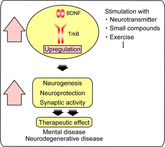
2. Activation of BDNF/TrkB System by Compounds

It is well known that BDNF and its high affinity receptor TrkB play essential roles to support survival in a variety of neuronal populations in the CNS and that they contribute to protect neurons against neurodegenerative disease, including AD [14,15,16]. Furthermore, downregulation of BDNF has been reported to be involved in the pathogenesis of mental disorders and neurodegenerative diseases, including AD, PD, and Huntington disease (HD). Therefore, activation of the BDNF/TrkB system is an attractive therapeutic target to treat these brain diseases.
Recently, evidence has demonstrated that induction of BDNF expression via stimulating/modulating activity of neurotransmitter receptors, and application of small molecules that activate TrkB are beneficial strategy for both neuroprotection and neuroregeneration. For example, activating 5-hydroxytryptophan 1A (5-HT1A) or 5-HT2A receptors that induce hippocampal BDNF expression has been examined. Afshar et al. investigated the effects of NAD-299 (an antagonist for 5-HT1A receptor) and TCB-2 (an agonist for 5-HT2A receptor) on learning and memory function in streptozotocin (STZ)-induced memory deficit animal models [17]. In their study, rats received intracerebroventricular (icv) injection of STZ exhibited decreased discrimination index in the novel object recognition test, reduced BDNF, and increased Aβ plaques in the brain, which were reversed by treatment with NAD-299 or TCB-2 [17]. Influence of neuropeptide Y (NPY), which is implicated as potential therapeutic target of HD, in expression of BDNF has been reported [18]. Activation of Y2 receptor (Y2R) by NPY or NPY13-16 (a selective agonist for Y2R) resulted in significant recovery from motor dysfunction in R6/2 mouse, a mouse model of HD. The activation of Y2R also decreased aggregated mutant huntingtin and increased levels of cAMP-regulated phosphoproteins, BDNF, and activated ERK1/2 in the HD model mice while selectively antagonizing Y2R by SF31 blocked the beneficial effects [18]. Recently, using R6/2 mice, Pardo et al. have shown that A-971432, a new selective agonist for sphingosine-1-phosphate (S1P) receptor 5, is a therapeutic candidate for the treatment of HD [19]. They found that chronic administration of A-971432 delayed the disease progression and prolonged the lifespan of symptomatic HD mice. Furthermore, they showed that the beneficial effects of A-971432 included the activation of the BDNF/TrkB system, with a significant decrease in mutant huntingtin aggregation [19].
In addition to stimulation of receptors with neurotransmitters, small compounds also induce upregulation of BDNF. Quercetin (3,5,7,30,40-pentahydroxyflavone), one of the flavonoids found in vegetables and fruits, has been shown protective effects against various diseases, including cancer, cardiovascular diseases, and neurodegenerative disorders [20,21]. Recently, increased expression of BDNF in the hippocampus of male rats that were received daily gavage with quercetin (20 or 50 mg/kg·bwt, for 30 days) has been reported, implying an involvement of BDNF in the neuroprotection by quercetin [22]. 7,8-Dihydroxyflavone (7,8-DHF), another flavonoid, stimulates TrkB receptor and exerts neuroprotective effect against traumatic brain injury [23]. Importantly, 7,8-DHF has a longer half-life time compared to BDNF and is able to pass the BBB due to its small molecular size, suggesting that the flavonoid is useful as a non-invasive clinical drug application [23]. Using HT22 cells, a mouse hippocampal neuronal cell line, positive effects of huperzine A (HupA) against oxidative/glutamate toxicity was reported. Cell death of HT22 cells after glutamate exposure was significantly restored by HupA treatment [24]. Such a HupA treatment reversed the downregulation of pAkt, p-mammalian target of rapamycin (pmTOR), pp70s6 kinase, and protein levels of both BDNF and pTrkB whereas NGF, NT-3, and NT-4 levels were not recovered, suggesting possible involvement of the BDNF/TrkB system in the effect of HupA [24]. In a rat model of acute spinal cord injury (SCI), Jisuikang, Chinese medicine compound, promoted recovery in neurological functions. Using the SCI model, in addition to increased survival rate of neurons, upregulated protein levels of BDNF was observed after Jisuikang administration [25].
It is well known that lipopolysaccharide (LPS), an endotoxin from Gram-negative bacteria, causes inflammation and deficits in learning and memory function in the CNS. Using mice treated with this endotoxin, Chowdhury et al. showed that imperatorin (IMP), one of the phytochemicals, exerted beneficial effects against poor memory retention (the Morris water maze and Y maze tests), via increasing TNF-α and IL-6 levels, and reducing hippocampal/cortical BDNF levels [26]. It has been reported that sulforaphane (SFN) is also effective for recovery from dysfunction of spatial memory caused by LPS [27]. Abnormal behaviors of the mice received LPS injection in the Morris water maze test was reversed by administration of SFN. Notably, an inhibitor for TrkB or mTOR signaling blocked the effects of SFN, suggesting that importance of BDNF/TrkB/mTOR signaling activation in the effect [27]. It has been reported that downregulation of PI3K/Akt/GSK3β signaling caused by sodium arsenite was ameliorated by curcumin application [28]. Rat received sodium arsenite showed decreased expression levels of BDNF, pAkt, pERK, pGSK3β, and p-cAMP response element binding protein (pCREB) in hippocampal region, which were reversed by curcumin application. In addition, curcumin was also effective on the learning and memory deficits in rats caused by sodium arsenite, suggesting that PI3K/Akt/GSK3β signaling stimulated by BDNF is involved in the effects of curcumin [28]. Interestingly, hippocampal Aβ accumulation and cognitive deficits in contextual fear conditioning in mice after repeated peripheral administration of LPS was observed, and the cognitive deficits associated with the increased hippocampal Aβ was reversed by a benzodiazepine agonist MRK-016 [29]. In their system, mRNA levels of BDNF decreased by LPS injections were also restored by MRK-016 [29]. Furthermore, effect of combined zinc and folic acid administration in a depression model established by chronic mild unpredictable stress was also studied [30]. Dou et al. compared the levels of 5-HT, dopamine and norepinephrine in four rat groups; control, paroxetine alone (P), zinc + folic acid (ZnY), and zinc + folic acid+ paroxetine (ZnYP), and found that 5-HT, dopamine and norepinephrine, NMDA, and TrkB mRNA levels in the frontal cortex were significantly increased in the ZnY group while no upregulation of BDNF was observed [30].

3. BDNF and Adult Neurogenesis

In several species, including rodents and human, generation of neurons persists throughout the life in some brain regions, such as the hippocampus. Growing evidence indicates that neurogenesis in the adult brain has been implicated in physiological brain function, such as learning and memory [31]. In addition, possible roles of adult neurogenesis in the pathogenesis of brain injury, neurodegenerative and psychiatric diseases have been extensively investigated [32,33]. In the adult rodent brain, neurogenic region is restricted in hippocampal DG and SVZ lining the lateral ventricles [31]. Interestingly, several lines of evidence demonstrate that BDNF plays an important role in adult neurogenesis although its effect is different between DG and SVZ regions [7].
In DG, neural progenitor cells (NPCs) constantly differentiate into excitatory granule neurons which migrate to the granule cell layer and are integrated into hippocampal neural circuits [7]. An in vitro study using neurosphere culture of NPCs isolated from adult rat hippocampus has demonstrated that BDNF promoted cell proliferation, neural differentiation and cell survival [34]. It has also been reported that the neurogenesis-promoting effect of BDNF in organotypic mice hippocampal slice culture was inhibited by pre-incubation with a TrkB antagonist, indicating important contribution of the BDNF/TrkB system [35]. Intrahippocampal infusion of BDNF in adult rats increased the number of newborn neurons and caused appearance of ectopic granule cells, suggesting that BDNF influences not only neurogenesis but also migration of newborn neurons [36]. In order to reveal a contribution of endogenous BDNF, several lines of genetically modified mice targeting BDNF or TrkB were generated. BDNF heterozygous mice (BDNF+/−) showed a decreased number of proliferative cells and newborn neurons in DG [37]. Genetic ablation of TrkB in NPCs (but not neurons) caused a reduction of proliferative hippocampal NPCs and newly generated granule neurons [38]. Inconsistent with these reports, another group reported a significant increase of proliferative cells and decrease of newborn neurons in DG in both BDNF +/− mice and dominant negative TrkB-overexpressing mice [39]. Although the reason of these discrepancies is unclear, consistent results on the reduction of newborn neurons have been highlighted the central role of BDNF in survival of newly generated neurons. Wang and colleagues showed that newborn granule neuron-specific BDNF deletion results in repression of dendrite growth and synaptogenesis to a similar extent of brain-specific BDNF conditional knockout mice [40]. Interestingly, they also found that newborn neuron-targeted BDNF gene transfer in brain-specific BDNF knockout mice recovered the deficit in dendrite and synapse in these neurons, demonstrating cell-autonomous actions of BDNF [40]. Such an abnormal dendrite growth and synaptogenesis is also observed in adult NPC-specific TrkB conditional knockout mice [41]. Note that this TrkB-deficient mouse exhibited disruption of neurogenesis-dependent long-term potentiation in DG, supporting the neurogenic effect of BDNF/TrkB signaling in the hippocampal circuits.
In SVZ regions, NPCs continuously differentiate into neuroblasts, and migrate into the granular cell layer or glomerular layers of olfactory bulb (OB), then differentiate into inhibitory interneurons or dopaminergic neurons [7]. Unlike NPCs in DG region, NPCs in SVZ and neuroblasts do not express TrkB although a few of them express pan-neurotrophin receptor p75NTR [42]. Young and colleagues reported that 0.3% of cells in SVZ region express p75NTR and these cells have ability to form neurosphere in vitro [42]. BDNF treatment to the neurospheres derived from p75NTR-positive cells increased neural production and this effect was disappeared in neurospheres derived from p75NTR-deficient mice [42]. The p75NTR-deficient mice have a reduced number of SVZ neuroblasts in vivo, suggesting that p75NTR signaling is essential for SVZ neurogenesis [42]. However, BDNF action on NPCs in SVZ and neuroblasts is still controversial because some group reported that infusion or overexpression of BDNF in SVZ region did not affect the number of newborn OB neurons whereas other group showed that intraventricular BDNF application increased the number of newly-generated OB neurons [43,44,45]. Thus, further study is required to clarify the contribution of BDNF on SVZ neurogenesis. After the migration of neuroblasts into the OB, these cells began to differentiate into neurons and express TrkB [46]. Similar to hippocampal DG, newborn neuron-targeted deletion of TrkB resulted in an impairment of dendritogenesis and spine formation in the OB newborn neurons, suggesting conserved role of the BDNF/TrkB system in neural maturation [46]. Furthermore, TrkB deletion severely decreased the survival of newborn dopaminergic neurons but not interneurons in glomerular layers [43]. Interestingly, the specific loss of dopaminergic neurons was mirrored by a corresponding increase in the number of interneurons, highlighting essential roles of TrkB in balancing the incorporation of defined classes of adult-born neurons in the OB [43].

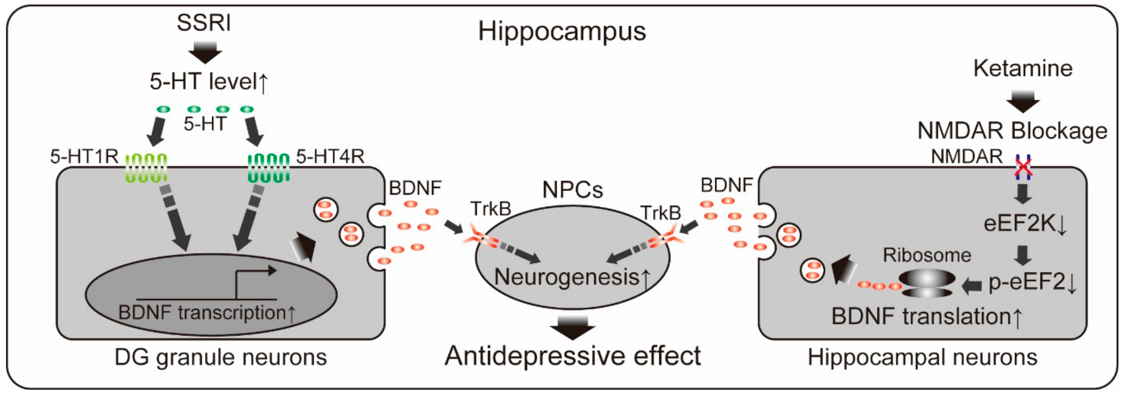
4. Monoaminergic System and BDNF-Mediated Neurogenesis

For over a decade, roles of neurogenic action of BDNF in the antidepressive effects of pharmacological and physical therapies have been well documented. Most of currently available antidepressants are categorized in monoaminergic antidepressants which primary act via increasing the activity of monoaminergic (serotonin, and norepinephrine) neural circuits usually within a few hours. In spite of the rapid increase of monoamine levels induced by these drugs, chronic administration for several weeks is required to achieve therapeutic response in patients with depression [47]. One of the possible explanations in this time-course discrepancy is a delayed induction of hippocampal neurogenesis by antidepressants. Chronic, but not acute, antidepressant treatment increases the number of proliferative cells and newborn neurons in rodents, which is consistent with the time-course of the antidepressive effect [48,49]. Investigation of post-mortal brain of depressive patients who had been treated with antidepressants also showed neurogenic action of antidepressants in the hippocampus [50,51,52]. Ablation of NPCs in adult DG by irradiation diminished antidepressant-induced antidepressive behavior in mice, demonstrating a requirement of hippocampal neurogenesis for the behavioral effect of antidepressants [48]. As expected, accumulating evidence indicates that antidepressant-induced neurogenic action is mediated by BDNF. It have been consistently reported that BDNF expression in the hippocampus after chronic (not acute) antidepressant treatment was increased in rodents and human [53,54,55]. Causative role of BDNF/TrkB signaling in the antidepressant-induced hippocampal neurogenesis was examined using genetic deletion of TrkB in hippocampal NPCs [38]. Conditional TrkB-deficient mice did not show both antidepressant-induced neurogenic effect and antidepressive behavior. Moreover, BDNF injection into hippocampal DG produced antidepressive effect as early as 3 days after its single administration and lasted for at least 10 days [56]. As BDNF infusion directly induces hippocampal neurogenesis, this antidepressive effect of BDNF may also be mediated by enhancement of neurogenesis [36]. Recent studies have addressed how antidepressants increase levels of BDNF and neurogenesis. Samuels and colleagues reported that genetic deletion of 5-HT1AR in DG granule neurons abolished antidepressive behaviors induced by selective serotonin reuptake inhibitors (SSRIs, widely used classes of antidepressants) and attenuated increased neurogenesis and BDNF [57]. Notably, they also found that specific deletion of 5-HT1AR in newborn granule cells in DG did not impair SSRI-induced behavioral and neurogenic responses, suggesting that 5-HT1AR signaling non-autonomously affects neurogenic responses. In addition to 5-HT1AR, contribution of 5-HT4R has also been reported. Pharmacological inhibition of 5-HT4R diminished antidepressive effects of SSRIs and 5-HT4R stimulation conversely exhibited antidepressant effects [58]. Imoto and colleagues showed that 5-HT4R knockout mice failed to respond to SSRI [59]. Interestingly, they found that chronic SSRI treatment decreased a maturity of preexisting granule neurons in DG (called “dematuration”) and this dematuration was not observed in 5-HT4R knockout mice. As BDNF induction and neurogenic action by SSRI were well correlated with the progress of dematuraion of granule neurons, the reduced maturity of granule neurons may be accountable for SSRI action although further studies to elucidate causal role of the dematuration by SSRI in neurogenesis may be required. Overall, current knowledge of SSRI actions in DG suggests that increased serotonin stimulates 5-HT1AR and/or 5-HT4R in preexisting granule neurons to induce neurogenesis via non-cell autonomous mechanism, possibly through enhanced production and secretion of BDNF (Figure 1).

5. Other Antidepressive Therapy and BDNF-Mediated Neurogenesis

Involvement of BDNF/TrkB signaling and hippocampal neurogenesis have also studied in not only monoaminergic antidepressants therapy but also in other types of antidepressive therapies including electroconvulsive shocks, exercise, enriched environment, flavonoids, and non-conventional antidepressants treatment [38,60,61,62,63,64,65]. It has been demonstrated that these therapies stimulated both the BDNF/TrkB system and hippocampal neurogenesis [38,60,61,62,63,64,65]. In addition, causative role of BDNF-mediated neurogenesis in antidepressive effects have also been demonstrated. NPC-specific TrkB knockout mice were insensitive to the exercise-induced improvement in depressive behaviors and showed no change in the number of proliferative cells and newborn neurons in DG after exercise [38]. Antidepressive effect and neurogenic action of silibinin, a polyphenolic flavonoid from Silybum marianum, was abrogated by icv injection of a TrkB antagonist in mice [63]. Recently, ketamine, an NMDA receptor antagonist, has been attracting researcher’s attention as a non-conventional antidepressant [66]. Single administration of ketamine produces rapid-onset antidepressant response in depressive patients within few hours (lasting 1–2 weeks) unlike effects by monoaminergic antidepressants. Therapy-response rate reaches over 60% of monoaminergic antidepressant drug non-responders and tolerability seems acceptable at least in the short term [66]. Recent animal studies revealed an involvement of BDNF and neurogenesis in the ketamine-induced antidepressive effects [64,65]. NMDA receptor blockage by ketamine led to deactivation of eukaryotic elongation factor 2 (eEF2) kinase, which resulted in decreased eEF2 phosphorylation and de-suppression of BDNF expression within 30 min after treatment [64]. BDNF heterozygous mice did not show antidepressive behavior at the time, suggesting rapid requirement of BDNF in the antidepressive effect of ketamine [64]. Increased hippocampal neurogenesis was also observed in the ketamine-treated mice at 24 h after treatment [65]. Deletion of TrkB in adult NPCs diminished the neurogenic effect of ketamine and antidepressive behaviors at 24 h after the treatment. Note that antidepressive behaviors at 1 h after ketamine treatment was not affected by adult NPCs-specific TrkB deletion, suggesting that the sustained antidepressive effect of ketamine depends on BDNF/TrkB-mediated neurogenesis while rapid antidepressive effect is occurred through neurogenesis-independent BDNF/TrkB action [65].
Although possible contributions of BDNF-mediated neurogenesis in the antidepressive therapy is likely, BDNF delivery into hippocampus is very difficult because of its inability to cross the BBB and short half-life time (<10min) [23]. Another option is to use other growth factors that have similar biological effects as BDNF. For example, insulin growth factor I (IGF-I) is one of the candidates because IGF-I can stimulate the same intracellular signaling as BDNF via activating the IGF-I receptor and penetrate the BBB [67]. It has been reported that IGF-I treatment enhances proliferation and differentiation of NPCs in vitro [68,69,70]. Recently, we reported that IGF-I application restored impairments in differentiation/maturation of newborn neurons caused by exposure to corticosterone, a stress-related hormone, in vitro [71]. Neurogenesis induced by IGF-I in adult hippocampal DG was revealed by in vivo studies using both subcutaneous (sc) and icv injection [72,73,74]. Furthermore, several animal studies indicate that icv injection of IGF-I produces antidepressive behavior in rodents [75,76]. However, requirement of neurogenic action in antidepressive effects by IGF-I is controversial because one study reported no change in neurogenesis when antidepressive behavior was appeared [75]. Therefore, precise research using irradiation technique to kill NPCs or adult NPC-specific IGF1R knockout mice is useful to clarify the relationship between neurogenesis and antidepressive behavior. It is also important to examine whether sc injection of IGF-I can achieve antidepressive effects. Although further studies were required, IGF-I has a potential to ameliorate depressive symptoms via non-conventional strategies.

6. Influence of Val66Met Polymorphism of BDNF on the Neurogenesis

Recently, many researchers have focused on possible relationship between BDNF polymorphism and brain disease including neuropsychiatric disorders [77]. A non-synonymous polymorphism which causes a valine (Val) to methionine (Met) substitution at position 66 of BDNF genes is carried by 40–50% and 25–32% of the Asian and Caucasian population, respectively [78]. Remarkably, several genetic studies have showed that Met carriers displayed smaller hippocampal volumes and lower recognition memory performance compared with Val homozygotes [79,80,81,82]. In vitro studies using mouse neurons reported that the Val66Met polymorphism impaired trafficking of BDNF mRNA to dendrites, localization of BDNF in secretory granules and activity-dependent secretion of BDNF [81,83]. In order to investigate possible impact of the Val66Met polymorphism on brain functions and behaviors, several animal studies using a variant BDNF knock-in mouse carrying BDNFMet/Met have been also reported. BDNFMet/Met mice exhibited defects in survival but not proliferation of adult-born neurons in both hippocampal DG and SVZ [84,85]. These mice exhibited increased anxiety-related behaviors when placed in stressful condition, and these behavioral abnormalities were not rescued by the treatment of a SSRI, fluoxetine [86]. It has been reported that chronic fluoxetine treatment failed to increase levels of BDNF, survival of newborn neurons, long-term potentiation in DG, and antidepressive behaviors in BDNFMet/Met mice [84]. Recently, it has been also reported that BDNFMet/Met mice did not show exercise-induced upregulation of BDNF, increase of newborn neurons, and antidepressive effects [87]. In addition to neurogenesis in the DG, dendritogenesis and synaptogenesis in the prefrontal cortex were also impaired in BDNFMet/Met mice [88]. BDNF Val66Met polymorphism caused constitutive atrophy of distal apical dendrites, decreased spine density/diameter and decrements in excitatory postsynaptic currents in layer V pyramidal neurons. Moreover, the ketamine-induced synaptogenesis and antidepressive behavior were also blocked in the mice [88]. It is possible that antidepressive effects induced by antidepressants and physical exercise are affected by alteration in the activity-dependent BDNF secretion due to Val66Met polymorphism.

7. Alzheimer’s Disease and BDNF

AD is a progressive neurodegenerative disorder that is accompanied by symptoms, such as memory and cognitive deterioration. Patients with AD show several neuropathological features, including extracellular amyloid plaques composed of aggregated Aβ peptide and intracellular neurofibrillary tangles of aggregates consisting of abnormally phosphorylated tau, a soluble microtubule-associated cytoskeletal protein. Degeneration especially in cholinergic neurons in basal forebrain regions is considered as the earliest pathological event in AD. Because these neurons innervate a variety of brain regions, including the hippocampus and cortex, a reduction in the retrograde neurotrophic signals are considered to be involved in the onset of AD [11].
Aβ peptides of either 40 or 42 amino acids are produced in the process of aberrant proteolytic cleavage of amyloid precursor protein (APP) by β- and γ-secretases [89]. Although α-secretase also cleaves APP effectively, the cleavage is occurred within the Aβ sequence and does not produce Aβ peptides. As the result of altered balance in the APP processing, the over-produced Aβ peptides aggregate to form soluble oligomers or insoluble beta-sheet fibrils accumulated in diffuse senile plaques, likely depending on the extracellular concentration of Aβ peptides [90]. Although it is still unclear how hyperphospholyration of tau is caused, there is a reciprocal relationship between Aβ peptides and tau hyperphospholyration and aggregation. Oxidative stress by Aβ oligomers was able to stimulate tau hyperphosphorylation [91,92] while site-specific phosphorylation of tau can protect neurons from Aβ toxiciticy [93].
A glycoprotein ApoE is also known as one of the risk factors for AD. It mediates the delivery of lipids like cholesterol via ApoE receptors in the membrane. Human ApoE proteins consist of three isoforms ApoE2, E3, and E4, with different isoelectric point as pH 5.4, 5.5, and 6.1, respectively [94]. There is no consensus on how only ApoE4 contributes to the pathogenesis of AD. However, a recent study showed that ApoE secreted from glial cells triggered the transcription of APP in neurons through MAPK pathway and AP-1 (activator protein 1), a transcription factor. The Aβ production induced by glial ApoE4 was most potent among the three types of ApoE [95].
Due to the essential roles in neuronal aspects of CNS function as mentioned above, possible contribution of the BDNF/TrkB dysfunction in the development of AD has been demonstrated. Interestingly, some reports showed reduced levels of BDNF and TrkB in patients with AD while others demonstrated the opposite results. Ferrer and colleagues reported decreased BDNF and full-length TrkB levels in the frontal cortex of patients with AD, and increased truncated TrkB that lacks its intracellular kinase domain and is mainly expressed in glial cells [96]. Downregulation of the BDNF transcripts in parietal cortex [97,98] and reduced protein levels of BDNF in the hippocampus and parietal cortex [99,100] have also been reported. Recent study showed a negative correlation between BDNF mRNA expression in the dorsal lateral prefrontal cortex and cognitive decline in the AD patients [101]. On the other hand, increased plasma concentration of BDNF has been observed in AD and mild cognitive impairment patients [102,103]. Similarly, it is suggested that the levels of BDNF are inconsistently regulated by Aβ in the preclinical studies. Xia et al. demonstrated that BDNF expression in the cerebral cortex and hippocampus was markedly reduced in an AD mouse model (APP/PS1 transgenic mice) at 3 and 9 months old [104]. Icv injections of Aβ oligomers to 3-month-old C57BL/6 mice dramatically decreased BDNF protein and mRNA levels within 24 h. They further clarified that peroxisome proliferators-activated receptor γ coactivator 1 α (PGC-1α) and fibronectin type III domain-containing 5 (FNDC5) were involved in the Aβ-dependent downregulation of BDNF [104]. In contrast, elevated BDNF protein and mRNA levels were observed in SH-SY5Y human neuroblastoma cells after exposure to Aβ peptides [105].
It has been reported that neuronal cell death and dysfunction in the animal models of AD were ameliorated through activating BDNF/TrkB signaling. Lentiviral BDNF gene delivery into the entorhinal cortex of transgenic mice expressing the APP with mutations at age 2 months improved hippocampal-dependent contextual fear conditioning after 3 months [106]. Obvious cell loss observed in the layers II-VI of the entorhinal cortex of these transgenic mice was also ameliorated by the BDNF delivery. Interestingly, the BDNF gene delivery did not change the number of amyloid plaque, indicating that the neuroprotective effect of BDNF was not through the downregulation of Aβ in the AD models [106]. Rutin (3,3’,4’,5,7-pentahydroxyflavone-3-rhamnoglucoside), one of the flavonoids, rescued reduction in the memory retrieval function in rats injected Aβ into the deep frontal cortex, through upregulation of BDNF and MAPK pathway activation [107]. Similarly, a 5 consecutive day intraperitoneal (ip) administration of curcumin, a major polyphenol from Curcumalonga, ameliorated cognitive deficits in rats induced by an icv injection of Aβ, via the upregulation of BDNF and activating MAPK pathway. Sole injection of BDNF into the hippocampus also improved the impairment in cognitive function [108]. Oral administration of SCM-198, an alkaloid extracted from Herba leonuri, for 3 months improved both recognition and spatial memory in the APP/PS1 transgenic mice without affecting the amount of Aβ. Importantly, SCM-198 ameliorated the decline in BDNF expression and TrkB activation both in in vivo and in cortical cultures [109].
Low-level laser therapy, a recently developed intervention, regulates neuronal function in neuronal cultures, animals, and clinical conditions [110] and rescues the neuronal loss and dendritic atrophy in cultured hippocampal neurons treated with Aβ and hippocampal neurons from the APP/PS1 mice via increasing BDNF expression [111]. As mentioned above, there is accumulating evidence for a potential use of 7,8-DHF, a small-molecule of TrkB agonist, in the therapeutic effect for AD patients. Importantly, 7,8-DHF can penetrate the BBB and systemic administrations of 7,8-DHF for 10 consecutive days restored the memory deficits of 5XFAD mice model of AD. 7,8-DHF blocked β-secretase elevation that causes Aβ accumulation in the 5XFAD mouse brain [112]. Furthermore, 7,8-DHF prevented the loss of hippocampal synapses and memory deficits in the 5XFAD mice [113,114,115].
Voluntary exercise that has been reported to increase BDNF in the several brain regions [116] was tested in the AD models. As expected, treadmill exercise saved neurons from Aβ-induced cell death in the cortex of the APP/PS1 mouse [117] and improved the spatial memory function of another AD model mouse, in both of which BDNF upregulation was confirmed to be involved in the effects [118]. However, there is no significant change in the accumulation of Aβ in either the cerebral cortex or hippocampus after exercise. It was also reported that BDNF reduced production of Aβ peptides through enhanced α-secretase-mediated processing of APP in the APP/PS1 transgenic mice [119]. The relationship between BDNF and APP/Aβ seems to be more complex. The pro-domain of BDNF protein exerted toxic effects only in the presence of Aβ in SH-SY5Y human neuroblastoma cells [120]. Ruiz-Leon et al. demonstrated that BDNF stimulated APP expression by activating promoter of APP gene through TrkB-, Ras/MAPK- and PI3K/Akt-dependent mechanisms in SH-SY5Y cells [121]. It was also reported that ApoE4 carriers have reduced serum BDNF levels [122]. Sen and colleagues showed that ApoE4 increased nuclear translocation of histone deacetylase 6 (HDAC6) in human neurons, which in turn suppressed BDNF transcription from the BDNF exon IV by binding of HDAC6 to the promoter region. In contrast, ApoE3 increased acetylation of histone 3 and stimulated BDNF expression [123]. They further reported regulatory roles of ApoE in BDNF secretion from cultured astrocytes. ApoE2 treatment for 24 h increased intracellular amount of pre-pro BDNF while ApoE4 reduced it. Astrocytes exposed to ApoE3 secreted 1.75-fold higher pro-BDNF than ApoE2-treated astrocytes. Furthermore, ApoE2-treated astrocytes secreted mature-BDNF 3 times higher than ApoE3-treated astrocytes. ApoE4 dramatically reduced secretion of both forms of BDNF from astrocytes [124]. These data shows reciprocal and complex interactions between BDNF/TrkB signals and pathogenesis of AD.
On the other hand, several studies indicated that phosphorylation of tau protein is also regulated by the BDNF/TrkB system. Elliott et al. reported that BDNF induced a dephosphorylation of tau in P19 mouse embryonic carcinoma cells within 15 min via PI3K signaling pathway [125]. 7,8-DHF was also reported to inhibit expression of abnormal tau via activating PI3K/Akt pathway [126]. MiR-322, the rodent homologue of human miR-424, is predicted to target the 3’-untranslated region (3’-UTR) of BDNF mRNA and found to be increased in AD mouse brain. Zhang et al. recently showed that overexpressed miR-322 promoted tau phosphorylation by negatively regulating BDNF levels and TrkB activation whereas silencing miR-322 increased TrkB activation and accelerated dephosphorylation of tau in Neuro2A mouse neuroblastoma cells [127]. Intralateral ventricle injection of adeno-associated virus carrying BDNF gene in P301L mouse model of tauopathy at 3 months of age before the occurrence of neurofibrillary tangle formation in the mice prevented the neuronal loss and the behavioral deficits without affect hyperphosphorylation levels of tau [128]. There are several studies that investigated the effect of abnormal tau on expression and secretion of BDNF. In a mouse model of tauopathy, P301S tau transgenic (P301S) mice show memory deficits, synaptic and neuronal loss in the cortex and spinal cord due to the fibrillary tau tangles emerged throughout the CNS [129]. Rosa and colleagues reported a secretion of BDNF was impaired in the vitreous of the mouse model while mRNA and protein levels of BDNF in retinas were not altered [130]. Both BDNF expression and secretion in rat primary cortical neurons were significantly suppressed by okadaic acid-induced tau hyperphosphorylation [131]. Although these findings suggest a limitation of the usage of serum BDNF levels as a biomarker of AD for early diagnosis, potentiation of BDNF/TrkB signals will be a one of the therapeutic targets for the treatment of AD symptoms. Future studies on other small molecules and the gene delivery, and cellular transplantation that stimulate BDNF/TrkB signaling pathways are important.

8. Parkinson’s Disease and BDNF

Parkinson’s disease (PD) is also progressive neurodegenerative disorder like AD. PD is clinically characterized by bradykinesia, rigidity and rest tremor, and is pathologically characterized by the loss of dopaminergic neurons in the substantia nigra pars compacta, which results in the decline of dopamine (DA) levels in the striatum. Dystrophic neurites and intracellular inclusions called “Lewy bodies” which mainly contain α-synuclein are also pathological hallmarks of PD [132,133]. It is well known that patients with PD also display mild cognitive deficits correlated with severity of symptoms. Because BDNF is an important mediator for neuronal survival and synaptic plasticity to maintain brain functions, possible contribution of BDNF/TrkB dysfunction in the cognitive deficits in PD patients was hypothesized. Positive correlations between serum BDNF concentration and severity of the symptoms [134,135] and cognitive deficits [136,137] in the patients with PD have been reported. Reduced levels of BDNF mRNA [12] and protein [13] in the substantia nigra of the postmortem PD patients were also demonstrated. Furthermore, serum BDNF levels was negatively correlated with degeneration levels of striatal dopaminergic neurons [138]. However, a large-scale study in which over 600 individuals were participated concluded that significant decrease of serum BDNF levels were observed in patients with AD, frontotemporal dementia, Lewy body dementia, vascular dementia whereas significant increase was confirmed in PD patients [139]. On the other hand, the latest study of the meta-analysis indeed supports an association between decreased blood concentration of BDNF and PD [140].
Preclinical studies using animal models of PD have been carried out to investigate whether BDNF dysfunction is linked to the PD symptoms. MPTP (1-methyl-4-phenyl-1,2,3,6-tetrahydropyridine), a dopamine neurotoxin, induces parkinsonism in animals. Using female rhesus monkeys received MPTP administration, Collier et al. found a decline of BDNF levels in striatal tissue of young and middle-aged animals compared with age-controlled animals while no significant change in the tissue of old animals where baseline BDNF levels were decreased [141]. Another model of preclinical stage of PD induced by 6-hydroxydopamine (6-OHDA) which causes moderate dopaminergic lesion has been examined. Bilateral injections of 6-OHDA into the rat ventral region of the caudate-putamen caused downregulation of BDNF and TrkB mRNA levels in the hippocampus, amygdala, and habenula [142]. Rats received 6-OHDA administration in the striatum exhibited reduction of BDNF but increase of TrkB [143].
Supporting the idea that BDNF dysfunction is involved in the PD symptoms, clinical rehabilitations and treatments likely ameliorates decline of BDNF in PD patients and animal models. A protocol of cognitive rehabilitation such as a 1-month 12-session treatment (three sessions a week) which focuses on the training of executive functioning increased BDNF levels in serum of PD patients affected by mild cognitive impairment, with improved cognitive performance [144,145]. Enhanced activation of the BDNF/TrkB system in lymphocyte after an intensive rehabilitation in PD patients was also reported [146]. Recent reports also showed that exercise or physical training ameliorated the reduced levels of BDNF in patients with PD [147]. In the animal model of PD, 6-OHDA-induced reductions of BDNF and TrkB in the striatum and hippocampus of mice were restored by physical training such as running on a treadmill [148]. The involvement of BDNF/TrkB signals in the ameliorating effect of physical exercise in the PD model was confirmed by using K252a, a blocker for Trk receptors [149]. There are several studies that show a protective effect of 7,8-DHF in degeneration of dopaminergic neurons through TrkB activation in the rodent models of PD induced by 6-OHDA or MPTP [150,151].
Recently, researchers are also focusing on the relationship between BDNF/TrkB functions and α-synuclein in the context of the development of PD. Goldberg and colleagues showed that transplantation of NPCs into the striatum of α-synuclein-expressing mouse model of PD that exhibits progressive development of Lewy body pathology with significant cognitive dysfunction significantly improved cognitive and motor functions. Importantly, transplantation of BDNF-depleted NPCs failed to improve these dysfunctions, and only BDNF gene delivery into the striatum mimicked the effects [152]. Overexpression of α-synuclein in PC12 cells led to suppression of BDNF transcription likely via downregulation of CREB [153]. Kang et al. found an interesting phenomenon that α-synuclein binds directly to the kinase domain of TrkB receptor. The interaction inhibited BDNF/TrkB signaling pathways and caused degeneration of dopaminergic neurons. TrkB ubiquitination was also induced by α-synuclein, which led a reduction in TrkB protein levels [154].

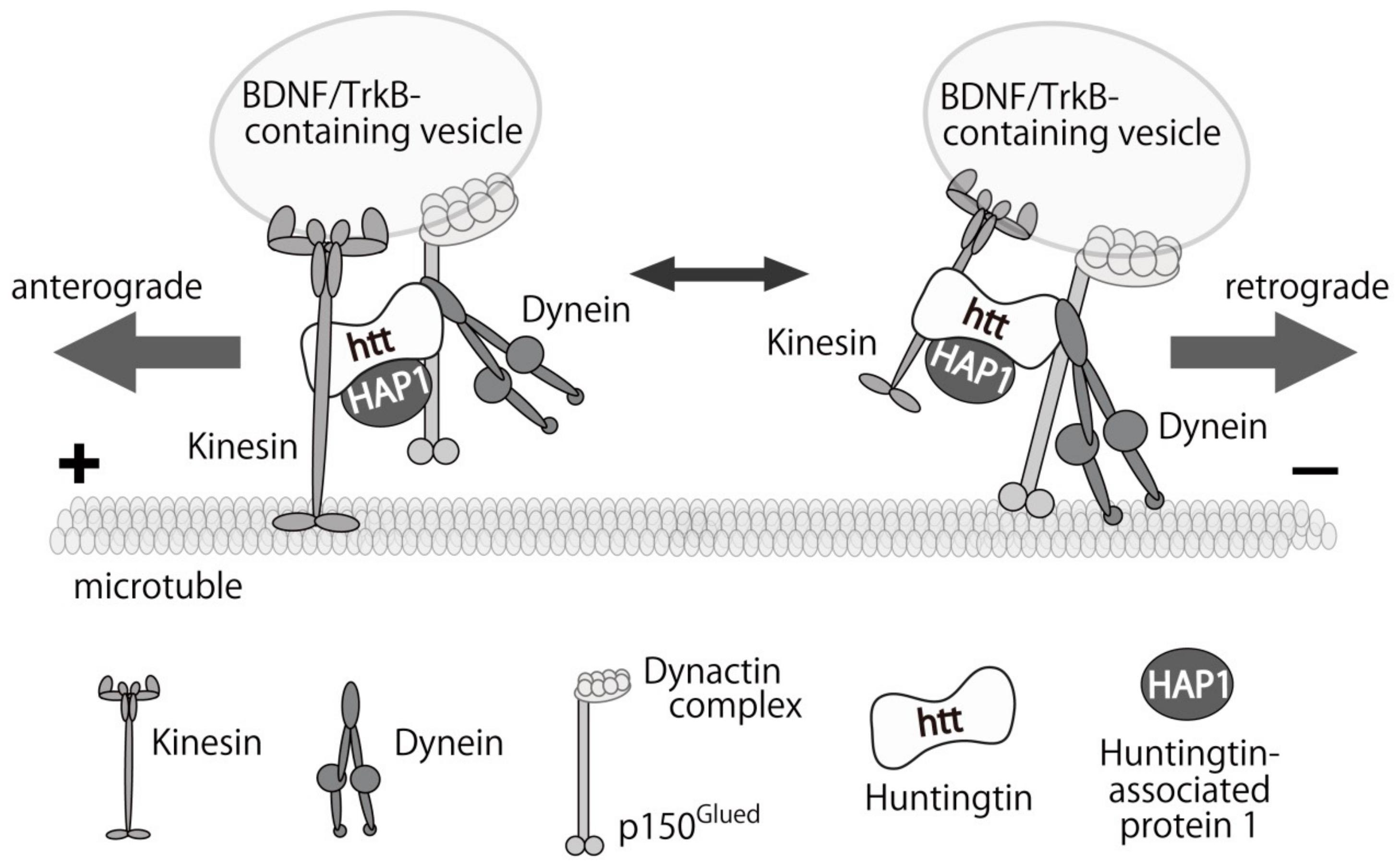
9. Huntington’s Disease and BDNF

Huntington’s disease (HD) is also a progressive neurodegenerative disease, characterized by abnormal involuntary movements and cognitive disability [155,156,157]. Patients with HD carry an expanded CAG-repeat in huntingtin (HTT) gene that generates abnormally elongated poly glutamine stretch in the N-terminal region of the huntingtin (htt) protein. Because progressive loss of striatal neurons is the most obvious pathological change in HD and these neurons can not produce BDNF, the impaired supply of BDNF from cortical neurons projecting their axons to the striatum is considered to be involved in the neuronal loss observed in HD patients.
Ferrer et al. reported reduced levels of BDNF in the caudate and putamen in the post-mortem brain of HD patients and normal levels in the cerebral cortex and hippocampus [155]. On the other hand, Zuccato et al. showed a significant reduction in BDNF mRNA and protein in the cortex of HD patients by using a systematic and quantitative assessment [156]. Many reports also have shown significant downregulation of BDNF in the several brain regions in animal models of HD [157]. Although correlations between serum levels of BDNF and HD have been controversial, a recent report concluded that levels of BDNF mRNA/protein in human blood samples were not suitable for biomarkers of HD [158]. The mutant htt protein has been considered to be toxic and cause neuronal death in striatal neurons. Recent findings, however, shed light on a beneficial function of wild-type htt protein. Cattaneo and colleagues demonstrated that wild-type htt stimulated transcription of BDNF in cortical neurons, which was critical for the survival of striatal neurons [159]. Conversely, Ma et al. showed normal pro-form, but reduced mature BDNF protein expression and activation of TrkB in the sensorimotor cortex and striatum of an aged HD mouse model, suggesting unaffected BDNF production in the cortical neurons [160]. Normal protein levels of BDNF and TrkB were also observed in another HD mouse, but levels of activated TrkB was reduced by unknown mechanisms in the model [161] while Plotkin et al. reported normal activation of TrkB but decreased downstream signals of TrkB via p75NTR-dependent mechanism [162].
Potential therapeutic effects of BDNF have been reported in rodent HD models. Bilateral expression of BDNF by adeno-associated virus in the striatum ameliorated the impairments in both motor and cognitive functions in transgenic HD rats, with increased volume of the striatum and the number of neural cells [163]. Improved motor function in quinolinic acid-induce HD mouse model was also accomplished by transplantation with embryonic stem cell-derived neural progenitors that overexpress BDNF [164]. These results suggest that at last in part of the cause of neuronal loss observed in the striatum of the HD patients could be attributed to the impairment of the BDNF/TrkB signals in the region and compensating these functions would be a promising strategy for the future treatment for HD.

10. BDNF/TrkB Transport As a Therapeutic Target of Neurodegenerative Diseases

Characterization of the relationship between BDNF and disease symptoms is difficult as there are several cellular processes (e.g., transcription, translation, and BDNF mRNA and protein stability) to regulate the amount of BDNF in tissues. Given that both reduced and elevated levels of BDNF/TrkB have been reported in the patients and animal models of neurodegenerative disorders, researchers in this field have been asked to find more reliable and direct impairments of the BDNF/TrkB functions in the pathogenesis of the diseases. Possible candidate of the BDNF/TrkB dysfunctions is the intracellular transport process of “synthesized” BDNF-containing vesicles for preparing secretion to surrounding cells and “received” BDNF-TrkB complex to send signals to the cell body.
Gauthier et al. revealed an important function of normal htt as a member of motor protein complex [165]. They showed that htt protein positively regulated the axonal transport of BDNF-containing vesicles in cortical neurons, and mutant htt expression or knockdown of normal htt hampered the transport. This report suggested that impaired trophic support from cortical neuron might contribute to the neuronal loss in the striatum, the main pathological change in HD [165]. The transport of BDNF/TrkB-containing vesicles is carried out by kinesin and dynein motor protein complex in a microtubule-dependent movement. The complex consists of normal htt, and huntingtin-associated protein (HAP), dynactin, and Rab GTPase family proteins [166] (Figure 2).
Using a mouse model of AD, Tg2576 transgenic mice, that express the Swedish mutation of the human APP, Poon et al. found significant reduction in the retrograde transport rate of BDNF, and suppression of TrkB signals in cultured cortical neurons. Importantly, they also confirmed that γ-secretase inhibitor restored the reduced BDNF transport and Aβ oligomers alone impaired the retrograde transport of BDNF in axon [167]. Seifert and colleagues also reported impairments in both the anterograde and retrograde transport of BDNF-containing vesicles in hippocampal neurons derived from another AD mouse model (5xFAD) with increased Aβ expression. In the study, a fast action (within 5 min) of Aβ on the transport was also revealed [168]. Gan et al. proposed Ca2+-dependent mechanisms of the disruption of BDNF transport induced by Aβ oligomers. Aβ enhanced Ca2+ influx via both axonal voltage-gated Ca2+ channels and dendritic glutamate receptors. Then, calmodulin binds to free Ca2+ and subsequently activates calcineurin-GSK3β, which may in turn phosphorylated motor proteins and disrupt BDNF transport [169]. Interestingly, similar defects in intracellular BDNF trafficking have been reported in PD models. Cultured hippocampal neurons exposed to α-synuclein aggregates disrupted axonal transport of BDNF/TrkB and Rab7-containing endosomes. In addition, TrkB/Rab7 and pERK5 which is responsible for propagation of the TrkB signaling were accumulated with α-synuclein aggregates [170]. Fang et al. showed the similar result that the retrograde axonal transport of BDNF in cortical neurons was dramatically impaired in an α-synuclein transgenic mice [171]. They found a direct interaction of α-synuclein with dynein, which inhibited the trafficking and BDNF/Trk-induced trophic signaling likely through activating Rab5 and Rab7 [171]. Furthermore, immunoreactivity of kinesin was significantly reduced in nigral neurons in the sporadic PD patients, and overexpression of α-synuclein in a PD mouse model resulted in reduced levels of motor proteins, including kinesin and dynein. Importantly, the reduction in kinesin levels preceded the degenerations in dopaminergic neurons, a phenotypic marker for the early stage of PD [172]. These results clearly suggest that preventing the impairments in the intracellular transport of BDNF/TrkB is a possible therapeutic target in these diseases. Recently, we reported a beneficial aspect of glucocorticoid to enhance the transport of BDNF-containing vesicles in cortical neurons. A synthetic glucocorticoid dexamethasone DEX, a potent agonist for glucocorticoid receptor (GR), increased huntingtin protein which forms motor complex with both kinesin and dynein and enhances the microtubule-dependent vesicular transport [173]. Supporting this, offspring from DEX-treated mothers were improved in their memory functions in a mouse model of AD [174], and DEX administration significantly ameliorated the degeneration of dopaminergic neurons in the substantia nigra in the MPTP-induced PD mouse [175]. Although it is possible that an inhibition of inflammatory activity by DEX was involved in these effects, restoring an impaired BDNF transport in model mice is also possible mechanism in the DEX effect. Because BDNF itself cannot cross the BBB, further studies focusing on the intracellular BDNF/TrkB transport would be important for the better understanding of the pathophysiology of neurodegenerative diseases and developing new medical treatments in the future.

11. Conclusion

In this review, we introduced the contribution of altered BDNF/TrkB system in the pathophysiology of brain diseases including mental disorders and neurodegenerative diseases. We discussed possible involvement of BDNF upregulation in the improvement of pathophysiological behaviors in disease models. In the process of BDNF upregulation, there are many steps, including transcription, translation, and degradation of both BDNF mRNA and protein. Thus, although characterization of the relationship among BDNF and disease symptoms is indeed difficult due to the multiple processes to regulate the amount of BDNF in tissues, detailed mechanisms underlying the upregulation of BDNF are need to be clarified to achieve more effective therapeutic approaches for neuroprotection. In addition to the cellular processes that regulate the amount of both BDNF mRNA and protein, the changes in the efficiency of secretion and transport of BDNF protein to the secretion sites in neurons may affect synaptic function and cell survival in the pathophysiology. Therefore, although studies concerning small compounds that can increase mRNA and/or protein expression levels of BDNF and regulate the related intracellular signaling molecules have been extensively carried out as mentioned above, to investigate secretion and/or transport of BDNF protein in the presence of these small compounds (e.g., a variety of flavonoid) is very important in the future study.

Author Contributions

T.N. wrote and edited the paper; H.O. and N.A. wrote the paper.

Funding

This study was supported by grants from the Grant-in-Aid for Scientific Research(C) (JSPS KAKENHI JP16K06996, T.N.; JP18K07891, N.A.) in the Ministry of Education, Culture, Sports, Science, and Technology of Japan, and supported by grants from by Takeda Science Foundation (T.N. and N.A.).

Conflicts of Interest

The authors declare no conflict of interest.

References

  1. Chao, M.V. Neurotrophins and their receptors: A convergence point for many signalling pathways. Nat. Rev. Neurosci. 2003, 4, 299–309. [Google Scholar] [CrossRef] [PubMed]
  2. Ohira, K.; Hayashi, M. A new aspect of the TrkB signaling pathway in neural plasticity. Curr. Neuropharmacol. 2009, 7, 276–285. [Google Scholar] [CrossRef] [PubMed]
  3. Lu, B.; Nagappan, G.; Guan, X.; Nathan, P.J.; Wren, P. BDNF-based synaptic repair as a disease-modifying strategy for neurodegenerative diseases. Nat. Rev. Neurosci. 2013, 14, 401–416. [Google Scholar] [CrossRef] [PubMed]
  4. Li, H.Y.; Zhao, Y.H.; Zeng, M.J.; Fang, F.; Li, M.; Qin, T.T.; Ye, L.Y.; Li, H.W.; Qu, R.; Ma, S.P. Saikosaponin D relieves unpredictable chronic mild stress induced depressive-like behavior in rats: Involvement of HPA axis and hippocampal neurogenesis. Psychopharmacology 2017, 234, 3385–3394. [Google Scholar] [CrossRef] [PubMed]
  5. Wang, H.; Zhao, Y.; Wang, Y.J.; Song, L.; Wang, J.L.; Huang, C.; Zhang, W.; Jiang, B. Antidepressant-like effects of tetrahydroxystilbene glucoside in mice: Involvement of BDNF signaling cascade in the hippocampus. Neurosci. Ther. 2017, 23, 627–636. [Google Scholar] [CrossRef] [PubMed]
  6. Numakawa, T.; Odaka, H.; Adachi, N. Actions of Brain-Derived Neurotrophic Factor and Glucocorticoid Stress in Neurogenesis. Int. J. Mol. Sci. 2017, 18, e2312. [Google Scholar] [CrossRef] [PubMed]
  7. Vilar, M.; Mira, H. Regulation of Neurogenesis by Neurotrophins during Adulthood: Expected and Unexpected Roles. Front. Neurosci. 2016, 10, 26. [Google Scholar] [CrossRef] [PubMed]
  8. Foltran, R.B.; Diaz, S.L. BDNF isoforms: A round trip ticket between neurogenesis and serotonin? J. Neurochem. 2016, 138, 204–221. [Google Scholar] [CrossRef] [PubMed]
  9. Lu, B. Pro-region of neurotrophins: Role in synaptic modulation. Neuron 2003, 39, 735–738. [Google Scholar] [CrossRef]
  10. Teng, K.K.; Hempstead, B.L. Neurotrophins and their receptors: Signaling trios in complex biological systems. Cell. Mol. Life Sci. 2004, 61, 35–48. [Google Scholar] [CrossRef] [PubMed]
  11. Serrano-Pozo, A.; Frosch, M.P.; Masliah, E.; Hyman, B.T. Neuropathological alterations in Alzheimer disease. Cold Spring Harb. Perspect. Med. 2011, 1, a006189. [Google Scholar] [CrossRef] [PubMed]
  12. Howells, D.W.; Porritt, M.J.; Wong, J.Y.; Batchelor, P.E.; Kalnins, R.; Hughes, A.J.; Donnan, G.A. Reduced BDNF mRNA expression in the Parkinson’s disease substantia nigra. Exp. Neurol. 2000, 166, 127–135. [Google Scholar] [CrossRef] [PubMed]
  13. Mogi, M.; Togari, A.; Kondo, T.; Mizuno, Y.; Komure, O.; Kuno, S.; Ichinose, H.; Nagatsu, T. Brain-derived growth factor and nerve growth factor concentrations are decreased in the substantia nigra in Parkinson's disease. Neurosci. Lett. 1999, 270, 45–48. [Google Scholar] [CrossRef]
  14. Guo, W.; Nagappan, G.; Lu, B. Differential effects of transient and sustained activation of BDNF-TrkB signaling. Dev. Neurobiol. 2018, 78, 647–659. [Google Scholar] [CrossRef] [PubMed]
  15. Sampaio, T.B.; Savall, A.S.; Gutierrez, M.E.Z.; Pinton, S. Neurotrophic factors in Alzheimer’s and Parkinson’s diseases: Implications for pathogenesis and therapy. Neural Regen. Res. 2017, 12, 549–557. [Google Scholar] [CrossRef] [PubMed]
  16. Mitre, M.; Mariga, A.; Chao, M.V. Neurotrophin signalling: Novel insights into mechanisms and pathophysiology. Clin. Sci. 2017, 131, 13–23. [Google Scholar] [CrossRef] [PubMed]
  17. Afshar, S.; Shahidi, S.; Rohani, A.H.; Komaki, A.; Asl, S.S. The effect of NAD-299 and TCB-2 on learning and memory, hippocampal BDNF levels and amyloid plaques in Streptozotocin-induced memory deficits in male rats. Psychopharmacology 2018, 235, 2809–2822. [Google Scholar] [CrossRef] [PubMed]
  18. Fatoba, O.; Kloster, E.; Reick, C.; Saft, C.; Gold, R.; Epplen, J.T.; Arning, L.; Ellrichmann, G. Activation of NPY-Y2 receptors ameliorates disease pathology in the R6/2 mouse and PC12 cell models of Huntington's disease. Exp. Neurol. 2018, 302, 112–128. [Google Scholar] [CrossRef] [PubMed]
  19. Di Pardo, A.; Castaldo, S.; Amico, E.; Pepe, G.; Marracino, F.; Capocci, L.; Giovannelli, A.; Madonna, M.; van Bergeijk, J.; Buttari, F.; et al. Stimulation of S1PR5 with A-971432, a selective agonist, preserves blood-brain barrier integrity and exerts therapeutic effect in an animal model of Huntington's disease. Hum. Mol. Genet. 2018, 27, 2490–2501. [Google Scholar] [CrossRef] [PubMed]
  20. Anand David, A.V.; Arulmoli, R.; Parasuraman, S. Overviews of Biological Importance of Quercetin: A Bioactive Flavonoid. Pharmacogn. Rev. 2016, 10, 84–89. [Google Scholar] [CrossRef] [PubMed]
  21. Oboh, G.; Ademosun, A.O.; Ogunsuyi, O.B. Quercetin and Its Role in Chronic Diseases. Adv. Exp. Med. Biol. 2016, 929, 377–387. [Google Scholar] [CrossRef] [PubMed]
  22. Rahvar, M.; Owji, A.A.; Mashayekhi, F.J. Effect of quercetin on the brain-derived neurotrophic factor gene expression in the rat brain. Bratisl. Lek. Listy. 2018, 119, 28–31. [Google Scholar] [CrossRef] [PubMed] [Green Version]
  23. Wurzelmann, M.; Romeika, J.; Sun, D. Therapeutic potential of brain-derived neurotrophic factor (BDNF) and a small molecular mimics of BDNF for traumatic brain injury. Neural Regen Res. 2017, 12, 7–12. [Google Scholar] [CrossRef] [PubMed]
  24. Mao, X.Y.; Zhou, H.H.; Li, X.; Liu, Z.Q. Huperzine A Alleviates Oxidative Glutamate Toxicity in Hippocampal HT22 Cells via Activating BDNF/TrkB-Dependent PI3K/Akt/mTOR Signaling Pathway. Cell Mol. Neurobiol. 2016, 36, 915–925. [Google Scholar] [CrossRef] [PubMed]
  25. Guo, Y.; Ma, Y.; Pan, Y.L.; Zheng, S.Y.; Wang, J.W.; Huang, G.C. Jisuikang, a Chinese herbal formula, increases neurotrophic factor expression and promotes the recovery of neurological function after spinal cord injury. Neural Regen Res. 2017, 12, 1519–1528. [Google Scholar] [CrossRef] [PubMed]
  26. Chowdhury, A.A.; Gawali, N.B.; Shinde, P.; Munshi, R.; Juvekar, A.R. Imperatorin ameliorates lipopolysaccharide induced memory deficit by mitigating proinflammatory cytokines, oxidative stress and modulating brain-derived neurotropic factor. Cytokine 2018, 110, 78–86. [Google Scholar] [CrossRef] [PubMed]
  27. Gao, J.; Xiong, B.; Zhang, B.; Li, S.; Huang, N.; Zhan, G.; Jiang, R.; Yang, L.; Wu, Y.; Miao, L.; et al. Sulforaphane alleviates lipopolysaccharide-induced spatial learning and memory dysfunction in mice: The role of BDNF-mTOR signaling pathway. Neuroscience 2018, 388, 357–366. [Google Scholar] [CrossRef] [PubMed]
  28. Srivastava, P.; Dhuriya, Y.K.; Kumar, V.; Srivastava, A.; Gupta, R.; Shukla, R.K.; Yadav, R.S.; Dwivedi, H.N.; Pant, A.B.; Khanna, V.K. PI3K/Akt/GSK3β induced CREB activation ameliorates arsenic mediated alterations in NMDA receptors and associated signaling in rat hippocampus: Neuroprotective role of curcumin. Neurotoxicology 2018, 67, 190–205. [Google Scholar] [CrossRef] [PubMed]
  29. Eimerbrink, M.J.; Pendry, R.J.; Hodges, S.L.; Wiles, J.D.; Peterman, J.L.; White, J.D.; Hayes, H.B.; Chumley, M.J.; Boehm, G.W. The α5-GABAAR inverse agonist MRK-016 upregulates hippocampal BDNF expression and prevents cognitive deficits in LPS-treated mice, despite elevations in hippocampal Aβ. Behav. Brain Res. 2018. [Google Scholar] [CrossRef] [PubMed]
  30. Dou, M.; Gong, A.J.; Liang, H.; Wang, Q.; Wu, Y.; Ma, A.; Han, L. Improvement of Symptoms in a Rat Model of Depression through combined Zinc and Folic Acid Administration via Up-regulation of the Trk B and NMDA. Neurosci. Lett. 2018. [Google Scholar] [CrossRef] [PubMed]
  31. Braun, S.M.; Jessberger, S. Adult neurogenesis: Mechanisms and functional significance. Development 2014, 141, 1983–1986. [Google Scholar] [CrossRef] [PubMed] [Green Version]
  32. Hayashi, Y.; Jinnou, H.; Sawamoto, K.; Hitoshi, S. Adult neurogenesis and its role in brain injury and psychiatric diseases. J. Neurochem. 2018. [Google Scholar] [CrossRef] [PubMed]
  33. Winner, B.; Winkler, J. Adult neurogenesis in neurodegenerative diseases. Cold Spring Harb. Perspect. Biol. 2015, 7, a021287. [Google Scholar] [CrossRef] [PubMed]
  34. Li, T.; Jiang, L.; Zhang, X.; Chen, H. In-vitro effects of brain-derived neurotrophic factor on neural progenitor/stem cells from rat hippocampus. Neuroreport 2009, 20, 295–300. [Google Scholar] [CrossRef] [PubMed]
  35. Wei, Z.; Liao, J.; Qi, F.; Meng, Z.; Pan, S. Evidence for the contribution of BDNF-TrkB signal strength in neurogenesis: An organotypic study. Neurosci. Lett. 2015, 606, 48–52. [Google Scholar] [CrossRef] [PubMed]
  36. Scharfman, H.; Goodman, J.; Macleod, A.; Phani, S.; Antonelli, C.; Croll, S. Increased neurogenesis and the ectopic granule cells after intrahippocampal BDNF infusion in adult rats. Exp. Neurol. 2005, 192, 348–356. [Google Scholar] [CrossRef] [PubMed]
  37. Lee, J.; Duan, W.; Mattson, M.P. Evidence that brain-derived neurotrophic factor is required for basal neurogenesis and mediates, in part, the enhancement of neurogenesis by dietary restriction in the hippocampus of adult mice. J. Neurochem. 2002, 82, 1367–1375. [Google Scholar] [CrossRef] [PubMed] [Green Version]
  38. Li, Y.; Luikart, B.W.; Birnbaum, S.; Chen, J.; Kwon, C.H.; Kernie, S.G.; Bassel-Duby, R.; Parada, L.F. TrkB regulates hippocampal neurogenesis and governs sensitivity to antidepressive treatment. Neuron 2008, 59, 399–412. [Google Scholar] [CrossRef] [PubMed]
  39. Sairanen, M.; Lucas, G.; Ernfors, P.; Castrén, M.; Castrén, E. Brain-derived neurotrophic factor and antidepressant drugs have different but coordinated effects on neuronal turnover, proliferation, and survival in the adult dentate gyrus. J. Neurosci. 2005, 25, 1089–1094. [Google Scholar] [CrossRef] [PubMed]
  40. Wang, L.; Chang, X.; She, L.; Xu, D.; Huang, W.; Poo, M.M. Autocrine Action of BDNF on Dendrite Development of Adult-Born Hippocampal Neurons. J. Neurosci. 2015, 35, 8384–8393. [Google Scholar] [CrossRef] [PubMed] [Green Version]
  41. Bergami, M.; Rimondini, R.; Santi, S.; Blum, R.; Götz, M.; Canossa, M. Deletion of TrkB in adult progenitors alters newborn neuron integration into hippocampal circuits and increases anxiety-like behavior. Proc. Natl. Acad. Sci. USA 2008, 105, 15570–15575. [Google Scholar] [CrossRef] [PubMed] [Green Version]
  42. Young, K.M.; Merson, T.D.; Sotthibundhu, A.; Coulson, E.J.; Bartlett, P.F. p75 neurotrophin receptor expression defines a population of BDNF-responsive neurogenic precursor cells. J. Neurosci. 2007, 27, 5146–5155. [Google Scholar] [CrossRef] [PubMed]
  43. Galvão, R.P.; Garcia-Verdugo, J.M.; Alvarez-Buylla, A. Brain-derived neurotrophic factor signaling does not stimulate subventricular zone neurogenesis in adult mice and rats. J. Neurosci. 2008, 28, 13368–13383. [Google Scholar] [CrossRef] [PubMed]
  44. Henry, R.A.; Hughes, S.M.; Connor, B. AAV-mediated delivery of BDNF augments neurogenesis in the normal and quinolinic acid-lesioned adult rat brain. Eur. J. Neurosci. 2007, 25, 3513–3525. [Google Scholar] [CrossRef] [PubMed]
  45. Zigova, T.; Pencea, V.; Wiegand, S.J.; Luskin, M.B. Intraventricular administration of BDNF increases the number of newly generated neurons in the adult olfactory bulb. Mol. Cell. Neurosci. 1998, 11, 234–245. [Google Scholar] [CrossRef] [PubMed]
  46. Bergami, M.; Vignoli, B.; Motori, E.; Pifferi, S.; Zuccaro, E.; Menini, A.; Canossa, M. TrkB signaling directs the incorporation of newly generated periglomerular cells in the adult olfactory bulb. J. Neurosci. 2013, 33, 11464–11478. [Google Scholar] [CrossRef] [PubMed]
  47. Boku, S.; Nakagawa, S.; Toda, H.; Hishimoto, A. Neural basis of major depressive disorder: Beyond monoamine hypothesis. Psychiatry Clin. Neurosci. 2018, 72, 3–12. [Google Scholar] [CrossRef] [PubMed]
  48. Surget, A.; Saxe, M.; Leman, S.; Ibarguen-Vargas, Y.; Chalon, S.; Griebel, G.; Hen, R.; Belzung, C. Drug-dependent requirement of hippocampal neurogenesis in a model of depression and of antidepressant reversal. Biol. Psychiatry 2008, 64, 293–301. [Google Scholar] [CrossRef] [PubMed]
  49. Santarelli, L.; Saxe, M.; Gross, C.; Surget, A.; Battaglia, F.; Dulawa, S.; Weisstaub, N.; Lee, J.; Duman, R.; Arancio, O.; et al. Requirement of hippocampal neurogenesis for the behavioral effects of antidepressants. Science 2003, 301, 805–809. [Google Scholar] [CrossRef] [PubMed]
  50. Boldrini, M.; Hen, R.; Underwood, M.D.; Rosoklija, G.B.; Dwork, A.J.; Mann, J.J.; Arango, V. Hippocampal angiogenesis and progenitor cell proliferation are increased with antidepressant use in major depression. Biol. Psychiatry 2012, 72, 562–571. [Google Scholar] [CrossRef] [PubMed]
  51. Boldrini, M.; Underwood, M.D.; Hen, R.; Rosoklija, G.B.; Dwork, A.J.; Mann, J.; Arango, V. Antidepressants increase neural progenitor cells in the human hippocampus. Neuropsychopharmacology 2009, 34, 2376–2389. [Google Scholar] [CrossRef] [PubMed] [Green Version]
  52. Boldrini, M.; Santiago, A.N.; Hen, R.; Dwork, A.J.; Rosoklija, G.B.; Tamir, H.; Arango, V.; Mann, J. Hippocampal granule neuron number and dentate gyrus volume in antidepressant-treated and untreated major depression. Neuropsychopharmacology 2013, 38, 1068–1077. [Google Scholar] [CrossRef] [PubMed]
  53. Nibuya, M.; Morinobu, S.; Duman, R.S. Regulation of BDNF and trkB mRNA in rat brain by chronic electroconvulsive seizure and antidepressant drug treatments. J. Neurosci. 1995, 15, 7539–7547. [Google Scholar] [CrossRef] [PubMed]
  54. Russo-Neustadt, A.A.; Beard, R.C.; Huang, Y.M.; Cotman, C.W. Physical activity and antidepressant treatment potentiate the expression of specific brain-derived neurotrophic factor transcripts in the rat hippocampus. Neuroscience 2000, 101, 305–312. [Google Scholar] [CrossRef]
  55. Chen, B.; Dowlatshahi, D.; MacQueen, G.M.; Wang, J.F.; Young, L.T. Increased hippocampal BDNF immunoreactivity in subjects treated with antidepressant medication. Biol. Psychiatry 2001, 50, 260–265. [Google Scholar] [CrossRef]
  56. Shirayama, Y.; Chen, A.C.; Nakagawa, S.; Russell, D.S.; Duman, R.S. Brain-derived neurotrophic factor produces antidepressant effects in behavioral models of depression. J. Neurosci. 2002, 22, 3251–3261. [Google Scholar] [CrossRef] [PubMed]
  57. Samuels, B.A.; Anacker, C.; Hu, A.; Levinstein, M.R.; Pickenhagen, A.; Tsetsenis, T.; Madroñal, N.; Donaldson, Z.R.; Drew, L.J.; Dranovsky, A.; et al. 5-HT1A receptors on mature dentate gyrus granule cells are critical for the antidepressant response. Nat. Neurosci. 2015, 18, 1606–1616. [Google Scholar] [CrossRef] [PubMed] [Green Version]
  58. Mendez-David, I.; David, D.J.; Darcet, F.; Wu, M.V.; Kerdine-Römer, S.; Gardier, A.M.; Hen, R. Rapid anxiolytic effects of a 5-HT4 receptor agonist are mediated by a neurogenesis-independent mechanism. Neuropsychopharmacology 2014, 39, 1366–1378. [Google Scholar] [CrossRef] [PubMed]
  59. Imoto, Y.; Kira, T.; Sukeno, M.; Nishitani, N.; Nagayasu, K.; Nakagawa, T.; Kaneko, S.; Kobayashi, K.; Segi-Nishida, E. Role of the 5-HT4 receptor in chronic fluoxetine treatment-induced neurogenic activity and granule cell dematuration in the dentate gyrus. Mol. Brain 2015, 8, 29. [Google Scholar] [CrossRef] [PubMed] [Green Version]
  60. Rotheneichner, P.; Lange, S.; O’Sullivan, A.; Marschallinger, J.; Zaunmair, P.; Geretsegger, C.; Aigner, L.; Couillard-Despres, S. Hippocampal neurogenesis and antidepressive therapy: Shocking relations. Neural Plast. 2014, 2014, 723915. [Google Scholar] [CrossRef] [PubMed]
  61. Rocha, R.B.; Dondossola, E.R.; Grande, A.J.; Colonetti, T.; Ceretta, L.B.; Passos, I.C.; Quevedo, J.; da Rosa, M.I. Increased BDNF levels after electroconvulsive therapy in patients with major depressive disorder: A meta-analysis study. J. Psychiatr. Res. 2016, 83, 47–53. [Google Scholar] [CrossRef] [PubMed]
  62. Rossi, C.; Angelucci, A.; Costantin, L.; Braschi, C.; Mazzantini, M.; Babbini, F.; Fabbri, M.E.; Tessarollo, L.; Maffei, L.; Berardi, N.; et al. Brain-derived neurotrophic factor (BDNF) is required for the enhancement of hippocampal neurogenesis following environmental enrichment. Eur. J. Neurosci. 2006, 24, 1850–1856. [Google Scholar] [CrossRef] [PubMed]
  63. Li, Y.J.; Yang, L.D.; Zhang, K.; Zheng, K.Y.; Wei, X.M.; Yang, Q.; Niu, W.M.; Zhao, M.G.; Wu, Y.M. Silibinin exerts antidepressant effects by improving neurogenesis through BDNF/TrkB pathway. Behav. Brain Res. 2018, 348, 184–191. [Google Scholar] [CrossRef] [PubMed]
  64. Autry, A.E.; Adachi, M.; Nosyreva, E.; Na, E.S.; Los, M.F.; Cheng, P.F.; Kavalali, E.T.; Monteggia, L.M. NMDA receptor blockade at rest triggers rapid behavioural antidepressant responses. Nature 2011, 475, 91–95. [Google Scholar] [CrossRef] [PubMed] [Green Version]
  65. Ma, Z.; Zang, T.; Birnbaum, S.G.; Wang, Z.; Johnson, J.E.; Zhang, C.L.; Parada, L.F. TrkB dependent adult hippocampal progenitor differentiation mediates sustained ketamine antidepressant response. Nat. Commun. 2017, 8, 1668. [Google Scholar] [CrossRef] [PubMed] [Green Version]
  66. Molero, P.; Ramos-Quiroga, J.A.; Martin-Santos, R.; Calvo-Sánchez, E.; Gutiérrez-Rojas, L.; Meana, J.J. Antidepressant Efficacy and Tolerability of Ketamine and Esketamine: A Critical Review. CNS Drugs 2018. [CrossRef] [PubMed]
  67. Szczęsny, E.; Slusarczyk, J.; Głombik, K.; Budziszewska, B.; Kubera, M.; Lasoń, W.; Basta-Kaim, A. Possible contribution of IGF-1 to depressive disorder. Pharmacol. Rep. 2013, 65, 1622–1631. [Google Scholar] [CrossRef]
  68. Zhang, X.; Zhang, L.; Cheng, X.; Guo.Y., *!!! REPLACE !!!*; Sun, X.; Chen, G.; Li, H.; Li, P.; Lu, X.; Tian, M.; et al. IGF-1 promotes Brn-4 expression and neuronal differentiation of neural stem cells via the PI3K/Akt pathway. PLoS ONE 2014, 9, e113801. [Google Scholar] [CrossRef] [PubMed]
  69. Aberg, M.A.; Aberg, N.D.; Palmer, T.D.; Alborn, A.M.; Carlsson-Skwirut, C.; Bang, P.; Rosengren, L.E.; Olsson, T.; Gage, F.H.; Eriksson, P.S. IGF-I has a direct proliferative effect in adult hippocampal progenitor cells. Mol. Cell Neurosci. 2003, 24, 23–40. [Google Scholar] [CrossRef]
  70. Mir, S.; Cai, W.; Carlson, S.W.; Saatman, K.E.; Andres, D.A. IGF-1 mediated Neurogenesis Involves a Novel RIT1/Akt/Sox2 Cascade. Sci. Rep. 2017, 7, 3283. [Google Scholar] [CrossRef] [PubMed]
  71. Odaka, H.; Numakawa, T.; Yoshimura, A.; Nakajima, S.; Adachi, N.; Ooshima, Y.; Inoue, T.; Kunugi, H. Chronic glucocorticoid exposure suppressed the differentiation and survival of embryonic neural stem/progenitor cells: Possible involvement of ERK and PI3K/Akt signaling in the neuronal differentiation. Neurosci. Res. 2016, 113, 28–36. [Google Scholar] [CrossRef] [PubMed] [Green Version]
  72. Aberg, M.A.; Aberg, N.D.; Hedbäcker, H.; Oscarsson, J.; Eriksson, P.S. Peripheral infusion of IGF-I selectively induces neurogenesis in the adult rat hippocampus. J. Neurosci. 2000, 20, 2896–2903. [Google Scholar] [CrossRef] [PubMed]
  73. Trejo, J.L.; Carro, E.; Torres-Aleman, I. Circulating insulin-like growth factor I mediates exercise-induced increases in the number of new neurons in the adult hippocampus. J. Neurosci. 2001, 21, 1628–1634. [Google Scholar] [CrossRef] [PubMed]
  74. Lichtenwalner, R.J.; Forbes, M.E.; Bennett, S.A.; Lynch, C.D.; Sonntag, W.E.; Riddle, D.R. Intracerebroventricular infusion of insulin-like growth factor-I ameliorates the age-related decline in hippocampal neurogenesis. Neuroscience 2001, 107, 603–613. [Google Scholar] [CrossRef]
  75. Hoshaw, B.A.; Hill, T.I.; Crowley, J.J.; Malberg, J.E.; Khawaja, X.; Rosenzweig-Lipson, S.; Schechter, L.E.; Lucki, I. Antidepressant-like behavioral effects of IGF-I produced by enhanced serotonin transmission. Eur. J. Pharmacol. 2008, 594, 109–116. [Google Scholar] [CrossRef] [PubMed] [Green Version]
  76. Hoshaw, B.A.; Malberg, J.E.; Lucki, I. Central administration of IGF-I and BDNF leads to long-lasting antidepressant-like effects. Brain Res. 2005, 1037, 204–208. [Google Scholar] [CrossRef] [PubMed]
  77. Tsai, S.J. Critical Issues in BDNF Val66Met Genetic Studies of Neuropsychiatric Disorders. Front Mol. Neurosci. 2018, 11, 156. [Google Scholar] [CrossRef] [PubMed]
  78. Balkaya, M.; Cho, S. Genetics of stroke recovery: BDNF val66met polymorphism in stroke recovery and its interaction with aging. Neurobiol. Dis. 2018. [Google Scholar] [CrossRef] [PubMed]
  79. Hajek, T.; Kopecek, M.; Höschl, C. Reduced hippocampal volumes in healthy carriers of brain-derived neurotrophic factor Val66Met polymorphism: Meta-analysis. World J. Biol. Psychiatry 2012, 13, 178–187. [Google Scholar] [CrossRef] [PubMed]
  80. Harrisberger, F.; Spalek, K.; Smieskova, R.; Schmidt, A.; Coynel, D.; Milnik, A.; Fastenrath, M.; Freytag, V.; Gschwind, L.; Walter, A.; et al. The association of the BDNF Val66Met polymorphism and the hippocampal volumes in healthy humans: A joint meta-analysis of published and new data. Neurosci. Biobehav. Rev. 2014, 42, 267–278. [Google Scholar] [CrossRef] [PubMed]
  81. Egan, M.F.; Kojima, M.; Callicott, J.H.; Goldberg, T.E.; Kolachana, B.S.; Bertolino, A.; Zaitsev, E.; Gold, B.; Goldman, D.; Dean, M.; et al. The BDNF val66met polymorphism affects activity-dependent secretion of BDNF and human memory and hippocampal function. Cell 2003, 112, 257–269. [Google Scholar] [CrossRef]
  82. Hariri, A.R.; Goldberg, T.E.; Mattay, V.S.; Kolachana, B.S.; Callicott, J.H.; Egan, M.F.; Weinberger, D.R. Brain-derived neurotrophic factor val66met polymorphism affects human memory-related hippocampal activity and predicts memory performance. J. Neurosci. 2003, 23, 6690–6694. [Google Scholar] [CrossRef] [PubMed]
  83. Chiaruttini, C.; Vicario, A.; Li, Z.; Baj, G.; Braiuca, P.; Wu, Y.; Lee, F.S.; Gardossi, L.; Baraban, J.M.; Tongiorgi, E. Dendritic trafficking of BDNF mRNA is mediated by translin and blocked by the G196A (Val66Met) mutation. Proc. Natl. Acad. Sci. USA 2009, 106, 16481–16486. [Google Scholar] [CrossRef] [PubMed] [Green Version]
  84. Bath, K.G.; Jing, D.Q.; Dincheva, I.; Neeb, C.C.; Pattwell, S.S.; Chao, M.V.; Lee, F.S.; Ninan, I. BDNF Val66Met impairs fluoxetine-induced enhancement of adult hippocampus plasticity. Neuropsychopharmacology 2012, 37, 1297–1304. [Google Scholar] [CrossRef] [PubMed]
  85. Bath, K.G.; Mandairon, N.; Jing, D.; Rajagopal, R.; Kapoor, R.; Chen, Z.Y.; Khan, T.; Proenca, C.C.; Kraemer, R.; Cleland, T.A.; et al. Variant brain-derived neurotrophic factor (Val66Met) alters adult olfactory bulb neurogenesis and spontaneous olfactory discrimination. J. Neurosci. 2008, 28, 2383–2393. [Google Scholar] [CrossRef] [PubMed]
  86. Chen, Z.Y.; Jing, D.; Bath, K.G.; Ieraci, A.; Khan, T.; Siao, C.J.; Herrera, D.G.; Toth, M.; Yang, C.; McEwen, B.S.; et al. Genetic variant BDNF (Val66Met) polymorphism alters anxiety-related behavior. Science 2006, 314, 140–143. [Google Scholar] [CrossRef] [PubMed]
  87. Ieraci, A.; Madaio, A.I.; Mallei, A.; Lee, F.S.; Popoli, M. Brain-Derived Neurotrophic Factor Val66Met Human Polymorphism Impairs the Beneficial Exercise-Induced Neurobiological Changes in Mice. Neuropsychopharmacology 2016, 41. [Google Scholar] [CrossRef] [PubMed]
  88. Liu, R.J.; Lee, F.S.; Li, X.Y.; Bambico, F.; Duman, R.S.; Aghajanian, G.K. Brain-derived neurotrophic factor Val66Met allele impairs basal and ketamine-stimulated synaptogenesis in prefrontal cortex. Biol. Psychiatry 2012, 71, 996–1005. [Google Scholar] [CrossRef] [PubMed]
  89. Zhang, Z.; Song, M.; Liu, X.; Su, K.S.; Duong, D.M.; Seyfried, N.T.; Cao, X.; Cheng, L.; Sun, Y.E.; Ping, Y.S.; et al. Delta-secretase cleaves amyloid precursor protein and regulates the pathogenesis in Alzheimer’s disease. Nat. Commun. 2015, 6, 8762. [Google Scholar] [CrossRef] [PubMed] [Green Version]
  90. Hardy, J. The discovery of Alzheimer-causing mutations in the APP gene and the formulation of the "amyloid cascade hypothesis. FEBS J. 2017, 284, 1040–1044. [Google Scholar] [CrossRef] [PubMed]
  91. Sultana, R.; Perluigi, M.; Butterfield, D.A. Oxidatively modified proteins in Alzheimer’s disease (AD), mild cognitive impairment and animal models of AD: Role of Abeta in pathogenesis. Acta. Neuropathol. 2009, 118, 131–150. [Google Scholar] [CrossRef] [PubMed]
  92. De Felice, F.G.; Wu, D.; Lambert, M.P.; Fernandez, S.J.; Velasco, P.T.; Lacor, P.N.; Bigio, E.H.; Jerecic, J.; Acton, P.J.; Shughrue, P.J.; et al. Alzheimer’s disease-type neuronal tau hyperphosphorylation induced by Abeta oligomers. Neurobiol. Aging 2008, 29, 1334–1347. [Google Scholar] [CrossRef] [PubMed]
  93. Ittner, A.; Chua, SW.; Bertz, J.; Volkerling, A.; van der Hoven, J.; Gladbach, A.; Przybyla, M.; Bi, M.; van Hummel, A.; Stevens, C.H.; et al. Site-specific phosphorylation of tau inhibits amyloid-β toxicity in Alzheimer's mice. Science 2016, 354, 904–908. [Google Scholar] [CrossRef] [PubMed]
  94. Kanekiyo, T.; Xu, H.; Bu, G. ApoE and Aβ in Alzheimer’s disease: Accidental encounters or partners? Neuron 2014, 81, 740–754. [Google Scholar] [CrossRef] [PubMed]
  95. Huang, Y.A.; Zhou, B.; Wernig, M.; Südhof, T.C. ApoE2, ApoE3, and ApoE4 Differentially Stimulate APP Transcription and Aβ Secretion. Cell 2017, 168, 427–441. [Google Scholar] [CrossRef] [PubMed]
  96. Ferrer, I.; Marín, C.; Rey, M.J.; Ribalta, T.; Goutan, E.; Blanco, R.; Tolosa, E.; Martí, E. BDNF and full-length and truncated TrkB expression in Alzheimer disease. Implications in therapeutic strategies. J. Neuropathol. Exp. Neurol. 1999, 58, 729–739. [Google Scholar] [CrossRef] [PubMed]
  97. Garzon, D.; Yu, G.; Fahnestock, M. A new brain-derived neurotrophic factor transcript and decrease in brain-derived neurotrophic factor transcripts 1, 2 and 3 in Alzheimer's disease parietal cortex. J. Neurochem. 2002, 82, 1058–1064. [Google Scholar] [CrossRef] [PubMed]
  98. Holsinger, R.M.; Schnarr, J.; Henry, P.; Castelo, V.T.; Fahnestock, M. Quantitation of BDNF mRNA in human parietal cortex by competitive reverse transcription-polymerase chain reaction: Decreased levels in Alzheimer’s disease. Brain Res. Mol. Brain Res. 2000, 76, 347–354. [Google Scholar] [CrossRef]
  99. Hock, C.; Heese, K.; Hulette, C.; Rosenberg, C.; Otten, U. Region-specific neurotrophin imbalances in Alzheimer disease: Decreased levels of brain-derived neurotrophic factor and increased levels of nerve growth factor in hippocampus and cortical areas. Arch. Neurol. 2000, 7, 846–851. [Google Scholar] [CrossRef]
  100. Peng, S.; Wuu, J.; Mufson, E.J.; Fahnestock, M. Precursor form of brain-derived neurotrophic factor and mature brain-derived neurotrophic factor are decreased in the pre-clinical stages of Alzheimer’s disease. J. Neurochem. 2005, 93, 1412–1421. [Google Scholar] [CrossRef] [PubMed]
  101. Buchman, A.S.; Yu, L.; Boyle, P.A.; Schneider, J.A.; De, Jager, P.L.; Bennett, D.A. Higher brain BDNF gene expression is associated with slower cognitive decline in older adults. Neurology 2016, 86, 735–741. [Google Scholar] [CrossRef] [PubMed]
  102. Angelucci, F.; Spalletta, G.; di Iulio, F.; Ciaramella, A.; Salani, F.; Colantoni, L.; Varsi, A.E.; Gianni, W.; Sancesario, G.; Caltagirone, C.; et al. Alzheimer's disease (AD) and Mild Cognitive Impairment (MCI) patients are characterized by increased BDNF serum levels. Curr. Alzheimer Res. 2010, 7, 15–20. [Google Scholar] [CrossRef] [PubMed]
  103. Faria, M.C.; Gonçalves, G.S.; Rocha, N.P.; Moraes, E.N.; Bicalho, M.A.; Gualberto, C.M.T.; Jardim de Paula, J.; de Miranda, L.F.; Clayton de Souza Ferreira, A.; Teixeira, A.L.; et al. Increased plasma levels of BDNF and inflammatory markers in Alzheimer's disease. J. Psychiatr. Res. 2014, 53, 166–172. [Google Scholar] [CrossRef] [PubMed]
  104. Xia, D.Y.; Huang, X.; Bi, C.; Mao, L.L.; Peng, L.J.; Qian, H.R. PGC-1α or FNDC5 Is Involved in Modulating the Effects of Aβ1-42 Oligomers on Suppressing the Expression of BDNF, a Beneficial Factor for Inhibiting Neuronal Apoptosis, Aβ Deposition and Cognitive Decline of APP/PS1 Tg Mice. Front Aging Neurosci. 2017, 9, 65. [Google Scholar] [CrossRef] [PubMed]
  105. Lattanzio, F.; Carboni, L.; Carretta, D.; Candeletti, S.; Romualdi, P. Treatment with the neurotoxic Aβ (25-35) peptide modulates the expression of neuroprotective factors Pin1, Sirtuin 1, and brain-derived neurotrophic factor in SH-SY5Y human neuroblastoma cells. Exp. Toxicol. Pathol. 2016, 68, 271–276. [Google Scholar] [CrossRef] [PubMed]
  106. Nagahara, A.H.; Mateling, M.; Kovacs, I.; Wang, L.; Eggert, S.; Rockenstein, E.; Koo, E.H.; Masliah, E.; Tuszynski, M.H. Early BDNF treatment ameliorates cell loss in the entorhinal cortex of APP transgenic mice. J. Neurosci. 2013, 33, 15596–15602. [Google Scholar] [CrossRef] [PubMed]
  107. Moghbelinejad, S.; Nassiri-Asl, M.; Farivar, T.N.; Abbasi, E.; Sheikhi, M.; Taghiloo, M.; Farsad, F.; Samimi, A.; Hajiali, F. Rutin activates the MAPK pathway and BDNF gene expression on beta-amyloid induced neurotoxicity in rats. Toxicol. Lett. 2014, 224, 108–113. [Google Scholar] [CrossRef] [PubMed]
  108. Zhang, L.; Fang, Y.; Xu, Y.; Lian, Y.; Xie, N.; Wu, T.; Zhang, H.; Sun, L.; Zhang, R.; Wang, Z. Curcumin Improves Amyloid β-Peptide (1–42) Induced Spatial Memory Deficits through BDNF-ERK Signaling Pathway. PLoS ONE 2015, 10, e0131525. [Google Scholar] [CrossRef] [PubMed]
  109. Hong, Z.Y.; Yu, S.S.; Wang, ZJ.; Zhu, Y.Z. SCM-198 Ameliorates Cognitive Deficits, Promotes Neuronal Survival and Enhances CREB/BDNF/TrkB Signaling without Affecting Aβ Burden in AβPP/PS1 Mice. Int. J. Mol. Sci. 2015, 16, 18544–18563. [Google Scholar] [CrossRef] [PubMed] [Green Version]
  110. Rojas, J.C.; Lee, J.; John, J.M.; Gonzalez-Lima, F. Neuroprotective effects of near-infrared light in an in vivo model of mitochondrial optic neuropathy. J. Neurosci. 2008, 28, 13511–13521. [Google Scholar] [CrossRef] [PubMed]
  111. Meng, C.; He, Z.; Xing, D. Low-level laser therapy rescues dendrite atrophy via upregulating BDNF expression: Implications for Alzheimer's disease. J. Neurosci. 2013, 33, 13505–13517. [Google Scholar] [CrossRef] [PubMed]
  112. Devi, L.; Ohno, M. 7,8-dihydroxyflavone, a small-molecule TrkB agonist, reverses memory deficits and BACE1 elevation in a mouse model of Alzheimer's disease. Neuropsychopharmacology 2012, 37, 434–444. [Google Scholar] [CrossRef] [PubMed]
  113. Zhang, Z.; Liu, X.; Schroeder, J.P.; Chan, C.B.; Song, M.; Yu, S.P.; Weinshenker, D.; Ye, K. 7,8-dihydroxyflavone prevents synaptic loss and memory deficits in a mouse model of Alzheimer's disease. Neuropsychopharmacology 2014, 39, 638–650. [Google Scholar] [CrossRef] [PubMed]
  114. Castello, N.A.; Nguyen, M.H.; Tran, J.D.; Cheng, D.; Green, K.N.; LaFerla, F.M. 7,8-Dihydroxyflavone, a small molecule TrkB agonist, improves spatial memory and increases thin spine density in a mouse model of Alzheimer disease-like neuronal loss. PLoS ONE 2014, 9, e91453. [Google Scholar] [CrossRef] [PubMed]
  115. Chen, C.; Wang, Z.; Zhang, Z.; Liu, X.; Kang, S.S.; Zhang, Y.; Ye, K. The prodrug of 7,8-dihydroxyflavone development and therapeutic efficacy for treating Alzheimer’s disease. Proc. Natl. Acad. Sci. USA 2018, 115, 578–583. [Google Scholar] [CrossRef] [PubMed]
  116. Oliff, H.S.; Berchtold, N.C.; Isackson, P.; Cotman, C.W. Exercise-induced regulation of brain-derived neurotrophic factor (BDNF) transcripts in the rat hippocampus. Brain Res. Mol. Brain Res. 1998, 61, 147–153. [Google Scholar] [CrossRef]
  117. Koo, J.H.; Kwon, I.S.; Kang, E.B.; Lee, C.K.; Lee, N.H.; Kwon, M.G.; Cho, I.H.; Cho, J.Y. Neuroprotective effects of treadmill exercise on BDNF and PI3-K/Akt signaling pathway in the cortex of transgenic mice model of Alzheimer’s disease. J. Exerc. Nutr. Biochem. 2013, 17, 151–160. [Google Scholar] [CrossRef] [PubMed]
  118. Xiong, J.Y.; Li, S.C.; Sun, Y.X.; Zhang, X.S.; Dong, Z.Z.; Zhong, P.; Sun, X.R. Long-term treadmill exercise improves spatial memory of male APPswe/PS1dE9 mice by regulation of BDNF expression and microglia activation. Biol. Sport 2015, 32, 295–300. [Google Scholar] [CrossRef] [PubMed]
  119. Nigam, S.M.; Xu, S.; Kritikou, J.S.; Marosi, K.; Brodin, L.; Mattson, M.P. Exercise and BDNF reduce Aβ production by enhancing α-secretase processing of APP. J. Neurochem. 2017, 142, 286–296. [Google Scholar] [CrossRef] [PubMed]
  120. Lim, J.Y.; Reighard, C.P.; Crowther, D.C. The pro-domains of neurotrophins, including BDNF, are linked to Alzheimer’s disease through a toxic synergy with Aβ. Hum. Mol. Genet. 2015, 24, 3929–3938. [Google Scholar] [CrossRef] [PubMed]
  121. Ruiz-León, Y.; Pascual, A. Brain-derived neurotrophic factor stimulates beta-amyloid gene promoter activity by a Ras-dependent/AP-1-independent mechanism in SH-SY5Y neuroblastoma cells. J. Neurochem. 2001, 79, 278–285. [Google Scholar] [CrossRef] [PubMed]
  122. Alvarez, A.; Aleixandre, M.; Linares, C.; Masliah, E.; Moessler, H. Apathy and APOE4 are associated with reduced BDNF levels in Alzheimer’s disease. J. Alzheimers Dis. 2014, 42, 1347–1355. [Google Scholar] [CrossRef] [PubMed]
  123. Sen, A.; Nelson, T.J.; Alkon, D.L. ApoE4 and Aβ Oligomers Reduce BDNF Expression via HDAC Nuclear Translocation. J. Neurosci. 2015, 35, 7538–7551. [Google Scholar] [CrossRef] [PubMed]
  124. Sen, A.; Nelson, T.J.; Alkon, D.L. ApoE isoforms differentially regulates cleavage and secretion of BDNF. Mol. Brain. 2017, 10, 19. [Google Scholar] [CrossRef] [PubMed]
  125. Elliott, E.; Atlas, R.; Lange, A.; Ginzburg, I. Brain-derived neurotrophic factor induces a rapid dephosphorylation of tau protein through a PI-3 Kinase signalling mechanism. Eur. J. Neurosci. 2005, 22, 1081–1089. [Google Scholar] [CrossRef] [PubMed]
  126. Tan, Y.; Nie, S.; Zhu, W.; Liu, F.; Guo, H.; Chu, J.; Cao, X.B.; Jiang, X.; Zhang, Y.; Li, Y. 7,8-Dihydroxyflavone Ameliorates Cognitive Impairment by Inhibiting Expression of Tau Pathology in ApoE-Knockout Mice. Front Aging Neurosci. 2016, 8, 287. [Google Scholar]
  127. Zhang, J.; Liu, Z.; Pei, Y.; Yang, W.; Xie, C.; Long, S. MicroRNA-322 Cluster Promotes Tau Phosphorylation via Targeting Brain-Derived Neurotrophic Factor. Neurochem. Res. 2018, 43, 736–744. [Google Scholar] [CrossRef] [PubMed]
  128. Jiao, S.S.; Shen, L.L.; Zhu, C.; Bu, X.L.; Liu, Y.H.; Liu, CH.; Yao, X.Q.; Zhang, L.L.; Zhou, H.D.; Walker, D.G.; et al. Brain-derived neurotrophic factor protects against tau-related neurodegeneration of Alzheimer’s disease. Transl. Psychiatry 2016, 6, e907. [Google Scholar] [CrossRef] [PubMed]
  129. Schindowski, K.; Bretteville, A.; Leroy, K.; Bégard, S.; Brion, J.P.; Hamdane, M.; Buée, L. Alzheimer's disease-like tau neuropathology leads to memory deficits and loss of functional synapses in a novel mutated tau transgenic mouse without any motor deficits. Am. J. Pathol. 2006, 169, 599–616. [Google Scholar] [CrossRef] [PubMed]
  130. Mazzaro, N.; Barini, E.; Spillantini, M.G.; Goedert, M.; Medini, P.; Gasparini, L. Tau-Driven Neuronal and Neurotrophic Dysfunction in a Mouse Model of Early Tauopathy. J. Neurosci. 2016, 6, 2086–2100. [Google Scholar] [CrossRef] [PubMed]
  131. Atasoy, I.L.; Dursun, E.; Gezen-Ak, D.; Metin-Armağan, D.; Öztürk, M.; Yılmazer, S. Both secreted and the cellular levels of BDNF attenuated due to tau hyperphosphorylation in primary cultures of cortical neurons. J. Chem. Neuroanat. 2017, 80, 19–26. [Google Scholar] [CrossRef] [PubMed]
  132. Hirsch, L.; Jette, N.; Frolkis, A.; Steeves, T.; Pringsheim, T. The Incidence of Parkinson's Disease: A Systematic Review and Meta-Analysis. Neuroepidemiology 2016, 46, 292–300. [Google Scholar] [CrossRef] [PubMed]
  133. Mack, J.M.; Schamne, M.G.; Sampaio, T.B.; Pértile, R.A.; Fernandes, P.A.; Markus, R.P.; Prediger, R.D. Melatoninergic System in Parkinson's Disease: From Neuroprotection to the Management of Motor and Nonmotor Symptoms. Oxid. Med. Cell Longev. 2016, 3472032. [Google Scholar] [CrossRef] [PubMed]
  134. Scalzo, P.; Kümmer, A.; Bretas, T.L.; Cardoso, F.; Teixeira, A.L. Serum levels of brain-derived neurotrophic factor correlate with motor impairment in Parkinson's disease. J. Neurol. 2010, 257, 540–545. [Google Scholar] [CrossRef] [PubMed]
  135. Zhao, H.; Alam, A.; San, C.Y.; Eguchi, S.; Chen, Q.; Lian, Q.; Ma, D. Molecular mechanisms of brain-derived neurotrophic factor in neuro-protection: Recent developments. Brain Res. 2017, 1665, 1–21. [Google Scholar] [CrossRef] [PubMed]
  136. Wang, Y.; Liu, H.; Zhang, B.S.; Soares, J.C.; Zhang, X.Y. Low BDNF is associated with cognitive impairments in patients with Parkinson's disease. Parkinsonism Relat. Disord. 2016, 29, 66–71. [Google Scholar] [CrossRef] [PubMed]
  137. Khalil, H.; Alomari, M.A.; Khabour, O.F.; Al-Hieshan, A.; Bajwa, J.A. Relationship of circulatory BDNF with cognitive deficits in people with Parkinson's disease. J. Neurol. Sci. 2016, 362, 217–220. [Google Scholar] [CrossRef] [PubMed]
  138. Ziebell, M.; Khalid, U.; Klein, A.B.; Aznar, S.; Thomsen, G.; Jensen, P.; Knudsen, G.M. Striatal dopamine transporter binding correlates with serum BDNF levels in patients with striatal dopaminergic neurodegeneration. Neurobiol. Aging 2012, 33. [Google Scholar] [CrossRef] [PubMed]
  139. Ventriglia, M.; Zanardini, R.; Bonomini, C.; Zanetti, O.; Volpe, D.; Pasqualetti, P.; Gennarelli, M.; Bocchio-Chiavetto, L. Serum brain-derived neurotrophic factor levels in different neurological diseases. Biomed. Res. Int. 2013, 901082. [Google Scholar] [CrossRef] [PubMed]
  140. Jiang, L.; Zhang, H.; Wang, C.; Ming, F.; Shi, X.; Yang, M. Serum level of brain-derived neurotrophic factor in Parkinson's disease: A meta-analysis. Prog. Neuropsychopharmacol. Biol. Psychiatry 2018, 88, 168–174. [Google Scholar] [CrossRef] [PubMed]
  141. Collier, T.J.; Dung, Ling, Z.; Carvey, P.M.; Fletcher-Turner, A.; Yurek, D.M.; Sladek, J.R., Jr.; Kordower, J.H. Striatal trophic factor activity in aging monkeys with unilateral MPTP-induced parkinsonism. Exp. Neurol. 2005, 191 Suppl. 1, S60–S67. [Google Scholar] [CrossRef] [PubMed]
  142. Berghauzen-Maciejewska, K.; Wardas, J.; Kosmowska, B.; Głowacka, U.; Kuter, K.; Ossowska, K. Alterations of BDNF and trkB mRNA expression in the 6-hydroxydopamine-induced model of preclinical stages of Parkinson's disease: An influence of chronic pramipexole in rats. PLoS ONE 2015, 10, e0117698. [Google Scholar] [CrossRef] [PubMed]
  143. Sampaio, T.B.; Pinton, S.; da Rocha, J.T.; Gai, B.M.; Nogueira, C.W. Involvement of BDNF/TrkB signaling in the effect of diphenyl diselenide on motor function in a Parkinson's disease rat model. Eur. J. Pharmacol. 2017, 795, 28–35. [Google Scholar] [CrossRef] [PubMed]
  144. Angelucci, F.; Peppe, A.; Carlesimo, G.A.; Serafini, F.; Zabberoni, S.; Barban, F.; Shofany, J.; Caltagirone, C.; Costa, A. A pilot study on the effect of cognitive training on BDNF serum levels in individuals with Parkinson's disease. Front. Hum. Neurosci. 2015, 9, 130. [Google Scholar] [CrossRef] [PubMed]
  145. Angelucci, F.; Piermaria, J.; Gelfo, F.; Shofany, J.; Tramontano, M.; Fiore, M.; Caltagirone, C.; Peppe, A. The effects of motor rehabilitation training on clinical symptoms and serum BDNF levels in Parkinson's disease subjects. Can. J. Physiol. Pharmacol. 2016, 94, 455–461. [Google Scholar] [CrossRef] [PubMed]
  146. Fontanesi, C.; Kvint, S.; Frazzitta, G.; Bera, R.; Ferrazzoli, D.; Di Rocco, A.; Rebholz, H.; Friedman, E.; Pezzoli, G.; Quartarone, A.; et al. Intensive Rehabilitation Enhances Lymphocyte BDNF-TrkB Signaling in Patients With Parkinson's Disease. Neurorehabil. Neural Repair 2016, 30, 411–418. [Google Scholar] [CrossRef] [PubMed]
  147. Hirsch, M.A.; van Wegen, E.E.H.; Newman, M.A.; Heyn, P.C. Exercise-induced increase in brain-derived neurotrophic factor in human Parkinson’s disease: A systematic review and meta-analysis. Transl. Neurodegener. 2018, 7, 7. [Google Scholar] [CrossRef] [PubMed]
  148. Tuon, T.; Valvassori, S.S.; Dal Pont, G.C.; Paganini, C.S.; Pozzi, B.G.; Luciano, T.F.; Souza, P.S.; Quevedo, J.; Souza, C.T.; Pinho, R.A. Physical training prevents depressive symptoms and a decrease in brain-derived neurotrophic factor in Parkinson’s disease. Brain Res. Bull. 2014, 108, 106–112. [Google Scholar] [CrossRef] [PubMed]
  149. Real, C.C.; Ferreira, A.F.; Chaves-Kirsten, G.P.; Torrão, A.S.; Pire, R.S.; Britto, L.R. BDNF receptor blockade hinders the beneficial effects of exercise in a rat model of Parkinson’s disease. Neuroscience 2013, 237, 118–129. [Google Scholar] [CrossRef] [PubMed]
  150. Nie, S.; Xu, Y.; Chen, G.; Ma, K.; Han, C.; Guo, Z.; Zhang, Z.; Ye, K.; Cao, X. Small molecule TrkB agonist deoxygedunin protects nigrostriatal dopaminergic neurons from 6-OHDA and MPTP induced neurotoxicity in rodents. Neuropharmacology 2015, 99, 448–458. [Google Scholar] [CrossRef] [PubMed]
  151. Luo, D.; Shi, Y.; Wang, J.; Lin, Q.; Sun, Y.; Ye, K.; Yan, Q.; Zhang, H. 7,8-dihydroxyflavone protects 6-OHDA and MPTP induced dopaminergic neurons degeneration through activation of TrkB in rodents. Neurosci. Lett. 2016, 620, 43–49. [Google Scholar] [CrossRef] [PubMed]
  152. Goldberg, N.R.S.; Caesar, J.; Park, A.; Sedgh, S.; Finogenov, G.; Masliah, E.; Davis, J.; Blurton-Jones, M. Neural Stem Cells Rescue Cognitive and Motor Dysfunction in a Transgenic Model of Dementia with Lewy Bodies through a BDNF-Dependent Mechanism. Stem Cell Rep. 2015, 5, 791–804. [Google Scholar] [CrossRef] [PubMed]
  153. Yuan, Y.; Sun, J.; Zhao, M.; Hu, J.; Wang, X.; Du, G.; Chen, N.H. Overexpression of alpha-synuclein down-regulates BDNF expression. Cell Mol. Neurobiol. 2010, 30, 939–946. [Google Scholar] [CrossRef] [PubMed]
  154. Kang, S.S.; Zhang, Z.; Liu, X.; Manfredsson, F.P.; Benskey, M.J.; Cao, X.; Xu, J.; Sun, Y.E.; Ye, K. TrkB neurotrophic activities are blocked by α-synuclein, triggering dopaminergic cell death in Parkinson's disease. Proc. Natl. Acad. Sci. USA 2017, 114, 10773–10778. [Google Scholar] [CrossRef] [PubMed]
  155. Ferrer, I.; Goutan, E.; Marín, C.; Rey, M.J.; Ribalta, T. Brain-derived neurotrophic factor in Huntington disease. Brain Res. 2000, 866, 257–261. [Google Scholar] [CrossRef]
  156. Zuccato, C.; Marullo, M.; Conforti, P.; MacDonald, M.E.; Tartari, M.; Cattaneo, E. Systematic assessment of BDNF and its receptor levels in human cortices affected by Huntington's disease. Brain Pathol. 2008, 18, 225–238. [Google Scholar] [CrossRef] [PubMed]
  157. Zuccato, C.; Cattaneo, E. Brain-derived neurotrophic factor in neurodegenerative diseases. Nat. Rev. Neurol. 2009, 5, 311–322. [Google Scholar] [CrossRef] [PubMed]
  158. Zuccato, C.; Marullo, M.; Vitali, B.; Tarditi, A.; Mariotti, C.; Valenza, M.; Lahiri, N.; Wild, E.J.; Sassone, J.; Ciammola, A.; et al. Brain-derived neurotrophic factor in patients with Huntington's disease. PLoS ONE 2011, 6, e22966. [Google Scholar] [CrossRef] [PubMed]
  159. Zuccato, C.; Ciammola, A.; Rigamonti, D.; Leavitt, B.R.; Goffredo, D.; Conti, L.; MacDonald, M.E.; Friedlander, R.M.; Silani, V.; Hayden, M.R.; et al. Loss of huntingtin-mediated BDNF gene transcription in Huntington’s disease. Science 2001, 293, 493–498. [Google Scholar] [CrossRef] [PubMed]
  160. Ma, Q.; Yang, J.; Li, T.; Milner, T.A.; Hempstead, B.L. Selective reduction of striatal mature BDNF without induction of proBDNF in the zQ175 mouse model of Huntington's disease. Neurobiol. Dis. 2015, 82, 466–477. [Google Scholar] [CrossRef] [PubMed] [Green Version]
  161. Nguyen, K.Q.; Rymar, V.V.; Sadikot, A.F. Impaired TrkB Signaling Underlies Reduced BDNF-Mediated Trophic Support of Striatal Neurons in the R6/2 Mouse Model of Huntington’s Disease. Front. Cell Neurosci. 2016, 10, 37. [Google Scholar] [CrossRef] [PubMed]
  162. Plotkin, J.L.; Day, M.; Peterson, J.D.; Xie, Z.; Kress, G.J.; Rafalovich, I.; Kondapalli, J.; Gertler, T.S.; Flajolet, M.; Greengard, P.; et al. Impaired TrkB receptor signaling underlies corticostriatal dysfunction in Huntington's disease. Neuron 2014, 83, 178–188. [Google Scholar] [CrossRef] [PubMed]
  163. Connor, B.; Sun, Y.; von Hieber, D.; Tang, S.K.; Jones, K.S.; Maucksch, C. AAV1/2-mediated BDNF gene therapy in a transgenic rat model of Huntington's disease. Gene Ther. 2016, 23, 283–295. [Google Scholar] [CrossRef] [PubMed]
  164. Zimmermann, T.; Remmers, F.; Lutz, B.; Leschik, J. ESC-Derived BDNF-Overexpressing Neural Progenitors Differentially Promote Recovery in Huntington’s Disease Models by Enhanced Striatal Differentiation. Stem Cell Rep. 2016, 7, 693–706. [Google Scholar] [CrossRef] [PubMed]
  165. Gauthier, L.R.; Charrin, B.C.; Borrell-Pagès, M.; Dompierre, J.P.; Rangone, H.; Cordelières, F.P.; De Mey, J.; MacDonald, M.E.; Lessmann, V.; Humbert, S.; et al. Huntingtin controls neurotrophic support and survival of neurons by enhancing BDNF vesicular transport along microtubules. Cell 2004, 118, 27–38. [Google Scholar] [CrossRef] [PubMed]
  166. Caviston, J.P.; Holzbaur, E.L. Huntingtin as an essential integrator of intracellular vesicular trafficking. Trends Cell Biol. 2009, 19, 147–155. [Google Scholar] [CrossRef] [PubMed] [Green Version]
  167. Poon, W.W.; Blurton-Jones, M.; Tu, C.H.; Feinberg, L.M.; Chabrier, M.A.; Harris, J.W.; Jeon, N.L.; Cotman, C.W. β-Amyloid impairs axonal BDNF retrograde trafficking. Neurobiol. Aging 2011, 32, 821–833. [Google Scholar] [CrossRef] [PubMed] [Green Version]
  168. Seifert, B.; Eckenstaler, R.; Rönicke, R.; Leschik, J.; Lutz, B.; Reymann, K.; Lessmann, V.; Brigadski, T. Amyloid-Beta Induced Changes in Vesicular Transport of BDNF in Hippocampal Neurons. Neural Plast. 2016, 4145708. [Google Scholar] [CrossRef] [PubMed]
  169. Gan, K.J.; Silverman, M.A. Dendritic and axonal mechanisms of Ca2+ elevation impair BDNF transport in Aβ oligomer-treated hippocampal neurons. Mol. Biol. Cell 2015, 26, 1058–1071. [Google Scholar] [CrossRef] [PubMed]
  170. Volpicelli-Daley, L.A.; Gamble, K.L.; Schultheiss, C.E.; Riddle, D.M.; West, A.B.; Lee, V.M. Formation of α-synuclein Lewy neurite-like aggregates in axons impedes the transport of distinct endosomes. Mol. Biol. Cell 2014, 25, 4010–4023. [Google Scholar] [CrossRef] [PubMed]
  171. Fang, F.; Yang, W.; Florio, J.B.; Rockenstein, E.; Spencer, B.; Orain, X.M.; Dong, S.X.; Li, H.; Chen, X.; Sung, K.; et al. Synuclein impairs trafficking and signaling of BDNF in a mouse model of Parkinson's disease. Sci. Rep. 2017, 7, 3868. [Google Scholar] [CrossRef] [PubMed]
  172. Chu, Y.; Morfini, G.A.; Langhamer, L.B.; He, Y.; Brady, S.T.; Kordower, J.H. Alterations in axonal transport motor proteins in sporadic and experimental Parkinson's disease. Brain 2012, 135 Pt 7, 2058–2073. [Google Scholar] [CrossRef]
  173. Adachi, N.; Numakawa, T.; Nakajima, S.; Fukuoka, M.; Odaka, H.; Katanuma, Y.; Ooshima, Y.; Hohjoh, H.; Kunugi, H. Glucocorticoid affects dendritic transport of BDNF-containing vesicles. Sci. Rep. 2015, 5, 12684. [Google Scholar] [CrossRef] [PubMed] [Green Version]
  174. Di Meco, A.; Joshi, Y.B.; Lauretti, E.; Praticò, D. Maternal dexamethasone exposure ameliorates cognition and tau pathology in the offspring of triple transgenic AD mice. Mol. Psychiatry 2016, 21, 403–410. [Google Scholar] [CrossRef] [PubMed]
  175. Kurkowska-Jastrzebska, I.; Litwin, T.; Joniec, I.; Ciesielska, A.; Przybyłkowski, A.; Członkowski, A.; Członkowska, A. Dexamethasone protects against dopaminergic neurons damage in a mouse model of Parkinson’s disease. Int. Immunopharmacol. 2004, 4, 1307–1318. [Google Scholar] [CrossRef] [PubMed]
Figure 1. Schematic illustration of a possible molecular mechanism of selective serotonin reuptake inhibitor (SSRI)- or ketamine-induced antidepressive effect. NCPS: neural progenitor cells; DG: dentate gyrus. Both antidepressants increase BDNF production in hippocampal neurons via distinct pathways and finally increased neurogenesis by stimulating TrkB in NPCs. ↑:Upregulation; ↓:Downregulation; × at NMDAR means blockage of NMDAR; → at BDNF transcription means transcription of BDNF genes.
Ijms 19 03650 g001
Figure 2. A proposed model of the bi-directional transport of BDNF/TrkB-containing vesicle on microtubules. The motor complex includes the motor proteins, huntingtin, huntingtin-associated protein 1 (HAP1), and dynactin complex. It can involve both kinesin and dynein simultaneously that are responsible for the anterograde (towards to the plus-end of microtubule) and the retrograde (to the minus-end) transport of cargos, respectively. Phosphorylation of huntingtin at serine 421 is possible to regulate the direction of transport, by stabilizing kinesin at the complex and dominantly move to the plus-end. Dynein-dependent retrograde transport becomes dominant when huntingtin is dephosphorylated (See also [166]). Because impaired intracellular transport of BDNF/TrkB-containing vesicles has been suggested as pathophysiology of neurodegenerative diseases, ameliorating the impairment would be a potential therapeutic target for the future medical treatment. +: plus end; -: minus end.
Ijms 19 03650 g002

