Article

Identification of ATP2B4 Regulatory Element Containing Functional Genetic Variants Associated with Severe Malaria

1
MarMara Institute, Aix-Marseille University INSERM, TAGC, UMR_S_1090, 13288 Marseille, France
2
Unité d’Immunogénétique, Institut Pasteur de Dakar, Dakar BP220, Senegal
3
Service d’Immunologie, Université Cheikh Anta Diop de Dakar, Dakar BP5005, Senegal
4
Service de Génétique Humaine, Faculté de Médecine, de Pharmacie et d’Odontologie, UCAD, Dakar BP5005, Senegal
*
Authors to whom correspondence should be addressed.
Current address: Sardar Bahadur Khan Women’s University, Quetta 1800, Balochistan, Pakistan.
Int. J. Mol. Sci. 2022, 23(9), 4849; https://0-doi-org.brum.beds.ac.uk/10.3390/ijms23094849
Submission received: 14 March 2022 / Revised: 15 April 2022 / Accepted: 22 April 2022 / Published: 27 April 2022

Abstract

:
Genome-wide association studies for severe malaria (SM) have identified 30 genetic variants mostly located in non-coding regions. Here, we aimed to identify potential causal genetic variants located in these loci and demonstrate their functional activity. We systematically investigated the regulatory effect of the SNPs in linkage disequilibrium (LD) with the malaria-associated genetic variants. Annotating and prioritizing genetic variants led to the identification of a regulatory region containing five ATP2B4 SNPs in LD with rs10900585. We found significant associations between SM and rs10900585 and our candidate SNPs (rs11240734, rs1541252, rs1541253, rs1541254, and rs1541255) in a Senegalese population. Then, we demonstrated that both individual SNPs and the combination of SNPs had regulatory effects. Moreover, CRISPR/Cas9-mediated deletion of this region decreased ATP2B4 transcript and protein levels and increased Ca2+ intracellular concentration in the K562 cell line. Our data demonstrate that severe malaria-associated genetic variants alter the expression of ATP2B4 encoding a plasma membrane calcium-transporting ATPase 4 (PMCA4) expressed on red blood cells. Altering the activity of this regulatory element affects the risk of SM, likely through calcium concentration effect on parasitaemia.

1. Introduction

Malaria caused by Plasmodium falciparum parasites is a major cause of morbidity and mortality in many developing countries, predominantly in endemic areas of Sub-Saharan Africa. The disease outcome is variable and may be subjective to a combination of various factors, including host genetic factors and parasite virulence, in addition to environmental factors [1,2]. P. falciparum infection causes various clinical phenotypes, from asymptomatic parasitemia and uncomplicated malaria to severe malaria (SM) [3].
There is a growing body of evidence that human genetic factors influence the outcome of infection since the discovery of the protective effect of the sickle cell mutant (rs334). Heritability studies reported that genetic factors explain 20% to 25% of the variations in malaria phenotypes [1,4,5,6]. Genetic linkage analyses conducted in Africa provided evidence of linkage of uncomplicated malaria with chromosome 6p21 [7,8,9], whereas parasitemia was genetically linked to chromosome 5q31-q33 [6,7,10,11,12,13]. Moreover, several GWAS of SM have identified malaria resistance loci [14,15,16,17,18,19]. However, a limited number of loci and genetic variants were identified and replicated in GWAS, which were as follows: ATP2B4 on chromosome 1, FREM3-GYP A/B on chromosome 4, EPHA7 on chromosome 6, ABO on chromosome 9, and HBB on chromosome 11. rs334 (a nonsense polymorphism) is the main causal SNP within the HBB locus [20], and a copy number variant within GYP A/B locus was proven to provide protection against malaria in East Africa [21]. The specific variant DUP4 encodes hybrid glycophorin proteins that may alter the invasion of red blood cells by the parasite [22,23]. Moreover, it is thought that regulatory genetic variants alter SM resistance. Lessard et al. (2017) evidenced that an enhancer containing rs10751450, rs10751451, and rs10751452 influences the expression of ATP2B4, which encodes for PMCA4 (Plasma membrane calcium transporting ATPase4), the major calcium pump of red blood cells [24]. Identifying causal variants from GWAS results remains challenging, although some important results have been obtained. It is thought that unknown non-coding variants may alter the expression of genes associated with malaria phenotypes.
Here we looked for regulatory SNPs in linkage disequilibrium (LD) with tagSNPs, for which association with SM has been replicated in independent populations. We prioritized and annotated the SNPs and further investigated the best candidates for their functional activity. We provide evidence that several SNPs were associated with SM in a Senegalese population, alter the activity of an Epromoter affecting the expression of ATP2B4 and modulate intracellular calcium concentration.

2. Results

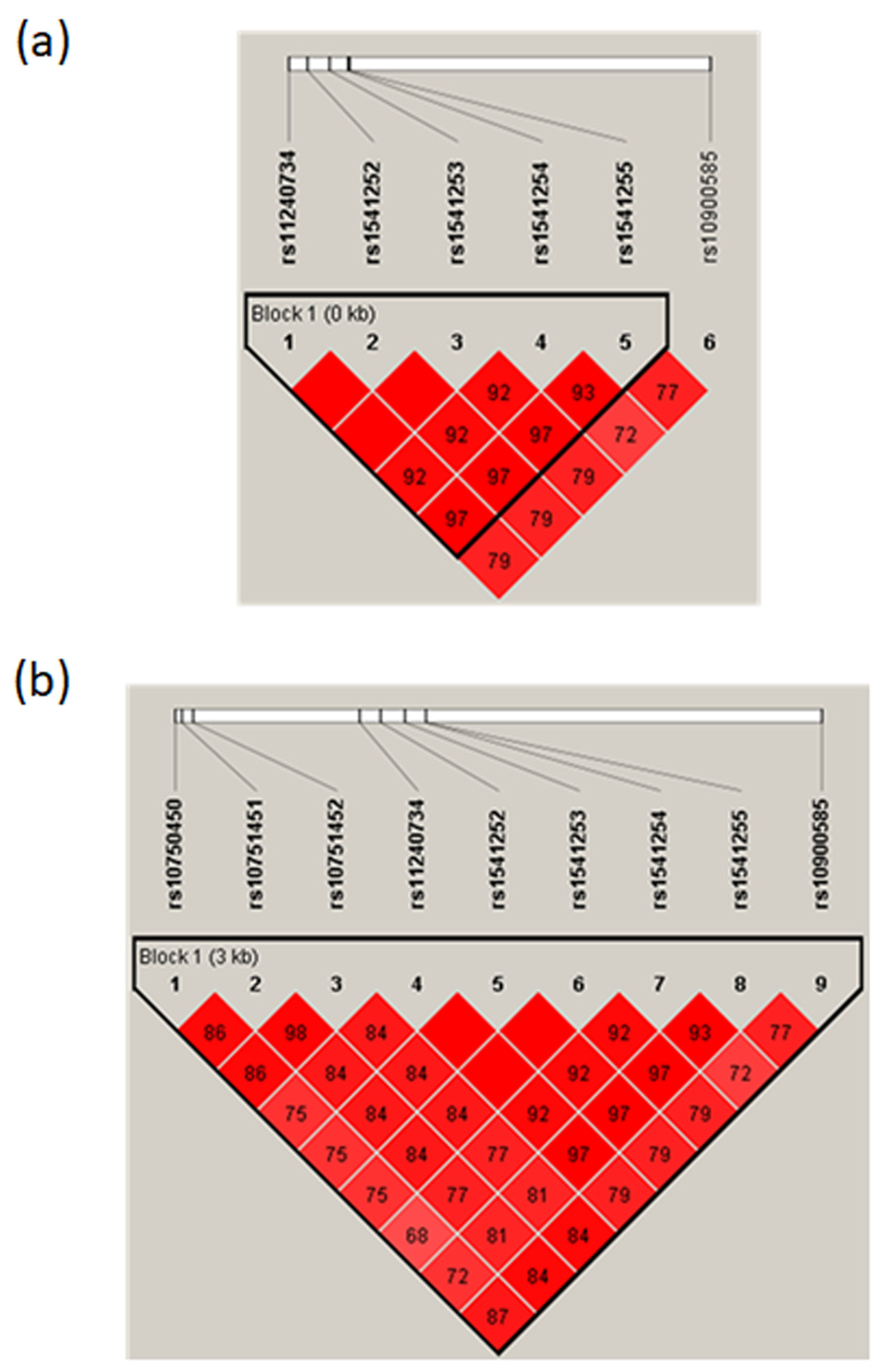
2.1. Prioritization and Annotation of Putative Regulatory SNPs in Linkage Disequilibrium with tagSNPs Associated with Severe Malaria

We focused our analysis on the SNPs for which significant GWAS signals have been replicated: rs4951377 and rs10900585 at the ATP2B4 locus, rs186873296 at the FREM3-GYP A/B locus, rs62418762 at the EPHA7 locus, and rs8176719 at the ABO locus. We excluded the coding variation rs334, which captured the GWAS signal at the HBB locus. We identified 126 SNPs in LD with the malaria-associated SNPs, based on an r2 higher than 0.6. We excluded rs181620317, which is a missense variant within the FREM3 coding sequence, and further prioritized the SNPs for their potential regulatory effect using IW scoring [25]. Moreover, we investigated the ability of sequences containing the SNPs to bind transcription factors using the ReMap tool [26], and we determined whether the 125 SNPs have been annotated as eQTLs or identified as regulatory SNPs [27]. (Table 1; Supplementary Table S1).
As displayed in Figure 1, the number of transcription factor peaks identified by ChIP-seq negatively correlated with the rank of the corresponding SNPs (rho = −0.652, p = 2.17·10−11). The highest numbers of peaks were observed for rs1541252, rs1541253, rs1541254, and rs1541255 (n > 90), which were ranked second, 10th, 6th, and 7th based on their IW scores, respectively (Table 1). The corresponding transcription factors are displayed in Supplementary Table S2; these include GATA1, RELA, GABPA, TCF3, ELF1, NFRKB, and RUNX1. The best SNP according to the IW scoring method was rs11240734, for which 54 peaks of ChIP-seq were registered in the ReMap catalog. Strikingly, those 5 SNPs are close to each other on chromosome 1 in the ATP2B4 region. These SNPs are located within a DNAse I hypersensitivity region and peaks of H3K4me1, H3K4me3, and H3K27ac marks in the K562 cell line, as displayed in Figure 2. Similar epigenomic marks have been found at these positions in erythroblasts (Supplementary Figure S1). rs11240734 and rs1541252 have been annotated as eQTLs, according to Haploreg, whereas rs1541255 was found to have a regulatory effect according to the Sure-seq method [28].
The prioritization was based on IW scoring analysis on the X-axis and the number of ChIP-seq peaks on the Y-axis. The four SNPs rs1541252, rs1541253, rs1541254, and rs1541255 have the highest number of ChIP-seq peaks (n > 90) as displayed. These SNPs are among the top 10 SNPs according to IW scoring.
Interestingly, the TaqSNPs rs4951377 and rs10900585 that are in LD with rs11240734, rs1541252, rs1541253, rs1541254, and rs1541255 had a low IW score and were ranked at positions 51 and 60, respectively (Supplementary Table S1). Furthermore, there were neither peaks of ChIP-seq in the ReMap catalog nor peaks of H3K4me1, H3K4me3, and H3K27ac marks in the K562 cell line for both SNPs (Figure 2), suggesting that they have no functional role. In contrast, rs10751450, ranked 3rd with 46 peaks of Chip-Seq, is located within a DNAse I hypersensitivity region, and peaks of H3K4me1 and H3K27ac marks in the K562 cell line (Figure 2), are located within a regulatory region [24]. Similar epigenomic profiles were observed in erythroblasts (Supplementary Figure S1). The SNPs rs10751451 and rs10751452, close to the rs10751450 [24], were ranked 12th and 13th, and rs10751451 was annotated as an eQTL according to Haploreg (Table 1). Figure 2 illustrates the position of the region containing rs10751450, rs10751451, and rs10751452 and the one containing rs11240734, rs1541252, rs1541253, rs1541254, and rs1541255. Overall, SNP prioritization and annotation allowed us to identify 5 new SNPs with potential regulatory effects, which may drive the observed association of their tagSNPs with malaria susceptibility.

2.2. Association between the ATP2B4 tagSNP rs10900585 and Severe Malaria in a Senegalese Cohort

One hundred and ninety-six individuals from the Senegalese cohort were successfully genotyped by sequencing. The frequencies of observed genotypes conformed to the Hardy–Weinberg equilibrium (p = 0.52) (Supplementary Table S3) and the allelic frequencies for rs10900585-T and G were found to be 0.59 and 0.41, respectively. We detected a significant association (p = 0.029; OR = 1.94) of ATP2B4 rs10900585 with SM (Table 2) without the effect of any covariate. The TT genotype that was more frequent among cases (48.7%) than controls (32.9%) was identified as the risk genotype. Nevertheless, there was no significant association when considering age as a covariate (p = 0.055) (Supplementary Table S4).
We added 13 independent populations to our study population for performing a meta-analysis, including 12,794 SM cases and 19,898 controls (Supplementary Table S5) [19,29,30]. There was no deviation from the Hardy–Weinberg equilibrium (p > 0.05) but there was a significant heterogeneity among the studies (I2 = 66%; tau2 = 0.0174; p = 0.0003). Thus, the random effect model was used for calculating the combined odd ratio and 95% confidence interval (CI). There was an association of rs10900585 with SM under the genetic dominant model (OR = 1.12; 95% CI = 1.02–1.23) (Figure 3).

2.3. Association of rs11240734, rs1541252, rs1541253, rs1541254, and rs1541255 with Severe Malaria in Senegalese Population

These five SNPs newly identified as potential regulatory variants were in a 600 bp DNA fragment that we sequenced to determine the genotypes of the subjects. None of the polymorphisms deviated from the Hardy–Weinberg equilibrium (Supplementary Table S3). We compared subjects carrying the homozygous genotype for the major alleles with those carrying either the heterozygous genotype or the homozygous genotype for the minor alleles. All these SNPs were significantly associated with the disease (p value = 0.002), with an estimated OR of 2.52 (1.39–4.58) (Table 2), indicating a stronger association compared to the tagSNP. Among the SM individuals, 53.8% carried the rs11240734TT, the rs1541252CC, the rs1541253CC, the rs1541254GG, and the rs1541255AA genotypes, whereas only 31.6% of the control individuals carried these genotypes. The association of the five SNPs with SM remained significant (p = 0.006) after considering age as a covariate (Supplementary Table S4). In addition, we found a strong LD between all five SNPs in addition to the tagSNP with an r2 varying from 0.72 to 1 (Figure 4a).

2.4. Association of rs10751450, rs10751451, and rs10751452 with Severe Malaria in Senegalese Population

Although suggested as regulatory polymorphisms [24], the SNPs rs10751450, rs10751451, and rs10751452 have never been genotyped and tested for their association with malaria. We tested whether these three SNPs were associated with SM. The frequencies of observed genotypes conformed to the Hardy–Weinberg equilibrium (Supplementary Table S3). The analysis indicated a significant association between malaria disease and these SNPs, rs10751450 (p = 0.016; OR 2.07), rs10751451 (p = 0.002; OR 2.49) and rs10751452 (p = 0.006; OR 2.27) (Table 2), which remained significant when considering age as a covariate (Supplementary Table S4). Noticeably, in our study population, these SNPs were found to be in strong LD between them in addition to our five new candidate SNPs (Figure 4b).

2.5. Haplotype Analysis of the ATP2B4 SNPs

Haplotype analysis, combining rs11240734, rs1541252, rs1541253, rs1541254, and rs1541255, revealed five haplotypes demonstrating various frequencies: 0.65 for haplotype (1) with the major alleles (rs11210734T-rs1541252C-rs1541253C-rs1541254G-rs1541255A), 0.33 for haplotype (2) with the minor alleles (rs11210734C-rs1541252T-rs1541253T-rs1541254C-rs1541255G), 0.015 for the haplotype (3) (rs11210734T-rs1541252C-rs1541253C-rs1541254C-rs1541255A), 0.002 for haplotype (4) (rs11210734C-rs1541252T-rs1541253T-rs1541254C-rs1541255A), and 0.002 for haplotype (5) (rs11210734C-rs1541252T-rs1541253T-rs1541254G-rs1541255A). We first investigated the relationship between SM and the two most frequent haplotypes (1 and 2). The haplotype (1) was found to be associated with an increased risk of developing SM (p = 0.005). Among SM subjects, 55.8% were homozygous for the susceptibility haplotype (1) and 34.5% were heterozygous, whereas only 31.6% of the control subjects carried this haplotype at homozygote state. Individuals homozygous for haplotype (1) with major alleles have a higher risk of developing SM (p = 0.002, OR = 2.52) compared to heterozygous and homozygous haplotypes for minor alleles. This association remained significant when including age as a covariate in the statistical model (p = 0.006) (Table 3). We further performed the haplotype analysis by including rs10751450, rs10751451, rs10751452, rs11240734, rs1541252, rs1541253, rs1541254, and rs1541255. Among SM individuals, 47.8% were homozygous and 27.4% were heterozygous for the haplotype (CCTTCCGA) while only 26.6% of the control subjects carried this haplotype in the homozygous state. We found that haplotype with major alleles (CCTTCCGA) at the homozygous state was associated with an increased risk of SM (p = 0.003, OR = 2.67), even when taking age as a covariate (p = 0.005) (Table 3).

2.6. Lower Promoter Activity and Higher Enhancer Activity Are Associated with the Risk Haplotype of Severe Malaria

Luciferase reporter assays were performed in K562 cell lines to evaluate the regulatory effect of the 5 SNPs on ATP2B4 expression. The candidate regulatory region and the position of the SNPs is portrayed in Figure 5. To assess whether this region may have a promoter function, we performed luciferase reporter gene assays by cloning it upstream the luciferase reporter gene. Interestingly, this regulatory region indicated strong promoter activity compared to the SV40 promoter for both the risk haplotype with major alleles (p < 1.10−4, 10.9-fold increase) and the non-risk haplotype with minor allele (p < 1.10−4, 17.7-fold increase) (Figure 6a). Moreover, the risk haplotype was shown to have reduced more transcriptional activity than the non-risk haplotype (p < 1.10−4). Hence, our results revealed differential allelic promoter activity in the K562 cell line, demonstrating that the non-risk haplotype exhibited a 1.6-fold increased transcriptional activity compared to the risk haplotype. In addition, we observed that each SNP has a specific impact on expression, and their combination reflects the combined effect observed. These results indicate that these five SNPs may be functional variants and that we must consider that it is their combined effect that is probably involved in the susceptibility to severe forms, as suggested by the haplotypes naturally present in individuals living in endemic areas.
To functionally confirm that this genomic region may have an enhancer function, we performed luciferase promoter assays by cloning it downstream of the luciferase reporter gene, whereas we cloned upstream a region of the main promoter of ATP2B4 (Figure 5 and Figure 6b). We first validated the promoter activity of this 780-bp region compared to the SV40 promoter (p < 1.10−4, 3.8-fold increase). We further confirmed the enhancer activity of the 601 bp region containing the five SNPs on the main promoter of ATP2B4 (p < 0.01, 1.3-fold increase for the construct with the 601 bp region as compared to the construct without this region, see Figure 6b). Moreover, we demonstrated that the region containing the major alleles exhibited an enhancer activity that was significantly higher than that of the region containing the minor alleles (p = 0.01). Also, there was a 1.2-fold increase in activity of the major allele haplotype relative to the minor allele haplotype. These data confirm that this regulatory region exhibits allele-dependent enhancer activity in the K562 cell line. Altogether, our results suggest that this regulatory region is a promoter with an enhancer function, also named Epromoter [31,32]. Furthermore, they suggest that genetic variants within this regulatory region orient the function of the region toward a promoter activity or toward an enhancer activity.

2.7. Genome Editing Confirmed the Regulatory Activity of the Region Containing the SNPs

To analyze the function of the Epromoter element, we deleted a DNA region of 506-bp containing the malaria-associated SNPs (rs11240734, rs1541252, rs1541253, rs1541254, and rs1541255) in the K562 cell line, using CRISPR/Cas9 mediated genome editing (Figure 5 and Figure 7a). We selected three deleted clones with the expected sequence and no additional edits. Expression analysis for total ATP2B4 [including long and short transcripts (Figure 2)] demonstrated a 3.3-fold decrease in gene expression in deleted clones as compared to WT cells (p < 1.10−4) (Figure 7b). We also estimated the expression of the two long transcripts demonstrated in Figure 2 (ATP2B4-203 and ATP2B4-204) of ATP2B4 (encoding for PMCA4a and PMCA4b) and we demonstrated a 2.8-fold decrease in deleted clones compared to WT cells (p < 1.10−4) (Figure 7b). These results support the hypothesis that this region has an enhancer function.
We deleted a region of 1262-bp to remove the eight SNPs (rs10751450, rs10751451, rs10751452, rs11240734, rs1541252, rs1541253, rs1541254, and rs1541255) (Figure 7a) and selected three clones validated by sequencing. Clones with this large 1262-bp deletion had a similar reduction in expression compared to the small 506-bp deletion, both for total expression and for long transcripts of ATP2B4, suggesting that the minimal region of 506 bp including rs11240734, rs1541252, rs1541253, rs1541254, and rs1541255 is enough to regulate the expression of ATP2B4 (Figure 7c).

2.8. K562 Deleted Clones Exhibited Higher Intracellular Calcium Concentration

Intracellular calcium concentration was measured in two clones (ΔATP2B4_2 and ΔATP2B4_3) deleted for the region containing rs11240734, rs1541252, rs1541253, rs1541254, and rs1541255 and a representative experiment is displayed in Figure 8 for each of the clones. Six and two experiments were performed with ΔATP2B4_2 and ΔATP2B4_3 clones, respectively. Supplementary Table S6 demonstrates descriptive statistics for each experiment performed with K562 wild-type (WT) cells and the two clones deleted for the genomic region of interest. First, we compared the mean of fluorescence intensity (MFI) of K562 WT cells with that of mutant clones, without considering the number of cells studied and the standard deviation (SD). The MFI of ΔATP2B4_2 and ΔATP2B4_3 clones was systematically higher than the MFI of K562 WT cells in each experiment (Supplementary Table S6). Non-parametric tests yielded significant differences when including only ΔATP2B4_2 clone results (p = 0.009) or including both ΔATP2B4_2 and ΔATP2B4_3 results (p = 0.029). Secondly, we tested whether K562 WT cells and the clones deleted for the genomic region of interest differed in their MFI, when considering the number of cells studied in each experiment and the corresponding standard deviation (SD). When analyzing each experiment, the wild-type cells and the clones deleted for the candidate region systematically differed in intracellular calcium concentration (p < 0.0001). The Cohen’s coefficient ranged from 0.14 to 0.56, demonstrating that the size effect was low. The statistical power was, however, close to 1 due to the large number of cells analyzed in each group (Table S6). Similarly, meta-analyses considering MFI, SD, and sample size provided evidence of an increase in calcium concentration in deleted clones when including only the results obtained with ΔATP2B4_2 clone (p < 0.0001) or including both ΔATP2B4_2 and ΔATP2B4_3 results (p < 0.0001). In all, the deletion of the region containing rs11240734, rs1541252, rs1541253, rs1541254, and rs1541255 was proven to cause a slight but significant increase in the intracellular calcium level.

2.9. PMCA4 Protein Is Not Expressed in K562 Deleted Clones

The expression of the protein PMCA4 has been assessed in the two deleted clones. Three independent experiments have been performed, indicating that the PMCA4 protein is not detected in the deleted clones (Figure 8), whereas this protein is expressed in K562 WT cells; the MFI difference between WT and deleted clones was significant (p = 0.002). Meta-analyses that considered MFI, SD, and sample size further provided evidence of a significant decrease of protein level in deleted clones (p < 0.0001). Overall, these results indicate that the deletion of the region containing rs11240734, rs1541252, rs1541253, rs1541254, and rs1541255 led to a dramatic fall in PMCA4 expression, a decrease of calcium efflux and an intracellular accumulation of calcium.

3. Discussion

Several GWAS have identified and replicated tagSNPs associated with SM. However, most of the causal variants remain unknown. Here we identified 5 ATP2B4 regulatory SNPs associated with SM in a Senegalese population and in LD with the tagSNPs identified by GWAS (rs4951377 and rs10900585). We demonstrated that these five SNPs are in a promoter region that displays enhancer activity (Epromoter) [31,32].
Bioinformatic prioritization of the 125 non-coding SNPs in LD with the tagSNPs associated with SM indicated that the top 10 ranked SNPs by the IW scoring were on chromosome 1 within ATP2B4. When combining the IW scoring with the annotation of transcriptional regulator ChIP-seq peaks, rs1541252, rs1541253, rs1541254, and rs1541255 appeared to be the most promising candidates. Interestingly, these SNPs were close to rs11240734, ranked 1st and 5th based on the IW score and on the number of ChIP-seq peaks, respectively, suggesting that they may be located within a regulatory element. This hypothesis is also supported by (1) the co-location of the SNPs with epigenomic marks associated with promoter or enhancer activity, (2) the characterization of rs11240734, rs1541252, rs1541253, and rs1541254 as eQTLs in blood cells [33], (3) the association of rs1541252 and rs1541253 with PMCA4 levels [34], and (4) the identification of rs1541255 as a regulatory variant based on a reporter assay [28].
Consistently, the tagSNPs rs4951377 and rs10900585 ranked 51st and 61st using the IW score, and neither were in a region with histone marks or chip-seq peaks, suggesting that they are not the causal SNPs. Nevertheless, rs4951377 was an eQTL in blood cells [33], illustrating that an eQTL is not necessarily a regulatory variant. The association signal of rs4951377 with gene expression is likely explained by the effect of functional SNPs in LD with it. We confirmed that rs10751450, rs10751451, and rs10751452 ranked 3rd, 12th, and 13th, respectively, they could be regulatory SNPs as previously suggested [24], and they were located within an enhancer altering ATP2B4 that codes for PMCA4 (Plasma membrane calcium transporting ATPase4) [24]. Noticeably, luciferase reporter assays provided evidence that these SNPs are regulatory variants [35]. This enhancer region is roughly 700 bp away from the region containing rs11240734, rs1541252, rs1541253, rs1541254, and rs1541255, which is potentially a regulatory region according to our results.
We confirmed the association between the tagSNP rs10900585 and SM in a Senegalese population with the same risk genotype and validated its association by meta-analysis, integrating all the SM populations [19,29,30]. In addition, we provided evidence of an association of the eight SNPs with SM in Senegal. Interestingly, these SNPs, except rs10751452, were imputed in the most recent GWAS of SM [17] and indicated a high Bayes Factor value (BF > 1.8 107) that was consistent with an association. Similarly, rs1541255 was associated with SM in Kenya [17], whereas the other SNPs were not genotyped in this population.
Furthermore, we found a strong haplotype association with SM, and we observed that most of the Senegalese individuals exhibited either the haplotype combination of all the major alleles or the haplotype combination of all the minor alleles, whereas only a few individuals carried other haplotypes. The deletion of the region containing rs11240734, rs1541252, rs1541253, rs1541254, and rs1541255 strongly reduced the level of long transcripts. As this region is located within intron 1, this suggests that it acts as an enhancer on the main promoter, which controls the expression of the long transcripts (Figure 9). Luciferase gene reporter assays further indicated that this region increased the activity of the main promoter of ATP2B4.We demonstrated that this region has a promoter activity using luciferase reporter assays; this supports the hypothesis that it is also an alternative promoter (Figure 9). In addition, we observed higher enhancer activity with the haplotype combining the major alleles whereas this haplotype indicated lower promoter activity compared to the haplotype with the minor alleles. This result supports a hypothetic model of regulation proposed recently by Gao et al., Hua et al., and Ying et al. [36,37,38], whereby genetic variants can impose a regulatory switch between the enhancer and the promoter activity (Figure 9). It implies that major allele haplotype would increase the level of long transcripts, the expression of which is controlled by the main ATP2B4 promoter, whereas minor allele haplotypes would increase the level of short transcripts, the expression of which is controlled by the alternative promoter containing the studied SNPs. It should be stressed, nevertheless, that mutagenesis or homologous recombination experiments in K562 cells are needed to confirm this hypothesis.
Our results further suggest that there was an effect of the combination of several SNP alleles on gene expression, thus reflecting the interaction between these SNPs. Although little is known about haplotypes’ influence on gene expression, Ying et al. [38] recently proposed a method to detect eQTL haplotypes and suggested that they are enriched in regulatory regions such as promoters or enhancers. Our finding is also supported by a study providing evidence at numerous loci that “multiple enhancer variants” cooperatively contribute to altered expression of their gene targets and that target transcript levels tend to be modest [39]. This raises the question of the natural combination of alleles in African populations under selective pressure, and supports our finding that the combination of these five SNPs was responsible for susceptibility to SM and that there is not a single causal variant.
We also demonstrated that the deletion of this regulatory region containing rs11240734, rs1541252, rs1541253, rs1541254, and rs1541255 decreased both the global ATP2B4 expression and specifically the expression of the long transcripts in the K562 cell line, suggesting that it is a promoter with an enhancer function (Epromoter). Consistently, decreasing expression of the transcripts in the deleted clone results in the absence of the PMCA4 protein and increased intracellular calcium.
After deleting a region containing rs10751450, rs10751451, and rs10751452, Lessard et al. demonstrated a reduction in ATP2B4 expression and a cytoplasmic calcium accumulation [24]. Hence, we included these SNPs in our study, and we obtained similar association results when adding rs10751450, rs10751451, and rs10751452 in the haplotype analysis, but also a similar decrease in ATP2B4 expression for the clones either with five or eight SNP deletions. This finding supports that rs11240734, rs1541252, rs1541253, rs1541254, and rs1541255 affect the ATP2B4 gene expression as much as rs10751450, rs10751451, and rs10751452 and contribute to the development of SM. Here, we have demonstrated the potential impact of these variants in regulating ATP2B4 expression and intracellular calcium levels. We hypothesize that changes in calcium homeostasis may affect the growth of the parasite in red blood cells, and may also affect the clumping of infected red blood cells and their sequestration to the brain microvessels, thus contributing to SM susceptibility. In addition, our results support the hypothesis that a calcium-activated potassium channel may be activated, resulting in potassium efflux, dehydration, red blood cell volume loss, increased mean corpuscular hemoglobin concentration, and reduced parasitemia in malaria patients [24]. Indeed, rs1541252 and rs1541255 were found to be associated with mean corpuscular hemoglobin concentration in African-American children and parasitemia in malaria patients, respectively [29,40]. However, this hypothesis was recently challenged by Villegas-Mendez et al. and Pance et al. [41,42]. Pance et al. reported that ATP2B4 deletion fully inhibited PMCA4 expression but had a very slight effect on the parasite growth in human stem cell-derived erythroid cells [42], whereas Villegas-Mendez et al. demonstrated that ATP2B4 gene targeting did not alter parasitemia in mice infected by Plasmodium berghei but did protect mice by against cerebral malaria induced by Plasmodium berghei [41]. Interestingly, PMCA1 expression was increased in ATP2B4−/− mice [41]. This suggests that PMCA1 expression and other calcium channels may compensate for a low expression of ATP2B4 gene and thus PMCA4 protein in erythrocytes. Also, calcium concentration and parasite growth may not be dramatically altered by a strong inhibition of ATP2B4 gene expression.
Conversely, the ATP2B4 gene is expressed in multiple cell types, and it is not excluded that the modulation of intracellular Ca2+ concentration alters the physiology of other cell types. For example, the regulation of intracellular Ca2+ signaling is a major determinant of CD8+ T cell responsiveness, which may have an important role in determining SM. Moreover, the PMCA4 protein encoded by ATP2B4 is expressed both in the red blood cell and the brain with PMCA4b and PMCA4a the most abundant isoforms in red blood cells and in the brain, respectively [43]. Because these isoforms are tightly and specifically regulated in tissues and cells, we can therefore propose that dysregulation of calcium homeostasis in the brain could also be directly involved in cerebral malaria susceptibility as for different brain disorders such as Alzheimer’s disease and Parkinson’s disease. PMCA4a, which is the major isoform present on endothelial cells, may also play a crucial role in SM through its role as a negative regulator of vascular endothelial growth factor (VEGF)-activated angiogenesis. In cerebral malaria, the binding of parasitized erythrocytes to the cerebral endothelium and the consequent angiogenic dysregulation play a key role in pathogenesis [44]. VEGF, a regulator of endothelial inflammation and integrity, is involved in modulating tissue pathology in response to P. berghei infection. Indeed, a potent inhibitor to the VEGF signaling pathway dramatically aggravated the course of P. berghei infection [45].

4. Materials and Methods

4.1. Study Subjects, Blood Samples, and Phenotypes

Malaria patients were recruited from two major Senegalese sites, the principal hospital of Dakar and the regional hospital of Tambacounda, including 90 cerebral malaria cases and 27 severe noncerebral cases, as described [46]. The control samples (n = 79) were obtained from healthy volunteers living in Dakar. Written informed consent was obtained for each patient and their accompanying family members. The study was approved by the institutional research ethics committee of the Université Cheikh Anta Diop.
Venous blood samples and biological data including parasite density, hematology, and other characteristics were collected on the day of admission. The presence of Plasmodium falciparum was determined by at least two trained biologists by microscope examination of thin and thick smears before antimalarial treatment. If asexual parasites were observed, then the slide was considered as positive, and the number of parasitized erythrocytes was counted per µL of blood.

4.2. Bioinformatic Prioritization and Functional Annotation of Genetic Variants

Significant GWAS signals replicated at least in an independent population were identified and the corresponding tagSNPs (rs4951377, rs10900585, rs186873296, rs62418762, and rs8176719) were selected for further analysis. Haploreg v4.1, a tool including LD information from the 1000 Genome project, was used to identify the SNPs in LD with these tagSNPs. This analysis of the African population and LD threshold r2 > 0.6 resulted in the selection of 125 SNPs. To prioritize the SNPs with putative functional significance, we performed bioinformatic analysis using the IW scoring annotation tool (https://www.snp-nexus.org/IW-Scoring/, accessed on 6 March 2019) [25] and ReMap 2018 based on DNA-binding ChIP-seq experiments [26]. We also checked whether these SNPs have been annotated as eQTLs (https://pubs.broadinstitute.org/mammals/haploreg/haploreg.php, accessed on 1 March 2019) [27] or identified as regulatory SNPs using the massive parallel reporter assay named Sure-seq [28]. Additional analysis has been performed to evaluate whether the candidate SNPs are located within a DNAse I hypersensitivity region, peaks of H3K4me1, H3K4me3, and H3K27ac marks in the K562 cell line, which were visualized in the UCSC genome browser.

4.3. DNA Extraction, DNA Amplification, and Genotyping

Genomic DNA was extracted and amplified as previously described [46]. In total, we genotyped nine SNPs of ATP2B4 including rs11240734, rs1541252, rs1541253, rs1541254, and rs1541255, the TaqSNP rs10900585, and the SNPs rs10751450, rs10751451, and rs10751452, previously described as regulatory variants [24]. Genotyping was performed using either the Sanger sequencing or the TaqMan SNP genotyping assays (Life Technologies, Waltham, MA, USA). All the primer pairs for sequencing were designed using Primer 3 software. PCR amplification of DNA fragments containing rs11240734, rs1541252, rs1541253, rs1541254, and rs1541255 was performed with forward (5′-TCAGGCCTAGCTATCAGTTCAG-3′) and reverse (5′-CGAGTAGCCGTCCGAAGTC-3′) primers, respectively. For the tagSNP rs10900585, the forward and the reverse primers were (5′-GGGATGAGGAGGCTTACAGG-3′) and (5′-GAGGTTGAGGTGAGCGGATC-3′), respectively.
PCR amplification was executed in 50µL reaction volume with 12.5 ng of genomic DNA, GoTaq G2 2X ready-to-use Master Mix (Promega, cat#M7433), and 10 µM of each primer. For rs10900585, the annealing temperature was 65 °C, while for rs1541252, annealing was at 60 °C. The PCR products were submitted to electrophoresis on 2% agarose gel to verify the product size, and then purified with a PCR Purification kit (Qiagen, Hilden, Germany) following the manufacturer’s protocol. Five µL of purified PCR product was sequenced using the Sanger method (GATC biotech, Ebersberg, Germany, or Eurofins Genomics, Ebersberg, Germany).
For rs10751450, rs10751451, and rs10751452, we used TaqMan genotyping assays C_31796478_10, C_31796479_10, and C_31796480_10, respectively. PCR amplification was performed with the QuantStudio 6 Flex Real-Time PCR Systems (Life Technologies, Waltham, MA, USA) in a final volume of 5 µL containing 2.1 µL of Master Mix (Life Technologies, Waltham, MA, USA), 0.06 µL of TaqMan probe, 1.84 µL H2O, and 1 µL of DNA (10–15 ng/µL).

4.4. Luciferase Gene Reporter Assay and Site-Directed Mutagenesis:

4.4.1. Promoter Activity

A 601bp DNA fragment upstream from the ATP2B4 translation start site (chromosome 1: 203682549–203683049 according to the hg38 assembly) was cloned into the MlulI-XhoI sites of the pGL3-basic vector (Promega, Madison, WI, USA), which contained the firefly luciferase coding sequence (GeneCust, Boynes, France). We obtained two different pGL3 constructs containing either minor or major alleles of the five SNPs (rs11240734, rs1541252, rs1541253, rs1541254, and rs1541255). From the minor allele construct, we generated additional constructs by replacing the minor allele with the major allele of each SNP individually. The site-directed mutagenesis was performed using the Q5 Site-Directed Mutagenesis Kit (New England Biolabs, Ipswich, MA, USA,) and primers were designed by NEBaseChanger tool and provided by the supplier (detail sequences provided in Supplementary Table S7). K562 cells (ATTC CLL-243) were grown in Gibco RPMI 1640 medium (Thermo Fisher Scientific, Waltham, MA, USA), supplemented with 10% FBS (fetal bovine serum). K562 transfection was performed with the NeonTM Transfection system (Invitrogen) according to the manufacturer’s instructions. For each of the nine tests performed, 106 cells were co-transfected with 1 µg of control vectors: (1) negative control vector (empty pGL3-basic vector (cat# E1751)) or (2) positive control vector (pGL3-promoter vector (cat# E1761)), or with 1 µg of the construct to be tested (a pGL3-basic vector containing either the minor alleles CTTCG or the major alleles TCCGA or one of these combinations: TTTCG, CCTCG, CTCCG, CTTGG, or CTTCA) and 200 ng of pRL-SV40 (a plasmid encoding renilla luciferase from Promega (cat# E2231)), which was used as a transfection efficiency control. Transfected cells were maintained at 37 °C in 5% CO2 for 24 h. Values of firefly and renilla luciferase were obtained by analyzing 20 µL of cell lysate according to the standard instructions provided in the Dual-Luciferase kit (Promega, Madison, WI, USA) in a TriStar LB 941 Multimode Microplate Reader (Berthold Technologies, Thermo Fisher Scientific, Waltham, MA, USA). Firefly luciferase activity of each sample was normalized to renilla luciferase and expressed as the fold change of the empty vector control.

4.4.2. Enhancer Activity

A 780-bp fragment of ATP2B4 promoter (chromosome1: 203626081-203626860 (hg38 assembly)) was cloned into pGL3-Enhancer vector (Promega, Madison, WI, USA) between MluI- Xho I sites and the 601 bp fragment of ATP2B4 enhancer (chromosome 1: 203682549-203683049 according to the hg38 assembly) containing respective minor and major alleles of the five SNPs was cloned into BamHI-SalI sites. Luciferase assays were performed in K562 cells as described above.

4.5. CRISPR-Cas9 Genome Editing in K562 Cells

Using the CRISPR design tool provided by IDT, we identified guide RNAs to target the ATP2B4-specific region. Chemically synthesized oligoribonucleotides were manufactured by IDT: the crRNAs (35 mer with specific part to DNA target sequence) and the universal tracrRNAs (67 mer). A two-part system where synthetic crRNA (5 µL of 100 µM) and tracrRNA (5 µL of 100 µM) were annealed to form an active gRNA complex. Cas9 RNP complexes were assembled in vitro by incubating 3.4 µL Cas9 protein (62 mM) with 4.8 µL active gRNA complex (cr:tracrRNA) and 1.8 µL of PBS. To generate the genomic deletion, two different RNP complexes were simultaneously electroporated with the Neon transfection system into 5.105 K562 cells. The small deletion of 506 bp (containing the five SNPs) was obtained by using 2 µL gRNA1 and 2 µL of gRNA2 (details of sequences in Supplementary Table S8). The large deletion of 1262 bp (containing the 8 SNPs) was performed by using gRNA2 and gRNA3 (Supplementary Table S8). Those bulk cultures transfected with tandem gRNA were plated clonally in 96-well plates at a limiting dilution less than 0.5 to avoid mixed clones. After approximately 14 days of clonal expansion, amplification of DNA was performed directly from the clones by using the 2X Phire Tissue Direct PCR Master Mix containing Phire Hot Start II DNA Polymerase (Thermo Fisher Scientific, Waltham, MA, USA), with 20 µL of dilution buffer and 0.5 µL of DNA Release additive, which allowed improved release of DNA from the cells. Clones were screened for deletion by PCR using primers F1 and R1 for the small deletion detection, which would produce a specific short fragment of 335 bp in the presence of deletion and a long fragment of 841 bp in the absence of deletion (Supplementary Table S8). Primers F2 and R2 were used for large deletion detection that would produce amplicons of 355 bp in presence of deletion and 1617 bp in the absence of deletion (Supplementary Table S8). PCR was performed using 10 µL of 2XMaster Mix and 10 µM of each primer, annealing at 60 °C. After electrophoresis on 1% agarose gel, we selected homozygote deleted clones displaying only amplicons of 335 bp or 355 bp for the small and the large deletions, respectively. After sequencing using the Sanger method, only the clones with the appropriate sequence with no additional edits were kept for gene expression analysis.

4.6. Reverse Transcription-Quantitative PCR

Total RNA was extracted for each selected clone and wild-type K562 cell using the RNeasy mini kit (Qiagen, Hilden, Germany). One µg of RNA per clone was converted into cDNA using Superscript VILO Master Mix (Invitrogen, Thermo Fisher Scientific, Waltham, MA, USA). Three independent RNA/cDNA preparations were performed for each clone. Real-time quantitative PCR (RT-qPCR) was subsequently performed using SYBR Select Master Mix (Thermo Fisher Scientific, Waltham, MA, USA) on a QuantStudio 6 Flex instrument. Primers were designed using Primer 3 software to span exon 1 of ATP2B4 (F3 and R3) to specifically quantify the long transcripts (ENST00000357681, ENST00000367218) or to span exon 19 and exon 20 (F4 and R4) to quantify all the ATP2B4 transcripts (ENST00000357681, ENST00000367218, ENST00000341360, ENST00000458092, and ENST00000356729) (Supplementary Table S9). PCR efficiency was validated by performing serial dilution analysis. Gene expression was normalized to that of actin and the relative expression was calculated by the ∆CT method. The data reported correspond to the mean of triplicates from three independent experiments per clone and expressed as fold change relative to wild-type cells. Three independently generated clones were analyzed for the gene expression quantification of ATP2B4.

4.7. Calcium Measurement by Flow Cytometry in K562 Clones and Wild-Type Cells

The intracellular quantity of calcium was measured in K562 cells and ΔATP2B4_2 and ΔATP2B4_3 clones by flow cytometry using the Fluo-4 AM kit (Molecular Probes/Thermo Fisher Scientific, Waltham, MA, USA), following manufacturer’s instructions. Briefly, samples of 106 cells were harvested for each cell type. The cells were pelleted and resuspended in 1 mL of culture medium without serum. One ml of either Fluo-4-AM (labeled cells) or kit buffer (control cells) was added to the cells, before 1 h incubation at 37 °C. The cells were then directly observed on an LSRFortessa X-20 cytometer (BD Biosciences, NJ, NJ, USA). Cells were gated on forward/side-light scatter and the fluorescence intensity of 20,000 cells was measured at the FITC excitation/emission wavelength for each sample.

4.8. Detection of PMCA4 by Flow Cytometry in K562 Clones and Wild-Type Cells

The presence of PMCA4 in K562 cells and clones ΔATP2B4_2 and ΔATP2B4_3 was detected by flow cytometry, using the specific antibody JA9 (ABCAM, ab2783). The labeling was realized according to a slight modification of the manufacturer’s protocol. For each cell type, 2 × 10⁶ cells were harvested, pelleted, and fixed in 2 mL of 80% methanol (5 min). After centrifugation, the cells were incubated in 2 mL of PBS with 1% BSA for 30 min at room temperature to block nonspecific interactions. Half of each cell type was then incubated with the JA9 antibody (2 µg/1 × 106 cells) for 30 min at room temperature. The other half was used for isotypic control (ab170190, 2 µg/1 × 106 cells). After centrifugation, the cells were incubated with the secondary antibody (goat anti-mouse IgG1 APC, ab 130786) at 1/200 dilution in PBS with 1% BSA, for 30 min at room temperature. Finally, cells were washed twice in PBS and observed on an LSR Fortessa X-20 cytometer (BD Biosciences). Cells were gated on forward/side-light scatter and the fluorescence intensity of 20 000 cells was measured at the APC excitation/emission wavelength for each sample.

4.9. Statistical Analysis

A Chi-squared test was used to determine whether the genotype distribution in healthy Senegalese subjects conformed to the Hardy–Weinberg equilibrium. Chi-squared tests and logistic regression analyses were carried out with SPSS (statistical software version 20) to assess the association between the SNPs and SM in Senegalese subjects. Differences were considered significant if the P-value obtained in a two-tailed test was <0.05. The odds ratio (OR), 95% confidence intervals (CIs), and the influence of covariates on the phenotype were evaluated by logistic regression analysis. A meta-analysis of the genetic association was performed using MetaGenyo [47]. Statistical differences between luciferase constructs or between clones were performed using Student’s t-tests. Mixed models were also used to consider the triplicates performed for each independent experiment. P values < 0.05 were considered statistically significant. Plot generation was performed using GraphPad Prism. Statistical analysis to evaluate the difference in the calcium concentration between deleted and wild-type clones was based on the non-parametric Wilcoxon test and the t-test using SPSS, and on a meta-analysis approach using Open Meta-Analyst software [48]. All the tests used were two-sided tests.

5. Conclusions

We identified a new regulatory region that controls the expression of both ATP2B4 mRNA and PMCA4 proteins, and that affects intracellular calcium levels. We also demonstrated that the activity of this regulatory region was perturbed by five genetic variants associated with SM. This suggests that we identified causal variants within a locus identified through GWAS of SM. This also fosters the development of therapeutic strategies based on the modulation of ATP2B4 expression and calcium levels. It should be stressed, however, that the effect size of ATP2B4 was modest in our study population, consistent with previous heritability studies [4,17,29]. More generally, genetic variants in ATP2B4, FREM3-GYP A/B, EPHA7, ABO, and HBB, which have been identified by several GWAS studies, explain 11% of the genetic contribution to variation in SM susceptibility [17]. In this way, a polygenic effect has been proposed by Damena et al. (2020), suggesting that many genetic variants with a small effect size remain to be discovered [4].

Supplementary Materials

The following are available online at https://www.mdpi.com/article/10.3390/ijms23094849/s1.

Author Contributions

S.M. and P.R. designed and supervised the project. S.N. genotyped most of the genetic variants, whereas M.T., A.T. and F.G. participated in genotyping. A.T. extracted the DNA samples in collaboration with R.N.D. and performed whole genome amplification. O.K., A.T. and B.M. were involved in the recruitment, the follow-up of Senegalese individuals, and the collection and interpretation of biological data including parasite density, hematology, and other characteristics from different areas (Dakar and Tambacounda), under the supervision of A.D.; M.T. guided CRISPR/Cas9 technology and gene reporter experiments. S.N. performed qPCR, CRISPR/Cas9, and gene reporter experiments. L.B. participated in gene reporter experiments. B.P. performed cytometry experiments for intracellular calcium levels and membrane protein levels. S.N., F.R. and P.R. performed bioinformatic analyses. S.N., P.R. and S.M. performed statistical analyses. S.S. provided useful comments on biological interpretation, whereas S.N., S.M. and P.R. interpreted the results and wrote the paper. S.M. and P.R. supervised the research and provided guidance. All authors have read and agreed to the published version of the manuscript.

Funding

This work was supported by the African Higher Education Centers of Excellence project (CEA-SAMEF) at UCAD, the Pasteur Institute in Dakar, the Pasteur Institute in Paris, the French embassy in Senegal, INSERM, and Aix-Marseille University. F.R. and S.N. were supported by a PhD fellowship from the French ministry of research and the Higher Education Commission (HEC) from Pakistan, respectively.

Institutional Review Board Statement

The study was conducted according to the guidelines of the Declaration of Helsinki, and approved by the Comité d’Ethique de la Recherche de l’Université Cheikh Anta Diop de Dakar on January 2015.

Informed Consent Statement

Informed consent was obtained from all subjects involved in the study. Each participant or their parents or legal guardians for any minors (before inclusion) has received written or verbal information in their native language and has given written consent.

Data Availability Statement

The datasets used and/or analyzed during the current study are available from the corresponding author on reasonable request.

Acknowledgments

We thank all the individuals who participated in the study, especially the patients from the regional hospital in Tambacounda and the Hospital Principal in Dakar. We thank Iris Manosalva and Charbel Souaid for helpful discussions, and Richard Redon for critical reading of the manuscript. We also thank Sabrina Baaklini and Mai Le Phuong Nguyen for the technical assistance. We thank Mouhamedou Mansour Fall for his technical assistance and follow-up of patients. We thank Laurence Borge for assistance and the use of the cell culture platform facility (CRCM U1068, Marseille).

Conflicts of Interest

The authors declare that there is no conflict of interest regarding the publication of this article.

References

  1. Mackinnon, M.J.; Mwangi, T.W.; Snow, R.W.; Marsh, K.; Williams, T.N. Heritability of malaria in Africa. PLoS Med. 2005, 2, e340. [Google Scholar] [CrossRef] [PubMed] [Green Version]
  2. Miller, L.H.; Baruch, D.I.; Marsh, K.; Doumbo, O.K. The pathogenic basis of malaria. Nature 2002, 415, 673–679. [Google Scholar] [CrossRef] [PubMed]
  3. Weatherall, D.; Clegg, J. Genetic variability in response to infection: Malaria and after. Genes Immun. 2002, 3, 331–337. [Google Scholar] [CrossRef] [PubMed] [Green Version]
  4. Damena, D.; Chimusa, E.R. Genome-wide heritability analysis of severe malaria resistance reveals evidence of polygenic inheritance. Hum. Mol. Genet. 2020, 29, 168–176. [Google Scholar] [CrossRef] [PubMed]
  5. Rihet, P.; Abel, L.; Traore, Y.; Traore-Leroux, T.; Aucan, C.; Fumoux, F. Human malaria: Segregation analysis of blood infection levels in a suburban area and a rural area in Burkina Faso. Genet. Epidemiol. 1998, 15, 435–450. [Google Scholar] [CrossRef]
  6. Rihet, P.; Traoré, Y.; Abel, L.; Aucan, C.; Traoré-Leroux, T.; Fumoux, F. Malaria in humans: Plasmodium falciparum blood infection levels are linked to chromosome 5q31–q33. Am. J. Hum. Genet. 1998, 63, 498–505. [Google Scholar] [CrossRef] [Green Version]
  7. Brisebarre, A.; Kumulungui, B.; Sawadogo, S.; Atkinson, A.; Garnier, S.; Fumoux, F.; Rihet, P. A genome scan for Plasmodium falciparum malaria identifies quantitative trait loci on chromosomes 5q31, 6p21. 3, 17p12, and 19p13. Malar. J. 2014, 13, 1–7. [Google Scholar] [CrossRef] [Green Version]
  8. Flori, L.; Sawadogo, S.; Esnault, C.; Delahaye, N.F.; Fumoux, F.; Rihet, P. Linkage of mild malaria to the major histocompatibility complex in families living in Burkina Faso. Hum. Mol. Genet. 2003, 12, 375–378. [Google Scholar] [CrossRef]
  9. Jepson, A.; Sisay-Joof, F.; Banya, W.; Hassan-King, M.; Frodsham, A.; Bennett, S.; Hill, A.; Whittle, H. Genetic linkage of mild malaria to the major histocompatibility complex in Gambian children: Study of affected sibling pairs. BMJ 1997, 315, 96–97. [Google Scholar] [CrossRef] [Green Version]
  10. Flori, L.; Kumulungui, B.; Aucan, C.; Esnault, C.; Traore, A.; Fumoux, F.; Rihet, P. Linkage and association between Plasmodium falciparum blood infection levels and chromosome 5q31–q33. Genes Immun. 2003, 4, 265–268. [Google Scholar] [CrossRef] [Green Version]
  11. Garcia, A.; Marquet, S.; Bucheton, B.; Hillaire, D.; Cot, M.; Fievet, N.; Dessein, A.J.; Abel, L. Linkage analysis of blood Plasmodium falciparum levels: Interest of the 5q31–q33 chromosome region. Am. J. Trop. Med. Hyg. 1998, 58, 705–709. [Google Scholar] [CrossRef] [PubMed]
  12. Milet, J.; Nuel, G.; Watier, L.; Courtin, D.; Slaoui, Y.; Senghor, P.; Migot-Nabias, F.; Gaye, O.; Garcia, A. Genome wide linkage study, using a 250K SNP map, of Plasmodium falciparum infection and mild malaria attack in a Senegalese population. PLoS ONE 2010, 5, e11616. [Google Scholar] [CrossRef] [PubMed]
  13. Sakuntabhai, A.; Ndiaye, R.; Casadémont, I.; Peerapittayamonkol, C.; Rogier, C.; Tortevoye, P.; Tall, A.; Paul, R.; Turbpaiboon, C.; Phimpraphi, W. Genetic determination and linkage mapping of Plasmodium falciparum malaria related traits in Senegal. PLoS ONE 2008, 3, e2000. [Google Scholar] [CrossRef]
  14. Band, G.; Le, Q.S.; Jostins, L.; Pirinen, M.; Kivinen, K.; Jallow, M.; Sisay-Joof, F.; Bojang, K.; Pinder, M.; Sirugo, G. Imputation-based meta-analysis of severe malaria in three African populations. PLoS Genet. 2013, 9, e1003509. [Google Scholar] [CrossRef]
  15. Jallow, M.; Teo, Y.Y.; Small, K.S.; Rockett, K.A.; Deloukas, P.; Clark, T.G.; Kivinen, K.; Bojang, K.A.; Conway, D.J.; Pinder, M. Genome-wide and fine-resolution association analysis of malaria in West Africa. Nat. Genet. 2009, 41, 657–665. [Google Scholar] [CrossRef] [PubMed] [Green Version]
  16. Network, M.G.E. A novel locus of resistance to severe malaria in a region of ancient balancing selection. Nature 2015, 526, 253–257. [Google Scholar]
  17. Network, M.G.E. Insights into malaria susceptibility using genome-wide data on 17,000 individuals from Africa, Asia and Oceania. Nat. Commun. 2019, 10, 5732. [Google Scholar]
  18. Ravenhall, M.; Campino, S.; Sepúlveda, N.; Manjurano, A.; Nadjm, B.; Mtove, G.; Wangai, H.; Maxwell, C.; Olomi, R.; Reyburn, H. Novel genetic polymorphisms associated with severe malaria and under selective pressure in North-eastern Tanzania. PLoS Genet. 2018, 14, e1007172. [Google Scholar] [CrossRef] [Green Version]
  19. Timmann, C.; Thye, T.; Vens, M.; Evans, J.; May, J.; Ehmen, C.; Sievertsen, J.; Muntau, B.; Ruge, G.; Loag, W. Genome-wide association study indicates two novel resistance loci for severe malaria. Nature 2012, 489, 443–446. [Google Scholar] [CrossRef]
  20. Pule, G.; Chimusa, E.; Mnika, K.; Mhandire, K.; Kampira, E.; Dandara, C.; Wonkam, A. Beta-globin gene haplotypes and selected Malaria-associated variants among black Southern African populations. Glob. Health Epidemiol. Genom. 2017, 2, e17. [Google Scholar] [CrossRef] [Green Version]
  21. Leffler, E.M.; Band, G.; Busby, G.B.; Kivinen, K.; Le, Q.S.; Clarke, G.M.; Bojang, K.A.; Conway, D.J.; Jallow, M.; Sisay-Joof, F. Resistance to malaria through structural variation of red blood cell invasion receptors. Science 2017, 356, eaam6393. [Google Scholar] [CrossRef] [PubMed] [Green Version]
  22. Mayer, D.G.; Cofie, J.; Jiang, L.; Hartl, D.L.; Tracy, E.; Kabat, J.; Mendoza, L.H.; Miller, L.H. Glycophorin B is the erythrocyte receptor of Plasmodium falciparum erythrocyte-binding ligand, EBL-1. Proc. Natl. Acad. Sci. USA 2009, 106, 5348–5352. [Google Scholar] [CrossRef] [Green Version]
  23. Sim, B.; Chitnis, C.; Wasniowska, K.; Hadley, T.; Miller, L. Receptor and ligand domains for invasion of erythrocytes by Plasmodium falciparum. Science 1994, 264, 1941–1944. [Google Scholar] [CrossRef] [PubMed]
  24. Lessard, S.; Gatof, E.S.; Beaudoin, M.; Schupp, P.G.; Sher, F.; Ali, A.; Prehar, S.; Kurita, R.; Nakamura, Y.; Baena, E. An erythroid-specific ATP2B4 enhancer mediates red blood cell hydration and malaria susceptibility. J. Clin. Investig. 2017, 127, 3065–3074. [Google Scholar] [CrossRef] [PubMed] [Green Version]
  25. Wang, J.; Dayem Ullah, A.Z.; Chelala, C. IW-Scoring: An Integrative Weighted Scoring framework for annotating and prioritizing genetic variations in the noncoding genome. Nucleic Acids Res. 2018, 46, e47. [Google Scholar] [CrossRef] [Green Version]
  26. Chèneby, J.; Gheorghe, M.; Artufel, M.; Mathelier, A.; Ballester, B. ReMap 2018: An updated atlas of regulatory regions from an integrative analysis of DNA-binding ChIP-seq experiments. Nucleic Acids Res. 2018, 46, D267–D275. [Google Scholar] [CrossRef] [PubMed]
  27. Ward, L.D.; Kellis, M. HaploReg: A resource for exploring chromatin states, conservation, and regulatory motif alterations within sets of genetically linked variants. Nucleic Acids Res. 2012, 40, D930–D934. [Google Scholar] [CrossRef] [Green Version]
  28. Van Arensbergen, J.; Pagie, L.; FitzPatrick, V.D.; de Haas, M.; Baltissen, M.P.; Comoglio, F.; van der Weide, R.H.; Teunissen, H.; Võsa, U.; Franke, L. High-throughput identification of human SNPs affecting regulatory element activity. Nat. Genet. 2019, 51, 1160. [Google Scholar] [CrossRef]
  29. Ndila, C.M.; Uyoga, S.; Macharia, A.W.; Nyutu, G.; Peshu, N.; Ojal, J.; Shebe, M.; Awuondo, K.O.; Mturi, N.; Tsofa, B.; et al. Human candidate gene polymorphisms and risk of severe malaria in children in Kilifi, Kenya: A case-control association study. Lancet Haematol. 2018, 5, e333–e345. [Google Scholar] [CrossRef] [Green Version]
  30. Rockett, K.A.; Clarke, G.M.; Fitzpatrick, K.; Hubbart, C.; Jeffreys, A.E.; Rowlands, K.; Craik, R.; Jallow, M.; Conway, D.J.; Bojang, K.A.; et al. Reappraisal of known malaria resistance loci in a large multicenter study. Nat. Genet. 2014, 46, 1197–1204. [Google Scholar]
  31. Dao, L.T.; Galindo-Albarrán, A.O.; Castro-Mondragon, J.A.; Andrieu-Soler, C.; Medina-Rivera, A.; Souaid, C.; Charbonnier, G.; Griffon, A.; Vanhille, L.; Stephen, T. Genome-wide characterization of mammalian promoters with distal enhancer functions. Nat. Genet. 2017, 49, 1073. [Google Scholar] [CrossRef] [PubMed]
  32. Dao, L.T.; Spicuglia, S. Transcriptional regulation by promoters with enhancer function. Transcription 2018, 9, 307–314. [Google Scholar] [CrossRef] [PubMed] [Green Version]
  33. Westra, H.-J.; Peters, M.J.; Esko, T.; Yaghootkar, H.; Schurmann, C.; Kettunen, J.; Christiansen, M.W.; Fairfax, B.P.; Schramm, K.; Powell, J.E. Systematic identification of trans eQTLs as putative drivers of known disease associations. Nat. Genet. 2013, 45, 1238–1243. [Google Scholar] [CrossRef] [PubMed] [Green Version]
  34. Zámbó, B.; Várady, G.; Padányi, R.; Szabó, E.; Németh, A.; Langó, T.; Enyedi, Á.; Sarkadi, B. Decreased calcium pump expression in human erythrocytes is connected to a minor haplotype in the ATP2B4 gene. Cell Calcium 2017, 65, 73–79. [Google Scholar] [CrossRef]
  35. Mozner, O.; Zambo, B.; Sarkadi, B. Modulation of the Human Erythroid Plasma Membrane Calcium Pump (PMCA4b) Expression by Polymorphic Genetic Variants. Membranes 2021, 11, 586. [Google Scholar] [CrossRef]
  36. Gao, P.; Xia, J.-H.; Sipeky, C.; Dong, X.-M.; Zhang, Q.; Yang, Y.; Zhang, P.; Cruz, S.P.; Zhang, K.; Zhu, J. Biology and clinical implications of the 19q13 aggressive prostate cancer susceptibility locus. Cell 2018, 174, 576–589. [Google Scholar] [CrossRef] [Green Version]
  37. Hua, J.T.; Ahmed, M.; Guo, H.; Zhang, Y.; Chen, S.; Soares, F.; Lu, J.; Zhou, S.; Wang, M.; Li, H. Risk SNP-mediated promoter-enhancer switching drives prostate cancer through lncRNA PCAT19. Cell 2018, 174, 564–575. [Google Scholar] [CrossRef] [Green Version]
  38. Ying, D.; Li, M.J.; Sham, P.C.; Li, M. A powerful approach reveals numerous expression quantitative trait haplotypes in multiple tissues. Bioinformatics 2018, 34, 3145–3150. [Google Scholar] [CrossRef]
  39. Corradin, O.; Saiakhova, A.; Akhtar-Zaidi, B.; Myeroff, L.; Willis, J.; Cowper-Sal, R.; Lupien, M.; Markowitz, S.; Scacheri, P.C. Combinatorial effects of multiple enhancer variants in linkage disequilibrium dictate levels of gene expression to confer susceptibility to common traits. Genome Res. 2014, 24, 1–13. [Google Scholar] [CrossRef] [Green Version]
  40. Li, J.; Glessner, J.T.; Zhang, H.; Hou, C.; Wei, Z.; Bradfield, J.P.; Mentch, F.D.; Guo, Y.; Kim, C.; Xia, Q. GWAS of blood cell traits identifies novel associated loci and epistatic interactions in Caucasian and African-American children. Hum. Mol. Genet. 2013, 22, 1457–1464. [Google Scholar] [CrossRef] [Green Version]
  41. Villegas-Mendez, A.; Stafford, N.; Haley, M.J.; Pravitasari, N.E.; Baudoin, F.; Ali, A.; Asih, P.B.S.; Siregar, J.E.; Baena, E.; Syafruddin, D.; et al. The plasma membrane calcium ATPase 4 does not influence parasite levels but partially promotes experimental cerebral malaria during murine blood stage malaria. Malar. J. 2021, 20, 297. [Google Scholar] [CrossRef] [PubMed]
  42. Pance, A.; Ling, B.; Mwikali, K.; Koutsourakis, M.; Agu, C.; Rouhani, F.; Ponstingl, H.; Montandon, R.; Law, F.; Rayner, J. Stem cell technology provides novel tools to understand human variation in Plasmodium falciparum MALARIA. BioRxiv 2021. [Google Scholar] [CrossRef]
  43. Caride, A.J.; Filoteo, A.G.; Penniston, J.T.; Strehler, E.E. The Plasma Membrane Ca2+ Pump Isoform 4a Differs from Isoform 4b in the Mechanism of Calmodulin Binding and Activation Kinetics IMPLICATIONS FOR Ca2+ SIGNALING. J. Biol. Chem. 2007, 282, 25640–25648. [Google Scholar] [CrossRef] [PubMed] [Green Version]
  44. Furuta, T.; Kimura, M.; Watanabe, N. Elevated levels of vascular endothelial growth factor (VEGF) and soluble vascular endothelial growth factor receptor (VEGFR)-2 in human malaria. Am. J. Trop. Med. Hyg. 2010, 82, 136–139. [Google Scholar] [CrossRef] [PubMed] [Green Version]
  45. Canavese, M.; Crisanti, A. Vascular endothelial growth factor (VEGF) and lovastatin suppress the inflammatory response to Plasmodium berghei infection and protect against experimental cerebral malaria. Pathog. Glob. Health 2015, 109, 266–274. [Google Scholar] [CrossRef] [Green Version]
  46. Thiam, A.; Baaklini, S.; Mbengue, B.; Nisar, S.; Diarra, M.; Marquet, S.; Fall, M.M.; Sanka, M.; Thiam, F.; Diallo, R.N. NCR3 polymorphism, haematological parameters, and severe malaria in Senegalese patients. PeerJ 2018, 6, e6048. [Google Scholar] [CrossRef] [Green Version]
  47. Martorell-Marugan, J.; Toro-Dominguez, D.; Alarcon-Riquelme, M.E.; Carmona-Saez, P. MetaGenyo: A web tool for meta-analysis of genetic association studies. BMC Bioinform. 2017, 18, 563. [Google Scholar] [CrossRef]
  48. Wallace, B.C.; Dahabreh, I.J.; Trikalinos, T.A.; Lau, J.; Trow, P.; Schmid, C.H. Closing the gap between methodologists and end-users: R as a computational back-end. J. Stat. Softw. 2012, 49, 1–15. [Google Scholar] [CrossRef] [Green Version]
Figure 1. Plot displaying the integrated results for the prioritization of best candidate SNPs. IW scoring method prioritizes functionally relevant noncoding variants. The graph displays the IW score ranks of 125 candidate SNPs. The number of ChIP-seq peaks was extracted from ReMap, which integrates the results of transcription factor ChIP-seq experiments. The graph displays the number of DNA-binding protein ChIP-seq peaks for each candidate SNP. The SNPs with the best IW score rank had the highest number of ChIP-seq peaks.
Ijms 23 04849 g001
Figure 2. Visual representation of the ATP2B4 locus and the epigenomic marks of the region containing the five ATP2B4 candidate variants. (a) Transcripts, TSS peaks, and studied SNPs are displayed. The two long transcripts (ATP2B4-203 and ATP2B4-204) and the short transcripts are displayed. The main TSS peaks are visualized. (b) The candidate SNPs (rs11240734, rs1541252, rs1541253, rs1541254, and rs1541255; encircled red) are on chromosome 1, located within a DNaseI hypersensitivity region and peaks of H3K4me3, H3K4me1, and H3K27ac histone marks. The two tagSNPs (rs10900585 and rs4951377; encircled green) are located neither in peaks of ChIP-seq in the ReMap catalog nor in other epigenomic marks. The three additional regulatory variants (rs10751450, rs10751451, and rs10751452; encircled blue), previously identified as functional SNPs, are displayed.
Ijms 23 04849 g002
Figure 3. Forest plot displaying meta-analysis for the association between rs10900585 and SM.
Ijms 23 04849 g003
Figure 4. Linkage disequilibrium (LD) (r-squared) plot for the ATP2B4 variants in the Senegalese population. (a) LD between the candidate five SNPs and the tagSNP rs10900585 (b) LD between the eight candidate SNPs and the tagSNP rs10900585 displaying r2 from 0.72–1.
Ijms 23 04849 g004
Figure 5. Description of the genomic regions used or deleted for luciferase gene reporter assay and CRISPR-Cas9 genome editing, respectively. The main ATP2B4 promoter is illustrated in blue, whereas the alternative promoter containing the five SNPs studied is illustrated in red. For the luciferase gene reporter assay, the 601 bp genomic region was cloned first into the promoter position in pGL3-basic vector with the minor or major alleles, and then into the enhancer position in pGL3-Enhancer vector using the 780 bp genomic region as a promoter. For CRISPR-Cas9 editing a 506 bp fragment containing the five SNPs was deleted using gRNA1 and gRNA2. We also deleted a larger region containing eight SNPs (our five SNPs and three SNPs studied by Lessard et al.) using gRNA3 and gRNA2.
Ijms 23 04849 g005
Figure 6. Luciferase reporter assay accessing the promoter and enhancer activity of ATP2B4 variants in K562 cells. (a) Promoter activity of the DNA region containing ATP2B4 Major and Minor haplotypes in addition to individual polymorphism. (b) Enhancer activity of ATP2B4 SNPs with Major and Minor haplotypes. SV40 promoter and basic vectors were used as positive and negative controls, respectively. Data presented as the mean ± standard deviation of three independent experiments. **** p < 0.0001, *** p < 0.001, ** p < 0.01, * p < 0.05.
Ijms 23 04849 g006
Figure 7. CRISPR-Cas9 mediated genome editing. (a) Principal strategy for the generation of DNA knockouts. gRNAs (gRNA1, gRNA2, and gRNA3) were designed flanking the genomic target to delete the two targeted DNA segments comprising either the five (506 bp) or the eight (1262 bp) candidate SNPs, creating double-strand breaks (DSBs) at 3 bp upstream of the PAM. The resulting DSB is repaired by the NHEJ pathway. The genomic deletion is detected by PCR using primers (F1, R1) and (F2, R2) providing the amplicons of 335 bp and 355 bp, respectively. (b) qPCR analysis of gene expression in wild-type K562 cells and the deleted ATP2B4 clones (for the five candidate SNPs) to quantify either all the transcripts or only the two long transcripts of ATP2B4. (c) qPCR analysis of gene expression in wild-type K562 cells and the deleted ATP2B4 clones (for the eight SNPs) to quantify either all the transcripts or only the two long transcripts of ATP2B4. Error bars illustrate standard deviation (n = 3 independent RNA/cDNA preparations): **** p < 0.0001.
Ijms 23 04849 g007
Figure 8. Visualization of calcium concentration and PMCA4 protein expression of K562 cells. (a,b) Visualization of calcium concentration of K562 WT (pink) and deleted cells (blue). An increased intracellular calcium concentration was displayed in clone ΔATP2B4_2 (a) and for clone ΔATP2B4_3 (b), respectively. (ce) PMCA4 protein expression using flow cytometry analysis. Representative FACs plot illustrating isotype (blue) and JA9 (pink) peaks visualized by an APC conjugated secondary antibody in K562 WT cells (c), ΔATP2B4_2 deleted clone (d), and ΔATP2B4_3 deleted clone (e). These were visualized using FlowJo software.
Ijms 23 04849 g008
Figure 9. A model for transcriptional regulation of ATP2B4 gene. The newly identified region containing the five SNPs studied (rs11240734, rs1541252, rs1541253, rs1541254, and rs1541255) corresponds to an alternative promoter with an enhancer function. Its activity is modulated by the combination of SNPs. The major haplotype had a stronger enhancer effect on the main ATP2B4 promoter, increasing transcription of long transcripts, whereas the minor allele haplotype had stronger promoter activity, resulting in an increase in short transcripts.
Ijms 23 04849 g009
Table 1. Numbers of transcription factor ChIP seq peaks and IW scoring rank for the promising candidates.
SNP rsIDGenomic Coordinate (hg38)IW Scoring RankNumber of ChIP-seq PeakseQTL Hits
rs112407342036826951543 hits
rs15412522036827982903 hits
rs10751450203681816346
rs22284452036982804183 hits
rs202111522203682602543
rs15412542036830116104
rs15412552036830127103
rs1059483820368554082
rs10625220203686622915
rs154125320368291110149
rs817671913325772111714 hits
rs1075145120368184912493 hits
rs107514522036819011350
Table 2. Association of ATP2B4 SNPs with severe malaria in the Senegalese population-based study.
SNPPosition aMinor AlleleMAF bRisk GenotypeControls%Severe Malaria %OR95% CIp
rs10900585 (T > G)203684896G0.41TT32.948.71.941.07–3.500.029
rs11240734 (T > C)203682696C0.40TT31.653.82.521.39–4.580.002
rs1541252 (C > T)203682799T0.40CC31.653.82.521.39–4.580.002
rs1541253 (C > T)203682912T0.40CC31.653.82.521.39–4.580.002
rs1541254 (G > C)203683012C0.43GG31.653.82.521.39–4.580.002
rs1541255 (A > G)203683013G0.40AA31.653.82.521.39–4.580.002
rs10751450 (C > T)203681817T0.41CC32.950.42.071.15–3.750.016
rs10751451 (C > T)203681850T0.40CC34.256.42.491.38–4.500.002
rs10751452 (T > C)203681902C0.39TT35.455.62.271.26–4.090.006
An association study has been performed in the Senegalese population including 117 severe malaria cases and 79 healthy controls. p-values and OR were calculated according to a logistic regression test without any covariate. a Data represent the position on chromosome 1 according to human hg38 coordinates. b MAF (Minor allele frequency) was estimated from Senegalese healthy controls. OR, odds ratio; CI, confidence interval.
Table 3. Haplotype association analysis for the five and eight SNPs in the Senegalese population.
SM vs. Ctrl
Major HaplotypeMinor HaplotypeRisk Haplotypep-ValueOR95% CI
TCCGACTTCGTCCGA/TCCGA0.002 a2.52 a1.39–4.58 a
0.006 b2.38 b1.29–4.40 b
CCTTCCGATTCCTTCGCCTTCCGA/CCTTCCGA0.003 a2.67 a1.41–5.04 a
0.005 b2.53 b1.32–4.86 b
Haplotype analysis has been performed on the two most frequent haplotypes including severe malaria cases and healthy controls. p-values and OR were calculated according to logistic regression. a The statistical model compares individuals homozygous for major allele haplotypes with individuals heterozygous or homozygous for minor haplotypes without any covariate. b The statistical model considers age as a covariate. OR, odds ratio; CI, confidence interval.
Publisher’s Note: MDPI stays neutral with regard to jurisdictional claims in published maps and institutional affiliations.

