Mastitis in Autoimmune Diseases: Review of the Literature, Diagnostic Pathway, and Pathophysiological Key Players

 , ,
, ,
Abstract
:1. Introduction
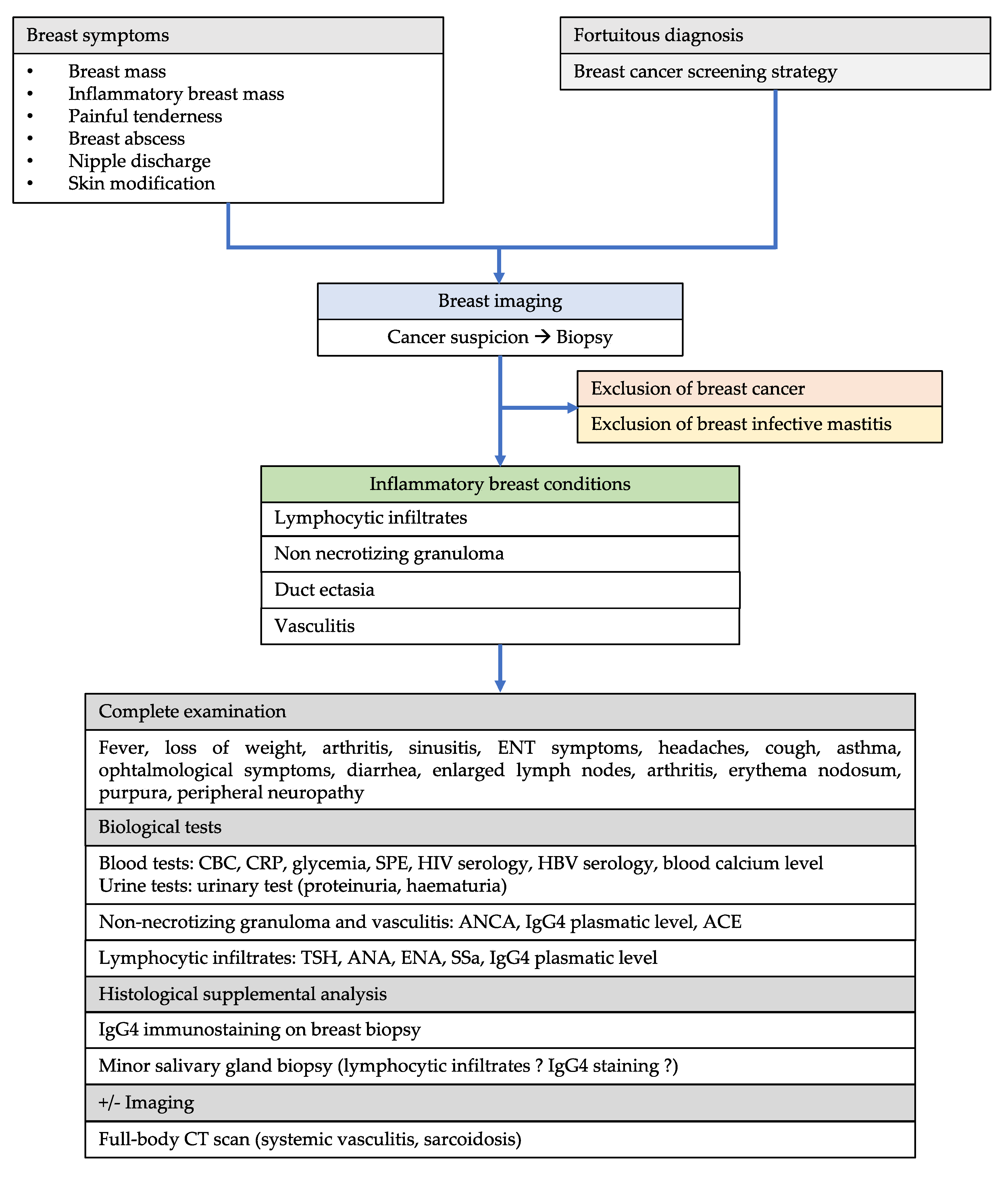
2. Methods
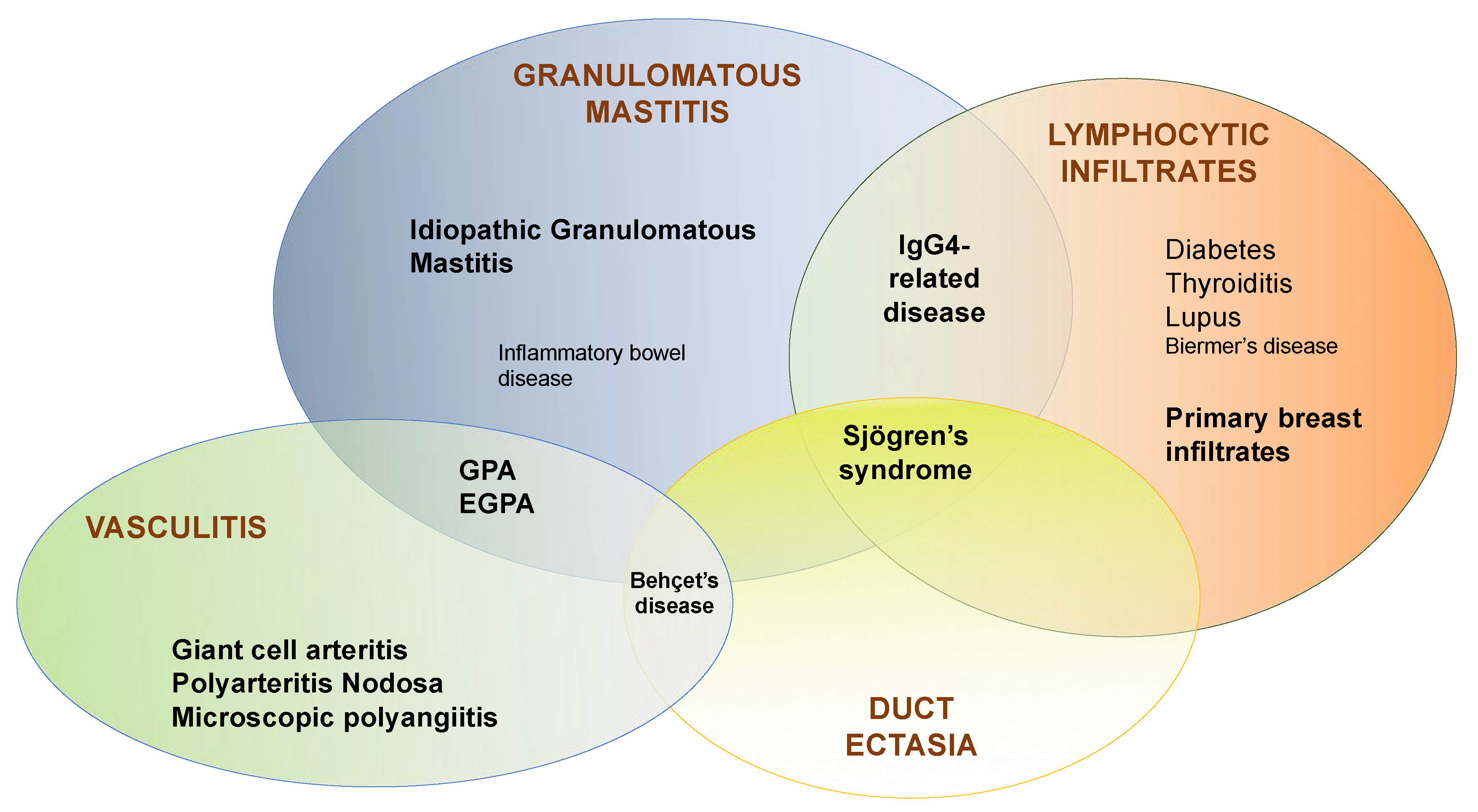
3. Breast Lymphocytic Infiltrates
3.1. Diabetes
3.2. Thyroiditis
3.3. Sjögren’s Syndrome
3.4. Lupus
3.5. Mixed Connective Tissue Disease
3.6. IgG4-Related Breast Disease
3.7. Biermer’s Disease
3.8. Primary Lymphocytic Infiltrates Involving the Breast
4. Mammary Duct Ectasia
5. Granulomatous Mastitis
5.1. Granulomatous Mastitis Associated with Systemic Granulomatous Diseases
5.1.1. Inflammatory Bowel Diseases
5.1.2. GPA
5.1.3. EGPA
5.1.4. Sarcoidosis
5.1.5. Granulomatous Mastitis Associated with IgG4-Disease
5.2. Granulomatous Mastitis Associated with Other Autoimmune Diseases
5.3. IdGM: “Idiopathic” or “Immunological” Granulomatous Mastitis?
6. Vasculitis
6.1. Polyarteritis Nodosa
6.2. Giant Cell Arteritis
6.3. Microscopic Polyangiitis
6.4. Behçet’s Disease
7. Discussion
7.1. Intrinsic Characteristics of the Mammary Gland
7.2. Environmental Factors
7.3. Hormonal Factors
7.4. The Role of Mammary Epithelial Cells
8. Conclusions
Author Contributions
Funding
Acknowledgments
Conflicts of Interest
References
- Rosa, M.; Mohammadi, A. Lupus mastitis: A review. Ann. Diagn. Pathol. 2013, 17, 230–233. [Google Scholar] [CrossRef] [PubMed]
- Ogiya, A.; Tanaka, K.; Tadokoro, Y.; Kikutani, M.; Uematsu, T.; Kashiwagi, H.; Kasami, M.; Takahashi, K. IgG4-related sclerosing disease of the breast successfully treated by steroid therapy. Breast Cancer 2014, 21, 231–235. [Google Scholar] [CrossRef] [PubMed]
- Goulabchand, R.; Hafidi, A.; Millet, I.; Morel, J.; Lukas, C.; Humbert, S.; Rivière, S.; Gény, C.; Jorgensen, C.; Le Quellec, A.; et al. Mastitis associated with Sjögren’s syndrome: A series of nine cases. Immunol. Res. 2016, 65. [Google Scholar] [CrossRef] [PubMed]
- Tan, E.S.; Friesen, B.; Loh, S.F.; Fox, J. Immunoglobulin-G4 related mastitis: A case report. Int. J. Surg. Case Rep. 2017, 37, 169–172. [Google Scholar] [CrossRef]
- Mason, C.; Yang, R.; Hamilton, R.; Punar, M.; Shah, Z.; Spigel, J.; Wang, J. Diagnosis of sarcoidosis from a biopsy of a dilated mammary duct. Proc. Bayl. Univ. Med. Cent. 2017, 30, 197–199. [Google Scholar] [CrossRef] [Green Version]
- Degnim, A.C.; Brahmbhatt, R.D.; Radisky, D.C.; Hoskin, T.L.; Stallings-Mann, M.; Laudenschlager, M.; Mansfield, A.; Frost, M.H.; Murphy, L.; Knutson, K.; et al. Immune cell quantitation in normal breast tissue lobules with and without lobulitis. Breast Cancer Res. Treat. 2014, 144, 539–549. [Google Scholar] [CrossRef] [Green Version]
- Hermsen, B.B.J.; von Mensdorff-Pouilly, S.; Fabry, H.F.J.; Winters, H.A.H.; Kenemans, P.; Verheijen, R.H.M.; van Diest, P.J. Lobulitis is a frequent finding in prophylactically removed breast tissue from women at hereditary high risk of breast cancer. J. Pathol. 2005, 206, 220–223. [Google Scholar] [CrossRef]
- Gulbahce, H.E.; Vanderwerf, S.; Blair, C.; Sweeney, C. Lobulitis in nonneoplastic breast tissue from breast cancer patients: Association with phenotypes that are common in hereditary breast cancer. Hum. Pathol. 2014, 45, 78–84. [Google Scholar] [CrossRef]
- De Boer, M.; van Leeuwen, F.E.; Hauptmann, M.; Overbeek, L.I.H.; de Boer, J.P.; Hijmering, N.J.; Sernee, A.; Klazen, C.A.H.; Lobbes, M.B.I.; van der Hulst, R.R.W.J.; et al. Breast Implants and the Risk of Anaplastic Large-Cell Lymphoma in the Breast. JAMA Oncol. 2018, 4, 335–341. [Google Scholar] [CrossRef]
- Fong, D.; Lann, M.A.; Finlayson, C.; Page, D.L.; Singh, M. Diabetic (lymphocytic) mastopathy with exuberant lymphohistiocytic and granulomatous response: A case report with review of the literature. Am. J. Surg. Pathol. 2006, 30, 1330–1336. [Google Scholar] [CrossRef]
- Kudva, Y.C.; Reynolds, C.; O’Brien, T.; Powell, C.; Oberg, A.L.; Crotty, T.B. “Diabetic mastopathy,” or sclerosing lymphocytic lobulitis, is strongly associated with type 1 diabetes. Diabetes Care 2002, 25, 121–126. [Google Scholar] [CrossRef] [PubMed] [Green Version]
- Lee, A.H.; Zafrani, B.; Kafiri, G.; Rozan, S.; Millis, R.R. Sclerosing lymphocytic lobulitis in the male breast. J. Clin. Pathol. 1996, 49, 609–611. [Google Scholar] [CrossRef] [PubMed] [Green Version]
- Hunfeld, K.P.; Bässler, R.; Kronsbein, H. “Diabetic mastopathy” in the male breast—A special type of gynecomastia. A comparative study of lymphocytic mastitis and gynecomastia. Pathol. Res. Pract. 1997, 193, 197–205. [Google Scholar] [PubMed]
- Boullu, S.; Andrac, L.; Piana, L.; Darmon, P.; Dutour, A.; Oliver, C. Diabetic mastopathy, complication of type 1 diabetes mellitus: Report of two cases and a review of the literature. Diabetes Metab. 1998, 24, 448–454. [Google Scholar] [PubMed]
- Valdez, R.; Thorson, J.; Finn, W.G.; Schnitzer, B.; Kleer, C.G. Lymphocytic mastitis and diabetic mastopathy: A molecular, immunophenotypic, and clinicopathologic evaluation of 11 cases. Mod. Pathol. 2003, 16, 223–228. [Google Scholar] [CrossRef] [PubMed] [Green Version]
- Andrews-Tang, D.; Diamond, A.B.; Rogers, L.; Butler, D. Diabetic Mastopathy: Adjunctive Use of Ultrasound and Utility of Core Biopsy in Diagnosis. Breast J. 2000, 6, 183–188. [Google Scholar] [CrossRef]
- Tuncbilek, N.; Karakas, H.M.; Okten, O. Diabetic fibrous mastopathy: Dynamic contrast-enhanced magnetic resonance imaging findings. Breast J. 2004, 10, 359–362. [Google Scholar] [CrossRef] [PubMed]
- Isomoto, I.; Wada, T.; Abe, K.; Uetani, M. Diagnostic utility of diffusion-weighted magnetic resonance imaging in diabetic mastopathy. Clin. Imaging 2009, 33, 146–149. [Google Scholar] [CrossRef]
- Sankaye, S.; Kachewar, S. Diabetic mastopathy. Australas Med. J. 2012, 5, 296–299. [Google Scholar] [CrossRef]
- Giusiano, S.; Andrac-Meyer, L.; Meunier-Carpentier, S.; Xerri, L.; Boubli, L.; Taranger-Charpin, C. A tumor-like lymphocytis mastitis. Ann. Pathol. 2005, 25, 231–234. [Google Scholar] [CrossRef]
- Douglas-Jones, A.G. Lymphocytic lobulitis in breast core biopsy: A peritumoral phenomenon. Histopathology 2006, 48, 209–212. [Google Scholar] [CrossRef] [PubMed]
- Oba, M.; Sasaki, M.; Ii, T.; Hoso, M.; Ajisaka, H.; Matsuki, N.; Miwa, K. A case of lymphocytic mastopathy requiring differential diagnosis from primary breast lymphoma. Breast Cancer 2009, 16, 141–146. [Google Scholar] [CrossRef] [PubMed]
- Murakami, R.; Kumita, S.; Yamaguchi, K.; Ueda, T. Diabetic mastopathy mimicking breast cancer. Clin. Imaging 2009, 33, 234–236. [Google Scholar] [CrossRef] [PubMed]
- Zhang, Y.; Zhong, D.; Sun, Q.; Zhou, Y.; Guan, J. Diabetic mastopathy mimicking breast cancer: Two case reports. Clin. Breast Cancer 2011, 11, 409–412. [Google Scholar] [CrossRef]
- Leroux-Stewart, J.; Rabasa-Lhoret, R. Diabetic mastopathy: Case report and summary of literature. Can. J. Diabetes 2014, 38, 305–306. [Google Scholar] [CrossRef]
- Tomaszewski, J.E.; Brooks, J.S.; Hicks, D.; Livolsi, V.A. Diabetic mastopathy: A distinctive clinicopathologic entity. Hum. Pathol. 1992, 23, 780–786. [Google Scholar] [CrossRef]
- Lammie, G.A.; Bobrow, L.G.; Staunton, M.D.; Levison, D.A.; Page, G.; Millis, R.R. Sclerosing lymphocytic lobulitis of the breast—Evidence for an autoimmune pathogenesis. Histopathology 1991, 19, 13–20. [Google Scholar] [CrossRef]
- Hunfeld, K.P.; Bässler, R. Lymphocytic mastitis and fibrosis of the breast in long-standing insulin-dependent diabetics. A histopathologic study on diabetic mastopathy and report of ten cases. Gen. Diagn. Pathol. 1997, 143, 49–58. [Google Scholar]
- Dorokhova, O.; Fineberg, S.; Koenigsberg, T.; Wang, Y. Diabetic mastopathy, a clinicopathological correlation of 34 cases. Pathol. Int. 2012, 62, 660–664. [Google Scholar] [CrossRef]
- Miura, K.; Teruya, C.; Hatsuko, N.; Ogura, H. Autoantibody with Cross-Reactivity between Insulin and Ductal Cells May Cause Diabetic Mastopathy: A Case Study. Case Rep. Med. 2012, 2012, 569040. [Google Scholar] [CrossRef]
- Dubenko, M.; Breining, D.; Surks, M.I. Sclerosing lymphocytic lobulitis of the breast in a patient with Graves’ disease. Thyroid 2003, 13, 309–311. [Google Scholar] [CrossRef] [PubMed]
- Park, S.H.; Choi, S.J.; Jung, H.K. Sclerosing lymphocytic lobulitis manifesting as suspicious microcalcifications with Hashimoto’s thyroiditis in a young woman. Breast J. 2013, 19, 539–541. [Google Scholar] [CrossRef] [PubMed]
- Gregoir, C.; Hilliquin, P.; Acar, F.; Lessana-Leibowitch, M.; Renoux, M.; Menkès, C.J. Acute mastitis in rheumatoid polyarthritis with Gougerot-Sjögren syndrome treated with tiopronin (Acadione). Rev. Rhum. Mal. Osteoartic. 1991, 58, 203–206. [Google Scholar] [PubMed]
- Ríos, G.; Peredo, R.A. Lymphocytic mastitis preceding Sjögren’s syndrome. Puerto Rico Health Sci. J. 2010, 29, 127–129. [Google Scholar]
- Kang, Y.K.; Jung, S.Y.; Qin, J.; Li, C.; Tsai, S.Y.; Tsai, M.-J.; O’Malley, B.W. E2/Estrogen receptor/sjogren syndrome-associated autoantigen relieves coactivator activator-induced G1/S arrest to promote breast tumorigenicity. Mol. Cell. Biol. 2014, 34, 1670–1681. [Google Scholar] [CrossRef] [PubMed] [Green Version]
- Moutsopoulos, H.M. Sjögren’s syndrome: Autoimmune epithelitis. Clin. Immunol. Immunopathol. 1994, 72, 162–165. [Google Scholar] [CrossRef]
- Chang, C.C.; Chang, Y.S.; Wang, S.H.; Lin, S.Y.; Chen, Y.H.; Chen, J.H. Primary Sjogren’s syndrome and the risk of acute pancreatitis: A nationwide cohort study. BMJ Open 2017, 7, e014807. [Google Scholar] [CrossRef]
- Ashton, M.A.; Lefkowitz, M.; Tavassoli, F.A. Epithelioid stromal cells in lymphocytic mastitis--a source of confusion with invasive carcinoma. Mod. Pathol. 1994, 7, 49–54. [Google Scholar]
- Kinonen, C.; Gattuso, P.; Reddy, V.B. Lupus mastitis: An uncommon complication of systemic or discoid lupus. Am. J. Surg. Pathol. 2010, 34, 901–906. [Google Scholar] [CrossRef]
- Warne, R.R.; Taylor, D.; Segal, A.; Irish, A. Lupus mastitis: A mimicker of breast carcinoma. BMJ Case Rep. 2011, 2011. [Google Scholar] [CrossRef] [Green Version]
- Lucivero, G.; Romano, C.; Ferraraccio, F.; Sellitto, A.; De Fanis, U.; Giunta, R.; Guarino, A.; Auriemma, P.P.; Benincasa, M.; Iovino, F. Lupus mastitis in systemic lupus erythematosus: A rare condition requiring a minimally invasive diagnostic approach. Int. J. Immunopathol. Pharmacol. 2011, 24, 1125–1129. [Google Scholar] [CrossRef] [PubMed] [Green Version]
- Sanders, L.M.; Lacz, N.L.; Blackwood, M.M.; Ongcapin, E.; Santos-Zabala, M.L. Lupus mastitis without systemic disease: Unusual imaging findings including MRI. Breast J. 2012, 18, 371–372. [Google Scholar] [CrossRef] [PubMed]
- Mosier, A.D.; Boldt, B.; Keylock, J.; Smith, D.V.; Graham, J. Serial MR Findings and Comprehensive Review of Bilateral Lupus Mastitis with an Additional Case Report. J. Radiol. Case Rep. 2013, 7, 48–58. [Google Scholar] [CrossRef] [PubMed]
- Tanaka, Y.; Manabe, H.; Shinzaki, W.; Hashimoto, Y.; Komoike, Y. A case of lupus mastitis in a patient with systemic lupus erythematosus. Breast J. 2019. [Google Scholar] [CrossRef] [PubMed]
- Xu, R.; Guo, Q.Q.; Yang, L.P.; Lai, M.L.; Tong, L. Variations of peripheral blood autoantibody, immunoglobuliln, and complement levels in patients with non-lactational mastitis and their clinical significances. Nan Fang Yi Ke Da Xue Xue Bao 2016, 36, 1157–1159. [Google Scholar]
- Varma, R.; Szilagyi, S.; Harshan, M. Breast involvement in mixed connective tissue disease. Radiol. Case Rep. 2019, 14, 430–435. [Google Scholar] [CrossRef]
- Zen, Y.; Kasahara, Y.; Horita, K.; Miyayama, S.; Miura, S.; Kitagawa, S.; Nakanuma, Y. Inflammatory pseudotumor of the breast in a patient with a high serum IgG4 level: Histologic similarity to sclerosing pancreatitis. Am. J. Surg. Pathol. 2005, 29, 275–278. [Google Scholar] [CrossRef]
- Cheuk, W.; Chan, A.C.L.; Lam, W.L.; Chow, S.M.; Crowley, P.; Lloydd, R.; Campbell, I.; Thorburn, M.; Chan, J.K.C. IgG4-related sclerosing mastitis: Description of a new member of the IgG4-related sclerosing diseases. Am. J. Surg. Pathol. 2009, 33, 1058–1064. [Google Scholar] [CrossRef]
- Chougule, A.; Bal, A.; Das, A.; Singh, G. IgG4 related sclerosing mastitis: Expanding the morphological spectrum of IgG4 related diseases. Pathology 2015, 47, 27–33. [Google Scholar] [CrossRef]
- Allen, S.G.; Soliman, A.S.; Toy, K.; Omar, O.S.; Youssef, T.; Karkouri, M.; Ayad, E.; Abdel-Aziz, A.; Hablas, A.; Tahri, A.; et al. Chronic Mastitis in Egypt and Morocco: Differentiating between Idiopathic Granulomatous Mastitis and IgG4-Related Disease. Breast J. 2016, 22, 501–509. [Google Scholar] [CrossRef]
- Yamada, R.; Horiguchi, S.; Yamashita, T.; Kamisawa, T. IgG4-related mastitis, a rare disease, can radiologically and histologically mimic malignancy. BMJ Case Rep. 2016, 2016. [Google Scholar] [CrossRef] [PubMed]
- Farooq, T.A.; Mudhar, H.; Sandramouli, S. A case of non-lacrimal immunoglobulin G4 (IgG4)-related orbital disease with mastitis. Orbit 2016, 35, 16–19. [Google Scholar] [CrossRef] [PubMed]
- Vitkovski, T.; Marder, G.S.; Filardi, D.A.; Gupta, E.; Breuer, F. IgG4-Related Sclerosing Disease of the Breast in a Male Patient. Int. J. Surg. Pathol. 2017, 25, 711–715. [Google Scholar] [CrossRef] [PubMed]
- Yokoe, T.; Hayashida, T.; Kikuchi, M.; Watanuki, R.; Nakashoji, A.; Maeda, H.; Toyota, T.; Seki, T.; Takahashi, M.; Iwasaki, E.; et al. IgG4-related mastopathy: A case report and literature review. Clin. Case Rep. 2018, 6, 1549–1553. [Google Scholar] [CrossRef] [PubMed] [Green Version]
- Kriegsmann, M.; Gomez, C.; Heil, J.; Schäfgen, B.; Gutjahr, E.; Kommoss, F.K.F.; Kriegsmann, K.; Flechtenmacher, C.; Goeppert, B.; Sinn, H.P. IgG4-related sclerosing mastitis in a 49-year-old patient with multiple, tumor-like nodules-Diagnostic accuracy of core needle biopsy. Breast J. 2019, 25, 1251–1253. [Google Scholar] [CrossRef]
- El-Nayir, M.; Subramanian, A.; Ali, Z.; Howlett, D. IgG4-mediated sclerosing fibroinflammatory disease presenting as inflammatory breast malignancy. BJR Case Rep. 2019, 5, 20180041. [Google Scholar] [CrossRef]
- Dítě, P.; Trna, J.; Kinkor, Z.; Novotný, I.; Lata, J.; Kianička, B.; Hermanová, M. Unusual Multiorgan Immunoglobulin G4 (IgG4) Inflammation: Autoimmune Pancreatitis, Mikulicz Syndrome, and IgG4 Mastitis. Gut Liver 2013, 7, 621–624. [Google Scholar] [CrossRef] [Green Version]
- Tsuda, B.; Kumaki, N.; Ishida, R.; Mizuno, M.; Yokoyama, K.; Oshitanai, R.; Terao, M.; Morioka, T.; Okamura, T.; Saito, Y.; et al. Distinction of IgG4-related mastitis from breast cancer: A case report. Surg. Case Rep. 2019, 5, 123. [Google Scholar] [CrossRef] [Green Version]
- Ely, K.A.; Tse, G.; Simpson, J.F.; Clarfeld, R.; Page, D.L. Diabetic mastopathy. A clinicopathologic review. Am. J. Clin. Pathol. 2000, 113, 541–545. [Google Scholar] [CrossRef]
- Rooney, N.; Snead, D.; Goodman, S.; Webb, A.J. Primary breast lymphoma with skin involvement arising in lymphocytic lobulitis. Histopathology 1994, 24, 81–84. [Google Scholar] [CrossRef]
- Rahal, R.M.S.; de Freitas-Júnior, R.; Carlos da Cunha, L.; Moreira, M.A.R.; Rosa, V.D.L.; Conde, D.M. Mammary Duct Ectasia: An Overview. Breast J. 2011, 17, 694–695. [Google Scholar] [CrossRef] [PubMed]
- O’Brien, P.H.; Kreutner, A. Another cause of nipple discharge. Mammary duct ectasia with periductal mastitis. Am. Surg. 1982, 48, 577–578. [Google Scholar] [PubMed]
- McHoney, M.; Munro, F.; Mackinlay, G. Mammary duct ectasia in children: Report of a short series and review of the literature. Early Hum. Dev. 2011, 87, 527–530. [Google Scholar] [CrossRef] [PubMed]
- D’Alfonso, T.M.; Ginter, P.S.; Shin, S.J. A Review of Inflammatory Processes of the Breast with a Focus on Diagnosis in Core Biopsy Samples. J. Pathol. Transl. Med. 2015, 49, 279–287. [Google Scholar] [CrossRef] [Green Version]
- Dixon, J.M.; Anderson, T.J.; Lumsden, A.B.; Elton, R.A.; Roberts, M.M.; Forrest, A.P. Mammary duct ectasia. Br. J. Surg. 1983, 70, 601–603. [Google Scholar] [CrossRef] [PubMed]
- Ramalingam, K.; Srivastava, A.; Vuthaluru, S.; Dhar, A.; Chaudhry, R. Duct Ectasia and Periductal Mastitis in Indian Women. Indian J. Surg. 2015, 77, 957–962. [Google Scholar] [CrossRef] [Green Version]
- Frantz, V.K.; Pickren, J.W.; Melcher, G.W.; Auchincloss, H. Indicence of chronic cystic disease in so-called “normal breasts; a study based on 225 postmortem examinations. Cancer 1951, 4, 762–783. [Google Scholar] [CrossRef]
- Dixon, J.M.; Ravisekar, O.; Chetty, U.; Anderson, T.J. Periductal mastitis and duct ectasia: Different conditions with different aetiologies. Br. J. Surg. 1996, 83, 820–822. [Google Scholar] [CrossRef]
- Al-Masad, J.K. Mammary duct ectasia and periductal mastitis in males. Saudi Med. J. 2001, 22, 1030–1033. [Google Scholar]
- Anil, C.; Guney, T.; Gursoy, A. The prevalence of benign breast diseases in patients with nodular goiter and Hashimoto’s thyroiditis. J. Endocrinol. Investig. 2015, 38, 971–975. [Google Scholar] [CrossRef]
- Choi, S.H.; Jang, K.S.; Chung, M.S. Bilateral granulomatous mastitis with a different etiology. Cancer Biomark. 2015, 15, 151–156. [Google Scholar] [CrossRef] [PubMed]
- Illman, J.E.; Terra, S.B.; Clapp, A.J.; Hunt, K.N.; Fazzio, R.T.; Shah, S.S.; Glazebrook, K.N. Granulomatous diseases of the breast and axilla: Radiological findings with pathological correlation. Insights Imaging 2018, 9, 59–71. [Google Scholar] [CrossRef] [PubMed] [Green Version]
- Gautham, I.; Radford, D.M.; Kovacs, C.S.; Calhoun, B.C.; Procop, G.W.; Shepardson, L.B.; Dawson, A.E.; Downs-Kelly, E.P.; Zhang, G.X.; Al-Hilli, Z.; et al. Cystic neutrophilic granulomatous mastitis: The Cleveland Clinic experience with diagnosis and management. Breast J. 2018, 25, 80–85. [Google Scholar] [CrossRef] [PubMed] [Green Version]
- Allende, D.S.; Booth, C.N. Wegener’s granulomatosis of the breast: A rare entity with daily clinical relevance. Ann. Diagn. Pathol. 2009, 13, 351–357. [Google Scholar] [CrossRef]
- Ojeda, H.; Sardi, A.; Totoonchie, A. Sarcoidosis of the breast: Implications for the general surgeon. Am. Surg. 2000, 66, 1144–1148. [Google Scholar]
- Ogura, K.; Matsumoto, T.; Aoki, Y.; Kitabatake, T.; Fujisawa, M.; Kojima, K. IgG4-related tumour-forming mastitis with histological appearances of granulomatous lobular mastitis: Comparison with other types of tumour-forming mastitis. Histopathology 2010, 57, 39–45. [Google Scholar] [CrossRef]
- Mathelin, C.; Riegel, P.; Chenard, M.P.; Brettes, J.P. Association of corynebacteria with granulomatous mastitis. Eur. J. Obstet. Gynecol. Reprod. Biol. 2005, 119, 260–261. [Google Scholar] [CrossRef]
- Renshaw, A.A.; Derhagopian, R.P.; Gould, E.W. Cystic neutrophilic granulomatous mastitis: An underappreciated pattern strongly associated with gram-positive bacilli. Am. J. Clin. Pathol. 2011, 136, 424–427. [Google Scholar] [CrossRef]
- Ladefoged, K.; Balslev, E.; Jemec, G.B. Crohn’s disease presenting as a breast abscess: A case report. J. Eur. Acad. Dermatol. Venereol. 2001, 15, 343–345. [Google Scholar] [CrossRef]
- Sallé de Chou, C.; Ortonne, N.; Hivelin, M.; Wolkenstein, P.; Chosidow, O.; Valeyrie-Allanore, L. Aseptic cutaneous breast abscesses associated with ulcerative colitis. Ann. Dermatol. Venereol. 2016, 143, 139–143. [Google Scholar] [CrossRef]
- Jennette, J.C.; Falk, R.J.; Bacon, P.A.; Basu, N.; Cid, M.C.; Ferrario, F.; Flores-Suarez, L.F.; Gross, W.L.; Guillevin, L.; Hagen, E.C.; et al. 2012 revised International Chapel Hill Consensus Conference Nomenclature of Vasculitides. Arthritis Rheum. 2013, 65, 1–11. [Google Scholar] [CrossRef] [PubMed]
- Pambakian, H.; Tighe, J.R. Breast involvement in Wegener’s granulomatosis. J. Clin. Pathol. 1971, 24, 343–347. [Google Scholar] [CrossRef] [PubMed] [Green Version]
- Trüeb, R.M.; Pericin, M.; Kohler, E.; Barandun, J.; Burg, G. Necrotizing granulomatosis of the breast. Br. J. Dermatol. 1997, 137, 799–803. [Google Scholar] [CrossRef] [PubMed]
- Ivanov, S. Wegner granulomatosis with initial manifestation of bilateral granulomatous purulent necrotic mastitis. Khirurgiia (Sofiia) 1999, 55, 54–57. [Google Scholar] [PubMed]
- Barczyńska, T.; Dankiewicz-Fares, I.; Bilińska-Reszkowska, H.; Zalewska, J.; Jeka, S. Atypical location of Wegener’s granulomatosis with breast involvement: Case report. Ann. Acad. Med. Stetin. 2011, 57, 70–76. [Google Scholar]
- Georgescu, R.; Podeanu, M.D.; Colcer, I.; Grigorescu, G.; Coroș, M.F.; Moldovan, C.; Ilyes, A.; Bârsan, I.; Moncea, D.; Stolnicu, S. Wegener’s Granulomatosis of the Breast with Peculiar Radiological Aspect Mimicking Breast Carcinoma. Breast J. 2015, 21, 550–552. [Google Scholar] [CrossRef]
- Ryba, M.; Konieczny, A.; Hruby, Z. Breast Involvement in Anti-Neutrophil Cytoplasmic Antibodies Positive Granulomatosis With Polyangiitis in a 64-Year-Old Female Patient. Arch. Rheumatol. 2017, 32, 358–360. [Google Scholar] [CrossRef]
- Daguzan, M.; Martinez-Mena, C.; Hermans, P. A case of breast necrosis. Rev. Med. Brux. 2012, 33, 112–115. [Google Scholar]
- Skandarajah, A.; Marley, L. Idiopathic granulomatous mastitis: A medical or surgical disease of the breast? ANZ J. Surg. 2015, 85, 979–982. [Google Scholar] [CrossRef]
- Ene, N.; Bélénotti, P.; Benyamine, A.; Sovaila, S.; Ben Sahla Talet, M.H.; Kaminsky, P.; Serratrice, J.; Weiller, P.J. Granulomatosis with polyangiitis (previously Wegener’s granulomatosis) mimicking malingering. Rev. Med. Interne 2014, 35, 540–542. [Google Scholar] [CrossRef]
- Gallais Sérézal, I.; Jennische, K. Image Gallery: Symmetrical necrosis of the breast as an inaugural manifestation of granulomatosis with polyangiitis (Wegener granulomatosis). Br. J. Dermatol. 2016, 175, e132. [Google Scholar] [CrossRef] [PubMed] [Green Version]
- Comas, A.G.V.; Diana, C.A.F.; Crespo, C.C.; Cebollada, M.M.; Liñán, M.A.L.; Vila, J.V.R. Wegener’s Granulomatosis Presented as Recurrent Breast Abscess. Breast J. 2010, 16, 82–84. [Google Scholar] [CrossRef] [PubMed]
- Giordano, D.; Dardani, L.; Ghidini, A. Atypical Presentation of Granulomatosis with Polyangiitis. J. Belg. Soc. Radiol. 2018, 102, 72. [Google Scholar] [CrossRef] [PubMed]
- Mengoli, M.C.; Ragazzi, M.; Lococo, F.; Mengoli, M.A.; Balli, M.C.; Marchioni, A.; Rossi, G. Breast granulomatosis with polyangiitis mimicking breast cancer. Pathologica 2017, 109, 405–407. [Google Scholar] [PubMed]
- Ren, J.; Liu, J.; Su, J.; Zhang, J.; Zhao, J. Systemic vasculitis involving the breast: A case report and literature review. Rheumatol. Int. 2019, 39, 1447–1455. [Google Scholar] [CrossRef]
- Stappaerts, I.; Colpaert, C.; Verbraecken, J.; Van Marck, E.; Vermeire, P. Granulomatous mastitis as presenting sign of Wegener’s granulomatosis. Acta Clin. Belg. 1999, 54, 207–210. [Google Scholar] [CrossRef]
- Villalba-Nuño, V.; Sabaté, J.M.; Gómez, A.; Vidaller, A.; Català, I.; Escobedo, A.; Torrubia, S. Churg-Strauss syndrome involving the breast: A rare cause of eosinophilic mastitis. Eur. Radiol. 2002, 12, 646–649. [Google Scholar] [CrossRef]
- Ben Dhaou, B.; Derbali, F.; Boussema, F.; Aydi, Z.; Baili, L.; Kochbati, S.; Cherif, O.; Rokbani, L. Eosinophilic nodule of the breast: A rare manifestation in the Churg-Strauss syndrome. Tunis. Med. 2012, 90, 752–753. [Google Scholar]
- Visentin, M.S.; Salmaso, R.; Modesti, V.; Ometto, F.; Ruffatti, A.; Punzi, L.; Doria, A. Parotid, breast, and fascial involvement in a patient who fulfilled the ACR criteria for Churg-Strauss syndrome. Scand. J. Rheumatol. 2012, 41, 319–321. [Google Scholar] [CrossRef]
- Dannepond, C.; Le Fourn, E.; de Muret, A.; Ouldamer, L.; Carmier, D.; Machet, L. Mastitis revealing Churg-Strauss syndrome. Ann. Dermatol. Venereol. 2014, 141, 30–33. [Google Scholar] [CrossRef]
- Lee, M.X.W.; Teng, G.G.; Raju, G.C.; Lim, A.Y.N. Catastrophic subarachnoid hemorrhage in eosinophilic granulomatosis with polyangiitis without asthma. Int. J. Rheum. Dis. 2017, 20, 2127–2131. [Google Scholar] [CrossRef] [PubMed]
- Donaldson, B.A.; Polynice, A.; Oluwole, S. Sarcoidosis of the breast: Case report and chart review. Am. Surg. 1995, 61, 778–780. [Google Scholar] [PubMed]
- Kenzel, P.P.; Hadijuana, J.; Hosten, N.; Minguillon, C.; Oellinger, H.; Siewert, C.; Thiel, T.; Lichtenegger, W.; Felix, R. Boeck sarcoidosis of the breast: Mammographic, ultrasound, and MR findings. J. Comput. Assist. Tomogr. 1997, 21, 439–441. [Google Scholar] [CrossRef] [PubMed]
- Kirshy, D.; Gluck, B.; Brancaccio, W. Sarcoidosis of the breast presenting as a spiculated lesion. AJR Am. J. Roentgenol. 1999, 172, 554–555. [Google Scholar] [CrossRef]
- Gisvold, J.J.; Crotty, T.B.; Johnson, R.E. Sarcoidosis presenting as spiculated breast masses. Mayo Clin. Proc. 2000, 75, 293–295. [Google Scholar] [CrossRef] [Green Version]
- Harris, K.P.; Faliakou, E.C.; Exon, D.J.; Nasiri, N.; Gui, G.P. Isolated sarcoidosis of the breast. J. R. Soc. Med. 2000, 93, 196–197. [Google Scholar] [CrossRef]
- Lower, E.E.; Hawkins, H.H.; Baughman, R.P. Breast disease in sarcoidosis. Sarcoidosis Vasc. Diffus. Lung Dis. 2001, 18, 301–306. [Google Scholar]
- Takahashi, R.; Shibuya, Y.; Shijubo, N.; Asaishi, K.; Abe, S. Mammary involvement in a patient with sarcoidosis. Intern. Med. 2001, 40, 769–771. [Google Scholar] [CrossRef] [Green Version]
- Mingins, C.; Williams, M.R.; Cox, N.H. Subcutaneous sarcoidosis mimicking breast carcinoma. Br. J. Dermatol. 2002, 146, 924–925. [Google Scholar] [CrossRef]
- Ishimaru, K.; Isomoto, I.; Okimoto, T.; Itoyanagi, A.; Uetani, M. Sarcoidosis of the breast. Eur. Radiol. 2002, 12 (Suppl. 3), S105–S108. [Google Scholar] [CrossRef]
- Mona, E.K.; Pascal, C.; Charley, H.; Françoise, B.; Véronique, B.; Marie-Madeleine, P. Quiz case. Breast sarcoidosis presenting as a metastatic breast cancer. Eur. J. Radiol. 2005, 54, 2–5. [Google Scholar] [CrossRef] [PubMed]
- Fiorucci, F.; Conti, V.; Lucantoni, G.; Patrizi, A.; Fiorucci, C.; Giannunzio, G.; Di Michele, L. Sarcoidosis of the breast: A rare case report and a review. Eur. Rev. Med. Pharmacol. Sci. 2006, 10, 47–50. [Google Scholar] [PubMed]
- Nicholson, B.T.; Mills, S.E. Sarcoidosis of the breast: An unusual presentation of a systemic disease. Breast J. 2007, 13, 99–100. [Google Scholar] [CrossRef] [PubMed]
- Hermann, G.; Nagi, C.; Mester, J.; Tierstein, A. Unusual presentation of sarcoidosis of the breast. Br. J. Radiol. 2008, 81, e231–e233. [Google Scholar] [CrossRef]
- Rishi, M.A.; Smith, M. Sarcoid of the breast associated with microcalcifications on mammogram. Intern. Med. J. 2009, 39, 134–135. [Google Scholar]
- Panzacchi, R.; Gallo, C.; Fois, F.; Dalpiaz, G.; Cucchi, M.C.; Degli Esposti, R.; Foschini, M.P. Primary sarcoidosis of the breast: Case description and review of the literature. Pathologica 2010, 102, 104–107. [Google Scholar]
- DeFilippis, E.M.; Arleo, E.K. New diagnosis of sarcoidosis during treatment for breast cancer, with radiologic-pathologic correlation. Clin. Imaging 2013, 37, 762–766. [Google Scholar] [CrossRef]
- Zivin, S.; David, O.; Lu, Y. Sarcoidosis mimicking metastatic breast cancer on FDG PET/CT. Intern. Med. 2014, 53, 2555–2556. [Google Scholar] [CrossRef] [Green Version]
- Endlich, J.L.; Souza, J.A.; de Toledo Osório, C.A.B.; Pinto, C.A.L.; Faria, E.P.; Bitencourt, A.G.V. Breast sarcoidosis as the first manifestation of the disease. Breast J. 2020, 26, 543–544. [Google Scholar] [CrossRef]
- Reis, J.; Boavida, J.; Lyngra, M.; Geitung, J.T. Radiological evaluation of primary breast sarcoidosis presenting as bilateral breast lesions. BMJ Case Rep. 2019, 12, e229591. [Google Scholar] [CrossRef]
- Tolaney, S.M.; Colson, Y.L.; Gill, R.R.; Schulte, S.; Duggan, M.M.; Shulman, L.N.; Winer, E.P. Sarcoidosis mimicking metastatic breast cancer. Clin. Breast Cancer 2007, 7, 804–810. [Google Scholar] [CrossRef] [PubMed]
- Zujić, P.V.; Grebić, D.; Valenčić, L. Chronic granulomatous inflammation of the breast as a first clinical manifestation of primary sarcoidosis. Breast Care (Basel) 2015, 10, 51–53. [Google Scholar] [CrossRef] [PubMed] [Green Version]
- Tareila, A.M.; Aswad, B.I.; Ward, R.C.; Hillstrom, M.M. Bilateral Breast Sarcoidosis Mimicking Synchronous Primary Breast Cancer. Rhode Isl. Med. J. 2013 2018, 101, 39–40. [Google Scholar]
- Maillet, L.; Chopin, N.; Treilleux, I.; Bachelot, T.; Tredan, O.; Faure, C.; Caux, C.; Beurrier, F. Spontaneous regression of breast cancer after biopsy. About two cases. Gynecol. Obstet. Fertil. 2014, 42, 269–272. [Google Scholar] [CrossRef] [PubMed]
- Akhtari, M.; Quesada, J.R.; Schwartz, M.R.; Chiang, S.B.; Teh, B.S. Sarcoidosis presenting as metastatic lymphadenopathy in breast cancer. Clin. Breast Cancer 2014, 14, e107–e110. [Google Scholar] [CrossRef]
- Grados, A.; Ebbo, M.; Bernit, E.; Veit, V.; Mazodier, K.; Jean, R.; Coso, D.; Aurran-Schleinitz, T.; Broussais, F.; Bouabdallah, R.; et al. Sarcoidosis Occurring After Solid Cancer: A Nonfortuitous Association: Report of 12 Cases and Review of the Literature. Medicine (Baltimore) 2015, 94, e928. [Google Scholar] [CrossRef]
- Kobak, S.; Yildiz, F.; Semiz, H.; Orman, M. Malignancy in Patients with Sarcoidosis. Reumatol. Clin. 2019. [Google Scholar] [CrossRef]
- Sun, H.H.; Sachanandani, N.S.; Jordan, B.; Myckatyn, T.M. Sarcoidosis of the Breasts following Silicone Implant Placement. Plast. Reconstr. Surg. 2013, 131, 939e–940e. [Google Scholar] [CrossRef]
- Yadlapati, S.; Verheyen, E.; Efthimiou, P. IgG4-related disease: A complex under-diagnosed clinical entity. Rheumatol. Int. 2018, 38, 169–177. [Google Scholar] [CrossRef]
- Herrera van Oostdam, D.A.; Jaimes Piñón, T.; Martínez-Martínez, M.U.; Oros-Ovalle, C.; Aléman-Sánchez, N.; Abud-Mendoza, C. IgG4-related disease, retrospective histopathological diagnosis. Prevalence in an university hospita. Reumatol. Clin. 2015, 11, 335–339. [Google Scholar] [CrossRef]
- Zhang, L.N.; Shi, T.Y.; Yang, Y.J.; Zhang, F.C. An SLE patient with prolactinoma and recurrent granulomatous mastitis successfully treated with hydroxychloroquine and bromocriptine. Lupus 2014, 23, 417–420. [Google Scholar] [CrossRef] [PubMed]
- Letourneux, C.; Diemunsch, P.; Korganow, A.-S.; Akladios, C.Y.; Bellocq, J.P.; Mathelin, C. First report of granulomatous mastitis associated with Sjögren’s syndrome. World J. Surg. Oncol. 2013, 11, 268. [Google Scholar] [CrossRef] [PubMed] [Green Version]
- Iqbal, F.M.; Ali, H.; Vidya, R. Breast lumps: A rare site for rheumatoid nodules. BMJ Case Rep. 2015, 2015. [Google Scholar] [CrossRef] [PubMed]
- Ramos-Casals, M.; Brito-Zerón, P.; García-Carrasco, M.; Font, J. Sarcoidosis or Sjögren syndrome? Clues to defining mimicry or coexistence in 59 cases. Medicine (Baltimore) 2004, 83, 85–95. [Google Scholar] [CrossRef]
- Yazigi, G.; Trieu, B.H.; Landis, M.; Parikh, J.G.; Mangal, M. Granulomatous Mastitis: A Rare Case with Sjogren’s Syndrome and Complications. Cureus 2019, 11, e5359. [Google Scholar] [CrossRef] [Green Version]
- Boufettal, H.; Essodegui, F.; Noun, M.; Hermas, S.; Samouh, N. Idiopathic granulomatous mastitis: A report of twenty cases. Diagn. Interv. Imaging 2012, 93, 586–596. [Google Scholar] [CrossRef] [Green Version]
- Altintoprak, F.; Kivilcim, T.; Ozkan, O.V. Aetiology of idiopathic granulomatous mastitis. World J. Clin. Cases 2014, 2, 852–858. [Google Scholar] [CrossRef]
- Aghajanzadeh, M.; Hassanzadeh, R.; Alizadeh Sefat, S.; Alavi, A.; Hemmati, H.; Esmaeili Delshad, M.S.; Emir Alavi, C.; Rimaz, S.; Geranmayeh, S.; Najafi Ashtiani, M.; et al. Granulomatous mastitis: Presentations, diagnosis, treatment and outcome in 206 patients from the north of Iran. Breast 2015, 24, 456–460. [Google Scholar] [CrossRef]
- Baslaim, M.M.; Khayat, H.A.; Al-Amoudi, S.A. Idiopathic granulomatous mastitis: A heterogeneous disease with variable clinical presentation. World J. Surg. 2007, 31, 1677–1681. [Google Scholar] [CrossRef]
- Lai, E.C.H.; Chan, W.C.; Ma, T.K.F.; Tang, A.P.Y.; Poon, C.S.P.; Leong, H.T. The role of conservative treatment in idiopathic granulomatous mastitis. Breast J. 2005, 11, 454–456. [Google Scholar] [CrossRef]
- Sheybani, F.; Naderi, H.R.; Gharib, M.; Sarvghad, M.; Mirfeizi, Z. Idiopathic granulomatous mastitis: Long-discussed but yet-to-be-known. Autoimmunity 2016, 49, 236–239. [Google Scholar] [CrossRef] [PubMed]
- Elzahaby, I.A.; Khater, A.; Fathi, A.; Hany, I.; Abdelkhalek, M.; Gaballah, K.; Elalfy, A.; Hamdy, O. Etiologic revelation and outcome of the surgical management of idiopathic granulomatous mastitis; An Egyptian centre experience. Breast Dis. 2016, 36, 115–122. [Google Scholar] [CrossRef] [PubMed]
- Patel, R.A.; Strickland, P.; Sankara, I.R.; Pinkston, G.; Many, W.; Rodriguez, M. Idiopathic granulomatous mastitis: Case reports and review of literature. J. Gen. Intern. Med. 2010, 25, 270–273. [Google Scholar] [CrossRef] [PubMed] [Green Version]
- Altintoprak, F.; Karakece, E.; Kivilcim, T.; Dikicier, E.; Cakmak, G.; Celebi, F.; Ciftci, I.H. Idiopathic granulomatous mastitis: An autoimmune disease? Sci. World J. 2013, 2013, 148727. [Google Scholar] [CrossRef]
- Co, M.; Cheng, V.C.C.; Wei, J.; Wong, S.C.Y.; Chan, S.M.S.; Shek, T.; Kwong, A. Idiopathic granulomatous mastitis: A 10-year study from a multicentre clinical database. Pathology 2018, 50, 742–747. [Google Scholar] [CrossRef]
- Helal, T.E.A.; Shash, L.S.; Saad El-Din, S.A.; Saber, S.M. Idiopathic Granulomatous Mastitis: Cytologic and Histologic Study of 65 Egyptian Patients. Acta Cytol. 2016, 60, 438–444. [Google Scholar] [CrossRef]
- Farouk, O.; Abdelkhalek, M.; Abdallah, A.; Shata, A.; Senbel, A.; Attia, E.; Elghaffar, M.A.; Mesbah, M.; Soliman, N.; Amin, M.; et al. Rifampicin for Idiopathic Granulomatous Lobular Mastitis: A Promising Alternative for Treatment. World J. Surg. 2017, 41, 1313–1321. [Google Scholar] [CrossRef]
- Martinez-Ramos, D.; Simon-Monterde, L.; Suelves-Piqueres, C.; Queralt-Martin, R.; Granel-Villach, L.; Laguna-Sastre, J.M.; Nicolau, M.J.; Escrig-Sos, J. Idiopathic granulomatous mastitis: A systematic review of 3060 patients. Breast J. 2019, 25, 1245–1250. [Google Scholar] [CrossRef]
- Tse, G.M.K.; Poon, C.S.P.; Ramachandram, K.; Ma, T.K.F.; Pang, L.M.; Law, B.K.B.; Chu, W.C.W.; Tang, A.P.Y.; Cheung, H.S. Granulomatous mastitis: A clinicopathological review of 26 cases. Pathology 2004, 36, 254–257. [Google Scholar] [CrossRef]
- Cheng, J.; Ding, H.; Du, Y. Granulomatous lobular mastitis associated with mammary duct ectasia: A clinicopathologic study of 32 cases with review of literature. Zhonghua Bing Li Xue Za Zhi 2013, 42, 665–668. [Google Scholar]
- Sheybani, F.; Sarvghad, M.; Naderi, H.; Gharib, M. Treatment for and clinical characteristics of granulomatous mastitis. Obstet. Gynecol. 2015, 125, 801–807. [Google Scholar] [CrossRef] [PubMed]
- Lin, C.H.; Hsu, C.W.; Tsao, T.Y.; Chou, J. Idiopathic granulomatous mastitis associated with risperidone-induced hyperprolactinemia. Diagn. Pathol. 2012, 7, 2. [Google Scholar] [CrossRef] [PubMed] [Green Version]
- Bellavia, M.; Damiano, G.; Palumbo, V.D.; Spinelli, G.; Tomasello, G.; Marrazzo, A.; Ficarella, S.; Bruno, A.; Sammartano, A.; Fiorentini, T.; et al. Granulomatous Mastitis during Chronic Antidepressant Therapy: Is It Possible a Conservative Therapeutic Approach? J. Breast Cancer 2012, 15, 371–372. [Google Scholar] [CrossRef] [PubMed]
- Holla, S.; Amberkar, M.B.; Kamath, A.; Kamalkishore, M.K.; Ommurugan, B. Risperidone Induced Granulomatous Mastitis Secondary to Hyperprolactinemia in a Non-Pregnant Woman-A Rare Case Report in a Bipolar Disorder. J. Clin. Diagn. Res. 2017, 11, FD01–FD03. [Google Scholar] [CrossRef]
- Salesi, M.; Karimifar, M.; Salimi, F.; Mahzouni, P. A case of granulomatous mastitis with erythema nodosum and arthritis. Rheumatol. Int. 2011, 31, 1093–1095. [Google Scholar] [CrossRef]
- Akın, M.; Karabacak, H.; Esendağlı, G.; Yavuz, A.; Gültekin, S.; Dikmen, K.; Bostancı, H. Coexistence of idiopathic granulomatous mastitis and erythemanodosum: Successful treatment with corticosteroids. Turk. J. Med. Sci. 2017, 47, 1590–1592. [Google Scholar] [CrossRef] [Green Version]
- Ozel, L.; Unal, A.; Unal, E.; Kara, M.; Erdoğdu, E.; Krand, O.; Güneş, P.; Karagül, H.; Demiral, S.; Titiz, M.I. Granulomatous mastitis: Is it an autoimmune disease? Diagnostic and therapeutic dilemmas. Surg. Today 2012, 42, 729–733. [Google Scholar] [CrossRef]
- Koksal, H. Human leukocyte antigens class I and II in patients with idiopathic granulomatous mastitis. Am. J. Surg. 2019, 218, 605–608. [Google Scholar] [CrossRef]
- Chirappapha, P.; Thaweepworadej, P.; Supsamutchai, C.; Biadul, N.; Lertsithichai, P. Idiopathic granulomatous mastitis: A retrospective cohort study between 44 patients with different treatment modalities. Ann. Med. Surg. (Lond.) 2018, 36, 162–167. [Google Scholar] [CrossRef]
- Deng, J.Q.; Yu, L.; Yang, Y.; Feng, X.J.; Sun, J.; Liu, J.; Fan, F.S.; Liao, L.Q. Steroids administered after vacuum-assisted biopsy in the management of idiopathic granulomatous mastitis. J. Clin. Pathol. 2017, 70, 827–831. [Google Scholar] [CrossRef]
- Postolova, A.; Troxell, M.L.; Wapnir, I.L.; Genovese, M.C. Methotrexate in the Treatment of Idiopathic Granulomatous Mastitis. J. Rheumatol. 2019. [Google Scholar] [CrossRef]
- Çetin, K.; Sıkar, H.E.; Göret, N.E.; Rona, G.; Barışık, N.Ö.; Küçük, H.F.; Gulluoglu, B.M. Comparison of Topical, Systemic, and Combined Therapy with Steroids on Idiopathic Granulomatous Mastitis: A Prospective Randomized Study. World J. Surg. 2019, 43, 2865–2873. [Google Scholar] [CrossRef] [PubMed]
- Zabetian, S.; Friedman, B.J.; McHargue, C. A case of idiopathic granulomatous mastitis associated with erythema nodosum, arthritis, and reactive cough. JAAD Case Rep. 2016, 2, 125–127. [Google Scholar] [CrossRef] [Green Version]
- Davis, J.; Cocco, D.; Matz, S.; Hsu, C.-H.; Brown, M.J.; Lee, J.; Bouton, M.E.; Caruso, D.M.; Komenaka, I.K. Re-evaluating if observation continues to be the best management of idiopathic granulomatous mastitis. Surgery 2019, 166, 1176–1180. [Google Scholar] [CrossRef] [PubMed]
- Yamaguchi, T. Polyarteritis nodosa limited to the breasts. Breast J. 2018, 24, 1088. [Google Scholar] [CrossRef] [PubMed]
- Griffiths, A.; Patel, A.; Roth, M.Z. Polyarteritis Nodosum of the Breast in a Patient with History of Bilateral Augmentation Mammoplasty. Aesthetic Plast. Surg. 2017, 41, 560–562. [Google Scholar] [CrossRef]
- Horne, D.; Crabtree, T.S.; Lewkonia, R.M. Breast arteritis in polymyalgia rheumatica. J. Rheumatol. 1987, 14, 613–615. [Google Scholar]
- Clement, P.B.; Senges, H.; How, A.R. Giant cell arteritis of the breast: Case report and literature review. Hum. Pathol. 1987, 18, 1186–1189. [Google Scholar] [CrossRef]
- McKendry, R.J.; Guindi, M.; Hill, D.P. Giant cell arteritis (temporal arteritis) affecting the breast: Report of two cases and review of published reports. Ann. Rheum. Dis. 1990, 49, 1001–1004. [Google Scholar] [CrossRef]
- Meriglier, E.; Belhadj Chaidi, R.; Debouverie, O.; Luca, L.; Roblot, P. Breast lesions as the presenting feature of giant cell arteritis. Rev. Med. Interne 2016, 37, 561–563. [Google Scholar] [CrossRef]
- Kafantari, E.; Sotiropoulou, M.; Sfikakis, P.; Dimitrakakis, K.; Zagouri, F.; Mandrekas, K.; Dimopoulos, S.; Dimopoulos, M.A.; Papadimitriou, C.A. Giant Cell Arteritis of the Breast and Breast Cancer: Paraneoplastic Manifestation or Concomitant Disease? A Case Report. Oncol. Res. Treat. 2008, 31, 685–688. [Google Scholar] [CrossRef] [PubMed]
- Devinck, M.; Vander Cruyssen, B.; Lambein, K.; De Keyser, F.; Praet, M.; Brusselle, G. Microscopic polyangiitis involving the breast. Acta Clin. Belg. 2011, 66, 139–141. [Google Scholar] [PubMed]
- Iyoda, M.; Ito, J.; Nagai, H.; Sato, K.; Kuroki, A.; Shibata, T.; Kitazawa, K.; Sugisaki, T. Microscopic polyangiitis after silicone breast implantation. Clin. Exp. Nephrol. 2005, 9, 252–254. [Google Scholar] [CrossRef] [PubMed]
- Dündar, S.V.; Sivri, B.; Gököz, A. Vasculitis of breast in Behçet’s disease—A case report. Angiology 1988, 39, 921–924. [Google Scholar] [CrossRef]
- Bergant, A.M.; Widschwendter, M.; Sepp, N. Bilateral nipple ulcers in a breastfeeding woman: A manifestation of Behcet’s disease? BJOG Int. J. Obstet. Gynaecol. 2000, 107, 1320–1322. [Google Scholar] [CrossRef]
- Soleto, M.J.; Marcos, L. Behçet’s disease involving the breast. Eur. Radiol. 2002, 12 (Suppl. 3), S98–S100. [Google Scholar] [CrossRef]
- Molès, J.P.; Tuaillon, E.; Kankasa, C.; Bedin, A.S.; Nagot, N.; Marchant, A.; McDermid, J.M.; Van de Perre, P. Breast milk cells trafficking induces microchimerism-mediated immune system maturation in the infant. Pediatr. Allergy Immunol. 2018, 29, 133–143. [Google Scholar] [CrossRef]
- Molès, J.P.; Tuaillon, E.; Kankasa, C.; Bedin, A.S.; Nagot, N.; Marchant, A.; McDermid, J.M.; Van de Perre, P. Breastfeeding-related maternal microchimerism. Nat. Rev. Immunol. 2017, 17, 721–729. [Google Scholar] [CrossRef] [Green Version]
- Cheah, C.Y.; Campbell, B.A.; Seymour, J.F. Primary breast lymphoma. Cancer Treat. Rev. 2014, 40, 900–908. [Google Scholar] [CrossRef]
- Brandtzaeg, P. The Mucosal Immune System and Its Integration with the Mammary Glands. J. Pediatri. 2010, 156, S8–S15. [Google Scholar] [CrossRef]
- Mackern-Oberti, J.P.; Jara, E.L.; Riedel, C.A.; Kalergis, A.M. Hormonal Modulation of Dendritic Cells Differentiation, Maturation and Function: Implications for the Initiation and Progress of Systemic Autoimmunity. Arch. Immunol. Ther. Exp. (Warsz.) 2017, 65, 123–136. [Google Scholar] [CrossRef] [PubMed]
- Laffont, S.; Seillet, C.; Guéry, J.-C. Estrogen Receptor-Dependent Regulation of Dendritic Cell Development and Function. Front. Immunol. 2017, 8, 108. [Google Scholar] [CrossRef] [PubMed] [Green Version]
- Scott, J.L.; Cunningham, M.A.; Naga, O.S.; Wirth, J.R.; Eudaly, J.G.; Gilkeson, G.S. Estrogen Receptor α Deficiency Modulates TLR Ligand-Mediated PDC-TREM Expression in Plasmacytoid Dendritic Cells in Lupus-Prone Mice. J. Immunol. 2015, 195, 5561–5571. [Google Scholar] [CrossRef] [PubMed] [Green Version]
- Stubelius, A.; Andersson, A.; Islander, U.; Carlsten, H. Ovarian hormones in innate inflammation. Immunobiology 2017, 222, 878–883. [Google Scholar] [CrossRef]
- Tsinti, M.; Kassi, E.; Korkolopoulou, P.; Kapsogeorgou, E.; Moutsatsou, P.; Patsouris, E.; Manoussakis, M.N. Functional estrogen receptors alpha and beta are expressed in normal human salivary gland epithelium and apparently mediate immunomodulatory effects. Eur. J. Oral Sci. 2009, 117, 498–505. [Google Scholar] [CrossRef]
- Savino, W. Prolactin: An Immunomodulator in Health and Disease. Front. Horm. Res. 2017, 48, 69–75. [Google Scholar]
- Jara, L.J.; Medina, G.; Saavedra, M.A.; Vera-Lastra, O.; Torres-Aguilar, H.; Navarro, C.; Vazquez Del Mercado, M.; Espinoza, L.R. Prolactin has a pathogenic role in systemic lupus erythematosus. Immunol. Res. 2017, 65, 512–523. [Google Scholar] [CrossRef]
- Song, G.G.; Lee, Y.H. Circulating prolactin level in systemic lupus erythematosus and its correlation with disease activity: A meta-analysis. Lupus 2017, 26, 1260–1268. [Google Scholar] [CrossRef]
- Tang, M.W.; Reedquist, K.A.; Garcia, S.; Fernandez, B.M.; Codullo, V.; Vieira-Sousa, E.; Goffin, V.; Reuwer, A.Q.; Twickler, M.T.; Gerlag, D.M.; et al. The prolactin receptor is expressed in rheumatoid arthritis and psoriatic arthritis synovial tissue and contributes to macrophage activation. Rheumatology (Oxford) 2016, 55, 2248–2259. [Google Scholar] [CrossRef] [Green Version]
- Vieira Borba, V.; Sharif, K.; Shoenfeld, Y. Breastfeeding and autoimmunity: Programing health from the beginning. Am. J. Reprod. Immunol. 2018, 79. [Google Scholar] [CrossRef]
- Legorreta-Haquet, M.V.; Chávez-Rueda, K.; Chávez-Sánchez, L.; Cervera-Castillo, H.; Zenteno-Galindo, E.; Barile-Fabris, L.; Burgos-Vargas, R.; Álvarez-Hernández, E.; Blanco-Favela, F. Function of Treg Cells Decreased in Patients With Systemic Lupus Erythematosus Due To the Effect of Prolactin. Medicine (Baltimore) 2016, 95, e2384. [Google Scholar] [CrossRef]
- Routsias, J.G.; Tzioufas, A.G. Autoimmune response and target autoantigens in Sjogren’s syndrome. Eur. J. Clin. Investig. 2010, 40, 1026–1036. [Google Scholar] [CrossRef] [PubMed]
- Kyriakidis, N.C.; Kapsogeorgou, E.K.; Tzioufas, A.G. A comprehensive review of autoantibodies in primary Sjögren’s syndrome: Clinical phenotypes and regulatory mechanisms. J. Autoimmun. 2014, 51, 67–74. [Google Scholar] [CrossRef] [PubMed]
- Gong, Y.Z.; Nititham, J.; Taylor, K.; Miceli-Richard, C.; Sordet, C.; Wachsmann, D.; Bahram, S.; Georgel, P.; Criswell, L.A.; Sibilia, J.; et al. Differentiation of follicular helper T cells by salivary gland epithelial cells in primary Sjögren’s syndrome. J. Autoimmun. 2014, 51, 57–66. [Google Scholar] [CrossRef] [PubMed]
- Ittah, M.; Miceli-Richard, C.; Eric Gottenberg, J.; Lavie, F.; Lazure, T.; Ba, N.; Sellam, J.; Lepajolec, C.; Mariette, X. B cell-activating factor of the tumor necrosis factor family (BAFF) is expressed under stimulation by interferon in salivary gland epithelial cells in primary Sjögren’s syndrome. Arthritis Res. Ther. 2006, 8, R51. [Google Scholar] [CrossRef] [Green Version]
- Botti, G.; Collina, F.; Scognamiglio, G.; Rao, F.; Peluso, V.; De Cecio, R.; Piezzo, M.; Landi, G.; De Laurentiis, M.; Cantile, M.; et al. Programmed Death Ligand 1 (PD-L1) Tumor Expression Is Associated with a Better Prognosis and Diabetic Disease in Triple Negative Breast Cancer Patients. Int. J. Mol. Sci. 2017, 18, 459. [Google Scholar] [CrossRef]
- Katsiougiannis, S.; Tenta, R.; Skopouli, F.N. Endoplasmic reticulum stress causes autophagy and apoptosis leading to cellular redistribution of the autoantigens Ro/Sjögren’s syndrome-related antigen A (SSA) and La/SSB in salivary gland epithelial cells. Clin. Exp. Immunol. 2015, 181, 244–252. [Google Scholar] [CrossRef] [Green Version]


| GPA | EGPA | Behçet’s Disease | PAN | GCA | Sarcoidosis | IgG4-RD | Lupus | SjS | IGM | |
|---|---|---|---|---|---|---|---|---|---|---|
| Clinical presentation | ||||||||||
| Fortuitous (breast cancer screening strategy) | ||||||||||
| Breast mass (uni- or bi-lateral) | ||||||||||
| Inflammatory breast mass; painful tenderness; breast abscess | ||||||||||
| Nipple discharge | ||||||||||
| Skin ulcers, skin modification | ||||||||||
| Immunological features (blood tests) | ANCA | ANCA | ACE | IgG4 | ANA | ANA, ENA, SSa | ANA | |||
| Breast histological features | ||||||||||
| Lymphocytic infiltrates | ||||||||||
| Hyaline fat necrosis | ||||||||||
| Panniculitis | ||||||||||
| Vasculitis | ||||||||||
| Non necrotizing granuloma | ||||||||||
| Duct ectasia | ||||||||||
| Fibrosis | ||||||||||
| Specific features | Eosino. infiltrates | TAB | IH: IgG4 staining | IH: IgG, C3 deposit | Negative microbial culture | |||||
| Breast initial or unique symptoms | ||||||||||
| Potential life-threatening damages | ||||||||||
| Reported related death |
© 2020 by the authors. Licensee MDPI, Basel, Switzerland. This article is an open access article distributed under the terms and conditions of the Creative Commons Attribution (CC BY) license (http://creativecommons.org/licenses/by/4.0/).
Share and Cite
Goulabchand, R.; Hafidi, A.; Van de Perre, P.; Millet, I.; Maria, A.T.J.; Morel, J.; Le Quellec, A.; Perrochia, H.; Guilpain, P. Mastitis in Autoimmune Diseases: Review of the Literature, Diagnostic Pathway, and Pathophysiological Key Players. J. Clin. Med. 2020, 9, 958. https://0-doi-org.brum.beds.ac.uk/10.3390/jcm9040958
Goulabchand R, Hafidi A, Van de Perre P, Millet I, Maria ATJ, Morel J, Le Quellec A, Perrochia H, Guilpain P. Mastitis in Autoimmune Diseases: Review of the Literature, Diagnostic Pathway, and Pathophysiological Key Players. Journal of Clinical Medicine. 2020; 9(4):958. https://0-doi-org.brum.beds.ac.uk/10.3390/jcm9040958
Chicago/Turabian StyleGoulabchand, Radjiv, Assia Hafidi, Philippe Van de Perre, Ingrid Millet, Alexandre Thibault Jacques Maria, Jacques Morel, Alain Le Quellec, Hélène Perrochia, and Philippe Guilpain. 2020. "Mastitis in Autoimmune Diseases: Review of the Literature, Diagnostic Pathway, and Pathophysiological Key Players" Journal of Clinical Medicine 9, no. 4: 958. https://0-doi-org.brum.beds.ac.uk/10.3390/jcm9040958