Review

Biosynthesis and Function of Extracellular Glycans in Cyanobacteria

University of Potsdam, Institute for Biochemistry and Biology, Department of Microbiology, Karl-Liebknecht-Str. 24/25, 14476 Potsdam-Golm, Germany
*
Author to whom correspondence should be addressed.
Submission received: 14 November 2014 / Accepted: 1 January 2015 / Published: 12 January 2015
(This article belongs to the Special Issue Cyanobacteria: Ecology, Physiology and Genetics)

Abstract

:
The cell surface of cyanobacteria is covered with glycans that confer versatility and adaptability to a multitude of environmental factors. The complex carbohydrates act as barriers against different types of stress and play a role in intra- as well as inter-species interactions. In this review, we summarize the current knowledge of the chemical composition, biosynthesis and biological function of exo- and lipo-polysaccharides from cyanobacteria and give an overview of sugar-binding lectins characterized from cyanobacteria. We discuss similarities with well-studied enterobacterial systems and highlight the unique features of cyanobacteria. We pay special attention to colony formation and EPS biosynthesis in the bloom-forming cyanobacterium, Microcystis aeruginosa.

1. Introduction

Cyanobacteria are a diverse group of photosynthetic bacteria that exhibit a wide range of morphological shapes. The phylum comprises species that are unicellular, grow in colonies or form true multicellular filaments. Many species produce high amounts of mucilage that is often used as criterion for species determination. Cyanobacteria of the genus Microcystis, as an example, occur in distinct colony morphotypes, which are differently shaped by the mucilage that embeds the cells (Figure 1A–C). This ubiquitous cyanobacterium frequently forms dense blooms (containing a mixture of morphotypes) in eutrophic freshwater lakes and represents a serious health threat due to the toxins produced by many strains [1]. Cyanobacteria may also change their extracellular glycan composition dynamically. A varying surface sugar composition was, for instance, shown for distinct differentiation steps of the symbiotic cyanobacterium, N. punctiforme [2]. Whereas these examples emphasize the importance of glycan structures in an environmental and physiological context, a systematic analysis is currently missing.
Figure 1. EPS in Microcystis. Light micrographs of characteristic morphotypes of (A) M. wesenbergii. (B) M. aeruginosa and (C) Microcystis sp. The colony morphology is determined by the mucilage embedding the cells. (D) The exopolysaccharides (EPS) are further classified as O-antigens of lipopolysaccharides (LPS) anchored in the outer membrane (OM), capsular polysaccharides (CPS), which are associated with the cell surface, and released polysaccharides (RPS), which are secreted to the culture medium without attachment to the producing cells. PG, peptidoglycan; IM, inner membrane. (E) The fluorescein isothiocyanate-labelled lectin, microvirin, bound to a Microcystis colony. Selective binding of MVN shows different exopolysaccharide composition in identical Microcystis morphotypes.
Life 05 00164 g001
Most cyanobacteria are surrounded by a matrix of polymeric substance, which forms a protective boundary between the bacterial cell and the immediate environment [3]. The secreted material is referred to as extracellular polymeric substances and is mainly composed of complex heteropolysaccharides. Extracellular polymeric substances, as well as exopolysaccharides are both commonly abbreviated as EPS, which might cause some confusion. In this article, the term EPS will be exclusively used to refer to exopolysaccharides. EPS are attached to the cell surface as capsular polysaccharides (CPS) or delivered to the culture medium as released polysaccharides (RPS) (Figure 1D). The CPS can appear as a sheath, usually a thin, defined layer loosely covering cells or assemblies of cells, a capsule, a thick layer tightly associated with a single cell, or slime, which surrounds the cells, but does not form a distinct shape [3]. In addition, the cells are covered by lipopolysaccharides (LPS) anchored in the outer membrane. Although many studies addressed the questions of the composition and function of cyanobacterial extracellular glycans [4,5,6,7,8,9], knowledge of them is still limited compared to other bacteria. In fact, there is intensive research on the biotechnological exploitation of cyanobacterial EPS, which led to the elucidation of the monosaccharide composition and the physico-chemical properties of EPS from many strains [10]. Nevertheless, the discovered complexity of EPS makes complete structure elucidation difficult. Therefore, it is not a surprise that cellulose, which is a component of the extracellular matrix of several cyanobacteria of Sections I, III and IV and consists only of glucose, is among the best-characterized polysaccharides in cyanobacteria [11]. The high diversity of monosaccharide building blocks defines the unique properties of cyanobacterial EPS and clearly sets them apart from other bacteria [12].
Glycans are by far the most complex repeating biomacromolecules in biological systems, and their ability to encode information is tremendous. Other than linear oligonucleotides or peptides, glycans can form branched molecules, where branching can occur on several positions of a monosaccharide (typically three or four). Werz et al. [13] have calculated that a trimer allowing the incorporation of the 10 most frequently occurring mammalian monosaccharides can have 126,000 possible combinations, exceeding the possible diversity of a trinucleotide (64) or tripeptide (8000) by far. Additionally, modifications, like methylation, acetylation and the addition of sulfate or pyruvate groups, can enhance diversity further [6]. Considering the high structural diversity that can be achieved by even a small number of building blocks, it is difficult to infer the properties or functions from just the monosaccharide composition of a polysaccharide.
In this review, we would like to present an overview of the current state of glycan research in cyanobacteria covering the composition and physico-chemical properties, the biosynthesis, as well as the function of extracellular polysaccharides.

2. Composition and Structure of Cyanobacterial EPS

Cyanobacterial exopolysaccharides consist of repeating units built from monosaccharides that result in molecules several hundred kDa in size, with the largest molecules reaching a molecular weight of 2 MDa [14]. The repeating units are typically made from five to eight monosaccharides, but few cyanobacteria exhibit a much higher complexity with repeating units comprised of up to 15 monosaccharides (an extensive summary is given in [12]). This is a unique feature of cyanobacteria, since other microorganism usually possess carbohydrate polymers that contain up to four monosaccharide building blocks only. Exopolysaccharides of cyanobacteria contain various hexoses (fructose, galactose, glucose and mannose), pentoses (arabinose, ribose and xylose) and deoxyhexoses (fucose and rhamnose), as well as the acidic sugars, glucuronic and galacturonic acid. Further modifications include methylation, sulphatation, acetylation and the introduction of peptide moieties. The presence of acidic sugars is only rarely observed in the EPS of other Gram-negative bacteria. Together with commonly occurring sulfate groups, acidic sugars are responsible for the anionic nature of cyanobacterial EPS, which enables the ability for metal sequestration [10]. Due to their polyanionic nature, cyanobacterial exopolysaccharides form hydrated gels.

3. Unique Features of Cyanobacterial Lipopolysaccharides

Lipopolysaccharides provide a permeability barrier to large, negatively-charged and/or hydrophobic molecules and contribute to the structural properties of the cell envelope [15]. LPS is an important surface structural component of Gram-negative bacteria and covers ~75% of the surface area of the outer membrane. It is a tripartite molecule consisting of lipid A, which is embedded in the outer membrane, a conserved glycan core attached to the lipid and a variable O-antigen extending the glycan core [16]. While in most bacteria, the LPS core is conserved and contains 3-deoxy-d-manno-octulosonic acid (KDO) and heptoses, these sugars are absent from cyanobacteria [9,17]. Additionally, lipid A is devoid of phosphate groups. Instead, galacturonic acid is linked to lipid A, introducing a negative charge [18]. The O-antigen is composed of glycans that show a high variability within and between species. The structural heterogeneity of the O-antigen portion confers versatility and adaptability to bacteria that are exposed to variable environmental conditions. The presence or absence of O-antigen defines either a smooth or rough phenotype [19].

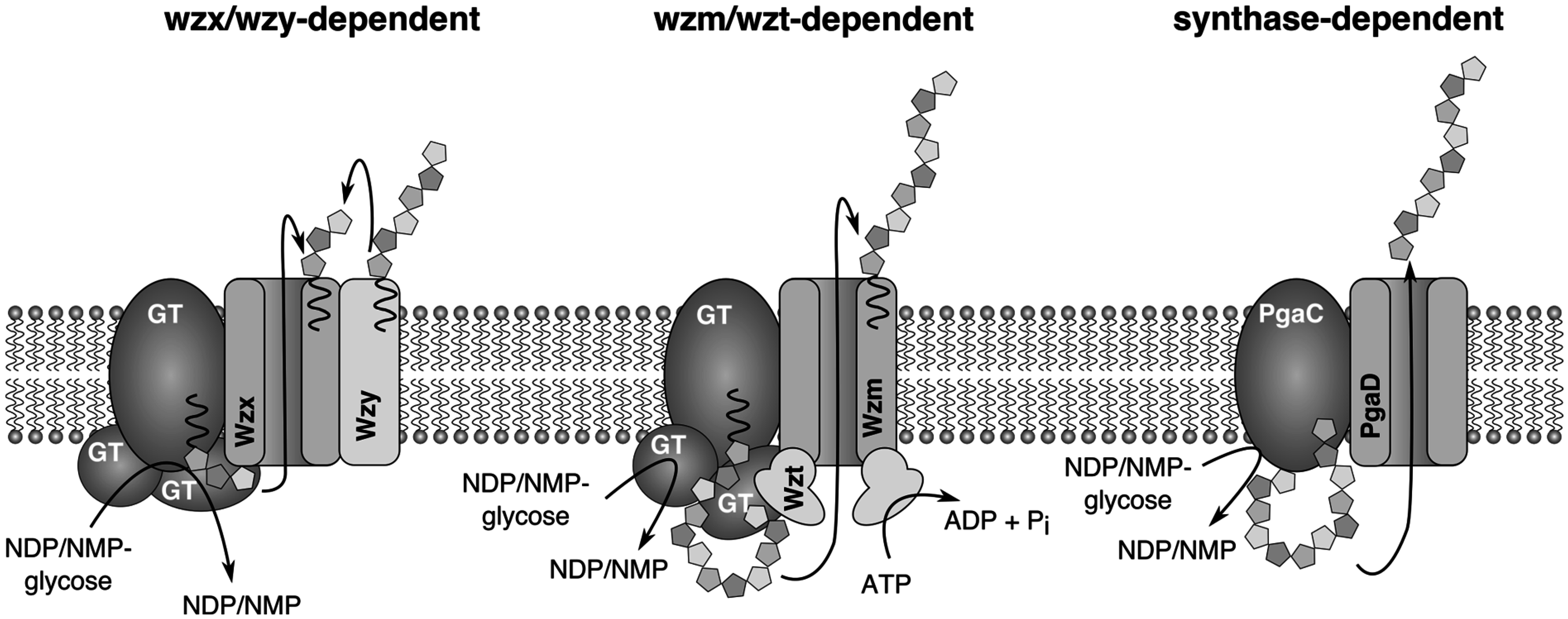
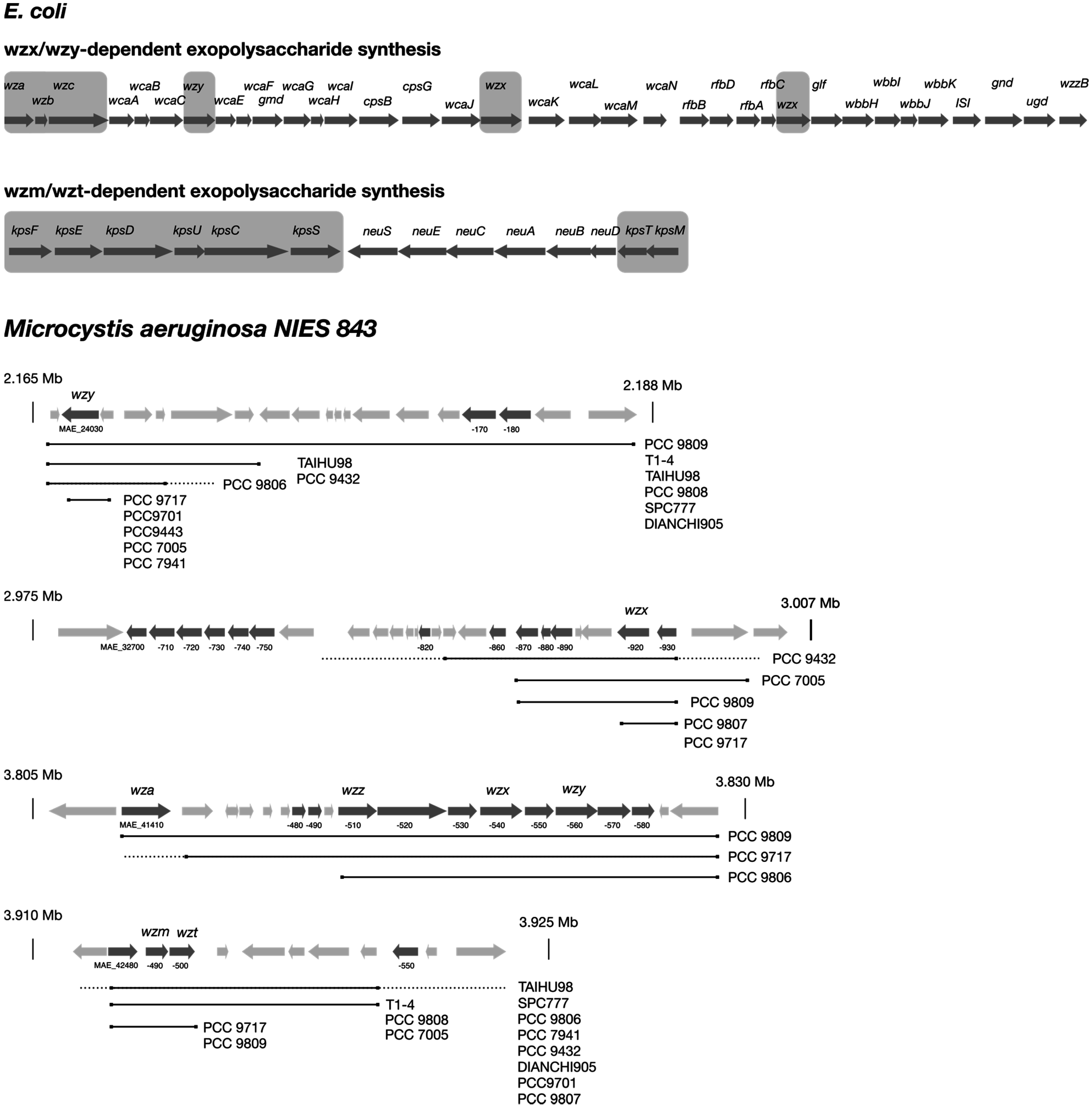
4. Biosynthesis of Extracellular Glycans

The biosynthesis of exopolysaccharides was shown to occur through very similar mechanisms throughout the bacterial kingdom. Three major biosynthetic routes are known, which share some similarities, but they are also characterized by fundamental differences. These pathways are distinguished by the enzymes responsible for the translocation of the polysaccharide or repeating units through the inner membrane. In a Wzx/Wzy-dependent system, repeating units are transferred to the periplasmic side of the inner membrane by a flippase (Wzx), where the final assembly of the nascent polysaccharide happens at the Wzy protein [20]. In an ABC transporter-dependent system (Wzm/Wzt-dependent), the polysaccharide is completely synthesized inside the cell before it is released through the ABC transporter, Wzm/Wzt [21]. In the third pathway (synthase-dependent), whose mechanistic details are not yet understood, a single enzyme that serves both as polymerase and an exporter facilitates the export [22].
Several biosynthetic routes in Gram-negative bacteria were elucidated, and the corresponding genes were identified [20,21,23,24,25,26]. Commonly, the genes involved in exopolysaccharide biosynthesis are clustered, and the nomenclature is consistent among different species, while in most cyanobacteria, the genes are clustered in smaller units or even orphaned and dispersed over the whole chromosome. Additionally, automated genome annotation led to misannotations and an inconsistent naming. Therefore, the detailed description of glycan biosynthesis below will follow the general scheme for Gram-negative bacteria and highlight differences described in cyanobacteria. Since only a few cyanobacterial pathways have been analyzed in detail, it is not clear if the mechanisms apply to all cyanobacterial genera. Capsular polysaccharides in E.coli are classified into four groups, where Groups 1 and 4 and Groups 2 and 3 share similar biosynthesis routes [25]. The proteins that facilitate initiation and transport of the glycopolymer are conserved and present in all members of each group, while the presence of serotype-specific enzymes providing monosaccharide building blocks and glycosyltransferases linking these to the growing polysaccharide chain determines the specific composition of the glycan [25]. Group 1 and 4 polysaccharides are assembled by the Wzx/Wzy systems, and Group 2 and 3 glycans depend on the Wzt/Wzm (ABC transporter-dependent) system (Figure 2). In both cases, biosynthesis is initiated at the cytosolic face of the inner membrane at an integral membrane glycosyltransferase by the transfer of the first building block to a lipid carrier. The following steps differ significantly between both systems. In the Wzx/Wzy system, individual repeating units are assembled and transferred to the periplasmic side of the inner membrane by the flippase Wzx and are subsequently linked by the Wzy protein to the growing polysaccharide chain. This process further requires the integral membrane protein Wzc and the associated phosphatase Wzb (Figure 2). Finally, the nascent polymer is translocated through the outer membrane by the channel Wza. In contrast, in the ABC transporter-dependent Wzt/Wzm (kpsT/kpsM)-dependent system, polysaccharides are completely assembled at the inner cytoplasmic membrane and secreted by the ABC transporter, Wzt/Wzm. Later, a synthase-dependent pathway was discovered, in which the export is directly linked to polysaccharide synthesis [27]. This type of polysaccharide biosynthesis was shown to be involved in the synthesis of poly-β-1,6-N-acetyl-d-glucosamine encoded by the pgaABCD gene cluster [28] and cellulose encoded by the bcsABZC gene cluster [29] in E. coli.
Figure 2. Models of three distinct EPS biosynthesis routes in E. coli. In the Wzx/Wzy-dependent system, repeat units are synthesized at the cytoplasmatic site of the inner membrane and translocated into the periplasm by the flippase, Wzx. Wzy assembles the repeat units to the final polysaccharide. In the Wzm/Wzt system, the complete polysaccharide is synthesized at the inner membrane and exported by the ABC transporter Wzm/Wzt. In the synthase-dependent system, chain elongation is directly linked to transport, but the details are unknown.
Life 05 00164 g002
To our knowledge, there are only a few studies that experimentally address exopolysaccharide biosynthesis and export in cyanobacteria [30,31,32]. Based on mutational and bioinformatic studies in Synechococcus elongatus PCC 7942, a model similar to the enterobacterial Wzm/Wzt system was proposed for the biosynthesis of the O-antigen part of lipopolysaccharides. It was further assumed that lipopolysaccharide biosynthesis occurs exclusively through this pathway, because orthologues of wzx and wzy were not found in S. elongatus [32]. In Synechocystis PCC 6803, ORFs encoding Wzm/Wzt-type ABC transporters (slr0977, slr0982, slr0574 and sll0575) were shown to be involved in EPS synthesis. Mutants of these proteins showed an altered EPS pattern, but the compositions of O-antigen was unaffected, corroborating that other genes are responsible for the synthesis of the polysaccharide part of LPS [30]. Another study showed that ORFs sll0923, sll1581 and slr1875, which encode proteins similar to components of the Wzx/Wzy pathway, are involved in CPS synthesis in Synechocystis PCC 6803 [31]. The biosynthesis of cellulose in Thermosynechococcus vulcanus occurs via a synthase-dependent pathway, which was confirmed by gene disruption of the putative cellulose synthase gene, TvTll0007 [33].
Figure 3. Genetic organization of EPS gene clusters in E. coli, as well as a representation of the loci harboring homologues of EPS core genes in the complete genome of Microcystis aeruginosa NIES 843 and draft genomes of other Microcystis strains. The typical organization of Group 1 and 4 and Group 2 and 3 capsule biosynthesis clusters is depicted for E. coli (top). Grey boxes highlight the conserved core genes. Genes encoding glycosyltransferases and precursor synthesis vary depending on the serotype. Wzx/Wzy and Wzm/Wzt gene homologues in Microcystis are shown in their genetic background (bottom). Additional genes putatively involved in EPS biosynthesis are shown in dark grey, and genes encoding unrelated functions are shown in light grey. The locus tags of relevant genes are depicted below. Solid black lines indicate similar sequences in Microcystis strains listed on the right end. Dotted lines represent upstream or downstream sequences, which do not show homology to corresponding flanking regions in NIES 843, and no line indicates missing sequence information due to unfinished genome status.
Life 05 00164 g003
Nevertheless, many cyanobacterial genome sequences were added to the databases in recent years, which offers the opportunity to systematically screen for genes involved in glycan synthesis. We used query sequences for conserved genes, as well as serotype-specific glycosyltransferases from E. coli to identify putative EPS genes in Microcystis aeruginosa NIES 843 (Figure 3). Indeed, the presence of a Wzm/Wzt system could be confirmed with identities of 32% and 41% compared to the E. coli enzymes, although the genes are not embedded in a gene cluster harboring further glycan biosynthesis genes. Putative wzx and wzy genes could be identified, as well, but these showed only partial (~40%) coverage and low (~27%) identity to the E. coli query sequences. However, glycosyltransferases and sugar epimerases were identified in direct or close vicinity, implying that these genes might be involved in the synthesis of exopolysaccharides. In addition, methyltransferases and sulfotransferases were identified, which may facilitate further modifications of the glycans. Furthermore, several glycosyltransferases spread all over the chromosome were found, many of which could not be assigned to EPS synthesis based on their genomic context. Previous studies showed that the sequence identity of Wzx and Wzy orthologues in different microorganisms is very low, and thus, BLAST searches with query sequences from distant species might fail to identify Wzx and Wzy proteins if search parameters are not adjusted properly [34,35]. We also tried to estimate the level of conservation of the putative EPS synthesis loci in other Microcystis genomes [36,37,38,39] by looking for homologues of the conserved pathway enzymes (Figure 3). Apparently, at least one wzm/wzt and wzx/wzy system could be identified in each strain, with some strains possessing multiple wzx/wzy systems. Furthermore, some variations in the genetic neighborhood were observed, although upstream and downstream regions of wzm/t/x/y genes were frequently not assessable due to the unfinished status of most genomes. However, a mosaic-like distribution, rather than a clustered organization, of genes implicated in EPS biosynthesis over the whole chromosome might indicate frequent recombination, which contributes to strain-specific glycan structure diversification.

5. Overview of Lectins in Cyanobacteria

Lectins are carbohydrate-binding proteins that recognize and attach to complex glycans. They often contain two binding sites or form oligomers effectively multiplying the number of binding sites, which makes them key players in interaction and recognition processes [40]. Though they are usually not glycosylated, they are important factors in glycan-mediated processes and, thus, must be covered by this article. Lectins show high specificity and can even discriminate between oligosaccharides built from the same monosaccharides, but linked by different glycosidic bonds [41]. Few cyanobacterial lectins have been described so far (Table 1), and most of them exhibit specificity to mannose-rich glycans (mannan). Mannan is the major glycan on the envelope of HIV, and some other viruses and cyanobacteria have been intensively screened for HIV entry inhibitors during the last decade [42]. Thus, the bias towards mannan-binding lectins does not necessarily reflect an abundant occurrence of mannan oligosaccharides in cyanobacteria. The interest in mannan-binding lectins led to an extensive biochemical characterization providing specificity, affinity and structural data for most of the isolated proteins, but virtually no information on the biological role of these lectins is available. Furthermore, many lectins contain signal peptides implying an extracellular function and association with cell surface carbohydrates. The lectin, microvirin (Mvn), from Microcystis aeruginosa PCC 7806, was shown to bind to LPS of the producing cells and was proposed to be involved in colony formation [43]. The fluorescently-labelled lectin could differentiate distinct Microcystis morphotypes in lectin binding analyses, thereby demonstrating the diverging glycan composition (Figure 1E). A lectin from M. viridis was detected only in a stationary phase culture grown without aeration [44,45].
Table 1. Cyanobacterial lectins with specificity for high-mannose glycans.
LectinOrganismReference
microvirin (MVN)Microcystis aeruginosa PCC 7806[44,46]
cyanovirin-N (CV-N)Nostoc ellipsosporum[47,48]
scytovirin (SVN)Scytonema varium[49]
Oscillatoria agardhii agglutinin (OAA)Oscillatoria agardhii[50,51]
Microcystis viridis lectin (MVL)Microcystis viridis[45]
MALMicrocystis aeruginosa M228[52]
Lectins also represent a useful tool for the analysis of glycans. In situ hybridization with labelled lectins of known specificity can be used to characterize biofilms and to determine the presence of certain polysaccharide types [53,54]. Data thus obtained can provide information of the composition and types of glycosidic linkages without a time-consuming and elaborate chemical analysis.

6. The Role of Exopolysaccharides in Cyanobacteria

Many functional assignments of cyanobacterial exopolysaccharides are related to their physico-chemical properties and describe their role in metal-binding, scaffolding in biofilms or as diffusion barriers [55], while the knowledge of glycans as specificity mediating agents in biological interactions is scarce compared to that of other bacteria.

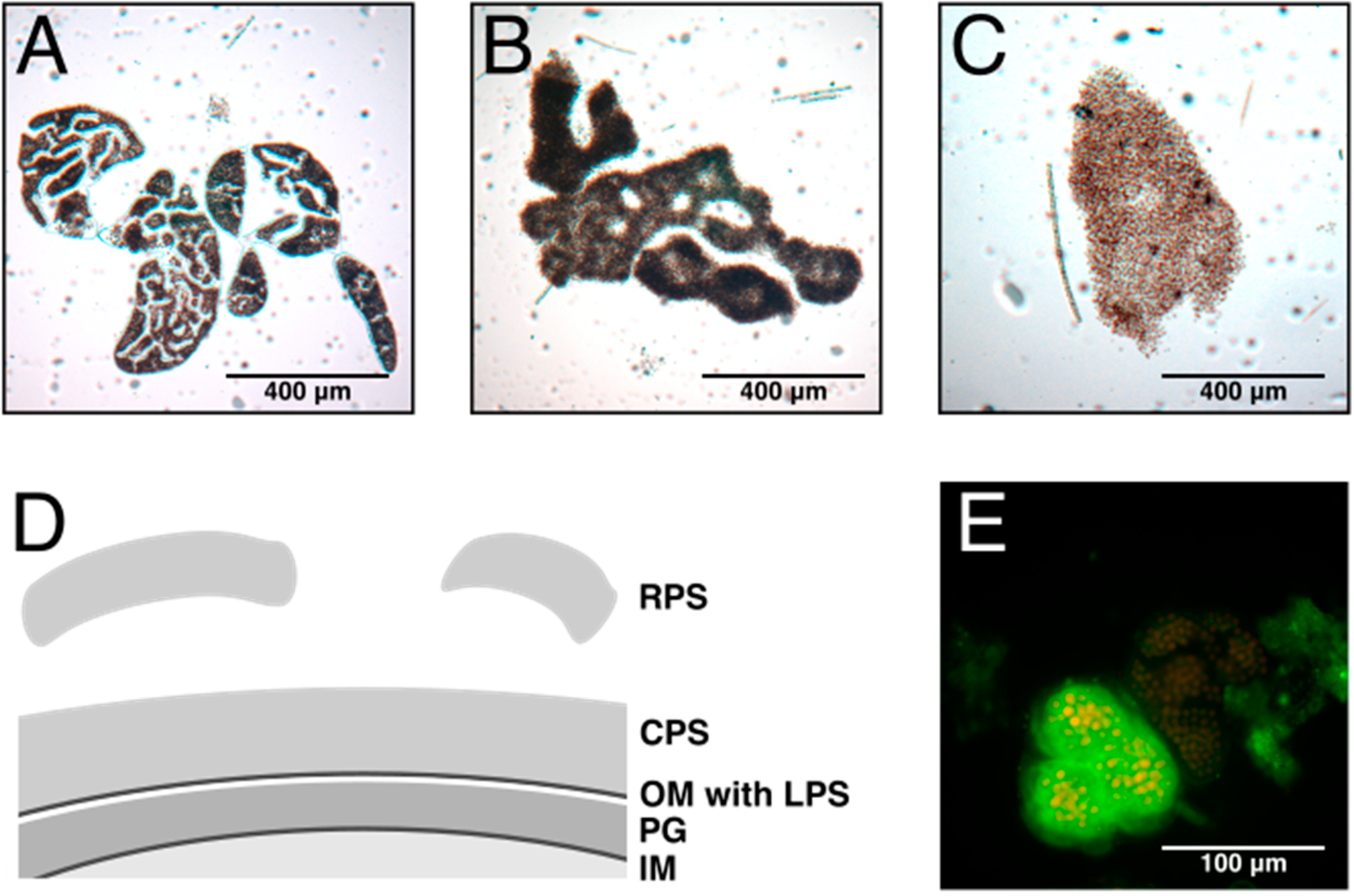
6.1. Colony Formation

The involvement of EPS in colony formation is evident for the species, Microcystis (Figure 1A), and several aspects of colony formation have been addressed in numerous studies [56,57,58,59,60,61]. The ability to form colonies seems to be beneficial in the natural environment, but Microcystis isolates cultivated in the laboratory under axenic conditions lose the ability to form colonies after a short period of time. Interestingly, the amount of EPS produced by laboratory strains was strongly reduced compared to fresh colonial growing isolates, which contained up to 10-fold higher amounts of EPS [62]. Various biotic and abiotic factors that could trigger EPS production were identified. It was shown that inter-species interactions could promote EPS production. Grazing pressure through flagellates led to the increase of EPS secretion [59], and cocultivation of an axenic culture of Microcystis with heterotrophic bacteria isolated from Microcystis field sample colonies reconstituted colony formation and stimulated EPS production [58]. Some studies report a correlation between toxin (microcystin) production and colony size [63], and indeed, the addition of microcystin led to increased colony size and EPS production, as well as induction of polysaccharide synthesis genes [56]. The addition of high, but not growth-inhibiting, concentrations of Ca2+ ions had a similar effect, which was attributed to the matrix stabilizing properties of divalent cations or the interactions with lectins, which often depend on Ca2+ to bind glycans [62]. Furthermore, the production of exopolysaccharides is negatively correlated with the specific growth, which might explain why the ability to form colonies is lost during laboratory culturing, where environmental growth rates are exceeded [64].

6.2. Symbiosis

A role of specific extracellular glycans in the recognition process of symbiotic partners was demonstrated for a broad number of phyla [65,66,67]. A dialogue of the partners involving glycan structures on one side and lectins on the other side is commonly anticipated [68]. Nitrogen-fixing heterocystous cyanobacteria are associates in manifold symbioses with plants and fungi. The infection process is facilitated by the differentiation of infectious motile hormogonia [69]. Lectins were applied to characterize the changing sugar composition at transition states between distinct developmental stages of Nostoc punctiforme. Schüssler et al. [2] could provide evidence that the differentiation from hormogonia to non-motile primordia correlates with the disappearance of β-d-galactosyl and α-d-fucosyl sugars and the appearance of a large amount of α-d-mannosyl or α-d-glycosyl sugars in the extracellular slime. Specific lectins correlated with the Anabaena azollae symbiosis. Only symbiotic Anabaena azollae strains contained a hemagglutinating factor that was not present in free-living Anabaena azollae strains. Antigenic cross-reactivity studies suggested a possible significance of common antigens of Azolla and Anabaena azollae and the involvement of a lectin [70]. The cyanolichen, Peltigera canina, was demonstrated to secrete an arginase with a lectin activity specifically recognizing a sugar receptor of the pre-symbiotic Nostoc cell surface, apparently using a polygalactosylated urease as a cell wall ligand [71]. Lectin secretion by the Nostoc host, Leptogium corniculatum, was also demonstrated, and the lectin was assumed to be a factor in recognition of a compatible host [72]. Although the composition of involved glycan structures is not fully understood, there is compelling evidence that glycans and lectins mediate the interaction of cyanobacterial symbionts and their hosts and act as specific recognition factors.

6.3 LPS Are Receptors for Cyanophages

Little is known about the specific role of cyanobacterial LPS, while in other bacteria, several examples emphasize their importance in intercellular interactions. In Gram-negative bacteria, the O-antigen is an important virulence factor of pathogenic bacteria, enabling infection, and it is required to establish successful symbiosis during host-microbe interactions [67]. In addition, bacterial surface structures can serve as receptors for bacteriophages. Besides surface-exposed proteins, like transporters and flagella, LPS are common targets for phage adhesion in bacteria [73]. Although many marine and freshwater cyanophages were described in recent years [74,75,76], little is known about the route of infection, but it can be assumed that phage infections in cyanobacteria follow the mechanisms established in other bacteria. Evidence for the involvement of LPS in phage adsorption was first provided for the cyanophage AS-1 infecting Anacystis nidulans [77]. Later, LPS were identified as the target of the phages A-1 and A-4 in Anabaena PCC 7120, because mutants with defective O-antigen synthesis became resistant against these phages [78]. Interestingly, the same mutant showed an aberrant heterocyst development, suggesting a role of LPS in cell differentiation.

6.4 EPS and Environmental Stress

Exopolysaccharides are implicated in a variety of stress conditions imposed by hazardous stimuli in the environment, as they constitute a physical barrier enveloping the cell. EPS production was increased by salt stress in Synechocystis strains [79], and Synechocystis mutants defective in EPS production were less tolerant to elevated salt concentrations [31]. The reports on the metal binding ability of cyanobacterial EPS and their biotechnological potential are numerous (a comprehensive review is given in [10]), but little is known about the biological significance. Jittawuttipoka et al. [31] could show that EPS offers protection against heavy metal stress (Co2+ and Cd2+), but can also contribute to the response to iron starvation. Investigation of photosynthetic biofilms from mine tailings that are rich in toxic metals showed that EPS production occurs in response to metal exposure [80]. However, the limited data do not allow drawing a general conclusion about the role of EPS in metal homeostasis. EPS may also play a role in retaining trace metals under conditions where the availability of these is limited, like in marine environments.
Several studies have reported an implication of EPS in UV-light adaptation. In Nostoc commune, EPS production was induced upon UV irradiation [81], and UV-absorbing of d-galacturonic acid, a common component of cyanobacterial EPS, might be responsible for the protective effects of exopolysaccharides [82]. In addition, EPS provide a compartment for UV-light absorbing compounds, like mycosporine-like amino acids (MAAs) or scytonemin, which are released by some cyanobacteria and accumulate in the extracellular matrix [81,83,84]. Some of the MAAs are glycosylated, which may be important to be retained in the mucilage of the producer [83,85]. In Microcoleus species, a desert-crust inhabiting cyanobacterium, EPS were shown to be important to reduce UV-induced ROS generation; although a precise mechanism was not provided [86].
Apart from the regular exposition to high UV irradiances, terrestrial cyanobacteria, like Nostoc species, are frequently exposed to desiccation [87], and the extracellular matrix plays a crucial role in adapting to these conditions. EPS can stabilize cells during the air-dried state and prevent membrane fusions [88]. Tamaru et al. [89] could show that O2 evolution in Nostoc commune was impaired after the removal of EPS and desiccation treatment, while cells with intact EPS showed normal O2 evolution. Furthermore, it was shown that EPS conferred heat resistance.
In Thermosynechococcus vulcanus, cellulose production was induced in response to low temperature and light illumination. The release of cellulose triggered cell aggregation as a means of physiological acclimation to avoid light stress, which could be reversed by cellulase treatment [33].
Metabolic imbalances can also represent a challenge for cyanobacteria, and exopolysaccharides can serve as a carbon sink if the C/N balance is shifted towards excess carbon. Otero and Vincencini [90] could induce either nude or capsulated cells in diazotrophic grown Nostoc sp. PCC 7936, depending on the availability of carbon.

7. Conclusions

Cyanobacterial exopolysaccharides feature some unique properties, like the high content of acidic sugar moieties or the complexity of their repeating unit building blocks. Tremendous progress has been made in the characterization of EPS, but studies on the biological function of EPS are still limited in number. In particular, the role of glycans in recognition processes has been rarely addressed, although several reports suggest an important participation of EPS in intra- and inter-species glycan-mediated interactions. Especially, the spatial and temporal dynamics of EPS production in cyanobacterial interactions, like colony formation or the interplay between host and cyanobiont during symbiosis, are not fully resolved. The large number of cyanobacterial genomes sequenced in recent years offers the opportunity to assess the EPS diversity at the molecular level and provides a basis for future studies in order to elucidate the function of glycans in cyanobacteria.

Author Contributions

J. C. Kehr and E. Dittmann conceived and wrote the manuscript. J. C. Kehr performed the literature search, bioinformatic analysis and prepared the figures. All authors have read and approved the final manuscript.

Conflicts of Interest

The authors declare no conflict of interest.

References

  1. Dittmann, E.; Wiegand, C. Cyanobacterial toxins—Occurrence, biosynthesis and impact on human affairs. Mol. Nut. Food Res. 2006, 50, 7–17. [Google Scholar] [CrossRef]
  2. Schussler, A.; Meyer, T.; Gehrig, H.; Kluge, M. Variations of lectin binding sites in extracellular glycoconjugates during the life cycle of Nostoc punctiforme, a potentially endosymbiotic cyanobacterium. Eur. J. Phycol. 1997, 32, 233–239. [Google Scholar] [CrossRef]
  3. De Philippis, R.; Sili, C.; Paperi, R.; Vincenzini, M. Exopolysaccharide-producing cyanobacteria and their possible exploitation: A review. J. Appl. Phycol. 2001, 13, 293–299. [Google Scholar]
  4. De Philippis, R.; Margheri, M.C.; Materassi, R.; Vincenzini, M. Potential of unicellular cyanobacteria from saline environments as exopolysaccharide producers. Appl Environ. Microb. 1998, 64, 1130–1132. [Google Scholar]
  5. De Philippis, R.; Sili, C.; Faraloni, C.; Vincenzini, M. Occurrence and significance of exopolysaccharide-producing cyanobacteria in the benthic mucilaginous aggregates of the Tyrrhenian Sea (Tuscan Archipelago). Ann. Microbiol. 2002, 52, 1–11. [Google Scholar]
  6. Di Pippo, F.; Ellwood, N.T.W.; Gismondi, A.; Bruno, L.; Rossi, F.; Magni, P.; De Philippis, R. Characterization of exopolysaccharides produced by seven biofilm-forming cyanobacterial strains for biotechnological applications. J. Appl. Phycol. 2013, 25, 1697–1708. [Google Scholar] [Green Version]
  7. Moreno, J.; Vargas, M.A.; Madiedo, J.M.; Munoz, J.; Rivas, J.; Guerrero, M.G. Chemical and rheological properties of an extracellular polysaccharide produced by the cyanobacterium Anabaena sp ATCC 33047. Biotechnol. Bioeng. 2000, 67, 283–290. [Google Scholar] [CrossRef] [PubMed]
  8. Rossi, F.; Micheletti, E.; Bruno, L.; Adhikary, S.P.; Albertano, P.; De Philippis, R. Characteristics and role of the exocellular polysaccharides produced by five cyanobacteria isolated from phototrophic biofilms growing on stone monuments. Biofouling 2012, 28, 215–224. [Google Scholar] [CrossRef] [PubMed] [Green Version]
  9. Weckesser, J.; Drews, G.; Mayer, H. Lipopolysaccharides of photosynthetic prokaryotes. Annu Rev. Microbiol. 1979, 33, 215–239. [Google Scholar] [CrossRef] [PubMed]
  10. De Philippis, R.; Colica, G.; Micheletti, E. Exopolysaccharide-producing cyanobacteria in heavy metal removal from water: Molecular basis and practical applicability of the biosorption process. Appl. Microbiol. Biotechol. 2011, 92, 697–708. [Google Scholar] [CrossRef]
  11. Nobles, D.R.; Romanovicz, D.K.; Brown, R.M., Jr. Cellulose in cyanobacteria. Origin of vascular plant cellulose synthase? Plant. Physiol. 2001, 127, 529–542. [Google Scholar] [CrossRef] [PubMed]
  12. Pereira, S.; Zille, A.; Micheletti, E.; Moradas-Ferreira, P.; De Philippis, R.; Tamagnini, P. Complexity of cyanobacterial exopolysaccharides: Composition, structures, inducing factors and putative genes involved in their biosynthesis and assembly. FEMS Microbiol. Rev. 2009, 33, 917–941. [Google Scholar] [CrossRef] [PubMed]
  13. Werz, D.B.; Ranzinger, R.; Herget, S.; Adibekian, A.; von der Lieth, C.W.; Seeberger, P.H. Exploring the structural diversity of mammalian carbohydrates (“glycospace”) by statistical databank analysis. ACS Chem. Biol. 2007, 2, 685–691. [Google Scholar] [CrossRef] [PubMed]
  14. Vicente-Garcia, V.; Rios-Leal, E.; Calderon-Dominguez, G.; Canizares-Villanueva, R.O.; Olvera-Ramirez, R. Detection, isolation, and characterization of exopolysaccharide produced by a strain of Phormidium 94a isolated from an arid zone of mexico. Biotechnol. Bioeng. 2004, 85, 306–310. [Google Scholar] [CrossRef] [PubMed]
  15. Snyder, D.S.; McIntosh, T.J. The lipopolysaccharide barrier: Correlation of antibiotic susceptibility with antibiotic permeability and fluorescent probe binding kinetics. Biochemistry (USA) 2000, 39, 11777–11787. [Google Scholar] [CrossRef]
  16. Sutherland, I.W. Microbial polysaccharides from Gram-negative bacteria. Int. Dairy J. 2001, 11, 663–674. [Google Scholar] [CrossRef]
  17. Keleti, G.; Sykora, J.L.; Lippy, E.C.; Shapiro, M.A. Composition and biological properties of lipopolysaccharides isolated from Schizothrix calcicola (Ag.) Gomont (Cyanobacteria). Appl Environ. Microb. 1979, 38, 471–477. [Google Scholar]
  18. Snyder, D.S.; Brahamsha, B.; Azadi, P.; Palenik, B. Structure of compositionally simple lipopolysaccharide from marine Synechococcus. J. Bacteriol. 2009, 191, 5499–5509. [Google Scholar] [CrossRef] [PubMed]
  19. Whitfield, C.; Kaniuk, N.; Frirdich, E. Molecular insights into the assembly and diversity of the outer core oligosaccharide in lipopolysaccharides from Escherichia coli and Salmonella. J. Endotoxin Res. 2003, 9, 244–249. [Google Scholar] [CrossRef] [PubMed]
  20. Islam, S.T.; Lam, J.S. Synthesis of bacterial polysaccharides via the Wzx/Wzy-dependent pathway. Can. J. Microbiol. 2014, 60, 697–716. [Google Scholar] [CrossRef] [PubMed]
  21. Greenfield, L.K.; Whitfield, C. Synthesis of lipopolysaccharide O-antigens by ABC transporter-dependent pathways. Carbohydr. Res. 2012, 356, 12–24. [Google Scholar] [CrossRef] [PubMed]
  22. Whitney, J.C.; Howell, P.L. Synthase-dependent exopolysaccharide secretion in Gram-negative bacteria. Trends Microbiol. 2013, 21, 63–72. [Google Scholar] [CrossRef] [PubMed]
  23. Cuthbertson, L.; Kos, V.; Whitfield, C. ABC transporters involved in export of cell surface glycoconjugates. Microbiol. Mol. Biol. Rev. 2010, 74, 341–362. [Google Scholar] [CrossRef] [PubMed]
  24. Liu, B.; Knirel, Y.A.; Feng, L.; Perepelov, A.V.; Senchenkova, S.N.; Wang, Q.; Reeves, P.R.; Wang, L. Structure and genetics of Shigella O antigens. FEMS Microbiol. Rev. 2008, 32, 627–653. [Google Scholar] [CrossRef] [PubMed]
  25. Whitfield, C. Biosynthesis and assembly of capsular polysaccharides in Escherichia coli. Annu. Rev. Biochem. 2006, 75, 39–68. [Google Scholar] [CrossRef] [PubMed]
  26. Willis, L.M.; Whitfield, C. Structure, biosynthesis, and function of bacterial capsular polysaccharides synthesized by ABC transporter-dependent pathways. Carbohydr. Res. 2013, 378, 35–44. [Google Scholar] [CrossRef] [PubMed]
  27. Keenleyside, W.J.; Whitfield, C. A novel pathway for O-polysaccharide biosynthesis in Salmonella enterica serovar borreze. J. Biol. Chem. 1996, 271, 28581–28592. [Google Scholar] [CrossRef] [PubMed]
  28. Itoh, Y.; Rice, J.D.; Goller, C.; Pannuri, A.; Taylor, J.; Meisner, J.; Beveridge, T.J.; Preston, J.F.; Romeo, T. Roles of pgaABCD genes in synthesis, modification, and export of the Escherichia coli biofilm adhesin poly-β-1,6-N-acetyl-d-glucosamine. J. Bacteriol. 2008, 190, 3670–3680. [Google Scholar] [CrossRef] [PubMed]
  29. Romling, U. Molecular biology of cellulose production in bacteria. Res. Microbiol. 2002, 153, 205–212. [Google Scholar] [CrossRef] [PubMed]
  30. Fisher, M.L.; Allen, R.; Luo, Y.Q.; Curtiss, R. Export of extracellular polysaccharides modulates adherence of the cyanobacterium Synechocystis. PloS One 2013, 8. [Google Scholar] [CrossRef] [PubMed]
  31. Jittawuttipoka, T.; Planchon, M.; Spalla, O.; Benzerara, K.; Guyot, F.; Cassier-Chauvat, C.; Chauvat, F. Multidisciplinary evidences that Synechocystis PCC6803 exopolysaccharides operate in cell sedimentation and protection against salt and metal stresses. PloS One 2013, 8. [Google Scholar] [CrossRef] [PubMed]
  32. Simkovsky, R.; Daniels, E.F.; Tang, K.; Huynh, S.C.; Golden, S.S.; Brahamsha, B. Impairment of O-antigen production confers resistance to grazing in a model amoeba-cyanobacterium predator-prey system. Proc. Natl. Acad. Sci. USA 2012, 109, 16678–16683. [Google Scholar] [CrossRef] [PubMed]
  33. Kawano, Y.; Saotome, T.; Ochiai, Y.; Katayama, M.; Narikawa, R.; Ikeuchi, M. Cellulose accumulation and a cellulose synthase gene are responsible for cell aggregation in the cyanobacterium Thermosynechococcus vulcanus RKN. Plant Cell Physiol. 2011, 52, 957–966. [Google Scholar] [CrossRef] [PubMed]
  34. Islam, S.T.; Lam, J.S. Wzx flippase-mediated membrane translocation of sugar polymer precursors in bacteria. Environ. Microbiol. 2013, 15, 1001–1015. [Google Scholar] [CrossRef] [PubMed]
  35. Islam, S.T.; Taylor, V.L.; Qi, M.; Lam, J.S. Membrane topology mapping of the O-antigen flippase (Wzx), polymerase (Wzy), and ligase (Waal) from Pseudomonas aeruginosa PAO1 reveals novel domain architectures. mBio 2010, 1. [Google Scholar] [CrossRef]
  36. Fiore, M.F.; Alvarenga, D.O.; Varani, A.M.; Hoff-Risseti, C.; Crespim, E.; Ramos, R.T.; Silva, A.; Schaker, P.D.; Heck, K.; Rigonato, J.; et al. Draft genome sequence of the brazilian toxic bloom-forming cyanobacterium Microcystis aeruginosa strain SPC777. Genome Announc. 2013, 1. [Google Scholar] [CrossRef]
  37. Gugger, H.; Jurich, M.; Swalen, J.D.; Sievers, A.J. Reply to “comment on ‘observation of an index-of-refraction-induced change in the drude parameters of ag films’”. Phys. Rev. B 1986, 34, 1322–1324. [Google Scholar] [CrossRef]
  38. Kaneko, T.; Nakajima, N.; Okamoto, S.; Suzuki, I.; Tanabe, Y.; Tamaoki, M.; Nakamura, Y.; Kasai, F.; Watanabe, A.; Kawashima, K.; et al. Complete genomic structure of the bloom-forming toxic cyanobacterium Microcystis aeruginosa NIES-843. DNA Res. 2007, 14, 247–256. [Google Scholar] [CrossRef] [PubMed]
  39. Yang, C.; Zhang, W.; Ren, M.; Song, L.; Li, T.; Zhao, J. Whole-genome sequence of Microcystis aeruginosa TAIHU98, a nontoxic bloom-forming strain isolated from Taihu Lake, China. Genome Announc. 2013, 1. [Google Scholar] [CrossRef]
  40. Sharon, N. Lectins: Carbohydrate-specific reagents and biological recognition molecules. J. Biol. Chem. 2007, 282, 2753–2764. [Google Scholar] [CrossRef] [PubMed]
  41. Sharon, N.; Lis, H. How proteins bind carbohydrates: Lessons from legume lectins. J. Agric. Food Chem. 2002, 50, 6586–6591. [Google Scholar] [CrossRef] [PubMed]
  42. Huskens, D.; Schols, D. Algal lectins as potential HIV microbicide candidates. Mar. Drugs 2012, 10, 1476–1497. [Google Scholar] [CrossRef] [PubMed]
  43. Kehr, J.C.; Zilliges, Y.; Springer, A.; Disney, M.D.; Ratner, D.D.; Bouchier, C.; Seeberger, P.H.; de Marsac, N.T.; Dittmann, E. A mannan binding lectin is involved in cell-cell attachment in a toxic strain of Microcystis aeruginosa. Mol. Microbiol. 2006, 59, 893–906. [Google Scholar] [CrossRef] [PubMed]
  44. Yamaguchi, M.; Ogawa, T.; Muramoto, K.; Jimbo, M.; Kamiya, H. Effects of culture conditions on the expression level of lectin in Microcystis aeruginosa (freshwater cyanobacterium). Fish. Sci. 2000, 66, 665–669. [Google Scholar] [CrossRef]
  45. Yamaguchi, M.; Ogawa, T.; Muramoto, K.; Kamio, Y.; Jimbo, M.; Kamiya, H. Isolation and characterization of a mannan-binding lectin from the freshwater cyanobacterium (blue-green algae) Microcystis viridis. Biochem. Biophys. Res. Commun. 1999, 265, 703–708. [Google Scholar] [CrossRef] [PubMed]
  46. Huskens, D.; Ferir, G.; Vermeire, K.; Kehr, J.C.; Balzarini, J.; Dittmann, E.; Schols, D. Microvirin, a novel α(1,2)-mannose-specific lectin isolated from Microcystis aeruginosa, has anti-HIV-1 activity comparable with that of cyanovirin-n but a much higher safety profile. J. Biol. Chem. 2010, 285, 24845–24854. [Google Scholar] [CrossRef] [PubMed]
  47. Boyd, M.R.; Gustafson, K.R.; McMahon, J.B.; Shoemaker, R.H.; O’Keefe, B.R.; Mori, T.; Gulakowski, R.J.; Wu, L.; Rivera, M.I.; Laurencot, C.M.; et al. Discovery of cyanovirin-N, a novel human immunodeficiency virus-inactivating protein that binds viral surface envelope glycoprotein gp120: Potential applications to microbicide development. Antimicrob. Agents Chemother. 1997, 41, 1521–1530. [Google Scholar] [PubMed]
  48. Bewley, C.A.; Gustafson, K.R.; Boyd, M.R.; Covell, D.G.; Bax, A.; Clore, G.M.; Gronenborn, A.M. Solution structure of cyanovirin-N, a potent HIV-inactivating protein. Nat. Struct. Biol. 1998, 5, 571–578. [Google Scholar] [CrossRef] [PubMed]
  49. Xiong, C.Y.; O’Keefe, B.R.; Botos, I.; Wlodawer, A.; McMahon, J.B. Overexpression and purification of scytovirin, a potent, novel anti-HIV protein from the cultured cyanobacterium Scytonema varium. Protein Expr. Purif. 2006, 46, 233–239. [Google Scholar] [CrossRef] [PubMed]
  50. Sato, Y.; Murakami, M.; Miyazawa, K.; Hori, K. Purification and characterization of a novel lectin from a freshwater cyanobacterium, Oscillatoria agardhii. Comp. Biochem. Phys. B 2000, 125, 169–177. [Google Scholar] [CrossRef]
  51. Sato, Y.; Okuyama, S.; Hori, K. Primary structure and carbohydrate binding specificity of a potent anti-HIV lectin isolated from the filamentous cyanobacterium Oscillatoria agardhii. J. Biol. Chem. 2007, 282, 11021–11029. [Google Scholar] [CrossRef] [PubMed]
  52. Jimbo, M.; Yamaguchi, M.; Muramoto, K.; Kamiya, H. Cloning of the Microcystis aeruginosa M228 lectin (MAL) gene. Biochem. Biophys. Res. Commun. 2000, 273, 499–504. [Google Scholar] [CrossRef] [PubMed]
  53. Tien, C.J.; Sigee, D.C.; White, K.N. Characterization of surface sugars on algal cells with fluorescein isothiocyanate-conjugated lectins. Protoplasma 2005, 225, 225–233. [Google Scholar] [CrossRef] [PubMed]
  54. Zippel, B.; Neu, T.R. Characterization of glycoconjugates of extracellular polymeric substances in Tufa-associated biofilms by using fluorescence lectin-binding analysis. Appl. Environ. Microb. 2011, 77, 505–516. [Google Scholar] [CrossRef]
  55. Baulina, O.I.; Titel, K.; Gorelova, O.A.; Malai, O.V.; Ehwald, R. Permeability of cyanobacterial mucous surface structures for macromolecules. Microbiology 2008, 77, 198–205. [Google Scholar] [CrossRef]
  56. Gan, N.Q.; Xiao, Y.; Zhu, L.; Wu, Z.X.; Liu, J.; Hu, C.L.; Song, L.R. The role of microcystins in maintaining colonies of bloom-forming Microcystis spp. Environ. Microbiol. 2012, 14, 730–742. [Google Scholar] [CrossRef] [PubMed]
  57. Rabouille, S.; Salencon, M.J.; Thebault, J.M. Functional analysis of Microcystis vertical migration: A dynamic model as a prospecting tool: I—Processes analysis. Ecol. Model. 2005, 188, 386–403. [Google Scholar] [CrossRef]
  58. Shen, H.; Niu, Y.; Xie, P.; Tao, M.; Yang, X. Morphological and physiological changes in Microcystis aeruginosa as a result of interactions with heterotrophic bacteria. Freshw. Biol. 2011, 56, 1065–1080. [Google Scholar] [CrossRef]
  59. Yang, Z.; Kong, F.X.; Shi, X.L.; Zhang, M.; Xing, P.; Cao, H.S. Changes in the morphology and polysaccharide content of Microcystis aeruginosa (cyanobacteria) during flagellate grazing. J. Phycol. 2008, 44, 716–720. [Google Scholar] [CrossRef]
  60. Zhang, M.; Kong, F.; Tan, X.; Yang, Z.; Cao, H.; Xing, P. Biochemical, morphological, and genetic variations in Microcystis aeruginosa due to colony disaggregation. World J. Microbiol. Biotechnol. 2007, 23, 663–670. [Google Scholar] [CrossRef]
  61. Zilliges, Y.; Kehr, J.C.; Mikkat, S.; Bouchier, C.; de Marsac, N.T.; Borner, T.; Dittmann, E. An extracellular glycoprotein is implicated in cell-cell contacts in the toxic cyanobacterium Microcystis aeruginosa PCC 7806. J. Bacteriol. 2008, 190, 2871–2879. [Google Scholar] [CrossRef] [PubMed]
  62. Wang, Y.W.; Zhao, J.; Li, J.H.; Li, S.S.; Zhang, L.H.; Wu, M. Effects of calcium levels on colonial aggregation and buoyancy of Microcystis aeruginosa. Curr. Microbiol. 2011, 62, 679–683. [Google Scholar] [CrossRef] [PubMed]
  63. Via-Ordorika, L.; Fastner, J.; Kurmayer, R.; Hisbergues, M.; Dittmann, E.; Komarek, J.; Erhard, M.; Chorus, I. Distribution of microcystin-producing and non-microcystin-producing Microcystis sp. in European freshwater bodies: Detection of microcystins and microcystin genes in individual colonies. Syst. Appl. Microbiol. 2004, 27, 592–602. [Google Scholar] [CrossRef] [PubMed]
  64. Li, M.; Zhu, W.; Gao, L.; Lu, L. Changes in extracellular polysaccharide content and morphology of Microcystis aeruginosa at different specific growth rates. J. Appl. Phycol. 2013, 25, 1023–1030. [Google Scholar] [CrossRef]
  65. Becker, A.; Fraysse, N.; Sharypova, L. Recent advances in studies on structure and symbiosis-related function of rhizobial K-antigens and lipopolysaccharides. Mol. Plant-Microbe Interact. 2005, 18, 899–905. [Google Scholar] [CrossRef] [PubMed]
  66. Bogino, P.C.; Oliva, M.D.; Sorroche, F.G.; Giordano, W. The role of bacterial biofilms and surface components in plant-bacterial associations. Int. J. Mol. Sci. 2013, 14, 15838–15859. [Google Scholar] [CrossRef] [PubMed] [Green Version]
  67. Lerouge, I.; Vanderleyden, J. O-antigen structural variation: Mechanisms and possible roles in animal/plant-microbe interactions. FEMS Microbiol. Rev. 2002, 26, 17–47. [Google Scholar] [CrossRef] [PubMed]
  68. Hirsch, A.M. Role of lectins (and rhizobial exopolysaccharides) in legume nodulation. Curr. Opin. Plant Biol. 1999, 2, 320–326. [Google Scholar] [CrossRef]
  69. Meeks, J.C.; Campbell, E.L.; Summers, M.L.; Wong, F.C. Cellular differentiation in the cyanobacterium Nostoc punctiforme. Arch. Microbiol. 2002, 178, 395–403. [Google Scholar] [CrossRef] [PubMed]
  70. Mellor, R.B.; Gadd, G.M.; Rowell, P.; Stewart, W.D. A phytohaemahglutinin from the azolla-Anabaena symbiosis. Biochem. Biophys. Res. Commun. 1981, 99, 1348–1353. [Google Scholar] [CrossRef] [PubMed]
  71. Diaz, E.M.; Sacristan, M.; Legaz, M.E.; Vicente, C. Isolation and characterization of a cyanobacterium-binding protein and its cell wall receptor in the lichen Peltigera canina. Plant Signal. Behav. 2009, 4, 598–603. [Google Scholar] [CrossRef] [PubMed]
  72. Vivas, M.; Sacristan, M.; Legaz, M.E.; Vicente, C. The cell recognition model in chlorolichens involving a fungal lectin binding to an algal ligand can be extended to cyanolichens. Plant Biol. 2010, 12, 615–621. [Google Scholar] [PubMed]
  73. Chaturongakul, S.; Ounjai, P. Phage-host interplay: Examples from tailed phages and Gram-negative bacterial pathogens. Front. Microbiol. 2014, 5. [Google Scholar] [CrossRef] [PubMed]
  74. Clokie, M.R.; Mann, N.H. Marine cyanophages and light. Environ. Microbiol. 2006, 8, 2074–2082. [Google Scholar] [CrossRef] [PubMed]
  75. Mann, N.H. Phages of the marine cyanobacterial picophytoplankton. FEMS Microbiol. Rev. 2003, 27, 17–34. [Google Scholar] [CrossRef] [PubMed]
  76. Xia, H.; Li, T.; Deng, F.; Hu, Z. Freshwater cyanophages. Virol. Sin. 2013, 28, 253–259. [Google Scholar] [CrossRef] [PubMed]
  77. Samimi, B.; Drews, G. Adsorption of cyanophage AS-1 to unicellular cyanobacteria and isolation of receptor material from Anacystis nidulans. J. Virol. 1978, 25, 164–174. [Google Scholar] [PubMed]
  78. Xu, X.D.; Khudyakov, I.; Wolk, C.P. Lipopolysaccharide dependence of cyanophage sensitivity and aerobic nitrogen fixation in Anabaena sp. strain PCC 7120. J. Bacteriol. 1997, 179, 2884–2891. [Google Scholar] [PubMed]
  79. Ozturk, S.; Aslim, B. Modification of exopolysaccharide composition and production by three cyanobacterial isolates under salt stress. Environ. Sci. Pollut. Res. 2010, 17, 595–602. [Google Scholar] [CrossRef]
  80. Garcia-Meza, J.V.; Barrangue, C.; Admiraal, W. Biofilm formation by algae as a mechanism for surviving on mine tailings. Environ. Toxicol. Chem. 2005, 24, 573–581. [Google Scholar] [CrossRef] [PubMed]
  81. Ehling-Schulz, M.; Bilger, W.; Scherer, S. UV-B-induced synthesis of photoprotective pigments and extracellular polysaccharides in the terrestrial cyanobacterium Nostoc commune. J. Bacteriol 1997, 179, 1940–1945. [Google Scholar] [PubMed]
  82. Sommaruga, R.; Chen, Y.; Liu, Z. Multiple strategies of bloom-forming Microcystis to minimize damage by solar ultraviolet radiation in surface waters. Microbial. Ecol. 2009, 57, 667–674. [Google Scholar] [CrossRef]
  83. Böhm, G.A.; Pfleiderer, W.; Böger, P.; Scherer, S. Structure of a novel oligosaccharide-mycosporine-amino acid ultraviolet A/B sunscreen pigment from the terrestrial cyanobacterium Nostoc commune. J. Biol. Chem. 1995, 270, 8536–8539. [Google Scholar] [CrossRef] [PubMed]
  84. Garcia-Pichel, F.; Wingard, C.E.; Castenholz, R.W. Evidence regarding the UV sunscreen role of a mycosporine-like compound in the cyanobacterium Gloeocapsa sp. Appl. Environ. Microbiol. 1993, 59, 170–176. [Google Scholar] [PubMed]
  85. Oren, A.; Gunde-Cimerman, N. Mycosporines and mycosporine-like amino acids: UV protectants or multipurpose secondary metabolites? FEMS Microbiol. Lett. 2007, 269, 1–10. [Google Scholar] [CrossRef] [PubMed]
  86. Chen, L.Z.; Wang, G.H.; Hong, S.; Liu, A.; Li, C.; Liu, Y.D. UV-B-induced oxidative damage and protective role of exopolysaccharides in desert cyanobacterium Microcoleus vaginatus. J. Integr. Plant. Biol. 2009, 51, 194–200. [Google Scholar] [CrossRef] [PubMed]
  87. Potts, M. Mechanisms of desiccation tolerance in cyanobacteria. Eur. J. Phycol. 1999, 34, 319–328. [Google Scholar] [CrossRef]
  88. Hill, D.R.; Keenan, T.W.; Helm, R.F.; Potts, M.; Crowe, L.M.; Crowe, J.H. Extracellular polysaccharide of Nostoc commune (cyanobacteria) inhibits fusion of membrane vesicles during desiccation. J. Appl. Phycol. 1997, 9, 237–248. [Google Scholar] [CrossRef]
  89. Tamaru, Y.; Takani, Y.; Yoshida, T.; Sakamoto, T. Crucial role of extracellular polysaccharides in desiccation and freezing tolerance in the terrestrial cyanobacterium Nostoc commune. Appl. Environ. Microbiol. 2005, 71, 7327–7333. [Google Scholar] [CrossRef] [PubMed]
  90. Otero, A.; Vincenzini, M. Nostoc (cyanophyceae) goes nude: Extracellular polysaccharides serve as a sink for reducing power under unbalanced C/N metabolism. J. Phycol. 2004, 40, 74–81. [Google Scholar] [CrossRef]

