Astaxanthin: A Potential Therapeutic Agent in Cardiovascular Disease

Abstract
:
1. Introduction
2. Oxidative Stress and Inflammation
3. Carotenoids
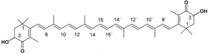
4. Astaxanthin
5. Astaxanthin Formulations
5.1. Astaxanthin of Marine Origin
5.2. Synthetic Astaxanthin
6. Astaxanthin-Experimental Studies
6.1. Cardiovascular Studies
6.2. Diabetes Studies
7. Astaxanthin Studies in Humans
7.1. Dosing
7.2. Bioavailability
7.3. Safety
7.4. Oxidative Stress and Inflammation
8. Clinical Trial Using Astaxanthin
9. Conclusions
Acknowledgments
- Samples Availability: Available from the authors.
References
- Dzau, VJ; Antman, EM; Black, HR; Hayes, DL; Manson, JE; Plutzky, J; Popma, JJ; Stevenson, W. The cardiovascular disease continuum validated: Clinical evidence of improved patient outcomes. Part II: Clinical trial evidence (acute coronary syndromes through renal disease) and future directions. Circulation 2006, 114, 2871–2891. [Google Scholar]
- Ellingsen, I; Seljeflot, I; Arnesen, H; Tonstad, S. Vitamin C consumption is associated with less progression in carotid intima media thickness in elderly men: A 3-year intervention study. Nutr Metab Cardiovasc Dis 2009, 19, 8–14. [Google Scholar]
- Carty, JL; Bevan, R; Waller, H; Mistry, N; Cooke, M; Lunec, J; Griffiths, HR. The effects of vitamin C supplementation on protein oxidation in healthy volunteers. Biochem Biophys Res Commun 2000, 273, 729–735. [Google Scholar]
- Carpenter, KL; Kirkpatrick, PJ; Weissberg, PL; Challis, IR; Dennis, IF; Freeman, MA; Mitchinson, MJ. Oral alpha-tocopherol supplementation inhibits lipid oxidation in established human atherosclerotic lesions. Free Radic Res 2003, 37, 1235–1244. [Google Scholar]
- Stampfer, MJ; Hennekens, CH; Manson, JE; Colditz, GA; Rosner, B; Willett, WC. Vitamin E consumption and the risk of coronary disease in women. N Engl J Med 1993, 328, 1444–1449. [Google Scholar]
- Rimm, EB; Stampfer, MJ; Ascherio, A; Giovannucci, E; Colditz, GA; Willett, WC. Vitamin E consumption and the risk of coronary heart disease in men. N Engl J Med 1993, 328, 1450–1456. [Google Scholar]
- Gey, KF; Puska, P. Plasma vitamins E and A inversely correlated to mortality from ischemic heart disease in cross-cultural epidemiology. Ann N Y Acad Sci 1989, 570, 268–282. [Google Scholar]
- Willcox, BJ; Curb, JD; Rodriguez, BL. Antioxidants in cardiovascular health and disease: Key lessons from epidemiologic studies. Am J Cardiol 2008, 101, 75D–86D. [Google Scholar]
- Frei, B. Cardiovascular disease and nutrient antioxidants: Role of low-density lipoprotein oxidation. Crit Rev Food Sci Nutr 1995, 35, 83–98. [Google Scholar]
- Steinberg, D. Antioxidants in the prevention of human atherosclerosis. Summary of the proceedings of a National Heart, Lung, and Blood Institute Workshop: September 5–6, 1991, Bethesda, Maryland. Circulation 1992, 85, 2337–2344. [Google Scholar]
- Helmersson, J; Arnlov, J; Larsson, A; Basu, S. Low dietary intake of beta-carotene, alpha-tocopherol and ascorbic acid is associated with increased inflammatory and oxidative stress status in a Swedish cohort. Br J Nutr 2009, 101, 1775–1782. [Google Scholar]
- Osganian, SK; Stampfer, MJ; Rimm, E; Spiegelman, D; Manson, JE; Willett, WC. Dietary carotenoids and risk of coronary artery disease in women. Am J Clin Nutr 2003, 77, 1390–1399. [Google Scholar]
- Ford, ES; Giles, WH. Serum vitamins, carotenoids, and angina pectoris: Findings from the National Health and Nutrition Examination Survey III. Ann Epidemiol 2000, 10, 106–116. [Google Scholar]
- Klipstein-Grobusch, K; Geleijnse, JM; den Breeijen, JH; Boeing, H; Hofman, A; Grobbee, DE; Witteman, JC. Dietary antioxidants and risk of myocardial infarction in the elderly: The Rotterdam Study. Am J Clin Nutr 1999, 69, 261–266. [Google Scholar]
- Gaziano, JM; Manson, JE; Branch, LG; Colditz, GA; Willett, WC; Buring, JE. A prospective study of consumption of carotenoids in fruits and vegetables and decreased cardiovascular mortality in the elderly. Ann Epidemiol 1995, 5, 255–260. [Google Scholar]
- Morris, DL; Kritchevsky, SB; Davis, CE. Serum carotenoids and coronary heart disease. The Lipid Research Clinics Coronary Primary Prevention Trial and Follow-up Study. JAMA 1994, 272, 1439–1441. [Google Scholar]
- Knekt, P; Reunanen, A; Jarvinen, R; Seppanen, R; Heliovaara, M; Aromaa, A. Antioxidant vitamin intake and coronary mortality in a longitudinal population study. Am J Epidemiol 1994, 139, 1180–1189. [Google Scholar]
- Stephens, NG; Parsons, A; Schofield, PM; Kelly, F; Cheeseman, K; Mitchinson, MJ. Randomised controlled trial of vitamin E in patients with coronary disease: Cambridge Heart Antioxidant Study (CHAOS). Lancet 1996, 347, 781–786. [Google Scholar]
- Tepel, M; van der Giet, M; Statz, M; Jankowski, J; Zidek, W. The antioxidant acetylcysteine reduces cardiovascular events in patients with end-stage renal failure: A randomized, controlled trial. Circulation 2003, 107, 992–995. [Google Scholar]
- Boaz, M; Smetana, S; Weinstein, T; Matas, Z; Gafter, U; Iaina, A; Knecht, A; Weissgarten, Y; Brunner, D; Fainaru, M; Green, MS. Secondary prevention with antioxidants of cardiovascular disease in endstage renal disease (SPACE): Randomised placebo-controlled trial. Lancet 2000, 356, 1213–1218. [Google Scholar]
- Steinhubl, SR. Why have antioxidants failed in clinical trials? Am J Cardiol 2008, 101, 14D–19D. [Google Scholar]
- Heart Protection Study Collaborative Group. MRC/BHF Heart Protection Study of antioxidant vitamin supplementation in 20,536 high-risk individuals: A randomised placebo-controlled trial. Lancet 2002, 360, 23–33. [Google Scholar]
- Yusuf, S; Dagenais, G; Pogue, J; Bosch, J; Sleight, P. Vitamin E supplementation and cardiovascular events in high-risk patients. The Heart Outcomes Prevention Evaluation Study Investigators. N Engl J Med 2000, 342, 154–160. [Google Scholar]
- Sandmann, G. Carotenoid biosynthesis in microorganisms and plants. Eur J Biochem 1994, 223, 7–24. [Google Scholar]
- McNulty, H; Jacob, RF; Mason, RP. Biologic activity of carotenoids related to distinct membrane physicochemical interactions. Am J Cardiol 2008, 101, 20D–29D. [Google Scholar]
- Jackson, H; Braun, CL; Ernst, H. The chemistry of novel xanthophyll carotenoids. Am J Cardiol 2008, 101, 50D–57D. [Google Scholar]
- McNulty, HP; Byun, J; Lockwood, SF; Jacob, RF; Mason, RP. Differential effects of carotenoids on lipid peroxidation due to membrane interactions: X-ray diffraction analysis. Biochim Biophys Acta 2007, 1768, 167–174. [Google Scholar]
- Brown, BG; Zhao, XQ; Chait, A; Fisher, LD; Cheung, MC; Morse, JS; Dowdy, AA; Marino, EK; Bolson, EL; Alaupovic, P; Frohlich, J; Albers, JJ. Simvastatin and niacin, antioxidant vitamins, or the combination for the prevention of coronary disease. N Engl J Med 2001, 345, 1583–1592. [Google Scholar]
- The Alpha-Tocopherol, Beta Carotene Cancer Prevention Study Group. The effect of vitamin E and beta carotene on the incidence of lung cancer and other cancers in male smokers. N Engl J Med 1994, 330, 1029–1035. [Google Scholar]
- Omenn, GS; Goodman, GE; Thornquist, MD; Balmes, J; Cullen, MR; Glass, A; Keogh, JP; Meyskens, FL; Valanis, B; Williams, JH; Barnhart, S; Hammar, S. Effects of a combination of beta carotene and vitamin A on lung cancer and cardiovascular disease. N Engl J Med 1996, 334, 1150–1155. [Google Scholar]
- Lee, IM; Cook, NR; Manson, JE; Buring, JE; Hennekens, CH. Beta-carotene supplementation and incidence of cancer and cardiovascular disease: The Women’s Health Study. J Natl Cancer Inst 1999, 91, 2102–2106. [Google Scholar]
- Hennekens, CH; Buring, JE; Manson, JE; Stampfer, M; Rosner, B; Cook, NR; Belanger, C; LaMotte, F; Gaziano, JM; Ridker, PM; Willett, W; Peto, R. Lack of effect of long-term supplementation with beta carotene on the incidence of malignant neoplasms and cardiovascular disease. N Engl J Med 1996, 334, 1145–1149. [Google Scholar]
- Burton, GW; Ingold, KU. beta-Carotene: An unusual type of lipid antioxidant. Science 1984, 224, 569–573. [Google Scholar]
- Lauver, DA; Driscoll, EM; Lucchesi, BR. Disodium disuccinate astaxanthin prevents carotid artery rethrombosis and ex vivo platelet activation. Pharmacology 2008, 82, 67–73. [Google Scholar]
- Gross, GJ; Lockwood, SF. Cardioprotection and myocardial salvage by a disodium disuccinate astaxanthin derivative (Cardax). Life Sci 2004, 75, 215–224. [Google Scholar]
- Gross, GJ; Hazen, SL; Lockwood, SF. Seven day oral supplementation with Cardax (disodium disuccinate astaxanthin) provides significant cardioprotection and reduces oxidative stress in rats. Mol Cell Biochem 2006, 283, 23–30. [Google Scholar]
- Wertz, K; Hunziker, PB; Seifert, N; Riss, G; Neeb, M; Steiner, G; Hunziker, W; Goralczyk, R. beta-Carotene interferes with ultraviolet light A-induced gene expression by multiple pathways. J Invest Dermatol 2005, 124, 428–434. [Google Scholar]
- Camera, E; Mastrofrancesco, A; Fabbri, C; Daubrawa, F; Picardo, M; Sies, H; Stahl, W. Astaxanthin, canthaxanthin and beta-carotene differently affect UVA-induced oxidative damage and expression of oxidative stress-responsive enzymes. Exp Dermatol 2009, 18, 222–231. [Google Scholar]
- Camara, B; Bouvier, F. Oxidative remodeling of plastid carotenoids. Arch Biochem Biophys 2004, 430, 16–21. [Google Scholar]
- Sharoni, Y; Agbaria, R; Amir, H; Ben-Dor, A; Bobilev, I; Doubi, N; Giat, Y; Hirsh, K; Izumchenko, G; Khanin, M; Kirilov, E; Krimer, R; Nahum, A; Steiner, M; Walfisch, Y; Walfisch, S; Zango, G; Danilenko, M; Levy, J. Modulation of transcriptional activity by antioxidant carotenoids. Mol Asp Med 2003, 24, 371–384. [Google Scholar]
- Guerin, M; Huntley, ME; Olaizola, M. Haematococcus astaxanthin: Applications for human health and nutrition. Trends Biotechnol 2003, 21, 210–216. [Google Scholar]
- Hussein, G; Sankawa, U; Goto, H; Matsumoto, K; Watanabe, H. Astaxanthin, a carotenoid with potential in human health and nutrition. J Nat Prod 2006, 69, 443–449. [Google Scholar]
- Schweigert, F. Metabolism of Carotenoids in Mammals; Birkhauser Verlag: Basel, Switzerland, 1998. [Google Scholar]
- Jyonouchi, H; Sun, S; Tomita, Y; Gross, MD. Astaxanthin, a carotenoid without vitamin A activity, augments antibody responses in cultures including T-helper cell clones and suboptimal doses of antigen. J Nutr 1995, 125, 2483–2492. [Google Scholar]
- Shimidzu, N. Carotenoids as singlet oxygen quenchers in marine organisms. Fish Sci 1996, 62, 134–137. [Google Scholar]
- Miki, W. Biological functions and activities of animal carotenoids. Pure Appl Chem 1991, 63, 141–146. [Google Scholar]
- Krinsky, NI. Antioxidant functions of carotenoids. Free Radic Biol Med 1989, 7, 617–635. [Google Scholar]
- Beutner, S; Bloedorn, B; Frixel, S; Blanco, IH; Hoffman, T; Martin, HD; Mayer, B; Noach, P; Rack, C; Schmidt, M; et al. Quantitative assessment of antioxidant properties of natural colorants and phytochemicals; carotenoids. flavonoids, phenols and indigoids: The role of β-carotene in antioxidant functions. J Sci Food Agric 2001, 81, 559–568. [Google Scholar]
- Pashkow, FJ; Watumull, DG; Campbell, CL. Astaxanthin: A novel potential treatment for oxidative stress and inflammation in cardiovascular disease. Am J Cardiol 2008, 101, 58D–68D. [Google Scholar]
- Kobayashi, M; Kakizono, T; Nishio, N; Nagai, S; Kurimura, Y; Tsuji, Y. Antioxidant role of astaxanthin in the green alga Haematococcus pluvialis. Appl Microbiol Biotechnol 1997, 48, 351–356. [Google Scholar]
- Ernst, H. Recent advances in industrial carotenoid synthesis. Pure Appl Chem 2002, 74, 2213–2226. [Google Scholar]
- Lauver, DA; Lockwood, SF; Lucchesi, BR. Disodium Disuccinate Astaxanthin (Cardax) attenuates complement activation and reduces myocardial injury following ischemia/reperfusion. J Pharmacol Exp Ther 2005, 314, 686–692. [Google Scholar]
- Lockwood, SF; Gross, GJ. Disodium disuccinate astaxanthin (Cardax): Antioxidant and antiinflammatory cardioprotection. Cardiovasc Drug Rev 2005, 23, 199–216. [Google Scholar]
- Gross, GJ; Lockwood, SF. Acute and chronic administration of disodium disuccinate astaxanthin (Cardax) produces marked cardioprotection in dog hearts. Mol Cell Biochem 2005, 272, 221–227. [Google Scholar]
- Khan, SK; Malinski, T; Mason, RP; Kubant, R; Jacob, RF; Fujioka, K; Denstaedt, SJ; King, TJ; Jackson, HL; Hieber, AD; Lockwood, SF; Goodin, TH; Pashkow, FJ; Bodary, PF. Novel astaxanthin prodrug (CDX-085) attenuates thrombosis in a mouse model. Thromb Res 2010, 126, 299–305. [Google Scholar]
- Shargel, L; Yu, A. Applied Biopharmaceutics and Pharmacokinetics; Appleton-Lange: Stamford, CT, USA, 1999. [Google Scholar]
- Rishton, GM. Natural products as a robust source of new drugs and drug leads: Past successes and present day issues. Am J Cardiol 2008, 101, 43D–49D. [Google Scholar]
- Kang, JO; Kim, SJ; Kim, H. Effect of astaxanthin on the hepatotoxicity, lipid peroxidation and antioxidative enzymes in the liver of CCl4-treated rats. Methods Find Exp Clin Pharmacol 2001, 23, 79–84. [Google Scholar]
- Kamath, BS; Srikanta, BM; Dharmesh, SM; Sarada, R; Ravishankar, GA. Ulcer preventive and antioxidative properties of astaxanthin from Haematococcus pluvialis. Eur J Pharmacol 2008, 590, 387–395. [Google Scholar]
- Naito, Y; Uchiyama, K; Aoi, W; Hasegawa, G; Nakamura, N; Yoshida, N; Maoka, T; Takahashi, J; Yoshikawa, T. Prevention of diabetic nephropathy by treatment with astaxanthin in diabetic db/db mice. Biofactors 2004, 20, 49–59. [Google Scholar]
- Ohgami, K; Shiratori, K; Kotake, S; Nishida, T; Mizuki, N; Yazawa, K; Ohno, S. Effects of astaxanthin on lipopolysaccharide-induced inflammation in vitro and in vivo. Invest Ophthalmol Vis Sci 2003, 44, 2694–2701. [Google Scholar]
- Lee, SJ; Bai, SK; Lee, KS; Namkoong, S; Na, HJ; Ha, KS; Han, JA; Yim, SV; Chang, K; Kwon, YG; Lee, SK; Kim, YM. Astaxanthin inhibits nitric oxide production and inflammatory gene expression by suppressing I(kappa)B kinase-dependent NF-kappaB activation. Mol Cells 2003, 16, 97–105. [Google Scholar]
- Aoi, W; Naito, Y; Sakuma, K; Kuchide, M; Tokuda, H; Maoka, T; Toyokuni, S; Oka, S; Yasuhara, M; Yoshikawa, T. Astaxanthin limits exercise-induced skeletal and cardiac muscle damage in mice. Antioxid Redox Signal 2003, 5, 139–144. [Google Scholar]
- Uchiyama, K; Naito, Y; Hasegawa, G; Nakamura, N; Takahashi, J; Yoshikawa, T. Astaxanthin protects beta-cells against glucose toxicity in diabetic db/db mice. Redox Rep 2002, 7, 290–293. [Google Scholar]
- Nakajima, Y; Inokuchi, Y; Shimazawa, M; Otsubo, K; Ishibashi, T; Hara, H. Astaxanthin, a dietary carotenoid, protects retinal cells against oxidative stress in vitro and in mice in vivo. J Pharm Pharmacol 2008, 60, 1365–1374. [Google Scholar]
- Manabe, E; Handa, O; Naito, Y; Mizushima, K; Akagiri, S; Adachi, S; Takagi, T; Kokura, S; Maoka, T; Yoshikawa, T. Astaxanthin protects mesangial cells from hyperglycemia-induced oxidative signaling. J Cell Biochem 2008, 103, 1925–1937. [Google Scholar]
- Nakano, M; Onodera, A; Saito, E; Tanabe, M; Yajima, K; Takahashi, J; Nguyen, VC. Effect of astaxanthin in combination with alpha-tocopherol or ascorbic acid against oxidative damage in diabetic ODS rats. J Nutr Sci Vitaminol (Tokyo) 2008, 54, 329–334. [Google Scholar]
- Choi, SK; Park, YS; Choi, DK; Chang, HI. Effects of astaxanthin on the production of NO and the expression of COX-2 and iNOS in LPS-stimulated BV2 microglial cells. J Microbiol Biotechnol 2008, 18, 1990–1996. [Google Scholar]
- Liu, X; Shibata, T; Hisaka, S; Osawa, T. Astaxanthin inhibits reactive oxygen species-mediated cellular toxicity in dopaminergic SH-SY5Y cells via mitochondria-targeted protective mechanism. Brain Res 2009, 1254, 18–27. [Google Scholar]
- Iwamoto, T; Hosoda, K; Hirano, R; Kurata, H; Matsumoto, A; Miki, W; Kamiyama, M; Itakura, H; Yamamoto, S; Kondo, K. Inhibition of low-density lipoprotein oxidation by astaxanthin. J Atheroscler Thromb 2000, 7, 216–222. [Google Scholar]
- Hussein, G; Nakamura, M; Zhao, Q; Iguchi, T; Goto, H; Sankawa, U; Watanabe, H. Antihypertensive and neuroprotective effects of astaxanthin in experimental animals. Biol Pharm Bull 2005, 28, 47–52. [Google Scholar]
- Hussein, G; Goto, H; Oda, S; Sankawa, U; Matsumoto, K; Watanabe, H. Antihypertensive potential and mechanism of action of astaxanthin: III. Antioxidant and histopathological effects in spontaneously hypertensive rats. Biol Pharm Bull 2006, 29, 684–688. [Google Scholar]
- Aoi, W; Naito, Y; Takanami, Y; Ishii, T; Kawai, Y; Akagiri, S; Kato, Y; Osawa, T; Yoshikawa, T. Astaxanthin improves muscle lipid metabolism in exercise via inhibitory effect of oxidative CPT I modification. Biochem Biophys Res Commun 2008, 366, 892–897. [Google Scholar]
- Nakao, R; Nelson, OL; Park, JS; Mathison, BD; Thompson, PA; Chew, BP. Effect of astaxanthin supplementation on inflammation and cardiac function in BALB/c mice. Anticancer Res 2010, 30, 2721–2725. [Google Scholar]
- Yamamoto, R; Kanazawa, A; Shimizu, T; Hirose, T; Tanaka, Y; Kawamori, R; Watada, H. Association between atherosclerosis and newly classified chronic kidney disease stage for Japanese patients with type 2 diabetes. Diabetes Res Clin Pract 2009, 84, 39–45. [Google Scholar]
- Osterlie, M; Bjerkeng, B; Liaaen-Jensen, S. Plasma appearance and distribution of astaxanthin E/Z and R/S isomers in plasma lipoproteins of men after single dose administration of astaxanthin. J Nutr Biochem 2000, 11, 482–490. [Google Scholar]
- Mercke Odeberg, J; Lignell, A; Pettersson, A; Hoglund, P. Oral bioavailability of the antioxidant astaxanthin in humans is enhanced by incorporation of lipid based formulations. Eur J Pharm Sci 2003, 19, 299–304. [Google Scholar]
- Spiller, GA; Dewell, A. Safety of an astaxanthin-rich Haematococcus pluvialis algal extract: A randomized clinical trial. J Med Food 2003, 6, 51–56. [Google Scholar]
- Coral-Hinostroza, GN; Ytrestoyl, T; Ruyter, B; Bjerkeng, B. Plasma appearance of unesterified astaxanthin geometrical E/Z and optical R/S isomers in men given single doses of a mixture of optical 3 and 3′R/S isomers of astaxanthin fatty acyl diesters. Comp Biochem Physiol C Toxicol Pharmacol 2004, 139, 99–110. [Google Scholar]
- Karppi, J; Rissanen, TH; Nyyssonen, K; Kaikkonen, J; Olsson, AG; Voutilainen, S; Salonen, JT. Effects of astaxanthin supplementation on lipid peroxidation. Int J Vitam Nutr Res 2007, 77, 3–11. [Google Scholar]
- Parisi, V; Tedeschi, M; Gallinaro, G; Varano, M; Saviano, S; Piermarocchi, S. Carotenoids and antioxidants in age-related maculopathy italian study: Multifocal electroretinogram modifications after 1 year. Ophthalmology 2008, 115, 324–333.e2. [Google Scholar]
- Miyawaki, H; Takahashi, J; Tsukahara, H; Takehara, I. Effects of astaxanthin on human blood rheology. J Clin Biochem Nutr 2008, 43, 69–74. [Google Scholar]
- Rufer, CE; Moeseneder, J; Briviba, K; Rechkemmer, G; Bub, A. Bioavailability of astaxanthin stereoisomers from wild (Oncorhynchus spp.) and aquacultured (Salmo salar) salmon in healthy men: A randomised, double-blind study. Br J Nutr 2008, 99, 1048–1054. [Google Scholar]
- Park, JS; Chyun, JH; Kim, YK; Line, LL; Chew, BP. Astaxanthin decreased oxidative stress and inflammation and enhanced immune response in humans. Nutr Metab (Lond) 2010, 7, 18. [Google Scholar]
- Serebruany, V; Malinin, A; Goodin, T; Pashkow, F. The in vitro effects of Xancor, a synthetic astaxanthine derivative, on hemostatic biomarkers in aspirin-naive and aspirin-treated subjects with multiple risk factors for vascular disease. Am J Ther 2010, 17, 125–132. [Google Scholar]
- Andersen, LP; Holck, S; Kupcinskas, L; Kiudelis, G; Jonaitis, L; Janciauskas, D; Permin, H; Wadstrom, T. Gastric inflammatory markers and interleukins in patients with functional dyspepsia treated with astaxanthin. FEMS Immunol Med Microbiol 2007, 50, 244–248. [Google Scholar]
- Fassett, RG; Healy, H; Driver, R; Robertson, IK; Geraghty, DP; Sharman, JE; Coombes, JS. Astaxanthin vs. placebo on arterial stiffness, oxidative stress and inflammation in renal transplant patients (Xanthin): A randomised controlled trial. BMC Nephrol 2008, 9, 17. [Google Scholar]


| Study | Model | Dosage | Duration/timing of supplementation | Effects of astaxanthin |
|---|---|---|---|---|
| Lauver et al. 2008 [34] | Dog with occlusive carotid artery thrombus | DDA 10, 30, or 50 mg/kg/body weight IV | 30 min after occlusion |
|
| Aoi et al. 2003 [63] | C57BL/6 mice | Diet supplemented with astaxanthin 0.02% weight/weight and food intake recorded | 3 weeks |
|
| Gross and Lockwood 2004 [35] | Myocardial infarct model Sprague-Dawley rats | DDA 25/50/75 mg/kg body weight intravenously daily | 4 days prior to myocardial infarction |
|
| Hussein et al. 2005 [71] | Stroke prone Spontaneously hypertensive rats | Astaxanthin 50 mg/kg body weight/day | 5 weeks |
|
| Lauver et al. 2005 [52] | Rabbit model of myocardial ischemia/reperfusion | DDA 50 mg/kg body weight/day intravenously | 5 days |
|
| Gross et al. 2005 [54] | Canine model of myocardial ischemia/reperfusion | DDA 50 mg/kg body weight/day intravenously | 2 h or daily for four days |
|
| Gross et al. 2006 [36] | Sprague-Dawley rats Left anterior descending coronary artery occlusion/reperfusion | DDA 125 or 500 mg/kg body weight/day orally | 7 days |
|
| Hussein et al. 2006 [72] | Spontaneously hypertensive rats | Astaxanthin 5% in olive oil (5 mg/kg/day orally) | 7 days |
|
| Aoi et al. 2008 [73] | ICR mice | Astaxanthin 0.02% w/w | 4 weeks |
|
| Nakao et al. 2010 [74] | BALC/c mice | Astaxanthin 0, 0.02, 0.08% orally/day | 8 weeks |
|
| Khan et al. 2010 [55] | C57BL/6 mice | CDX-085 500 mg/kg body weight/day | 14 days |
|
| Human umbilical vein endothelial cells and platelets from Wistar-Kyoto rats |
| Study | Study population (n = subject numbers) | Dosage | Study design | Duration of supplementation | Effects of astaxanthin |
|---|---|---|---|---|---|
| Iwamoto et al. 2000 [70] | Volunteers (n = 24) | Different doses: 1.8, 3.6, 14.4, 21.6 mg/day | Open labelled | 2 weeks |
|
| Osterlie et al. 2000 [76] | Middle aged male volunteers (n = 3) | 100 mg | Open labelled | Single dose |
|
| Mercke Odeberg et al. 2003 [77] | Healthy male volunteers (n = 32) | 40 mg | Open labelled parallel | Single dose |
|
| Spiller et al. 2003 [78] | Healthy adults (n = 35) | 6 mg/day (3 × 2 mg tablets/day) | Randomised, double blind, placebo controlled | 8 weeks |
|
| Coral-Hinostroza et al. 2005 [79] | Healthy adult males (n = 3) | 10 mg and 100 mg | Open labelled | Single dose or 4 weeks |
|
| Karppi et al. 2007 [80] | Healthy non-smoking Finnish males (n = 40) | 8 mg/day | Randomised, double blind, placebo controlled | 12 weeks |
|
| Parisi et al. 2008 [81] | Non-advanced age related macular degeneration (n = 27) | 4 mg/day | Randomised controlled trial open labelled no placebo | 12 months |
|
| Miyawaki et al. 2008 [82] | Healthy males (n = 20) | 6 mg/day | Single blind, placebo controlled | 10 days |
|
| Rufer et al. 2008 [83] | Healthy males (n = 28) | 5 μg/g salmon flesh (wild vs. aquacultured) | Randomised, double blind, placebo controlled | 4 weeks |
|
| Park et al. 2010 [84] | Healthy females (n = 14) | 0, 2, 8 mg/day | Randomised, double blind, placebo controlled | 8 weeks |
|
© 2011 by the authors; licensee MDPI, Basel, Switzerland. This article is an open-access article distributed under the terms and conditions of the Creative Commons Attribution license (http://creativecommons.org/licenses/by/3.0/).
Share and Cite
Fassett, R.G.; Coombes, J.S. Astaxanthin: A Potential Therapeutic Agent in Cardiovascular Disease. Mar. Drugs 2011, 9, 447-465. https://0-doi-org.brum.beds.ac.uk/10.3390/md9030447
Fassett RG, Coombes JS. Astaxanthin: A Potential Therapeutic Agent in Cardiovascular Disease. Marine Drugs. 2011; 9(3):447-465. https://0-doi-org.brum.beds.ac.uk/10.3390/md9030447
Chicago/Turabian StyleFassett, Robert G., and Jeff S. Coombes. 2011. "Astaxanthin: A Potential Therapeutic Agent in Cardiovascular Disease" Marine Drugs 9, no. 3: 447-465. https://0-doi-org.brum.beds.ac.uk/10.3390/md9030447