Article

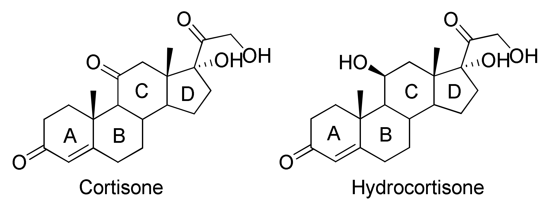
Δ1-Dehydrogenation and C20 Reduction of Cortisone and Hydrocortisone Catalyzed by Rhodococcus Strains

1
Department of Life Sciences and Biotechnology, University of Ferrara, Via L. Borsari, 46, 44121 Ferrara, Italy
2
GATE SRL, Via L. Borsari, 46, 44121 Ferrara, Italy
3
Department of Chemical and Pharmaceutical Sciences, University of Ferrara, Via L. Borsari, 46, 44121 Ferrara, Italy
*
Author to whom correspondence should be addressed.
Submission received: 26 March 2020 / Revised: 4 May 2020 / Accepted: 4 May 2020 / Published: 7 May 2020

Abstract

:
Prednisone and prednisolone are steroids widely used as anti-inflammatory drugs. Development of the pharmaceutical industry is currently aimed at introducing biotechnological processes and replacing multiple-stage chemical syntheses. In this work we evaluated the ability of bacteria belonging to the Rhodococcus genus to biotransform substrates, such as cortisone and hydrocortisone, to obtain prednisone and prednisolone, respectively. These products are of great interest from a pharmaceutical point of view as they have higher anti-inflammatory activity than the starting substrates. After an initial lab-scale screening of 13 Rhodococcus strains, to select the highest producers of prednisone and prednisolone, we reported the 200 ml-batch scale-up to test the process efficiency and productivity of the most promising Rhodococcus strains. R. ruber, R. globerulus and R. coprophilus gave the Δ1-dehydrogenation products of cortisone and hydrocortisone (prednisone and prednisolone) in variable amounts. In these biotransformations, the formation of products with the reduced carbonyl group in position C20 of the lateral chain of the steroid nucleus was also observed (i.e., 20β-hydroxy-prednisone and 20β-hydroxy-prednisolone). The yields, the absence of collateral products, and in some cases the absence of starting products allow us to say that cortisone and hydrocortisone are partly degraded.

1. Introduction

Steroids are lipids belonging to the terpenes class, and from a chemical-structural point of view they contain a tetracyclic system of carbon atoms (cyclopentanoperhydrophenanthrene). This type of compound is widespread in nature: thousands of steroids have been identified in living systems. Over 250 sterols and related compounds have been reported to occur in plants (e.g., phytosterols, diosgenin, and brassinosteroids), insects (e.g., ecdysteroids), vertebrates (e.g., cholesterol; corticosteroids: glucocorticoids, mineralocorticoids; sex hormones: androgens, estrogens; bile acids, vitamin D; and neurosteroids), and lower eukaryotes: yeasts and fungi (e.g., ergosterol and ergosteroids) [1,2,3].
Steroid-based drugs present a broad range of therapeutic applications and represent the highest marketed category of pharmaceuticals, after antibiotics, with an annual production of more than one million tons. Currently, about 300 steroid drugs are known, and this number tends to grow. Their production represents the second category in the pharmaceutical market after antibiotics [4].
A large number of steroids are used as anti-inflammatory agents, immunosuppressants, progestational agents, diuretics, anabolics, and contraceptives [5,6,7]. Some steroid compounds are used for the treatment of prostate and breast cancer [8], as replacement therapy in patients with primary or secondary adrenal insufficiency, as adrenal suppression therapy in congenital adrenal hyperplasia and glucocorticoid resistance for adrenal insufficiency [9], for the prevention of heart disease [10], as antifungal agents [11], and as active ingredients useful for the treatment of obesity [12] and AIDS [13]. Recently, the antiviral activity against the herpes simplex virus type I of some steroid glycosides was determined [14].
The structure of steroids is closely related to their biological activity, i.e., the type, number, and regio and stereo position of the functional groups attached to the steroid core and the oxidation state of the rings. For example, the presence of an oxygen-containing functional group at C11β is crucial for inflammatory activity, the hydroxyl group linked to C17β determines the androgenic properties, the aromatization of ring A results in estrogenic effects, and the corticoids have a 3-oxo-5-ene moiety and the pregnane side chain at C17 [5,15].
In this field, corticosteroids are a group of hormones, produced by the adrenal gland cortex, belonging to the steroid class. They are used for their anti-inflammatory and immunosuppressive properties and for their effects on metabolism. They are divided into glucocorticoids, which control the metabolism of carbohydrates, lipids, and proteins, and mineralcorticoids, which control the electrolyte levels and the amount of water present in the blood.
Cortisone and cortisol (hydrocortisone) belong to the glucocorticoid class. They are characterized by the presence of a ketone group in position C3, a double bond in position C4, and a chain COCH2OH in C17, and they differ from each other by the fact that in the C11 position cortisone has a carbonyl group while in the structure of hydrocortisone there is a hydroxyl group (Figure 1).
Cortisone and hydrocortisone have a well-known anti-inflammatory activity, and it is equally known that the presence of additional/second double bond at the C1-C2 position in prednisone and prednisolone increases the mentioned activity: in particular Δ1-hydrocortisone (prednisolone) acetate is four times more active than hydrocortisone acetate [16].
Therapeutic effects of steroids can often parallel undesirable side effects, especially when high doses and long-term therapy are required. For this reason the use of more powerful drugs, such as prednisone and prednisolone, can reduce these effects as a lower dose of drug is administered to obtain the same therapeutic effect; prednisone and prednisolone have in fact proved to be four to five times more active than cortisone and hydrocortisone [17].
The synthesis of prednisone and prednisolone by chemical methods requires various reaction steps, in particular regarding prednisone as there are various ways of synthesizing it. In one of these, it is synthesized from dihydrocortisone acetate. In the given example, this compound undergoes dibromination by molecular bromine, giving a 2,4-dibromo derivative of dihydrocortisone. Dehydrobromination with 3,5-lutidine, followed by subsequent hydrolysis of the acetyl group using potassium bicarbonate, gives prednisone [18] (Figure 2).
As for the chemical synthesis of prednisolone, one of various strategies starts from 21-acetoxy-11β,17α-dihydroxy-5α-pregnan-3,20-dione, which undergoes dibromination by molecular bromine in acetic acid at positions C2 and C4, and then the resulting dibromide is dehydrobrominated by heating it in collidine, which gives prednisolone as an acetate at position C21. Hydrolyzing this compound leads to the formation of prednisolone [18] (Figure 3).
In order to overcome all the problems deriving from chemical synthesis, the potential of microbial steroid biotransformation has been known for several decades, as its application offers a number of advantages over chemical synthesis. These advantages concern (i) the regio- and/or stereospecific functionalization of molecules at positions not always available for chemical agents, (ii) how multiple consecutive reactions are carried out in a single operation step, and (iii) more ecofriendly processes (i.e., mild reaction conditions and aqueousmedia), as several studies have been conducted on the biotransformations of steroid compounds [19].
Recently the biotransformation of cortisone acetate and hydrocortisone acetate with Arthrobacter simplex (Pimelobacter simplex) to the corresponding prednisone acetate [20,21,22] and prednisolone acetate [23] has been reported. Moreover, prednisolone is also obtained from hydrocortisone by biotransformation with Arthrobacter simplex [24] using recombinant 3-ketosteroid-Δ1-dehydrogenases [25] and as a hydrocortisone biometabolite from unicellular microalgal cultures [26]. Furthermore, recently the 11β-reduction of prednisone by biotransformation capacities were confirmed to produce prednisolone [27].
On the other hand, biotransformations are widely used to obtain new steroidal derivatives that can have some therapeutic advantages, such as higher activity, longer half-life, and reduced side effects. In this field, various microorganisms afford the 20β-hydroxy-prednisone from prednisone [28] and 20β-hydroxy-prednisolone from prednisolone [29,30].
The Actinobacteria have been widely studied and their biotransformation capacities confirmed in various types of transformations of steroid rings, such as the reduction of the C-C and C-O double bonds, oxidation of the hydroxyl (OH) group, dehydrogenation and hydroxylation, and in many cases also in the lateral-chain degradation processes or the total degradation of the steroid ring [31].
Rhodococci belong to the order of Actinomycetales, they are gram-positive, obligate aerobic, non-mobile, and non-sporulating bacteria that are usually found in soil and aquatic environments and are considered very important as they can be used industrially [32]. Several studies have shown that the species belonging to this genus are able to grow both in mesophilic conditions [33] and in psychrophilic conditions [34].
In particular, Rhodococcus include aerobic bacterial species important for their ability to biotransform a wide range of steroids via hydroxylation or Bayer–Villiger oxidation, while the Corynebacterium and Nocardia genera are known to provide dehydrogenation products [31].
Moreover, in previous works the biotransformations of various bile acids with Rhodococcus spp. have been reported and Δ1-dehydrogenation, the partial degradation of the C17-side chain together with new 9,10-secosteroids derivatives, has been achieved [35,36]. The highlighted ability of these microorganisms to hydroxylate and dehydrogenate the steroid ring system prompted us to test the capabilities of various Rhodococcus strains towards the biotransformation of cortisone and hydrocortisone.
The purpose of this work is to evaluate the biotransformative capacity of some bacteria belonging to the Rhodococcus genus using cortisone and hydrocortisone as substrates with the aim of isolating products with greater therapeutic activity than the starting compounds. In this study we present the results of a preliminary screening followed by the isolation and characterization of the products obtained from batch cultures of the microorganisms that tested positive for the screening (strains with capacity to transform the cortisone and/or hydrocortisone).
After an initial lab-scale screening where 13 representatives of the Rhodococcus genus were tested towards the biotransformation of cortisone and hydrocortisone, preparative cultures of the strains that were found to be positive to verify the biotransformation of the products and their yields were set up.

2. Results and Discussion

2.1. Rhodococcus Screening and Selection

Due to the numerous harmful and dangerous effects of the reagents used in the chemical synthesis of prednisone and prednisolone, research is increasingly turning towards greener approaches. The use of bromine in the chemical synthesis can cause an explosive risk as well as irritating and respiratory effects if you come into contact with this reagent, also as regards 3,5-lutidine there are particular risks as it is flammable corrosive and toxic. To overcome all these problems, the biotransformative approach using non-pathogenic microorganisms has proved to be of great interest for the mild reaction conditions and the water-based culture medium of the microorganisms. In previous studies it has been shown that there is much evidence of the capacity of Rhodococcus strains to biotransform steroidal substrates to have Δ1-dehydrogenation products. [35,36]. Starting from these results, 13 Rhodococcus strains in the biotransformations of cortisone and hydrocortisone were tested. Screening tests were carried out in order to select microrganisms able to biotransformate these substrates. In Table 1 results of this screening are reported with the relative retention factors (Rf) of the product obtained.
Some of the tested bacteria were able to biotransform the administered substrates in particular, regarding the biotransformations of cortisone, R. globerulus, R. aetherivorans, R. coprophilus, R. ruber, and R. rhodochrous DSM 43273 converted the starting substrate into a product having Rf equal to 0.55. The reactions conducted with R. erythropholis and R. baikonurensis, in addition to the product just mentioned, generated another compound with an Rf of 0.23.
Biotransformation activity has not been verified with R. opacus, R. fascians, Rhodococcus sp. R312, R. rhodochrous NCIMB 11216, and R. equi.
In the biotransformation of cortisone R. zopfii exhibited a particular behavior leading to a complete degradation of the substrate without providing biotransformation products. In this regard, it is known that some microorganisms are able to use steroids as a carbon source, degrading the lateral chain and subsequently the steroid nucleus, reaching complete mineralization [36].
Figure 4 shows the probable degradation pathway of cortisone by Actinobacteria [31] based on the literature data.
Steroids with 3-oxo-4-ene structure, such as cortisone and hydrocortisone, are subjected to the cleavage of ring B. The Δ1-dehydrogenation of cortisone to give prednisone is the initial stage that leads to the complete elimination of the C17 chain with the ADD (androstadienedione) formation. This is a classic step in the degradation of other steroids, such as sitosterols, testosterone, and bile acids [37].
ADD is subjected to hydroxylation at the C9-position catalyzed by 3-ketosteroid-9α-hydroxylase (KSH), a two-component iron-sulfur-containing monooxygenase [38,39,40]. This reaction leads to the spontaneous cleavage of the ring B by a retro-aldol cleavage and the formation of the aromatic 9,10-secosteroids, which undergo further degradation [41].
Because of this knowledge we can, therefore, assume that the Δ1-dehydrogenation is the initial stage that leads to the degradation of the C17-chain and to the opening of the ring B and subsequently to the total degradation of cortisone. Furthermore non-quantitative yields and the absence of collateral products and starting product confirm this assumption.
As for the biotransformations of hydrocortisone, in most cases a single product with an Rf of 0.45 was obtained, only in one case with R. baikonurensis a compound with an Rf of 0.21 was detected, while R. erythropholis was the only microorganism able to produce two compounds (Rf 0.45 and 0.21). R. rhodochrous DSM 43273 had completely metabolized the starting substrate without providing detectable products through thin-layer chromatography (TLC) analysis. Most likely, R. rhodochrous DSM 43273 completely degraded the carbon source following a metabolic pathway similar to that of cortisone as previously described. Finally, R. opacus, R. fascians, R. aetherivorans, R. rhodochrous NCIMB 11216, Rhodococcus sp. R312, and R. equi showed no biotransformative capacity towards hydrocortisone.

2.2. Characterization of the Biotransformation Products of Cortisone

Semi-preparative cultures were necessary to isolate and identify the biotransformation products. The identification of the products was carried out by NMR analysis, comparing the signals obtained with those of the spectra present in the literature. In general, from the biotransformation of cortisone with Rhodococcus species, two products were obtained: prednisone and 20β-hydroxy-prednisone, the first being the product of the dehydrogenation of the C1-C2 bond in cortisone, while 20β-hydroxy-prednisone in addition to Δ1-dehydrogenation presents the reduction of the carbonyl group in the position C20β as shown in Figure 5.
Table 2 shows the products of the biotransformations of cortisone with the relative yields and reaction times; the quantity of residual cortisone is also specified. The biotransformation yields were obtained by working out the percentage of the ratio between the moles of the product obtained compared to the moles of the administered substrate. The relative standard deviation value for statistical analysis was also reported.
The product of Δ1-dehydrogenation (prednisone) was the only one obtained from the biotransformations with R. globerulus, R. aetherivorans, R. coprophilus, R. ruber, and R. rhodochrous DSM 43273. Among these bacteria, only in one case (R. coprophilus after 24 h of biotransformation), was no residual cortisone observed, and the yield was very high (94%). This result clearly underlines this strategy as a valid alternative to the chemical synthesis of prednisone, since the time of biotransformation is rather short when whole cells are rather short, and the yield of the product is almost quantitative. Regarding the biotransformations of cortisone with R. globerulus, R. aetherivorans, and R. ruber after 72 h of incubation with the substrate, yields of prednisone production were quite similar: 33% with R. globerulus, and 35% and 52% with R. aetherivorans and R. ruber, respectively. In these cases, variable amounts of cortisone residue were observed (47–65%). The 1H- and 13C-NMR spectra showed the characteristic signals of the C1-C2 double bond at 6.19 (C2-H), 7.63 (C1-H), 127.7 (C2), and 158.4 (C1) ppm of prednisone. On the other hand, in biotransformations carried out with R. baikonurensis and R. erythropolis, after 72 h of reaction, in addition to prednisone with yields of 27% and 33% respectively, both bacteria produced a new compound corresponding to 20β-hydroxy-prednisone (yield 68% for R. baikonurensis and 37% for R. erythropolis). The stereochemistry of the C20-reduction is confirmed by comparing the 1H-NMR data (i.e., 4.61 (C20-H) and 4.52 (C21-H) ppm in C5D5N) and 13C-NMR data (i.e., 65.0 (C21) and 72.5 (C20) ppm) with data recorded for the commercially available compound. This product could be an interesting derivative of the bioactive prednisone.

2.3. Characterization of the Biotransformation Products of Hydrocortisone

As in the biotransformation of cortisone, in the case of hydrocortisone two types of compound were also obtained: prednisolone, deriving from the Δ1-dehydrogenation of the ring A, and 20β-hydroxy-prednisolone, which is a product of the reduction of the carbonyl group in the side chain (C20) as shown in Figure 6.
The products obtained from the biotransformations of hydrocortisone with their relative yields and reaction times are reported in Table 3, and the quantities of residual hydrocortisone are also reported (where present). Yield percentages of biotrasformation have been calculated on the basis of the weights of the products obtained after chromatography separation. The relative standard deviation value for statistical analysis was also reported.
R. coprophilus is the only microrganism able to bioconvert hydrocortisone after 24 h in prednisolone with a yield equal to 97% without the presence of residual hydrocortisone; the 1H and 13C-NMR spectra show the characteristic signals 6.27 (C2-H), 7.24 (C1-H), 127.1 (C2) and 156.8 (C1) ppm of prednisolone. R. globerulus and R. ruber afforded the same product after 72 h of reaction with different yields (76% and 56%, respectively), but in these cases a certain amount of residual hydrocortisone was observed.
R. baikonurensis transformed hydrocortisone into 20β-hydroxy-prednisolone without recovering hydrocortisone. The stereochemistry of the C20-reduction is supported by the comparison of the recorded NMR data with the data given in the literature [29]: 3.85 (C20-H), 3.91 (C21-H), 65.3 (C21), and 79.1 (C20) ppm. The yield after 72 h of biotransformation is quite low (32%), and a possible explanation may be that the enzymes of the microorganism are able to form 20β-hydroxy-prednisolone and subsequently the substrate is metabolized via the steroid degradation pathway described previously. This process includes the initial oxidation of the ring A, degradation of the side chain, and finally complete mineralization.
Finally, R. erythropolis provided both the prednisolone and the 20β-hydroxy-prednisolone biotransformation products with yields of 37% and 40%, respectively, after a 72-hour-reaction. In this case, even using the maximum biotransformation time, 18% of residual hydrocortisone was recovered.

3. Materials and Methods

3.1. Chemicals and Rhodococcus Strains

Cortisone and hydrocortisone were purchased from Merck KGaA (Darmstadt, Germany), and 20β-hydroxy-prednisone was purchased from Toronto Research Chemical Inc. (Toronto, Canada). Plate count broth (PCB) is commercially available from Oxoid (Basingstoke, UK) and contains tryptone enzymatic digest from casein 5 g/L, yeast extract 2.5 g/L, and glucose 1 g/L. All the other chemicals were purchased from Fluka Analytical (Steinheim, Germany) unless otherwise stated.
The 13 Rhodococcus strains belong to the collection of microorganisms of the Life Sciences and Biotechnology Department of the University of Ferrara and they were purchased from the DSMZ (Leibniz Institute DSMZ-German Collection of Microorganisms and Cell Cultures GmbH) company (Table 4). The master cell bank of every strain was maintained at −20 °C in cryovials in Plate count broth medium (1 mL) mixed with glycerol (0.5 mL) as a crioprotectant agent. The working cell bank was conserved at 4 °C in plate count agar (PCA) slants for 6 months and used for seed cultures.

3.2. Rhodococcus Biotransformation Screening Conditions

For each of the selected Rhodococcus strains, a loopful of bacterial cells cultured on the plate count agar was transferred to 50 mL Erlenmeyer flasks containing 10 mL of sterile PCB medium and incubated at 30 °C and 110 rpm in an orbital shaker for 48 h. Each microorganism was inoculated in three Erlenmeyer flasks, closed with cotton plugs, and submitted to the same protocol.
After the bacteria were grown, a solution of the substrate (cortisone or hydrocortisone) in dimethylsulfoxide (DMSO) (0.1 mL containing 10 mg of substrate) was added. The course of the reactions was followed by withdrawing samples (1 mL) every 24 h up to 72 h from the administration of the substrate. The reactions were stopped after 72 h, as no significant differences were observed between the results of the TLC analysis performed at 48 h and 72 h. Samples were then centrifuged (6720 RCF; 10 min) in order to remove the cells, and the supernatants were extracted with ethyl acetate and immediately analyzed by TLC on silica gel using ethyl acetate as an eluent.

3.3. Semi-Preparative Biotransformation Protocol

The biotransformations showing additional spots other than the starting material were repeated on a semipreparative scale (200 mL) in order to isolate the products. For each of the selected Rhodococcus strains, a loopful of bacterial cells cultured on PCA was transferred to 50 mL Erlenmeyer flasks containing 20 mL of sterile PCB medium and incubated at 30 °C and 110 rpm in an orbital shaker for 48 h. The whole culture was introduced into a 500 mL Erlenmeyer flask containing 200 mL of sterile PCB, and after 48 h incubation at 30 °C and 110 rpm, the substrate (0.2 g) in DMSO (2 mL) was added and the culture was maintained in the same conditions for a maximum of 72 h. All tests were carried out in triplicate for statistical significance. A blank sample was also incubated containing all the reagents but no bacteria. The reaction course was monitored by withdrawing samples (1 mL) every 24 h up to 72 h of reaction, and the semipreparative reactions were followed by TLC analysis and stopped when the higher conversion was reached. TLC analysis was performed using 1 ml of sample taken every 24 h from the flask in which the reaction was conducted. The sample was subsequently subjected to centrifugation, extraction with ethyl acetate, and immediately analyzed using TLC on silica gel using ethyl acetate as an eluent, following the same protocol used in the screening procedure.
The cells were removed by centrifugation (5242 RCF, 20 min) and the supernatant was extracted with ethyl acetate (3 × 80 mL). The organic layer was dried over anhydrous Na2SO4, the solvent was evaporated, and the crude mixture was purified on a chromatographic column (silica gel, ethyl acetate/cyclohexane 50/50 as an eluent). The biotransformation yields were obtained by working out the percentage of the ratio between the moles of the product obtained compared to the moles of the administered substrate.

3.4. Analitical Methods

TLC was performed on precoated silica gel plates (thickness 0.25 mm, Merck) and silica gel (Fluka, Kiesegel 60, 70–230 mesh) was used for preparative column chromatography.
Melting points are uncorrected and were determined on a 510 Büchi melting point instrument. 1H and 13C NMR spectra were obtained with a Varian Gemini 300 spectrometer operating at 300 MHz (1H) and 100 MHz (13C), with Me4Si as an internal standard.

3.5. Compound Data

17α,21-dihydroxy-1,4-pregnadiene-3,11,20-trione (prednisone): m.p. 234 °C; 1H-NMR (CDCl3): δ 0.67 (s, 3H, C18-CH3); 1.42 (s, 3H, C19-CH3); 1.69 (m, 1H, C7-H); 1.73 (m, 1H, C7-H); 1.92 (m 1H; C16-H); 1.94 (m, 2H, C15-H); 1.98 (m, 1 H, C8-H); 1.98 (m, 1H, C6-H); 2.01 (m, 1H, C6-H); 1.99 (d, 1H, J = 3.1, C9-H); 2.06 (m, 1H, C14-H); 2.38 (m, 1H, C12-H); 2.72 (m 1H; C16-H); 2.77 (m, 1H, C12-H); 4.24 (d, 1H, J = 20 Hz, C21-H); 4.62 (d, 1H, J = 20 Hz, C21-H); 6.07 (t, 1H, J = 1.8 Hz, C4-H); 6.19 (dd, 1H, J = 10.2 and 1.8 Hz, C2-H); 7.63 (d, 1H, J = 10.2 Hz, C1-H); 13C-NMR (CDCl3): δ 16.2 (C18); 19.3 (C19); 24.1 (C15); 33.3 (C7); 34.9 (C6); 35.2 (C16); 37.4 (C8); 44.1 (C10); 50.8 (C14); 51.1 (C12); 52.3 (C13); 61.1 (C9); 67.8 (C21); 89.1 (C17); 124.7 (C4); 127.8 (C2); 158.2 (C1); 170.9 (C5); 188.6 (C3); 211.4 (C11); 212.7 (C20).
11α,17β,21-trihydroxy-1,4-pregnadiene-3,20-dione (prednisolone): m.p. 235 °C; 1H-NMR (CDCl3): δ 0.97 (s, 3H, C18-CH3); 1.08 (d, 1H, C9-H); 1.10 (m, 1H, C7-H); 1.45 (s, 3H, C19-CH3); 1.45 (m, 1H, C15-H); 1.55 (m, 1H, C16-H); 1.56 (m, 1H, C12-H); 1.71 (m, 1H, C14-H); 1.72 (m, 1H, C15-H); 2.09 (m, 1H, C12-H); 2.13 (m, 1H, C7-H); 2.15 (m, 1H, C8-H); 2.35 (m, 1H, C6-H); 2.57 (m, 1H, C6-H); 2.71 (m, 1H, C16-H); 4.28 (d, 2H, C21-H); 4.50 (s, 1H, C11-H); 6.02 (s, 1H, C4-H); 6.27 (d, 1H, C2-H); 7.24 (d, 1H, C1-H); 13C-NMR (CDCl3): δ 16.9 (C18); 20.9 (C19); 23.8 (C15); 31.2 (C7); 31.8 (C6); 33.5 (C16); 34 (C8); 38.9 (C10); 44.1(C14); 47.1 (C12); 51.1 (C13); 55.4 (C9); 66.5 (C21); 68.9 (C11); 88.6 (C17); 121.8 (C4); 127.1 (C2); 156.8 (C1); 170.5 (C5); 185.8 (C3); 212.1 (C20).
17α,20β,21-trihydroxy-1,4-pregnadiene-3,11-dione (20β-hydroxy-prednisone): m.p.240–242 °C; 1H-NMR (CDCl3): δ 0.81 (s, 3H, C18-CH3); 1.03 (m, 1H, C15-H); 1.43 (s, 3H, C19-CH3); 1.50 (m, 1H, C7-H); 1.77 (m, 1H, C6-H); 1.81 (m, 1H, C7-H); 1.82 (m, 1H, C15-H); 1.90 (m, 1H, C14-H); 1.92 (m, 1H, C6-H); 1.93 (m, 1H, C8-H); 2.01 (m, 1H, C16-H); 2.45 (m, 1H, C9-H); 2.48 (m, 1H, C16-H); 2.52 (m, 1H, C12-H); 2.56 (m, 1H, C12-H); 2.70 (m, 1H, C12-H); 3.76 (brs, 1H, C20-H) (4.61, m in C5D5N); 3.76 (brs, 1H, C21-H) (4.52, m in C5D5N); 6.06 (t, 1H, J = 1.26, C4-H); 6.18 (dd, 1H, J = 10.3, 1.9, C2-H); 7.67 (d, 1H, J = 10.2, C1-H); 13C-NMR (CDCl3): δ 15.9 (C18); 19.3 (C19); 24.5 (C15); 34.8 (C7); 35.0 (C6); 33.4 (C16); 37.7 (C8); 44.1 (C10); 49.5 (C14); 52.7 (C12); 52.8 (C13); 61.3 (C9); 65.0 (C21); 72.5 (C20); 85.0 (C17); 124.6 (C4); 127.7 (C2); 158.4 (C1); 171.2 (C5); 188.7 (C3); 213.3 (C11).
11β,17α,20β,21-tetrahydroxy-1,4-pregnadiene-3-one (20β-hydroxy-prednisolone): mp 185–187 °C; 1H-NMR (CDCl3): δ 1.06 (s, 3H, C18-CH3); 1.11 (m, 1H, C7-H); 1.12 (d, J = 10, 1H, C9-H); 1.44 (s, 3H, C19-CH3); 1.52 (m, 1H, C15-H); 1.54 (m, 1H, C16-H); 1.6 (m, 1H, C12-H); 1.77 (m, 1H, C14-H); 1.8 (m, 1H, C15-H); 2.07 (m, 1H, C12-H); 2.12 (m, 1H, C7-H); 2.18 (m, 1H, C8-H); 2.23 (m, 1H, C6-H); 2.35 (m, 1H, C6-H); 2.45 (m, 1H, C16-H); 3.85 (m, 1H, C20-H); 3.91 (m, 2H, C21-H); 4.41 (s, 1H, C11-H); 6.02 (s, 1H, C4-H); 6.27 (dd, J = 10 and 1.5, 1H, C2-H); 7.29 (d, J = 10, 1H, C1-H); 13C-NMR (CDCl3): δ 16.39 (C18); 21.1 (C19); 23.2 (C15); 33.5 (C7); 31.9 (C6); 35.3 (C16); 33.2 (C8); 42.2 (C10); 55.1 (C14); 47.4 (C12); 46.9 (C13); 55.5 (C9); 65.3 (C21); 69.1 (C11); 79.1 (C20); 84.9 (C17); 120.9 (C4); 126.1 (C2); 159.2 (C1); 173.8 (C5); 187.8 (C3).

4. Conclusions

In this work the ability of some strains belonging to the Rhodococcus genus to biotransform cortisone and hydrocortisone was tested. The aim of this work was to find new active compounds and alternative methods to synthesize chemical products already known. To reach this goal, we performed a screening of several Rhodococcus strains as it has been shown in the literature that they have a high ability to biotransform many substances including steroids. Lab-scale experiments allowed us to identify the positive strains (strains with the capacity to transform the cortisone and/or hydrocortisone), and subsequently, the preparation on a semi-preparative scale of the same allowed for the characterization of the products obtained with the relative biotransformation yields. For both substrates used, Δ1-dehydrogenation products (e.g., prednisone and prednisolone) were obtained, in particular with R. coprophilus where the bioconversion yields were practically quantitative, making it a good candidate for the synthesis of prednisone and prednisolone using a method that follows the philosophy of green chemistry.
With regard to the reduction products at the level of the carbonyl group in C20, the stereochemistry of the hydroxyl group was carried out by comparing NMR spectra of known substances or commercial standards.
In these biotransformations, this type of product has been obtained with lower yields than those previously described. If of interest, it could be useful to study the reaction conditions in order to optimize the yield of generation of these compounds.

Author Contributions

Conceptualization and writing—review and editing S.C., investigation and analysis F.Z., B.S. and D.S., supervision and project administration G.F. All authors have read and agreed to the published version of the manuscript.

Funding

Ambrosialab: Ferrara, Italy (Grant 2019), Italian Ministry of Education University and Research (Grant MIUR-PRIN 2017E84AA4_002).

Acknowledgments

The authors would like to acknowledge Elisa Durini and Elisa Buzzi for technical assistance.

Conflicts of Interest

The authors declare no conflict of interest.

References

  1. Fernandes, P.; Cabral, J.M.S. Phytosterols: Applications and recovery methods. Bioresour. Technol. 2007, 98, 2335–2350. [Google Scholar] [CrossRef] [PubMed]
  2. Hannich, J.T.; Umebayashi, K.; Riezman, H. Additional Perspectives on The Biology of Lipids. Cold Spring Harbour Perspect. Biol. 2011, 3, 4762–4763. [Google Scholar] [CrossRef] [PubMed]
  3. Muthukrishnan, S.; Merzendorfer, H.; Arakane, Y.; Kramer, K.J. Chitin Metabolism in Insects. In Insect Molecular Biology and Biochemistry; Gilbert, L.I., Ed.; Elsevier: Amsterdam, The Netherlands, 2012; pp. 193–235. [Google Scholar]
  4. Tong, W.Y.; Dong, X. Microbial Biotransformation: Recent Developments on Steroid Drugs. Recent Pat. Biotechnol. 2009, 3, 141–153. [Google Scholar] [CrossRef]
  5. Fragkaki, A.G.; Angelis, Y.S.; Koupparis, M.; Tsantili-Kakoulidou, A.; Kokotos, G.; Georgakopoulos, C. Structural characteristics of anabolic androgenic steroids contributing to binding to the androgen receptor and to their anabolic and androgenic activities. Applied modifications in the steroidal structure. Steroids 2009, 74, 172–197. [Google Scholar] [CrossRef] [PubMed]
  6. Agoston, E.S.; Hatcher, M.A.; Kensler, T.W.; Posner, G.H. Vitamin D Analogs as Anti-Carcinogenic Agents. Anti. Cancer Agents Med. Chem. 2006, 3, 53–71. [Google Scholar] [CrossRef]
  7. Tuba, Z.; Bardin, C.W.; Dancsi, A.; Francsics–Czinege, E.; Molnár, C.; Csörgei, J.; Falkay, G.; Samuel, S. Synthesis and biological activity of a new progestogen, 16-methylene- 17α-hydroxy-18-methyl-19-norpregn-4-ene-3,20-dione acetate. Steroids 2000, 65, 266–274. [Google Scholar] [CrossRef]
  8. Díaz-Chico, N.; Germán Rodríguez, F.; González, A.; Ramírez, R.; Bilbao, C.; Cabrera de León, A.; Aguirre Jaime, A.; Chirino, R.; Navarro, D.; Díaz-Chico, J.C. Androgens and androgen receptors in breast cancer. J.Steroid Biochem. 2007, 105, 1–15. [Google Scholar] [CrossRef]
  9. Chrousos, G.P. Basic & Clinical Pharmacology. In Adrenocorticosteroids & Adrenocortical Antagonists, 13th ed.; McGraw-Hill Medical: New York, NY, USA, 2015. [Google Scholar]
  10. Craigie, E.; Mullins, J.J.; Bailey, M.A. Glucocorticoids and mineralocorticoids. In Cardiovascular Hormone Systems: From Molecular Mechanisms to Novel Therapeutics; Bader, M., Ed.; Wiley-VCH Verlag GmbH & Co: Weinheim, Germany, 2009; pp. 1–64. [Google Scholar]
  11. Chung, S.K.; Ryooa, C.H.; Yang, H.W.; Shim, J.-Y.; Kang, M.G.; Lee, K.W.; Kang, H.I. Synthesis and bioactivities of steroid derivatives as antifungal agents. Tetrahedron 1998, 54, 1589–1591. [Google Scholar] [CrossRef]
  12. Suzuki, K.; Nakata, T.; Shimizu, T. Anti-Obesity Agents Patent 5,846,962, 8 December 1998.
  13. Dombrowski, A.W.; Hazuda, D.J.; Polishook, J.D.; Felock, P.J.; Singh, S.B.; Zink, D.L. HIV-Integrase-Inhibitors. World Patent 0036132, 22 June 2000. [Google Scholar]
  14. Arthan, D.; Svasti, J.; Kittakoop, P.; Pittayakhachonwut, D.; Tanticharoen, M.; Thebtaranonth, Y. Antiviral isoflavonoid sulfate and steroidal glycosides from the fruits of Solanum torvum. Phytochemistry 2002, 59, 459–463. [Google Scholar] [CrossRef]
  15. Funder, J.W. Minireview: Aldosterone and mineralocorticoid receptors: Past, present, and future. Endocrinology 2010, 151, 5098–5102. [Google Scholar] [CrossRef] [Green Version]
  16. Zhang, H.; Tian, Y.; Wang, J.; Li, Y.; Wang, H.; Mao, S.; Liu, X.; Wang, C.; Bie, S.; Lu, F. Construction of engineered Arthrobacter simplex with improved performance for cortisone acetate biotransformation. Appl. Microbiol. Biotechnol. 2013, 97, 9503–9514. [Google Scholar] [CrossRef] [PubMed]
  17. Samuel, S.; Nguyen, T.; Choi, H.A. Pharmacologic Characteristics of Corticosteroids. J. Neurocritical Care 2017, 10, 53–59. [Google Scholar] [CrossRef] [Green Version]
  18. Olivetq, E.P.; Gould, D.H. US Patent 2.897.216, 28 July 1959.
  19. Fernández-Cabezón, L.; Galán, B.; García, J. New Insights on Steroid Biotechnology. Front Microbiol. 2018, 15, 958–973. [Google Scholar] [CrossRef]
  20. Mao, S.; Yu, L.; Ji, S.; Liu, X.; Lu, F. Evaluation of deep eutectic solvents as co-solvent for steroids 1-en-dehydrogenation biotransformation by Arthrobacter simplex. J. Chem. Technol. Biotechnol. 2016, 91, 1099–1104. [Google Scholar] [CrossRef]
  21. Gao, Q.; Shen, Y.B.; Huang, W.; Wang, M. Effect of Natural Cyclodextrins on Cell Growth, Activity and Permeability of Arthrobacter simplex. Appl. Mech. Mater. 2015, 730, 249–253. [Google Scholar] [CrossRef]
  22. Luo, J.; Ning, J.; Wang, Y.; Cheng, Y.; Zheng, Y.; Shen, Y.; Wang, M. The effect of ethanol on cell properties and steroid 1-en-dehydrogenation biotransformation of Arthrobacter simplex. Biotechnol. Appl. Biochem. 2014, 61, 555–564. [Google Scholar] [CrossRef]
  23. Spassov, G.; Krützfeldt, R.; Sheldrick, W.S.; Wania, W.; Vlahov, R.; Snatzke, G. Crystallographic monitoring of microbiological steroid transformations. Eur. J. Appl. Microbiol. Biotechnol. 1983, 17, 80–84. [Google Scholar] [CrossRef]
  24. Vlahov, R.; Pramatarova, V.; Spassov, G.; Suchodolskaya, G.V.; Koshcheenko, K.A. Transformation of microcrystalline hydrocortisone by free and immobilized cells of Arthrobacter simplex. Appl. Microbiol. Biotechnol. 1990, 33, 172–175. [Google Scholar] [CrossRef]
  25. Wang, X.; Feng, J.; Zhang, D.; Wu, Q.; Zhu, D.; Ma, Y. Characterization of new recombinant 3-ketosteroid-Δ1-dehydrogenases for the biotransformation of steroids. Appl. Microbiol. Biotechnol. 2017, 101, 6049–6060. [Google Scholar] [CrossRef]
  26. Ghasemi, Y.; Rasoul-Amini, S.; Morowvat, M.H.; Raee, M.J.; Ghoshoon, M.B.; Nouri, F.; Negintaji, N.; Parvizi, R.; Mosavi-Azam, S.B. Characterization of Hydrocortisone Biometabolites and 18S rRNA Gene in Chlamydomonas reinhardtii Cultures. Molecules 2008, 13, 2416–2425. [Google Scholar] [CrossRef] [Green Version]
  27. Bie, S.; Lu, F.; Liu, X.; Mao, S.; Li, J. Method for Manufacturing Prednisolone with Biotransformation. Patent CN 2012 102505024 A 20120620, 20 June 2012. [Google Scholar]
  28. Choudhary, M.I.; Siddiqui, Z.A.; Musharraf, S.G.; Nawaz, S.A. Microbial transformation of prednisone. Nat. Prod. Res. 2005, 19, 311–317. [Google Scholar] [CrossRef] [PubMed]
  29. Zhang, W.; Cui, L.; Wu, M. Transformation of prednisolone to a 20β-hydroxy-prednisolone compound by Streptomyces roseochromogenes TS79. Appl. Microbiol. Biotechnol. 2011, 92, 727–735. [Google Scholar] [CrossRef] [PubMed]
  30. Mohamed, S.S.; El-Hadi, A.A. Biotransformation of prednisolone to hydroxyl derivatives by Penicillium aurantium. Biocatal. Biotransformation 2017, 35, 215–222. [Google Scholar] [CrossRef]
  31. Donova, M.V. Transformation of steroids by actinobacteria: A review. Appl. Biochem. Micro. 2007, 43, 1–14. [Google Scholar] [CrossRef]
  32. Krivoruchko, A.; Kuyukina, M.; Ivshina, I. Advanced Rhodococcus Biocatalysts for Environmental Biotechnologies. Catalysts 2019, 9, 236–255. [Google Scholar] [CrossRef] [Green Version]
  33. Lichtinger, T.; Reiss, G.; Benz, R. Biochemical identification and biophysical characterization of a channel-forming protein from Rhodococcus erythropolis. J. Bacteriol. 2000, 182, 764–770. [Google Scholar] [CrossRef] [Green Version]
  34. Patrauchan, M.A.; Florizone, C.; Dosanjh, M.; Mohn, W.W.; Davies, J.; Eltis, L.D. Catabolism of benzoate and phthalate in Rhodococcus sp. strain RHA1: Redundancies and convergence. J. Bacteriol. 2005, 187, 4050–4063. [Google Scholar] [CrossRef] [Green Version]
  35. Costa, S.; Giovannini, P.P.; Fantin, G.; Medici, A.; Pedrini, P. New 9,10-secosteroids from biotransformations of bile acids with Rhodococcus ruber. Helv. Chim. Acta 2013, 96, 2124–2133. [Google Scholar] [CrossRef]
  36. Costa, S.; Giovannini, P.P.; Fantin, G.; Medici, A.; Pedrini, P. New 9,10-secosteroids from biotransformations of hyodeoxycholic acid with Rhodococcus spp. Helv. Chim. Acta 2013, 96, 1062–1071. [Google Scholar] [CrossRef]
  37. Horinouchi, M.; Hayashi, T.; Kudo, T. Steroid degradation in Comamonas testosteroni. J. Steroid Biochem. Mol. Biol. 2012, 129, 4–14. [Google Scholar] [CrossRef] [Green Version]
  38. Hu, Y.; Van Der Geize, R.; Besra, G.S.; Gurcha, S.S.; Liu, A.; Rohde, M.; Singh, M.; Coates, A. 3-Ketosteroid 9α-hydroxylase is an essential factor in the pathogenesis of Mycobacterium tuberculosis. Mol. Microbiol. 2010, 75, 107–121. [Google Scholar] [CrossRef] [PubMed]
  39. Capyk, J.K.; D’Angelo, I.; Strynadka, N.C.; Eltis, L.D. Characterization of 3-ketosteroid 9α-hydroxylase, a Rieske oxygenase in the cholesterol degradation pathway of Mycobacterium tuberculosis. J. Biol. Chem. 2009, 284, 9937–9946. [Google Scholar] [CrossRef] [PubMed] [Green Version]
  40. Petrusma, M.; Dijkhuizen, L.; Van Der Geize, R. Rhodococcus rhodochrous DSM 43269 3-ketosteroid 9α-hydroxylase, a two-component iron-sulfur-containing monooxygenase with subtle steroid substrate specificity. Appl. Environ. Microbiol. 2009, 75, 5300–5307. [Google Scholar] [CrossRef] [PubMed] [Green Version]
  41. Park, R.J. Phenolic 9,10-secosteroids as products of the catabolism of bile acids by a Pseudomonas sp. Steroids 1984, 44, 175–193. [Google Scholar] [CrossRef]
Figure 1. Chemical structures of cortisone and hydrocortisone.
Molecules 25 02192 g001
Figure 2. Synthesis of prednisone starting from dihydrocortisone acetate.
Molecules 25 02192 g002
Figure 3. Synthesis of prednisolone starting from 21-acetoxy-11β,17α-dihydroxy-5α-pregnan-3,20-dione.
Molecules 25 02192 g003
Figure 4. Metabolic pathway of steroid degradation.
Molecules 25 02192 g004
Figure 5. Products obtained from biotransformation of cortisone with Rhodococcus spp.
Molecules 25 02192 g005
Figure 6. Products obtained from biotransformation of hydrocortisone with Rhodococcus spp.
Molecules 25 02192 g006
Table 1. Analitical results of Rhodococcus screening in biotransformation of cortisone and hydrocortisone.
BacteriaProducts from CortisoneProducts from Hydrocortisone
R. zopfiiDegradationRf 0.45
R. opacusno productno product
R. fasciansno productno product
R. baikonurensisRf 0.23, 0.55Rf 0.21
R. globerulusRf 0.55Rf 0.45
R. aetherivoransRf 0.55no product
R. coprophilusRf 0.55Rf 0.45
R. rhodochrous NCIMB 11216no productno product
Rhodococcus sp. R312no productno product
R. equino productno product
R. ruberRf 0.55Rf 0.45
R. rhodochrous DSM 43273Rf 0.55degradation
R. erythropholisRf 0.23, 0.55Rf 0.45, 0.21
Table 2. Biotransformations of cortisone with Rhodococcus spp.
BacteriaTime of Biotransformation
(h)
Prednisone
(% Yield)
20β-Hydroxy-Prednisone
(% Yield)
Cortisone Residue
(%)
R. baikonurensis7227 ± 3
(0.053 ± 0.005 g )
68 ± 4
(0.134 ± 0.007 g)
--
R. globerulus7233 ± 6
(0.065 ± 0.012 g)
--65 ± 3
(0.129 ± 0.006 g)
R. aetherivorans7235 ± 3
(0.069 ± 0.006 g)
--63 ± 7
(0.125 ± 0.014 g)
R. coprophilus2494 ± 2
(0.185 ± 0.004 g)
----
R. ruber7252 ± 4
(0.102 ± 0.008 g)
--47 ± 4
(0.093 ± 0.008 g)
R. rhodochrous DSM 432737231 ± 3
(0.0065 ± 0.006)
53 ± 3
(0.105 ± 0.006)
R. erythropolis7233 ± 5
(0.065 ± 0.01 g)
37 ± 6
(0.073 ± 0.011 g)
28 ± 6
(0.056 ± 0.012 g)
Table 3. Biotransformations of hydrocortisone with Rhodococcus spp.
BacteriaTime of Biotransformation
(h)
Prednisolone
(% Yield)
20β-Hydroxy-Prednisolone
(% Yield)
Hydrocortisone Residue
(%)
R. baikonurensis72--32 ± 3
(0.063 ± 0.006 g)
--
R. globerulus7276 ± 2
(0.151 ± 0.004 g)
--23 ± 1
(0.045 ± 0.002 g)
R. coprophilus2497 ± 2
(0.192 ± 0.004 g)
----
R. ruber7256 ± 4
(0.111 ± 0.009 g)
--40 ± 4
(0.072 ± 0.007 g)
R. erythropolis7237 ± 5
(0.073 ± 0.01 g)
40 ± 8
(0.079 ± 0.016 g)
18 ± 6
(0.035 ± 0.012 g)
Table 4. Rhodococcus strains used for biotransformation screening of cortisone and hydrocortisone.
#GenusSpeciesIdentification Code
1RhodococcuszopfiiDSM 44108
2RhodococcusopacusDSM 43250
3RhodococcusfasciansDSM 43985
4RhodococcusbaikonurensisDSM 44587
5RhodococcusgloberulusDSM 43954
6RhodococcusaetherivoransDSM 44752
7RhodococcuscoprophilusDSM 43347
8RhodococcusrhodochrousNCIMB 11216
9Rhodococcussp. R312CBS 717.73
10RhodococcusequiIFO 3730
11RhodococcusruberDSM 6264
12RhodococcusrhodochrousDSM 43273
13RhodococcuserythropholisDSM 43188

