Dentistry 4.0 Concept in the Design and Manufacturing of Prosthetic Dental Restorations


Medical and Dental Engineering Centre for Research, Design and Production ASKLEPIOS, 13 D Krolowej Bony St., 44-100 Gliwice, Poland
*
Authors to whom correspondence should be addressed.
Processes 2020, 8(5), 525; https://0-doi-org.brum.beds.ac.uk/10.3390/pr8050525
Submission received: 18 March 2020
/
Revised: 10 April 2020
/
Accepted: 17 April 2020
/
Published: 29 April 2020
(This article belongs to the Special Issue Synthesis and Characterization of Biomedical Materials)
Abstract
:The paper is a comprehensive but compact review of the literature on the state of illnesses of the human stomatognathic system, related consequences in the form of dental deficiencies, and the resulting need for prosthetic treatment. Types of prosthetic restorations, including implants, as well as new classes of implantable devices called implant-scaffolds with a porous part integrated with a solid core, as well as biological engineering materials with the use of living cells, have been characterized. A review of works on current trends in the technical development of dental prosthetics aiding, called Dentistry 4.0, analogous to the concept of the highest stage of Industry 4.0 of the industrial revolution, has been presented. Authors’ own augmented holistic model of Industry 4.0 has been developed and presented. The studies on the significance of cone-beam computed tomography (CBCT) in planning prosthetic treatment, as well as in the design and manufacture of prosthetic restorations, have been described. The presented and fully digital approach is a radical turnaround in both clinical procedures and the technologies of implant preparation using computer-aided design and manufacturing methods (CAD/CAM) and additive manufacturing (AM) technologies, including selective laser sintering (SLS). The authors’ research illustrates the practical application of the Dentistry 4.0 approach for several types of prosthetic restorations. The development process of the modern approach is being observed all over the world. The use of the principles of the augmented holistic model of Industry 4.0 in advanced dental engineering indicates a change in the traditional relationship between a dentist and a dental engineer. The overall conclusion demonstrates that it is inevitable and extremely beneficial to implement the idea of Dentistry 4.0 following the assumptions of the authors’ own, holistic Industry 4.0 model.
Keywords:
stomatognathic system; prosthetic restorations; surgical guide; dental prosthesis restoration manufacturing center; CBCT tomography; dental implants; implant-scaffolds; hybrid multilayer biological-engineering composites biomaterials; CAD/CAM methods; additive manufacturing technologies; selective laser sintering; stereolithography; Dentistry 4.0; Industry 4.01. Introduction
Modern medicine uses numerous engineering solutions strictly [1,2,3]. It applies to many aspects, and very often, the production of devices replaces the body’s natural functions and even some of its components. Biomaterials, also called biomedical or bioengineering materials, are used for this. An interest in the group of materials and the possibilities of their use in dentistry is the subject of this paper. Biomaterials are used to produce implants and other prosthetic restorations, for a long time, staying in direct contact with the tissues of the human body. The function of implants is to replace the hard and soft tissues of the body and reconstruct their natural functions (Table 1). They have artificially manufactured medical devices placed entirely or partially under the epithelial surface and remaining in the body for a more extended period to perform the mentioned functions [4,5,6,7,8,9,10,11,12]. Implants are made of one or more biomaterials. A separate group of biomaterials is surface layers on other materials used to manufacture implantable devices for the human body. Biocompatibility and non-hemolysis tendency, if the predictions of the implantable device with blood, are the most important properties of biomaterials. Such requirements are met by various materials, including different metal alloys, ceramic, and polymeric materials [4,5,6,7,8,9,10,11,12,13,14].
A dental prosthetic is a part of dentistry related to the restoration of the physiological shape and function of teeth along with maintaining appropriate occlusal conditions, enabling the appropriate grinding of food. Prosthetic restorations can refer to both prosthetic restorations based solely on own teeth, not restoring lost teeth (crowns, veneers, inlay/onlay), as well as restoring lost teeth (prosthetic bridges, skeletal, and acrylate prostheses). It is also possible to restore lost teeth as a result of extraction by performing prosthetic implant reconstruction based only on implants without disturbing the neighboring teeth. A unique case is a toothless reconstruction, which requires reconstruction of the correct relation between the lower and upper arch before proceeding with the prosthetic restoration. Modern dental engineering allows the use of advanced, accurate, and repeatable technologies, such as subtractive manufacturing, including machining with the use of numerically controlled centers, additive manufacturing, including selective laser sintering (SLS) for the manufacturing of prosthetic restorations designed using software of computer-aided design/manufacturing (CAD/CAM).
To ensure the highest quality of services are also provided for patients who, without the use of the latest technologies, are condemned to basic prosthetic solutions, such as settling dentures, the normal practice of dentists is in the area of interest of which dental prosthetics is daily in close cooperation with dental engineers that are having access to the latest achievements in engineering in prosthetics and dental implantology. A broad scope of engineering aiding in contemporary dentistry is associated with the use of the achievements of material engineering, manufacturing engineering, and tissue engineering, as part of the current stage of the Industrial 4.0 of the industrial revolution [1,15,16,17,18,19,20,21,22,23,24,25,26,27,28,29,30,31,32,33,34,35,36,37,38,39,40,41,42,43,44,45,46,47,48,49,50,51,52,53,54,55,56,57,58,59,60,61,62,63,64,65,66,67,68,69,70,71,72,73,74,75,76,77,78,79,80,81,82]. The industrial revolution began with the invention of the steam machine in the Industry 1.0 stage, followed by electricity and a production line, then automation and computerization, and now cyber-physical systems have been introduced. It is inextricably linked to the introduction of the analogous approach of Materials 4.0 [1,20,22,24,37,78,79,80,81,82,83,84,85,86,87,88,89,90]. Idea Industry 4.0 is a clamp connecting those issues, although the currently developed model has met with criticism as incomplete. Therefore, the authors of this paper have developed the augmented holistic Industry 4.0 model. In the technological plane of the model, the current one appears after the necessary additions as one of the four complementary components [1,20,22,37,78,79,80,81,91,92]. As part of the approach, digitization and computerization in dentistry, named as Dentistry 4.0, are becoming more common [1,93,94]. This paper addresses those issues and their practical application. It presents selected results of the authors’ own studies and technological and clinical research, as well as practical examples of the use of biomaterials and advanced digitized technologies to solve various clinical problems [1,20,22,37,78,79,80,81,91,92].
The purpose of the paper is to indicate the premises and evidence for the inevitability of the application of the Dentistry 4.0 model in dental engineering and clinical practice following the extended holistic Industry 4.0 model corresponding to the current stage of development. It is expected to ensure very high-quality standards and tight dimensional tolerances of the prosthetic restorations. It is achieved by appropriate material design and, at the same time, correct technological design with the right structural design of the analyzed prosthetic restorations and implants, as well as the consistent implementation of technical assumptions in practice. It is the main focus of this paper.
2. General Characteristics of Diseases in the Human Stomatognathic System and Its Prosthetic Treatment
This paper deals with the design and production of prosthetic restorations under the Dentistry 4.0 approach, the adaptation of the augmented holistic Industry 4.0 model in the conditions of a micro-enterprise cooperating with a network of dental clinics, usually private. Therefore, an analysis of the condition in the human stomatognathic system, leading to partial or complete loss of natural dentition and the possibility of prosthetic treatment, has been made.
The stomatognathic system includes soft and hard tissues of the mouth and surrounding areas. Prosthetics of the system involves the introduction of artificial devices and substances to replace the mentioned tissues with dental prosthetic restorations. It ensures that the system will continue to function, while not only improving the patient’s appearance but also his comfort and health. The scope of dental prosthetics includes not only the restoration of the proper functioning of the stomatognathic system but also prophylaxis [4].
Prosthetic treatment may be necessary for several reasons. It is most often associated with damage or loss of teeth. It may be a consequence of the tooth tissue abrasion as a result of bruxism, the need to strengthen the crown on previously treated root canal teeth in which degrading lesions, periodontal diseases, as well as caries and changes caused by it, require the implementation of restoration a prosthetic tooth crown, both in the form of a prosthetic crown and partial restorations, such as onlay, inlay, or veneer, and ultimately also single or multiple missing teeth or completely toothless. Dental deficiencies can be congenital, including due to a lack of tooth buds. However, the causes of missing teeth are most often acquired. The first case is extractions of teeth, which are directly caused by diseases in the oral cavity, such as periodontal disease. However, the leading cause of dental extractions is caries. The results of epidemiological studies illustrating examples of the extent of the disease and the causes of dental extractions caused by it are collated in publications [1,3] and are presented in Table 2, Table 3, Table 4 and Table 5.
Caries is a bacterial infectious disease of the hard tissues of a tooth. Bacteria that cause the development of dentin disease include anaerobes, Gram-negative bacteria, and streptococci [99]. As a result of tooth tissue degradation and reaching the pulp, it causes an inflammatory process and, finally, necrosis. Low oxidoreductive potential and low oxygen levels promote colonization by subsequent anaerobic bacteria [100], intensifying the severity of the disease. Those bacteria produce acids in the plaque, which is the result of intra- and extracorporeal metabolism of sugars. It is a reason for the demineralization of inorganic substances and the subsequent proteolysis of organic substances. The course of such a pathological process affects the successive decalcification and breakdown of the hard tissues of the tooth, which results in inflammatory changes and irreversible destruction of the periodontal tissue, leading to loss of dentition [97,101,102]. At present, caries is a human disease in many countries around the entire globe [13,95,96,97,98,99,100,101,102,103,104,105,106,107,108,109,110,111]. In addition to local lesions in the stomatognathic system, caries also causes numerous systemic complications in many distant organs. Caries affects most adults and 60–90% of schoolchildren [110,112] in most countries. For example, in Poland, with a high share of tooth decay [102,113], only 0.1% of adults aged 35–44 and only 19.3% of 12-year-old children are free from caries [111,112] (Table 3).
The level of caries severity intensifies when the diet of a given society has an excess of purified sugars [97,102,109,110,111]. It is considered the most common and expensive chronic disease. Caries is a human disease, and the scale of its prevalence depends, among others, on the economic situation and the general level of culture in society and may also be the result of many years of systemic neglect in the organization of health care in a given country [97,109,111,114,115]. Minimizing the impact of caries bacteria on teeth requires strict observance of the principles of oral hygiene and proper prevention, as well as early detection of tooth decay initiation and periodontal disease [97,114,116]. Teeth with irreversible pulpitis and pulp necrosis are now effectively left in the mouth for many years after modern endodontic treatment using liquid gutta-percha [117,118]. Peri-apical lesions, such as granulomas or cysts, may develop in treated endodontic teeth [119]; however, according to statistical studies, it only applies to 3% of cases 8 years after treatment [120,121,122,123] (Table 4).
Table 5, on the other hand, indicates epidemiological data on the number of teeth treated endodontically with periapical lesions revealed in pantomographic images [119,120,121,122,123,124,125,126,127,128,129,130,131,132,133]. Of the 3% of failures, most were revealed within the first three years of discontinuation of treatment and resulted from improperly developed and inaccurately filled root canals [119,120,121,122,123,124,125,126], which concerns 91% of teeth treated endodontically with revealed periapical lesions [119].
Numerous tooth extractions are very often a necessity in the treatment of tooth decay [13,95,96,98,103,104], in particular, if the patient is negligent in prevention, in the case of cancer or as a result of injuries during an accident [134] or if there are errors in treatment, tooth fractures after endodontic treatment. Other reasons for the failure of endodontic treatment include iatrogenic causes, such as root or crown fractures [120]. The endodontic treatment of teeth is statistically more often performed in women, and the least biting teeth of the jaw are subjected to it, while most often premolars and molars of the jaw [104] and molars of the mandible [126]. Figure 1 illustrates the extent and diversity of the most common dental defects of a patient. It presents as an example of the classical classification of upper and lower jaw dental defects, as well as left and right arches.
The tooth as a result of conservative or endodontic treatment often requires prosthetic reconstruction due to the significant loss of hard tissues and the limitations of composite materials for direct reconstruction in the oral cavity. Teeth with living pulp are also subjected to prosthetic treatment in case of tooth wear and the need to increase the occlusion height, desire to improve the cosmetic effects, or the need to reproduce the missing tooth or teeth when creating a structure in the form of a prosthetic bridge. Tooth deficiencies from single to complete toothlessness are still prevalent and inevitable. Extractions of teeth temporarily solve problems but, at the same time, affect the formation of other issues related to the violation of the structure and balance of the stomatognathic system. It can function properly only if it ensures an even distribution of chewing forces on the teeth in all sections of the dental arches. Improving the health condition, and mainly food chewing conditions and treatment of associated digestive system diseases, elimination of frequent headaches caused by masticatory dysfunctions, as well as the desire to improve the appearance of patients’ faces, requires the use of modern dental prosthetics and the use of appropriate biomaterials and appropriate technologies [3,4,5,6,7,8,9,10,11,12,79,135].
Prosthetic restorations are each time individually designed and manufactured in a way dedicated to each patient. Both alive and dead teeth after endodontic treatment are subjected to prosthetic treatment. The need for prosthetic reconstruction is often a consequence of endodontic or conservative treatment associated with significant loss of hard tissues and the limitations of tooth reconstruction using composite materials. The possibility of making a prosthetic bridge reproducing missing teeth is closely related to the presence of prosthetic pillars characterized by the required quality and correct placement. Those restorations can be based on the patient’s own teeth or implants [136], but they should not be simultaneously attached to implants and their own teeth. Devices permanently attached indelibly in the mouth to stop the dysfunction of the system are generally classified as dental prostheses [6].
Out of the four groups (Table 6), they include permanent prosthetic restorations not removable from the oral cavity by the patient and forming a kind of unity with the living organism, including bridges, onlays, crown-root inlays, and crowns, as well as crowns and bridges based on implants screwed directly or indirectly through implant platforms or cemented to implantology abutments embedded in implants. The second group is mobile prosthetic restorations that can be removed by the patient from the mouth. Those include skeletal, acetal, acrylate, and nylon dentures based not only on own teeth and mucosa or exclusively on the mucosa but also on dentures based on implants on telescopes integrated with the implant connector or mounted on a beam screwed to the implants. The use of removable dentures based on teeth and mucosa or only on the mucosa is still necessary in some cases. However, it involves limited retention of those elements in the planned place during use and significant degradation of tissues affected by such a prosthesis. The financial aspect still determines its use. They are also performed for medical reasons in patients with a tooth system that is not indicated for the introduction of bridges based on their own teeth, and bone base reconstruction using a bone substitute or autograft or both of these materials is not possible at the same time, or it does not give satisfactory results. Also, during such treatment, long-term, temporary prostheses are used. The third case is combined with prosthetic restorations. Then, removable dentures and permanent restorations, which is the case with precision dentures, are connected. The connecting elements are bolts and latches.
The last case is implantology hybrid solutions, combining the advantages of withdrawn prosthetic restorations with the perfect mounting of permanent prosthetic restorations. Such a type of prosthetic restoration includes prosthetic implant bridges based on telescopic crowns. The prosthetic restoration can be removed by the patient for hygienic procedures once a week or even once a month. Such a solution prevents bone base degradation, which can occur in the case of permanent prosthetic restorations and, in particular, in patients with periodontal disease. An example of such a prosthetic restoration is the design of the authors’ titanium hygienic bridge, which combines the advantages of a titanium prosthetic restoration core with the option of its withdrawal.
The extent of the patient’s dental defects determines the scope of prosthetic treatment and the choice of the type of prosthetic restoration (compare Figure 1). It indicates a large number of possibilities and diversity of the manufactured prosthetic restorations [1].
Implants of the stomatognathic system are used to supplement teeth defects and replace, improve, or increase any hard tissue of the jaw, mandible, or temporomandibular joint in the mouth [135]. For years, the most convenient shape of dental implants has been worked on, and their general classification is presented in Table 7.
A correctly designed dental restoration meets biological, strength, and technological expectations. The materials used and the design features of the implant introduced to the body must, first and foremost, guarantee compliance with the bio tolerance requirements. An implant cannot initiate inflammation, nor can it promote chronic and acute immune responses and toxicological reactions, nor can it irritate adjacent tissues [13]. In such cases, various substances differing from medicines, or a mixture of various natural and synthetic substances, are classified as biomaterials, often also referred to as biomedical materials, which can replace or supplement living tissues in performing all or some of their functions [14]. Besides, depending on the intended use of implants or other dental restorations, dental biomaterials must meet different requirements [1]. Table 8 presents guidelines regarding the desired properties of implants and related requirements for the biomaterials used for them. However, in the case of a dental crown, the high compressive strength and high tribological properties are expected. Still, other expectations are concerning biomaterials used for orthopedic implants.
Implants must ensure a combination of natural body tissues, including in the mouth with artificially manufactured implants and prosthetic restorations. In addition to the requirements for the structure and mechanical properties and fulfillment of operational requirements, an essential criterion for the selection of biomaterials for individual implants and prosthetic restorations, including dental, is the diversity of technological and economic conditions, as well as the availability of clinical methods of varying levels, and crucial considerations, in this case, aesthetic. Practice indicates that the most frequently mentioned criteria in dental engineering fill foundry cobalt alloys, titanium and its alloys, and ceramics based on zirconia [83,137,138,139,140,141,142,143,144,145,146,147], and there are serious reasons to expand the canon with magnesium alloys.
Dental implants have gained a cylindrical shape since the 1960s and are screwed into the bone, according to the concept of P.I. Braenemark [5,7,8,9,10,11,12]. It was found out that it is the most advantageous of the possible connections. In 1965, titanium was used for dental implants, and the phenomenon of osseointegration confirmed at that time became a breakthrough and practically initiated the development of modern implantology. Osteointegration involves the fusion of implant surfaces with trabecular bone structures [5,7,8,9,10,11,12,148,149,150,151,152,153,154,155,156]. Starting from the 1980s, studies have shown that titanium oxide formed as a layer on the surface of titanium enables bone tissue regeneration by stimulating its growth and formation of epithelial attachment, which is a bone defensive reaction, guaranteeing implant fixation and stabilization [157,158,159]. The dental implantation procedure includes, in the first stage, examination, diagnostics, and preparation for implantation, in the second stage, implant placement, and the third stage consists of prosthetic loading of the implant. As part of the primary stabilization, a hole is made in the bone into which the implant is screwed, causing its immobilization with the assumed force. Ultimately, secondary stabilization by osseointegration takes place (Figure 2).
In the first stage of the dental implantation procedure, inflammation occurs as a consequence of bone trauma as a result of surgical intervention. The second stage involves bone healing, starting from supplying blood through small blood vessels damaged during surgery to local osteonecrosis compacted to a depth of 1 mm along the entire length of the implant. The body is stimulated to differentiate stem cells into osteoblasts that stimulate bone healing and osteoid or bone matrix formation in newly created connective tissue. After about 2–3 days of the process, bone mineralization takes place over the next week, thanks to osteoblasts, so that after 4–6 weeks, there may be a repair bone, though with poor mineralization, but already connected to the patient’s bone. The prosthetic restoration can take place after at least three months after the implant’s placement. A temporary immediate reconstruction should be performed. It is also currently accepted that patients who meet stringent requirements can have direct implantation involving the removal of a tooth that does not have large periapical lesions and must be removed (e.g., as a result of root fracture, which often occurs after root canal treatment) and its replacement with an implant. A temporary reconstruction will be placed in the remaining alveolus, which, due to the risk of non-acceptance of the implant, must remain excluded from the occlusion.
Regardless of the widespread use of cylindrical implants with a helical surface, currently, several works are being carried out, including the own work of the authors of this article [81,91,92,160]. They deal with implants for whose manufacturing, it is necessary to use the cone-beam computed tomography (CBCT) method, to assess the state of deficiencies to patients’ teeth and individual design of adequate individual prosthetic restorations. The widespread use of CBCT tomography allows planning prosthetic implant treatment and individual design of appropriate prosthetic restoration [91,92]. The technology of producing prosthetic restorations and engineering materials used for the purpose is systematically progressing. Regardless of the development of the digitization of dental diagnostics through the use of CBCT tomography, in the last decade, the role of computer-aided design/manufacturing CAD/CAM in dental engineering has been increasing [91,92,161,162,163,164,165,166,167,168].
Metal foundations made of cobalt–chromium alloys, titanium alloys, zirconium oxides, and aluminum oxides, as well as poly (methyl methacrylate) (PMMA), can be manufactured by milling in numerically controlled machining centers (CNC). Frameworks made of metal alloys are increasingly manufactured by additive methods. The digital prosthetic model is individually designed with the use of computer-aided design (CAD) and scan results of an intraoral scanner, under the supervision of the attending dentist. Such an approach allows planning the placement of the implants, ensuring entirely aesthetic reconstruction, with optimal use of the patient’s bone base. Implantology abutments should not fit in the interdental spaces but the lumen of the prosthetic crown. It is necessary to use the surgical guide, developed by authors [92]. It allows the dentist to place the implants in places strictly designated for such a purpose. The authors’ procedure was developed as a result of cooperation between a dentist and a dental engineer. According to the procedure, the implantation area requires a combination of a three-dimensional bone base model made with the use of a CBCT tomogram with a three-dimensional scan directly from the oral cavity or an impression scan used for a three-dimensional model of the dentition and soft tissues in the oral cavity. On this basis, the prosthetic restoration design is made, and the placement of the implants is planned. It involves planning technological holes to guide the pilot drill in such a way that holes are made in the bone in the right direction, which is achieved by creating a surgical guide using the stereolithographic (SLA) method. The guide is made of PMMA, and the pilot tunnels are filled with a titanium sleeve, produced by the selective laser sintering (SLS) method. It is a radical turnaround in the CAD/CAM design of the implant device system and in clinical procedures. On the other hand, the finite element method (FEM) is commonly used as a computational simulation tool for prosthetic restorations and enables the prediction of their behavior in application conditions [169,170,171,172,173,174,175,176,177,178,179,180,181,182,183,184,185,186,187,188,189,190,191,192].
Completion of the design of the prosthetic restoration by choosing the shape and material design by choosing, e.g., cobalt or titanium alloys, the numerical CNC milling machine performs machining of the appropriately selected material disc, when, in the case of choosing ceramic materials, e.g., ZrO2, coloring paints and sintering are also applied using resistance or microwave ovens, followed by surface application and firing of facing ceramics, regardless of the material [91,92].
Computer-aided technologies for both design and manufacturing (CAD/CAM) are widely used in the field of additive manufacturing (AM) technologies. Among the full range of known additive technologies, mainly selective laser sintering (SLS) and stereolithography (SLA) are used in dental prosthetics [2,3,80,160,162,166,193,194,195,196,197,198,199,200,201,202,203,204,205,206,207,208,209,210,211,212,213,214,215,216,217,218,219,220,221,222,223]. The use of additive manufacturing technologies is unrivaled for the design and manufacturing of prosthetic restorations from solid and microporous materials compared to casting technologies, powder metallurgy, and the production of metallic foams. Selective laser sintering (SLS) is the only one that allows for a full mapping of the designed shape, which positively affects both the shape of the framework and then the entire prosthetic restoration, as well as its mass, which is crucial for the patient, especially in the case of extensive prosthetic restorations. Additionally, only by means of selective laser sintering, the porous skeletal structures of implants can be produced, e.g., teeth, but also bones from metal alloys with the required biocompatible properties can be produced [208].
Specifically, the relevant material properties include the absence of allergic and cytotoxic reactions in contact with living tissues, osteoconductivity, and osteoinductivity when the pore diameter is 50–500 µm, ensuring bone ingrowth, mechanical stabilization, the possibility of sterilization and sterility as elements of biological safety, resorptive and tissue substitution capacity with bone reconstruction, and the ability to fill the defect space [3,224,225].
The results of procedural benchmarking [85,226,227,228,229] and analysis of technology value using the weighted scoring method and dendrological matrix using a 10-stage universal unipolar positive scale without zero relative states create the conditions for the comparative assessment of individual technologies in the process of manufacturing porous materials [85,226,227,228,229]. The analysis perfectly objectifies the made estimations. Detailed attribution of weights to individual criteria of technology assessment in terms of their potential as a measure of the objective value and attractiveness indicated by customers and [230] has enabled to accurately compare those technologies in the interesting area of prosthetic restorations. The potential and attractiveness of technologies of additive manufacturing (TAM) with successively given coordinates of potential and attractiveness (8.6; 6.6) are much higher than other analyzed technologies. The determined coordinates ensure the location of the technology in the quarter-spreading oak of the said dendrological matrix, which means very favorable development prospects for those technologies. Other obtained technologies lower coordinates, that is why their development prospects in the analyzed area of application are not as desirable as technologies of powder metallurgy (TPM) (3.5; 5.0), technologies of casting (TC) (4.9; 4.4), and the technology of metallic foams (TMF) manufacturing (6.5; 4.3). The results of foresight studies [231,232] also confirm the very high development prospects of selective laser sintering (SLS) of metal and ceramic powders, which still belongs to embryonic, experimental, or prototype technologies [209,232,233,234] with very high potential and attractiveness. It justifies the excellent research interest in this issue concerning dental prosthetics.
It should be noted that, as in any case of sintering, among others, in powder metallurgy, sintering occurs in the liquid phase in 90% of the commercial value of sintered products [163] because mass transport in the liquid phase is hundreds of times faster than in the case of sintering in solids [163,235,236,237,238,239,240]. The liquid phase is formed under those conditions, and the liquid phase wets the particles of the remaining undissolved powders [209,223], inducing capillary forces [220,228], which affects the thickening and, as a result, the melting of a solid with high surface energy, even in relatively distant areas, and then their solidification [127]. Liquid phase sintering (LPS) includes coexisting liquid and solid during part or all of the sintering process [127]. The structure of sintered materials with a liquid phase consists of evenly distributed solid phase particles in the matrix of solidified liquid, including during SLS (Figure 3).
Very modern technologies used in the manufacturing of implants and other prosthetic restorations include surface engineering technologies [211,241,242,243], e.g., atomic layers deposition ALD (Figure 4), and less preferably physical vapor deposition (PVD). The particular benefits of the use of those technologies in dental prosthetics include barrier prevention by applied surface layers of unfavorable greying of cover ceramics as a result of revival diffusion of titanium atoms and other metals to ceramic surfaces of crowns and bridges, and even cracking of the porcelain facing layer. In turn, inside the pores of the skeletal porous structure selectively sintered by laser, e.g., from Co–Cr alloys, titanium, or its alloys powders, thin layers, e.g., TiO2, ZrO2, or Al2O3, can be applied inside the pores (Figure 4), ensuring the proliferation and growth of living osteoblast cells. Studies using human cells from the hFOB 1.19 osteoblast culture line (Human ATCC-CRL-11372) American Type Culture Collection ATCC (USA) have been performed. In the example of the Ti6Al4V alloy, it has been indicated that coverage with ALD layers promotes the growth of viable cells, and their viability increases by up to more than 12% [244,245]. The stereolithographic (SLA) method is applicable to the implant template model.
A new class of implantable devices that, by all means, are desirable to use, among others, in dental implantology are implant-scaffolds. Those are new applications for which, due to the inability to depart from medical indications to classical prosthesis/implantation, it is necessary for natural cells to grow into the implanted elements and not just to generate living cells on their surface [2,3,80,244,245]. The wholly innovative and patent-protected implant-scaffolds, developed by the authors of this paper, contain a solid zone, like all implants used so far, and a hybrid porous zone is connected to it, fulfilling the function of scaffolds. The zone contains 100–600 µm micropores of various shapes.
Personalized implant-scaffolds (Figure 5) are made from powders of biomaterials, e.g., titanium and its alloys or Co–Cr alloys, through selective laser sintering (SLS). Inside the pores of implant-scaffolds, layers are applied using the atomic layers deposition (ALD) method, providing favorable conditions for the growth and proliferation of live cells after implantation. Implant-scaffolds can also be dedicated to dental implantology. The idea is developed by the author’s concept of hybrid multilayer biological-engineering composites [244,245]. The structure of those hybrid multilayer composites is the artificial part, produced in the form of a porous skeleton by the SLS method with optional ALD layers inside the pores, and the biological part, consisting of living cells filling the micropores of skeletal engineering materials [244,245]. Such hybrid multilayer biological-engineering composites after completing in-vitro and in-vivo tests will find their use in dentistry and regenerative medicine, in general, to replace damaged and removed parts of the body, including teeth.
Implantology treatment is teamwork characterized by the cooperation of a dentist, often a surgeon, prosthetist, often an orthodontist, and dental engineer, using tissue engineering methods. In each case, prosthetic restorations are individually designed and manufactured for a specific patient. The activities and operations of planning, designing, and manufacturing them constitute a complex clinical and manufacturing process. The synergy of advanced interdisciplinary knowledge in the field of imaging medicine and dentistry, tissue engineering, dental engineering, material engineering, and computer-aided design currently allows the dentist to implement complex treatment plans. The detailed design and management plan prepared in the dental engineering laboratory, resulting from the treatment plan, are the conditions for the effectiveness of clinical management. It applies, especially to the design and manufacturing of individual prosthetic components and surgical guides. Those works must be carried out in such a way that the chance factor is minimized as much as possible during the implant surgery. At the same time, it is necessary to ensure the full repeatability of medical procedures and ensure the least invasive procedure. It requires a mutual understanding by the dentist and dental engineer of the specifics and limitations of both technological and mid-surgical, creating a framework of trust and cooperation.
It should be taken into account that, as in any technical or industrial operation, it is now necessary to use the principles of the augmented holistic Industry 4.0 model in modern dental engineering. It applies to various spheres of activity, equally on both engineering design of different types of prosthetic restorations and their production. It also indicates a significant increase in the role and importance of the dental engineer, his knowledge, and experience in modern prosthetic treatment in dentistry. Thanks to this, it is not only possible to individually design prosthetic restorations, taking into account the patient’s anatomical features, but the process of manufacturing these restorations is subject to all the rigors of modern industry, using the most modern technologies. In this respect, the production of prosthetic restorations does not differ in principle from the manufacturing of the most advanced products available on the market, e.g., car, aircraft, or robot components.
3. The Dentistry 4.0 Concept as a Consequence of the Current State of Civilization and Technology Development
This paper analyzes the possibilities of adapting the extended holistic Industry 4.0 model to the design and production of prosthetic restorations based on extensive literature studies. The implementation of the Industry 4.0 stage of the industrial revolution is progressing dynamically in individual industrial sectors. The process primarily involves creating so-called smart factories and, in fact, transforming existing manufacturing plants in such a way that, among others, digital technologies, data analysis, cloud computing, and additive technologies gain the practical significance. Undoubtedly, it also includes dental clinics and the processes of transformation of dental prosthesis restoration manufacturing centers into smart factories. The whole complex process is taking place in many places around the world and is termed Dentistry 4.0 [1,94] with varying degrees of advancement.
Systematic progress includes an increasing number of dental clinics, mainly centers for the production of prosthetic restorations. The progress is inevitable, and besides the technical aspect, it has a deep moral and ethical sense for those entities, which, for various reasons, but always consciously shirk active participation in the process, actually act to the detriment of patients. Patients receive less well-designed and manufactured prosthetic restorations, and the clinical procedure can be more invasive and expose them to greater pain sensations and a higher risk of complications. Those changes are evolutionary, and digital dentistry is as if absorbed successively by conventional dentistry. Over the past two decades, these changes have become increasingly dynamic and widespread [1,93]. It is undoubtedly a breakthrough, although such development depends on many factors. Those factors include the development of dental materials, manufacturing processes, and the level of equipment in technological machines. It involves ever-increasing requirements for financial commitment. It is even more challenging to overcome the barriers associated with mastering and adapting to the needs of dental prosthetics’ very complex tools, machines, and cyber-IT systems being customized. It forces the cooperation of dentists with dental engineers, creating a specific supply chain. Those factors are typical attributes of the generalized Industry 4.0 holistic model. For this reason, considerations on such issues are presented later in this paper.
The value of society, as well as the increase in people’s productivity and efficiency, depend on innovation and technological processes. Various strategic programs, including sustainable development goals (SDG) established by the UN, have been created in various countries. The Society 5.0 program deals with the development of the information society and human well-being. It was implemented in 2016 by the Government of Japan [22,246,247,248,249,250,251,252]. The Industry 4.0 program introduced in Germany [15,16,17,18] concerns the technological development of industry and IT aiding for manufacturing. The approach has been widespread in the European Union [19,21] and in many other countries of the world [20]. Currently, the most advanced developmental stage of the industrial revolution is developing, the detailed aspects of which have been extensively described in the literature [22,23,24,25,26,27,28,29,30,31,32,33,34,35,36,37,38,39,40,41,42,43,44,45,46,47,48,49,50,51,52,53,54,55,56,57,58,59,60,61,62,63,64,65,66,67,68,69,70,71,72,73,74,75,76]. The first stage of Industry 1.0 is associated with the invention of steam machines at the end of the 18th century [77]. Computer-aided design and production using electronic devices but also an electronic cloud, augmented reality, machine learning, and advanced robotization applies to the current stage of Industry 4.0. It is accompanied by care for the full implementation of the sustainable development principle and zeroes technology emissivity [253]. It applies to the so-called green life cycle of products and services from design to the recycling of post-production and post-operational waste. The Economy 4.0 model, taking into account the development stage of Industry 4.0, models the relationship between economic growth, concern for the climate and the human environment, and ensuring the quality of human health and life [254,255].
Due to the use of extensive information and sensory systems and technologies, with the necessary participation of people and the aiding of production automation along with the introduction of the Industry 4.0 stage of industrial development, the digital age in the industry was initiated [15,18,19,20,21,22,25,26,27,28,29,30,31,32,33,34,35,36,37,38,39,40,41,42,43,44,45,46,47,48,49,50,51,52,53,54,55,56,57,58,59,60,61,62,63,64,65,66,67,68,69,70,71,72,73,74,75,76]. The Internet of things (IoT), together with the Internet of people and services, integrate internal and inter-organizational networks [22,27,29,32,39,41,43,45,47,49,66], enabling connecting participants in the value chain when previously, despite the use of various other technologies’ operations connecting different machines, the full visibility of processes at the factory was impossible. Currently, high computing power, metadata processing, and other cyberspace attributes, enabling the creation of social, business, biological, and cyber-physical systems (CPS), with a close connection with the technological activity, are used [22,28,33,44,46,57,58,60,63]. Those systems perform various tasks using the Internet of things, as well as sensors, radiofrequency, and identification elements, triggering devices, and even cell phones [22,27,29,32,39,41,43,45,47,66].
The Industry 4.0 concept significantly improves the manufacturing process and reduces the costs of materials, storage, and management. Smart factory systems allow improving operational flexibility with various production configurations and supply chain efficiency. The system mentioned above of sensors cooperating with the Internet of things, the use of big data sets, and results-oriented orientation ensures improvement of supply chain efficiency and accuracy of predictions in this respect, guaranteeing a sustainable increase in productivity [256]. Many elements related to the application of the Industry 4.0 concept also improve the competitiveness of not only companies but also regions. Beneficial acceleration of industrial growth, improvement of production economics, increase of productivity, and changes in the profile of labor force can be specified here [42,48].
Comparison of Industry 4.0 and Society 5.0 models indicates that the latter is much broader in terms of not only technical aspects but also includes a longer historical perspective. On the contrary to the name, the Industry 4.0 model does not even cover all cyber-physical systems. Still, in fact, it only concerns a fragment of the issue related to cyber-IT systems [22,34]. The model presented in the report [17] did not take into account engineering materials that are necessary for the manufacturing of any product or manufacturing processes, except for additive technologies. Those technologies often turn out to be impossible to use, they are rarely unrivaled, and in many cases, only alternative or complementary. There are also no technological machines in the model. In fact, the model [17] is an IT component of industrial development, but not the only one [1]. Although it is popularized by many official documents, including in the European Industry Digitization Strategy [19], through the development of so-called Digital Innovation Hubs [21] and in four flagship projects of the European Institute of Innovation and Technology (EIT) [257], the model contained in work [17] is incomplete and requires supplementation and augmentation. It is not true that by paying attention to the development of cyber-IT systems [18,258], one can ignore the need for progress in the field of materials used to manufacture any product, as well as machines and production technologies. It is also advisable to take into account the anticipated development trend of living and bioengineering machines [259]. Dynamic development of additive technologies [1,15,16,17,18,19,20,21,25,26,27,28,29,30,31,32,33,34,35,36,37,38,39,40,41,42,43,44,45,46,47,48,50,51,53,54,55,56,57,59,60,62,64,66,202,203,209,224,225,230,232,233,234,241,246,247,248,249,250,251,252,260,261,262,263,264,265,266,267,268,269,270,271,272,273,274,275,276,277,278,279,280,281,282,283,284,285,286,287,288,289,290,291,292,293,294,295] justifies their inclusion in the industrial development model, but it is necessary to take into account other technologies that make it possible to manufacture many products and which also currently require very advanced computer support [1,22,24,25,34]. Currently, various hybrid technologies are also used, combining conventional, although advanced technologies for the production of materials or products with incremental technologies of their production are commonly used. The model should also take such an aspect into account.
The authors’ views on the full augmented holistic Industry 4.0 model (Figure 6) and the need to augment the current model has already been presented previously [20,37,296]. The model in the form of an octahedron contains people playing a leading role, a technological platform with four complementary components, and products that are the target of the action. Unbalance of the advanced level of the contractual level of impact of each of the individual components in the technological platform of the extended holistic model of Industry 4.0 indicates the need to increase the intensity of development of the particular component, and not only cyber-IT systems, which is allowed in the current model or any other developed in an unsustainable way.
Such a point of view is indirectly confirmed in a recently published report because the idea of Industry 4.0 is based on a combination of modern manufacturing processes of various technology components, i.e., digital production technology, nanotechnology, biotechnology, and new materials [82]. In practice, according to the holistic augmented Industry 4.0 model presented by the authors, all technologies, machines, and materials used in practice decide on real progress. However, each of them individually determines growth to varying degrees, and no progress is possible without any of them. New technologies bring about integration and sustainable industrial development (ISID) by bringing new goods to the market and improving production efficiency. The evolutionary introduction of advanced digital production (ADP) technology of the Industry 4.0 stage of the industrial revolution consists of a combination of hardware, software, and connectivity. The dissemination of advanced digital production technologies (ADP) involves the development and use of artificial intelligence, analysis of big data sets, cloud computing, Internet of things (IoT), and advanced robotics and successively combines physical and digital production systems. Currently, only ten countries in the world introduce ADP technologies, having 90% patents and exporting up to 70%; the idea should be expected to develop in the economies of other 40 countries over time and perhaps the next ones in the future [82].
On the contrary to appearances, the augmented holistic Industry 4.0 model is used not only in large manufacturing organizations in the high technology industry, such as space, aviation, automotive, machine, defense, or electronics, but also in small and medium enterprises and smart factories created there. It is a guarantee of real progress as, e.g., as much as 99% of all enterprises in the European Union are micro and small and medium-sized enterprises (SMEs). For example, more than half of the total value added of EUR 3.9 billion in 2015 generated almost 23 million small and medium enterprises in the European Union [297]. From this point of view, the issue is significant for centers for the production of prosthetic restorations, which usually belong to the group of entrepreneurs.
The basis of manufacturing being launched and performed in engineering designing is the basis for the production of all products. Engineering designing inseparably includes structural designing (including the development of the shape and geometric form of the product or its element), material (related to shaping the structure and properties of materials corresponding to the requirements in working conditions), and technological (related to the processing of engineering materials to manufacture a product) [23,83,231,298,299].
All products require the use of engineering materials for their manufacturing, which, therefore, play a crucial role [83]. The material is solid with properties that enable it to be used by man in the manufacturing of products and can be natural or engineered when the natural resources used to require complex manufacturing processes to adapt them to technical needs. The expected functional properties of the product are ensured as a result of designing the expected material, processed in the expected manufacturing process, ensuring the expected shape and geometric features of the product, the expected material structure, ensuring the expected mechanical, physical, and/or chemical properties of the material, which guarantees the expected operating functions of products [83,299] (Figure 7). It is the 6xE principle of the six expectations, exposing the material science and material engineering paradigm.
Multi-criteria optimization, inevitably computer-aided one, enables material design and selection of one or more alternative materials for a particular product or its element from among metals, ceramics, polymers, composites, and special materials, e.g., porous or foams. The criteria are taken into account relating to the chemical composition, manufacturing conditions, operating and maintenance conditions, as well as the method of removing material waste in the post-use phase. Economic aspects and price conditions regarding its acquisition, processing into a product, and its exploitation, as well as the costs of removing post-production and post-operational waste, are also important. The modeling of processes and material properties is also included. Today, over 100,000 engineering materials are used. Initially, throughout almost the entire history of human civilization until the early twentieth century, the materials were selected by trial and error. Over time, a material selection methodology was developed, followed by the material’s design in recent decades. The currently avant-garde Materials 4.0 method not only covers human systems but uses advanced cyber-physical systems containing material data, artificial intelligence tools, and intelligent machine learning algorithms (Figure 8) [24,83,84,85,86,87,88,89].
Manufacturing products from raw materials requires the use of various processes, the use of many machines, and the organization of operations according to a well-prepared plan with properly used materials, energy, capital, and people. Manufacturing requires the involvement of people of different professions and to perform various tasks and the use of multiple machines, equipment, and tools. Currently, the degree of automation is usually high, and the use of computers to control the processes and products as well as computer networks and robots is significant [83,231,299]. A general diagram of technological processes is shown in Figure 9.
As it was previously indicated, additive technologies play an important role [77,136,202,300,301,302,303,304,305,306,307,308,309,310,311,312,313,314,315,316,317,318,319,320,321,322,323,324,325,326,327,328,329,330,331,332,333]. They rely on joining together layer by layer of properly prepared powders or liquids, thin fibers, and sometimes in the form of a rolled role of metals and alloys, ceramics, and polymeric materials. Additive technologies are currently very popular in many applications in various industrial sectors [135,266,271,278,279,281,283,290,312] in the photovoltaic industry [285,295]. Additive technologies also find numerous applications in medicine and medical industry, among others, for planning operations by producing models of anatomical organs, for epitheses, e.g., of the eye, nose, ear, taking into account the individual anatomical features of the patient [232,284,286,288,289,293]. Additive technologies are also used to produce scaffolds with a porous structure corresponding to the anatomical shape of a natural organ [194,197,209,233,234,241,262,265,287,292,294,316]. Stereolithography (SLA) [135,202,334,335,336,337] and selective laser sintering (SLS) [2,3,80,193,194,195,196,197,198,199,200,201,202,219,224,243,244,245,260,262,263,264,265,267,270,274,277,280,282,293,338,339,340,341,342,343,344,345,346,347] (Figure 10) and selective laser melting (SLM) [202,225,261,264,266,268,269,272,273,275,276,291] are wrongly treated as a separate group, although it is only a variation of the previous one in the case of sintering with liquid phase used in engineering and dental prosthetics [2,3,80,193,195,196,197,198,199,200,201,287,294]. Selective laser sintering (SLS) is used to manufacture metal prosthetic restorations. SLA is used for the manufacturing of models and rapid prototyping and some polymeric temporary and mobile restorations.
Additive technologies greatly simplify the technological process as no molds or matrices are required. The product manufacturing in other processes, e.g., casting, powder metallurgy, or plastic working or other tools in the case of machining, requires considerable financial outlays and time to prepare the production. It is particularly burdensome in unit manufacturing, and, as a result, determines that in such cases, additive technologies are extremely competitive [202,203]. However, it is necessary to make supports that attach the manufactured product to the work table. At the end of the process, the supports are cut off, and the product surface is processed by grinding or sanding.
In a large number of cases, additive technologies are complementary to other traditional technologies, so the latter still has a non-threatened position. Those technologies are considered as conventional, but now also require very advanced computer aiding and are usually highly automated and computerized. It should be noted that it is not currently and in the foreseeable future to eliminate those traditional technologies by replacing them with additive technologies. That fact must be taken into account in the current model of industrial development.
The classification of technological machines corresponds to the classification of technological processes to enable their implementation (Figure 11). Cyber-physical systems (CPS) ensure that those machines communicate with other smart components, as well as with products via the Internet of things (IoT) [22,53], and create digital twins as a virtual copy of the physical world. The existing Industry 4.0 model includes nine technologies [15,18,19,20,21,25,26,27,28,29,30,31,32,33,34,35,36,37,38,39,40,41,42,43,44,45,46,47,48,49,50,51,52,53,54,55,56,57,58,59,60,61,62,63,64,65,66,67,68,69,70,71,72,73,74,75,76,348]. The industrial Internet of things [22,27,29,32,39,41,43,45,47,66], big data sets [55,57,59], cloud computing for data processing [35,36,62] simulations, self-learning processes, and artificial intelligence [26,62] are used. The existing Industry 4.0 model also includes autonomous robots, horizontal and vertical systems integration, cybersecurity, additive production, and augmented reality [348]. However, the model should be supplemented, as previously mentioned, with engineering materials, living and bioengineering machines, and other technology groups, in addition to additive technologies (Figure 12).
The Industry 4.0 system is based on smart factories [51,55] developing smart production. Smart factories use the embedded cyber-physical system and smart exchange data through smart networks connecting value creation modules with smart factories. Comprehensive engineering includes connections of stakeholders (clients, employees, or suppliers), products, and production machines, which are connected through the exchange of data from a virtual network with the participation of cloud computing [35,36,62]. Identification systems, including radiofrequency identification (RFID) chips and quick reference codes (QR), provide wireless identification and location of materials, intermediates, and products in the value chain. The actuator system using the collected data in real-time enables products, people, or product changes.
A similar situation applies to medicine and dentistry, in particular, to technical aiding centers for medicine and centers for the production of prosthetic restorations. Well-known and used tools in the area include digital documentation, online prescriptions, digital X-ray tomograms and photos, online media marketing, patient registration and treatment progress software, and the use of cloud storage [93]. The Dentistry 4.0 includes, among others, electronic medical records, digital patient records, diagnostic information, including 3D imaging using cone-beam computed tomography CBCT and intraoral and optical scanning, electronic development of treatment plans, software improved workflow tools, and increasingly available CNC milling and additive technologies, such as stereolithography and SLS. With regard to the development of Dentistry 4.0, all elements of the technological platform of the holistic augmented Industry 4.0 model regarding prosthetic and implantology dental engineering restorations are relevant, both in clinical practice and the production phase. Therefore, Dentistry 4.0 model includes digitization and computerizations of the design and manufacturing processes of prosthetic restorations for the use of cloud computing. The system contains biomaterials, as well as engineering design of prosthetic restorations and technologies for their production, including additive technologies, as well as machines and devices and supported cyber-IT systems. The issue of Dentistry 4.0 has been presented in the next subchapters of this paper.
4. General Characteristics of the Dentistry 4.0 Stage of Design and Manufacture of Prosthetic Restorations
Dentistry 4.0 is the imperative of the moment in modern dentistry and, at the same time, a unique opportunity for its development. A significant determinant of the development is the increased awareness of dentists regarding the need to use the model and the formation of a new generation of dental engineers. In addition to knowledge of the latest manufacturing technologies using appropriate machines, their competence also includes perfect IT preparation. It is not only about the ability to program and use advanced IT tools but also the use of cloud computing, the Internet of things, augmented reality, and medical imaging techniques, including cone-beam computed tomography (CBCT). It also requires knowledge of the appropriate software to connect computer networks of dental clinics and prosthetic restoration manufacturing centers. CBCT tomography (Figure 13) uses divergent cone-shaped X-rays for medical imaging. The scanner rotates around the patient’s head, and, at that time, about 600 different images are recorded, each of which contains a set of volumetric data after a single rotation by another 200 degrees. Scanning software creates a digital volume composed of three-dimensional voxels of anatomical data, which are collected, reconstructed, and visualized by specialized software. The importance of this CBCT method is increasing.
Currently, the traditional relationship between a dentist and a laboratory or a center for the production of prosthetic restorations is changing. To date, the dentist has planned treatment and, according to his/her plan, commissioned the manufacturing of prosthetic restorations. The qualitative change is currently taking place. A dentist still supervises the treatment and is making the diagnosis of the patient’s teeth state and registration of the situation of the teeth with the use of CBCT tomography. However, the planning, designing, and manufacturing of the prosthetic restoration are in the competence of the dental engineer cooperating with the dentist. It ensures that the highest possible quality standards and narrow dimensional tolerances are achieved. In many cases, it requires the design of a surgical guide, which, due to the diagnosis of the actual state of the bony processes of the maxilla and the mandible, allows the correct execution of holes in which implants and/or implant-scaffolds should be fixed. The idea of a digital twin gives a chance to do virtual experiments during the design and digital verification of the prosthetic restoration. On this basis, a real stereolithographic (SLA) model of rapid prototyping can be made in order to determine the correctness of short-circuit height in the articulator. In this way, the roles change because the competence of the dental engineer cooperating with the dentist undergoes the full scope of design and manufacturing of prosthetic restorations. The dentist invariably supervises the entire treatment from the medical side and assembles the manufactured prosthetic restoration in the patient’s mouth.
The relations according to the Dentistry 4.0 concept between the medical tasks of the dental clinic and the design and manufacturing capabilities of the manufacturing center for the design and manufacturing of dental prosthetic restorations concerning the patient’s expectations are schematically shown in Figure 14. Significant progress has now been made compared to the previous historical Dentistry 3.0 stage, when, in addition to the evident improvement in conservative dentistry, X-ray evolution occurred in the diagnosis of patients’ dentition [349]. In contrast, the improved level of oral care with a significant reduction in the cost of implant-prosthetic treatment and, in particular, the production of prosthetic restorations is associated with 3D imaging manifested in the use of CBCT, data processing using cloud computing, and personalization of additive technologies. The 3D printing, mainly SLS, constitutes the essence of the current very advanced stage of Dentistry 4.0. The approach appropriate for the Dentistry 4.0 stage ensures business efficiency. Digital technology affects the minimization of the cost of manufacturing personalized prosthetic restorations in some cases by more than 70% compared with conventional methods [349].
The application of the Dentistry 4.0 idea also reduces the time spent on the design and manufacturing of the prosthetic restoration. It is also associated with a reduction in the amount of time spent by the dentist and medical staff, and what is extremely important, the patient’s time, logistics, and the time and expenditure required to match prosthetic restorations during necessary tests with the participation of the patient. At the same time, it is one of the reasons for reducing the total costs of the procedure, enabling the production and implementation of non-standard restorations during one visit to the dentist [349]. However, prior to the use of CBCT tomography, it requires the visualization of the state of teeth [349]. The attributes given correspond to the standards of the dental version of the smart factory. A prerequisite for the success of this modern approach and, at the same time, its opportunity is to use networks and other computer tools to significantly extend the reach and integration of the network of suppliers, cooperating dental clinics, and patients. Undoubtedly, the determinants of the possibility of implementing the Dentistry 4.0 idea are the investment limitations of the technological machines and computer software used and very high costs, incomparably greater than in the case of using conventional technologies, when the costs are even several dozen times lower. Both the horizontal and vertical technological line integration system in intelligent centers for the production of dental prosthetic restorations and the cyber-physical system ensures the creation of a value chain. Sensors in technological machines make the identification and location of the factors, creating value, things, and people, and the system for monitoring the processes of generating and collecting smart data in real-time takes into account changes in technological processes, people, and products and activates executive devices.
The concept of “digital twins” of the physical world reduces the risk of design and technological errors. Data collection enables the use of artificial intelligence tools for virtual experiments and simulations. Network production integration systems in various areas are related to intelligent implant connections and prosthetic restorations as products with machines and people. In essence, those centers are connected to many dental clinics with cooperation bonds, and cloud computing can be used more and more commonly to send electronic patient data, including CBCT images. The information obtained and collected in this way creates the conditions for the design of prosthetic restorations, models, and surgical guides by means of computer-aided design (CAD) and production planning using numerically controlled CNC machines, machines for SLS additive manufacturing, and coating of internal surfaces with ALD methods. Table 9 lists materials used in dental prosthetics as an example of the authors’ own experience.
The Dentistry 4.0 approach requires the dental engineer to start treatment planning with the dentist, who diagnoses the condition of the teeth based on the CBCT tomogram. The assessment of the patient’s teeth condition by a dentist allows the teeth to be qualified for removal. The dental engineer and the dentist who is responsible for treatment are planning the type of prosthetic restoration and how to mount it on existing teeth or planned dental implants. In the case of restorations based on the patient’s own teeth, the dental engineer can prepare wax-up or mock-up restorations in order to agree with the patient’s assumed aesthetic effect. Performing those works with PMMA allows imitation of restorations faced with composite or porcelain and allows the dentist to make corrections. Preparation of the pillar teeth with the degree enables verification of the position of prosthetic restoration in the oral cavity and thus ensuring the correct occlusion position. In the case of restoration mounted on implants, the dental engineer prepares surgical guides, determining the placement of the implants in cooperation with the dentist and then, using the CAD program, determines their final positions in the mouth. In the case of individual implants, the teeth are qualified for extraction, a project of a copy of the tooth is made by reverse engineering methods using CAD software, and then the implant is made using the SLS method. An implant insertion surgical guide and customized instruments are also manufactured. In all those activities, the dental engineer works closely with the dentist. The associated clinical activity places high demands on dental engineering in all related areas from the selection of engineering materials to manufacture. It is necessary to ensure repeatability and adequacy of prosthetic restorations, including implants, to the patient’s individual anatomical features. The use of conventional technologies is currently unacceptable, including related to the production of gypsum models and subsequent casting. Fully digitized technologies are highly desirable and require virtual intraoral scans. Own tests have shown that the inaccuracy of impressions is about 0.3%, dimensions differing by about ± 0.05–0.1 mm. The tested masses include polyether and polyvinyl siloxane masses—the additive silicones used for impressions reflecting the patient’s teeth. The accuracy of the working model can be achieved by increasing the volume of gypsum during setting by about 0.08%. The change in the volume of mapped teeth on the working model is –0.2% to 0.4%.
Ensuring the required accuracy needs eliminating factors that increase the inaccuracy of the working model. It is possible to scan an impression directly from the impression tray using a prosthetic scanner or based on the results of scanning with an intraoral scanner. The accuracy of 3D models made with an intraoral scanner varies in the range of 50–70 μm and is close to the maximum accuracy of CNC numerically controlled milling machines. However, the technology still does not allow performing repetitive scans of the entire arch. In the case of casting methods, accuracy is included in a wide range and depends on many factors. As a result, the inaccuracy of the procedure of duplicating the teeth to the working model reduces the accuracy of the prosthetic restoration significantly.
To eliminate the lack of dimensional inaccuracy of manufactured prosthetic restorations, computer-aided design/computer-aided manufacturing CAD/CAM and full digitization of the manufacturing technologies [91,171,176,178,185,187,192,349,350,351,352,353,354,355,356,357,358,359,360,361,362,363,364,365] are used. The manufacturing of metal foundations is increasingly used by machining with milling Co–Cr alloys, titanium alloys, as well as prefabricated ceramics, including oxides zirconia and alumina, and reinforced with lithium disilicate, as well as poly (methyl methacrylate) (PMMA) and wax. Additive manufacturing technologies, including selective laser sintering, have a wide area of use, including dental engineering of computer-aided design/manufacturing technology [27,29,178,180,201,224,244,245,260,262,264,265,267,269,277,280,293,366,367,368,369,370,371].
An essential goal of using CAD technology in dental prosthetics is to increase the accuracy of the production process [349,350,351,352,353,354,355,356,357,358,359,360,361,362,363,364,365,370,372,373,374,375,376,377]. One of the essential criteria for the long-term success of prosthetic restorations [378,379,380,381,382] is the highest possible compatibility of prosthetic restorations with the patient’s anatomical features. Digitization of the prosthetic restoration is carried out in one of two ways using extraoral or intraoral scanners [383,384,385,386,387,388,389,390,391,392,393,394,395,396,397,398,399,400,401,402,403,404,405,406,407]. The first one consists of the extraoral scanning [361,364] of a traditional classical impression. Extraoral scanners use three main methods, including using laser radiation, structured light, and direct contact. Optical scanners using laser radiation and structural light do not require physical contact, do not affect the density and quality of the surface of the scanned object [361], and usually provide a shorter scanning process than contact scanners [355]. Those scanners do not fully eliminate the impact of surface gloss, brightness, and other optical properties of the scanned object [363]. The second way to digitize a prosthetic restoration is to make a digital prosthetic model by scanning dental arches using an intraoral scanner [387,389,401]. Both types of scanners using CAD software, in turn, allow for the production of a physical model using CAM technologies using milling or additive 3D printing technologies and appropriate engineering materials, including ceramics, polymers, or metals [388,390,392,393,394,405].
A radical turnaround in clinical procedures and in the way of technical preparation of implantation using CAD/CAM methods is worked. They include the authors’ own methods [3,206,207,208,210,212,223,244,245] regarding data acquisition by means of cone-beam computed tomography (CBCT) in planning and performing implant prosthetic treatment and also in endodontics and orthodontics [408,409,410,411,412,413,414,415,416,417,418,419,420,421,422,423,424,425,426,427].
Using CAM software, it is possible to manufacture precisely the prosthesis with an accuracy of up to 0.05 mm [136]. CAD/CAM techniques can also be used to prepare temporary restorations made of acrylic materials for intraoral placement. The widespread use of CAD/CAM methods and the full digitization of manufacturing technology create broad premises for the successive introduction of the assumptions of the augmented holistic Industry 4.0 model among manufacturing centers and other dental prosthetics manufacturers.
5. Examples of Digitization of the Design and Manufacture of Prosthetic Restorations and Dental Implants under the Dentistry 4.0 Model
This subchapter presents, for example, the authors’ own projects regarding the application of the augmented holistic Industry 4.0 concept at the dental prosthetic restoration center according to idea Dentistry 4.0. Table 10 summarizes how the requirements of the technological platform of this model are met in the case of producing fully personalized prosthetic restorations and made on-demand.
The first of these examples concerns digitization under the Dentistry 4.0 model related to the manufacturing of dental bridges using CAD/CAM design technology and manufacturing using 3D printing in SLS technology. When designing a bridge or crown on a Co–Cr alloy or titanium framework, attention should be paid to the arrangement of the abutment teeth and ensuring adequate distribution of occlusal forces on the alveolar ridge while providing an even load on the abutment teeth. In particular, in the case of long bridges, including circular bridges, it is crucial to ensure the right mass of the manufactured element while ensuring high accuracy that will preserve the expected strength of these elements. The most crucial step in the procedure for producing a prosthetic restoration is to provide the highest possible efficiency in the reproduction of the oral situation. According to the authors’ research, the best results are still achieved when performing a traditional impression using silicone impression masses and performing a 3D scan directly from the impression spoon [160].
The procedure ensures that the prosthetic elements are kept to the highest position with the minimum distance for the introduction of cement connecting tooth tissues with prosthetic restoration while maintaining the most upper marginal tightness for works in which the preparation is performed with the degree. It is crucial to ensure the highest strength of the designed element, in particular, so that the cross-sections of the connectors maintain the appropriate assumed surface area. Currently, SLS laser sintering of titanium and Co–Cr alloy powders is increasingly used in the manufacturing of prosthetic restorations. The technology is gaining recognition because it allows reducing the time necessary to manufacture the foundation by three times and, at the same time, allows to increase the accuracy of the manufactured element relative to the design, which reduces weight and post-production time (Figure 15).
The dentist conducting the treatment performs the preparation of the teeth with the formation of a degree and maintaining the parallelism of all pillar teeth (a). Dental engineer using a scanner that allows making a scan of the impression spoon makes a virtual model of teeth along with models of posts (b), which will be needed to make appropriate faced porcelain. The designer must then consider pillars. Then, a prosthetic restoration project is created using libraries, which are then verified and corrected by the designer (d). The finished model (e) is transferred to the computer-aided manufacturing (CAM) software and placed on the platform where then the supports (f) are designed. After manufacturing the foundation on the SLS device, the ceramist technician performs the characterization of the foundation by forming teeth (g). The ready effect of these efforts is visible in the picture (h).
The second example concerns the digitization under the Dentistry 4.0 model related to the planning, design, and manufacturing of prosthetic elements and the surgical guide in the case of immediate implantation in place of the planned tooth extraction [81] (Figure 16). The entire design process is performed on the basis of a 3D tomogram of the patient obtained in the CBCT tomography (a) available in many implantology clinics. A digital twin of the patient’s entire dentition is created. After the tomogram is converted to a 3D digital model, the bone base models and intraoral scans (b) are assembled, and the tooth (c,d) is separated, which will be removed. It is then removed from the model, and the remaining model after this operation is a virtual model of the situation after the really successful and safe removal of the tooth by the dentist (e). Then, in consultation with the attending dentist, the dental engineer plans the optimal location of the implant in the alveolus (f). After accepting the place, the implant guide (g) is designed. Then, a working model with the implant analog implemented is created. Using a typical CAD prosthetic design software, one can design an individual abutment (h), filling the extraction socket. The design of the connector is transferred to the CAM software, and then the individual abutment is made by milling using a CNC milling center. Further, it is necessary to design, also in a typical way, the design of the PMMA temporary crown (i). The crown is made in CNC technology using a milling machine.
The presented procedure is an avant-garde, authors’ own method of performing the entire immediate implantation procedure, which is designed solely on the basis of data obtained in digital form. The dentist proceeds to medical procedures already having all the prosthetic elements necessary to perform the treatment (surgical guide, individual abutment, temporary crown). Thanks to the procedure, after its completion, the patient comes out with the tooth replaced with a fully useful prosthetic restoration. In addition, such a procedure may be 60% shorter compared to the same procedure performed in the traditional way using impression materials.
The third example concerns the preparation of a surgical guide for implantation of four implants, which will be the basis for the restoration of the entire arch using a permanent hybrid construction, which can be pulled out by the patient for hygiene (Figure 17). The surgical guide is made for implantation of all implants planned for placement. It is prepared for introducing first of all pilot drill, although it is also possible to replace guide sleeves with sleeves for subsequent drills. The exact surgical guide for implant must be prepared based on detailed data on the volume of bone base obtained from the 3D tomogram, which is performed before planning the procedure. A dental engineer using CAD software creates a bone base model. Throughout the entire design process, it is possible to verify the accuracy of the model, as shown in the picture (a1–a4). Then, the bone base and intraoral situation models are assembled. The correct connection of the intraoral scan with the bone base model is significant for the success of the whole procedure.
Next, the bone base and intraoral situation models are assembled. The correct connection of the intraoral scan with the bone base model is significant for the success of the whole procedure. It is necessary to make a scanning plate that is visible on the 3D tomogram pattern (reflects X rays) (b). The CBCT pattern should be performed with this plate. Using the CAD software using the 3-point folding method, the position of soft tissues relative to the bone base can be mapped. A dental engineer, in consultation and under the supervision of a dentist, plans the placement of implants (c). The thickness and distribution of the cortical layer and spongy bone base are verified, and the optimal placement of the implants is planned to take into account the anatomy of the appendix and their optimal location, taking into account the position relative to the mandibular nerve canal. Next, the surgical guide is made using 3D SLA printing technology. Using advanced 3D printing technology allows ensuring tight adherence of the surgical guide to soft tissues. It is essential because the surgical guide during the procedure is fixed only on soft tissues. The dentist only makes a small incision of soft tissue only around the holes through which the implant will be inserted. Those holes can be made in soft tissues using a dedicated drill with a diameter close to the width of the implant being inserted. Thanks to that, recovery after the procedure is mild. The assembly model of the intraoral model and bone base model is shown in the photo (d).
The quick surgical guide printed in SLA technology with individually designed guide sleeves is shown in Figure 17e. After drilling the holes using the surgical guide, a CBCT control test with control pins (f1–f4) is carried out, confirming the correct placement of the holes in accordance with the plan presented in the project.
6. Summary
Based on the extensive literature review and selected results from the authors’ own professional dental engineering practice, the scope of the modern idea of Dentistry 4.0 regarding technical aiding for dental prosthetics is indicated as part of the overall concept of the currently highest stage of Industry 4.0 of the industrial revolution. The progress of civilization depends on many factors associated with numerous inventions of humanity by inventing the steam machine recognized as the Industry 1.0 stage to the present stage of very advanced computerization, robotization, and automation. Since the progress is continuously being made, the UN has set 17 sustainable development goals (SDG), and several strategic programs have been developed, including the Society 5.0 program in Japan and Industry 4.0 in Germany, adopted in the European Union and widespread in many countries. While the Japanese program includes an extensive concern for the well-being of people, the German program mainly focuses on the development of industry, and both are based on very advanced computer-aiding using cyber-physical systems. The existing Industry 4.0 model is only an IT element of industrial development and, therefore, had to be augmented and supplemented. The authors of this paper have developed a full holistic model of technological development at the Industry 4.0 stage. The technology platform of the model includes four aspects, including materials, manufacturing processes, and technological machines, as well as their progress, and as the fourth element regarding cyber-information systems and the extensive use of information technology. Those issues have been generally discussed in this paper. It has been noted that this holistic augmented model has universal significance and can be used both in large manufacturing organizations in various industries, e.g., automotive, space, aviation, electronics, household appliances, and also in micro, small, and medium enterprises. Therefore, the possibility of adapting all assumptions of this universal model to the issues of designing and manufacturing prosthetic restorations as well as the cooperation of centers and laboratories for the manufacturing of those restorations with a network of generally private dental clinics has been considered in detail. An analysis of interrelationships between entities participating in the prosthetic treatment process has been made, paying attention to the relationship between patient expectations, design, and manufacturing capabilities of dental prosthesis restoration centers and the medical tasks of dental clinics. The basis of the considerations is a detailed literature analysis of diseases of the human stomatognathic system. The prevalence of caries and methods of its treatment have been pointed out, including endodontic, which allows the patient to leave his/her cured teeth for a long time in the mouth. Caries is the leading cause of dental extraction, and, in addition to periodontal disease and congenital disabilities, it mainly determines the need for prosthetic treatment. The classification of dental deficiencies has been made, and the possibilities of prosthetic treatment have been indicated along with the designation of prosthetic restorations in the event of partial or complete loss of natural dentition. Advanced prosthetic supplements offered by modern dentistry allow rebuilding all types of missing teeth. In the case of complete toothlessness, implants, permanent, and removable restorations are used for such a purpose. In the case of partial deficiencies, in particular, bridges based on their own teeth or implants are installed. It is also possible to manufacture bridges covering the full arch. In each case, dental prostheses are individually designed and manufactured for a specific patient and can be mounted alternatively on healthy or endodontically treated their own teeth or dental implants. In modern dentistry, cylindrical implants with a spiral surface are widely used. In several centers, independent work is also carried out, including own work carried out by the authors of this paper, on the introduction of individual implants produced using information obtained using cone-beam computed tomography (CBCT). Authors’ own research indicates a change in the traditional relationship between a dentist and a dental engineer, which requires engineering knowledge in the field of biomaterials used, manufacturing technology, extensive knowledge in the field of IT support in design and manufacturing, as well as networks enabling internal integration and with numerous external stakeholders and with cloud computing.
The methodology in those cases is based on close cooperation between the implant dentist responsible for diagnosis and treatment and the dental engineer. His/her tasks include determining implantation areas, creating a three-dimensional model of the bone base based on a CBCT tomogram registered by the dentist and its connection with the three-dimensional model of the dentition and soft tissues in the oral cavity, prepared based on impression material, and most preferably a three-dimensional scan directly from the oral cavity. Those activities enable the design of prosthetic reconstruction and implant placement of one or several implants by planning technological holes for guiding the pilot drill to the bone based on a specific implant system and finally implementing the final design. This is necessary to design and create the surgical guide. Therefore, the dentist diagnoses the patient’s condition, records the state of dental health using CBCT, and all prosthetic reconstruction is planned, designed, and manufactured by a dental engineer in consultation with the dentist while maintaining the highest possible quality standards and very narrow dimensional tolerances. Finally, the dentist assembles the manufactured prosthetic restoration in the patient’s mouth in full accordance with the precise plan designed by the dental engineer using the prosthetic restorations he/she made and surgical guides. The procedure also enables virtual experiments at the design stage and digital verification of the designed prosthetic restoration, in line with the idea of a digital twin. Besides, it is possible to simultaneously make a model using rapid prototyping by stereolithographic (SLA) method and check the correctness of maintaining the correct fault height using an articulator. The presented approach is a radical turnaround in both clinical procedures and engineering techniques of implant preparation using computer-aided design and CAD/CAM manufacturing methods and AM additive manufacturing technology, including mainly selective laser sintering and stereolithography. Selective laser sintering technology also allows for the production of entirely innovative implant-scaffolds developed by the authors. They are built of a solid zone, typical of previously used implants, and a porous zone that acts as a scaffold, as hybrid connected into a uniform whole. The pores of implant-scaffolds are internally covered with bioactive thin atomic layers deposition (ALD), e.g., TiO2 or Al2O3, providing favorable conditions for the proliferation and growth of living cells inside the pores of the device after implantation. The concept of hybrid multilayer engineering-biological composites developed by the authors, which is at the stage of in vitro and in vivo research, is a much more technologically advanced version of the concept. Surgical guides for the controlled making of holes in the bones are produced by methods of additive manufacturing (AM) or subtractive manufacturing by machining on a computerized numerical control (CNC) milling machine. For technological reasons, the surgical guide can be made of PMMA, in which pilot sleeves are filled with steel or titanium tubes dedicated to the selected implantation system. The authors’ own designs illustrate the practical application of the augmented holistic concept of Industry 4.0 for the prosthetic restoration center, called Dentistry 4.0. The first example is the manufacturing of dental bridges using solid metal discs milling technology. Alternatively, their manufacturing is made with selective laser sintering (SLS) using metal powders hybridized with mechanical treatment using a center numerically controlled (CNC). The additive manufacturing by stereolithography (SLA) technology has been used for manufacturing the surgical guide for implantation of four implants for the assembly of a full arch bridge. Thanks to digital technology, it is possible to reduce the cost of manufacturing some prosthetic restorations compared to conventional methods. The overall conclusion of the research and analyses carried out indicates that on the one hand, unavoidable, and on the other hand, extremely beneficial, mainly due to maintaining very high-quality standards and strict dimensional tolerances, is the implementation of the idea of Dentistry 4.0 in accordance with the assumptions of the authors’ own holistic Industry 4.0 model.
7. General Conclusions
The literature studies and analyses carried out take the following general conclusions:
- The implementation of the idea of Dentistry 4.0 following the assumptions of the extended holistic model of Industry 4.0 is the imperative of the moment and the main direction of the development of dental prosthetic treatment, consisting mainly of the digitization of clinical diagnostics and the technology of designing and producing prosthetic restorations, gaining a dominant position, which is not only inevitable but also extraordinarily beneficial both from an economic and therapeutic point of view, as well as because of patient comfort.
- The dissemination of the idea of Dentistry 4.0 poses new challenges in cooperation and mutual responsibility of dentists and dental engineers in the scope of treatment planning and implementation, which have undergone significant changes and require from dentists a wide range of self-education in IT and technical aspects with a considerable share of digitization and new challenges in creating a new generation of dental engineers and improving their competences and qualifications.
- The engineering aspects of Dentistry 4.0 include both materials used for prosthetic restorations, manufacturing technologies using CAD/CAM, including, in particular, selective laser sintering and stereolithography, milling on center numerically controlled machines as hybrid or competitive technologies, as well as the use of atomic layers depositions in porous implants and implant-scaffolds, application of appropriate machines and devices or highly specialized computer-aided design and manufacturing software, use of computer networks, cloud computing, analysis of large data sets, augmented reality, artificial intelligence tools, and learning machines, whose range is systematically increasing in the given area.
- Very high-quality standards and narrow dimensional tolerances of prosthetic restorations manufactured following the assumptions of Dentistry 4.0 significantly reduce the total duration of dental treatment, and patients reduce the nuisance associated with reducing the number of visits to the dental clinic in connection with the optimization of the term of implant treatment.
Author Contributions
Conceptualization, Methodology, literature review, Software, Formal Analysis, Investigation, Resources, Data Curation, Writing—Original Draft Preparation, Writing—Review & Editing, Visualization, Supervision, Project Administration, Funding Acquisition—all tasks performed jointly to half by both authors L.A.D. and L.B.D. All authors have read and agreed to the published version of the manuscript.
Funding
This research was prepared in the framework of the Project POIR.01.01-00-0485/16-00 on ‘IMSKA-MAT Innovative dental and maxillofacial implants manufactured using the innovative additive technology supported by computer-aided materials design ADD-MAT’ realized by the Medical and Dental Engineering Center for Research, Design, and Production ASKLEPIOS in Gliwice, Poland. The project implemented in 2017–2021 is co-financed by the Operational Programme Intelligent Development of the European Union.
Conflicts of Interest
The authors declare no conflict of interest.
References
- Dobrzański, L.A.; Dobrzański, L.B. Approach to the design and manufacturing of prosthetic dental restorations according to the rules of the Industry 4.0 industrial revolution stage. MPC 2020. prepared for printing. [Google Scholar]
- Dobrzański, L.A. (Ed.) Biomaterials in Regenerative Medicine; InTech: Rijeka, Croatia, 2018. [Google Scholar]
- Dobrzański, L.A.; Dobrzańska-Danikiewicz, A.D. (Eds.) Metalowe Materiały Mikroporowate i lite do Zastosowań Medycznych i Stomatologicznych; International OCSCOWorld Press: Gliwice, Poland, 2017. [Google Scholar]
- Vieira, C.L.Z.; Caramelli, B. The history of dentistry and medicine relationship: Could the mouth finally return to the body? Oral Dis. 2009, 15, 538–546. [Google Scholar] [CrossRef] [PubMed]
- Brånemark, P.-I.; Rydevik, B.L.; Skalak, R. (Eds.) Osseointegration in Skeletal Reconstruction and Joint Replacement; Quintessence Publishing Co.: Carol Stream, IL, USA, 1997. [Google Scholar]
- Marxkors, R. Rozwój protetyki stomatologicznej w Europie Środkowej od drugiej połowy XX wieku. Protet. Stomatol. 2011, 61, 138–152, (translated from Germany: B. Płonka). [Google Scholar]
- Brånemark, P.-I.; Hansson, B.O.; Adell, R.; Breine, U.; Lindström, J.; Hallén, O.; Ohman, A. Osseointegrated implants in the treatment of the edentulous jaw. Experience from a 10-year period. Scand. J. Plast. Reconstr. Surg. 1977, 11 (Suppl. 16), 1–132. [Google Scholar]
- Brånemark, R.; Brånemark, P.-I.; Rydevik, B.; Myers, R.R. Osseointegration in skeletal reconstruction and rehabilitation: A review. J. Rehabil. Res. Dev. 2001, 38, 175–181. [Google Scholar] [PubMed]
- Brånemark, P.-I.; Adell, R.; Breine, U.; Hansson, B.O.; Lindström, J.; Ohlsson, A. Intra-osseous anchorage of dental prosthesis. I Experimental studies. Scand. J. Plast. Reconstr. Surg. 1969, 3, 81–100. [Google Scholar] [CrossRef] [PubMed]
- Brånemark, P.-I. Osseointegration and its experimental studies. J. Prosthet. Dent. 1983, 50, 399–410. [Google Scholar] [CrossRef]
- Brånemark, P.-I.; Hansson, B.O.; Adell, R.; Breine, U.; Lindström, J.; Hallén, O.; Ohman, A. Osseointegr. Implant. Treat. Edentulous Jaw; Almqvist and Wiksell: Stockholm, Sweden, 1977. [Google Scholar]
- Brånemark, P.-I. Osseointegration in orthopaedic surgery. In Proceedings of the First World Congress of Osseointegration, Venice, Italy, 2 October 1994; pp. 17–18. [Google Scholar]
- Angelillo, I.F.; Nobile, C.G.A.; Pavia, M. Survey of reasons for extraction of permanent teeth in Italy. Community Dent. Oral Epidemiol. 1996, 24, 336–340. [Google Scholar] [CrossRef]
- Maciejewska-Szaniec, Z.; Maciejewska, B.; Piotrowski, P.; Wiskirska-Woźnica, B. Charakterystyka zaburzeń czynnościowych układu stomatognatycznego u pacjentów audiologicznych. Fam. Med. Prim. Care Rev. 2014, 16, 255–256. [Google Scholar]
- Rüßmann, M.; Lorenz, M.; Gerbert, P.; Waldner, M.; Justus, J.; Engel, P.; Harnisch, M. Industry 4.0: The Future of Productivity and Growth in Manufacturing Industries; Boston Consulting Group: Boston, MA, USA, 2015; Available online: http://web.archive.org/web/20190711124617/https://www.zvw.de/media.media.72e472fb-1698-4a15-8858-344351c8902f.original.pdf (accessed on 15 December 2019).
- Kagermann, H. Chancen von Industrie 4.0 Nutzen. In Industrie 4.0 in Produktion, Automatisierung und Logistik; Springer Fachmedien Wiesbaden: Wiesbaden, Germany, 2014; pp. 603–614. [Google Scholar]
- Kagermann, H.; Wahlster, W.; Helbig, J. Recommendations for Implementing the Strategic Initiative Industrie 4.0: Final Report of the Industrie 4.0 Working Group; Federal Ministry of Education and Research: Bonn, Germany, 2013.
- Hermann, M.; Pentek, T.; Otto, B. Design Principles for Industrie 4.0 Scenarios: A Literature Review; Technische Universität Dortmund: Dortmund, Germany, 2015. [Google Scholar]
- European Commision. Commission sets out path to digitise European industry. Press release on 19 April 2016, Brussels. Available online: https://ec.europa.eu/commission/presscorner/detail/en/IP_16_1407 (accessed on 15 December 2019).
- Dobrzański, L.A. Effect of Heat and Surface Treatment on the Structure and Properties of the Mg-Al-Zn-Mn Casting Alloys. In Magnesium and Its Alloys: Technology and Applications; Dobrzański, L.A., Totten, G.E., Bamberger, M., Eds.; CRC Press: Boca Raton, FL, USA, 2019; pp. 91–202. [Google Scholar]
- Implementing the Digitising European Industry actions. “Digital Innovation Hubs on Smart Factories in new EU Member States,” the project managed by the EC to support the European Parliament. 2017. Available online: https://ec.europa.eu/futurium/en/implementing-digitising-european-industry-actions/digital-innovation-hubs-smart-factories-new-eu (accessed on 15 December 2019).
- Dobrzański, L.A.; Dobrzańska-Danikiewicz, A.D. Why are Carbon-Based Materials Important in Civilization Progress and Especially in the Industry 4.0 Stage of the Industrial Revolution? Mater. Perform. Charact. 2019, 8, 337–370. [Google Scholar] [CrossRef]
- Ruehle, M.; Dosch, H.; Mittemeijer, E.J.; van de Voorde, M.H. (Eds.) European White Book on Fundamental Research in Materials Science; Max-Planck-Institute fuer Metallforschung: Stuttgart, Germany, 2001. [Google Scholar]
- Jose, R.; Ramakrishna, S. Materials 4.0: Materials Big Data Enabled Materials Discovery. Appl. Mater. Today 2018, 10, 127–132. [Google Scholar] [CrossRef]
- Lu, B.H.; Bateman, R.J.; Cheng, K. RFID Enabled Manufacturing: Fundamentals, Methodology and Applications. Int. J. Agil. Syst. Manag. 2006, 1, 73–92. [Google Scholar] [CrossRef]
- Brettel, M.; Friederichsen, N.; Keller, M.; Rosenberg, M. How Virtualization, Decentralization and Network Building Change the Manufacturing Landscape: An Industry 4.0 Perspective. Int. J. Mech. Aerosp. Ind. Mechatron. Manuf. Eng. 2014, 8, 37–44. [Google Scholar]
- Wan, J.; Yan, H.; Liu, Q.; Zhou, K.; Lu, R.; Li, D. Enabling Cyber-Physical Systems with Machine-to-Machine Technologies. Int. J. Ad Hoc Ubiquitous Comput. 2013, 13, 187–196. [Google Scholar] [CrossRef]
- Zhong, R.Y.; Li, Z.; Pang, L.Y.; Pan, Y.; Qu, T.; Huang, G.Q. RFID-Enabled Real-Time Advanced Planning and Scheduling Shell for Production Decision Making. Int. J. Comput. Integr. Manuf. 2013, 26, 649–662. [Google Scholar] [CrossRef]
- Gubbi, J.; Buyya, R.; Marusic, S.; Palaniswami, M. Internet of Things (IoT): A Vision, Architectural Elements, and Future Directions. Future Gener. Comput. Syst. 2013, 29, 1645–1660. [Google Scholar] [CrossRef] [Green Version]
- Zhu, Q.; Wang, R.; Chen, Q.; Liu, Y.; Qin, W. IOT Gateway: Bridging Wireless Sensor Networks in to Internet of Things. In 2010 IEEE/IFIP International Conference on Embedded and Ubiquitous Computing; The Institute of Electrical and Electronics Engineers: Piscataway, NJ, USA, 2010; pp. 347–352. [Google Scholar]
- Wu, D.Z.; Greer, M.J.; Rosen, D.W.; Schaefer, D. Cloud Manufacturing: Strategic Vision and State-of-the-Art. J. Manuf. Syst. 2013, 32, 564–579. [Google Scholar] [CrossRef] [Green Version]
- Buer, S.V.; Strandhagen, J.O.; Chan, T.S. The link between Industry 4.0 and lean manufacturing: Mapping current research and establishing a research agenda. Int. J. Prod. Res. 2018, 56, 2924–2940. [Google Scholar] [CrossRef] [Green Version]
- Moreno-Vozmediano, R.; Montero, R.S.; Llorente, I.M. Key Challenges in Cloud Computing: Enabling the Future Internet of Services. IEEE Internet Comput. 2013, 17, 18–25. [Google Scholar] [CrossRef]
- Giusto, D.; Iera, A.; Morabito, G.; Atzori, L. (Eds.) The Internet of Things; Springer: New York, NY, USA, 2010. [Google Scholar]
- Lee, J.; Kao, H.-A.; Yang, S. Service Innovation and Smart Analytics for Industry 4.0 and Big Data Environment. Procedia CIRP 2014, 16, 3–8. [Google Scholar] [CrossRef] [Green Version]
- Bi, Z.; Xu, L.D.; Wang, C. Internet of Things for Enterprise Systems of Modern Manufacturing. IEEE Trans. Ind. Inform. 2014, 10, 1537–1546. [Google Scholar] [CrossRef]
- Dobrzański, L.A.; Dobrzańska-Danikiewicz, A.D. Applications of Laser Processing of Materials in Surface Engineering in the Industry 4.0 Stage of the Industrial Revolution. Mater. Perform. Charact. 2019, 8, 1091–1129. [Google Scholar] [CrossRef]
- Patel, P.; Cassou, D. Enabling High-Level Application Development for the Internet of Things. J. Syst. Softw. 2015, 103, 62–84. [Google Scholar] [CrossRef]
- Qiu, X.; Luo, H.; Xu, G.; Zhong, R.Y.; Huang, G.Q. Physical Assets and Service Sharing for IoT-enabled Supply Hub in Industrial Park (SHIP). Int. J. Prod. Econ. 2015, 159, 4–15. [Google Scholar] [CrossRef]
- Wamba, S.F.; Akter, S.; Edwards, A.; Chopin, G.; Gnanzou, D. How ‘Big Data’ Can Make Big Impact: Findings from a Systematic Review and a Longitudinal Case Study. Int. J. Prod. Econ. 2015, 165, 234–246. [Google Scholar] [CrossRef]
- Posada, J.; Toro, C.; Barandiaran, I.; Oyarzun, D.; Stricker, D.; de Amicis, R.; Pinto, E.B.; Eisert, P.; Döllner, J.; Vallarino, I. Visual Computing as a Key Enabling Technology for Industrie 4.0 and Industrial Internet. IEEE Comput. Graph. Appl. 2015, 35, 26–40. [Google Scholar] [CrossRef]
- Zhang, Y.; Zhang, G.; Wang, J.; Sun, S.; Si, S.; Yang, T. Real-Time Information Capturing and Integration Framework of the Internet of Manufacturing Things. Int. J. mputer Integr. Manuf. 2015, 28, 811–822. [Google Scholar] [CrossRef]
- Lee, J.; Bagheri, B.; Kao, H.-A. A Cyber-Physical Systems Architecture for Industry 4.0-Based Manufacturing Systems. Manuf. Lett. 2015, 3, 18–23. [Google Scholar] [CrossRef]
- Georgakopoulos, D.; Jayaraman, P.P.; Fazia, M.; Villari, M.; Ranjan, R. Internet of Things and Edge Cloud Computing Roadmap for Manufacturing. IEEE Cloud Comput. 2016, 3, 66–73. [Google Scholar] [CrossRef]
- Almada-Lobo, F. The Industry 4.0 Revolution and the Future of Manufacturing Execution Systems (MES). J. Innov. Manag. 2015, 3, 16–21. [Google Scholar] [CrossRef]
- Gerbert, P.; Lorenz, M.; Rüßmann, M.; Waldner, M.; Justus, J.; Engel, P.; Harnisch, M. Industry 4.0: The Future of Productivity and Growth in Manufacturing Industries; Boston Consulting Group: Boston, MA, USA, 2015; Available online: http://web.archive.org/web/20190711125458/https://www.bcg.com/publications/2015/engineered_products_project_business_industry_4_future_productivity_growth_manufacturing_industries.aspx (accessed on 15 December 2019).
- Farooq, M.U.; Waseem, M.; Mazhar, S.; Khairi, A.; Kamal, T. A Review on Internet of Things (IoT). Int. J. Comput. Appl. 2015, 113, 1–7. [Google Scholar] [CrossRef]
- Yin, Y.H.; Nee, A.Y.C.; Ong, S.K.; Zhu, J.Y.; Gu, P.H.; Chen, L.J. Automating Design with Intelligent Human-Machine Integration. CIRP Ann. 2015, 64, 655–677. [Google Scholar] [CrossRef]
- Li, B.-H.; Hou, B.-C.; Yu, W.-T.; Lu, X.-B.; Yang, C.-W. Applications of Artificial Intelligence in Intelligent Manufacturing: A Review. Front. Inf. Technol. Ectronic Eng. 2017, 18, 86–96. [Google Scholar] [CrossRef]
- Colin, M.; Galindo, R.; Hernández, O. Information and Communication Technology as a Key Strategy for Efficient Supply Chain Management in Manufacturing SMEs. Procedia Comput. Sci. 2015, 55, 833–842. [Google Scholar] [CrossRef] [Green Version]
- Hozdić, E. Smart Factory for Industry 4.0: A Review. Int. J. Mod. Manuf. Technol. 2015, 7, 28–35. [Google Scholar]
- Kumar, K.; Zindani, D.; Davim, J.P. Industry 4.0: Developments Towards the Fourth Industrial Revolution; Springer Nature Singapore Pte Ltd.: Singapore, 2019. [Google Scholar]
- Zhong, R.Y.; Huang, G.Q.; Lan, S.; Dai, Q.Y.; Zhang, T.; Xu, C. A Two-Level Advanced Production Planning and Scheduling Model for RFID-Enabled Ubiquitous Manufacturing. Adv. Eng. Inform. 2015, 29, 799–812. [Google Scholar] [CrossRef]
- Qin, J.; Liu, Y.; Grosvenor, R. A Categorical Framework of Manufacturing for Industry 4.0 and Beyond. Procedia CIRP 2016, 52, 173–178. [Google Scholar] [CrossRef] [Green Version]
- Wang, S.; Wan, J.; Zhang, D.; Li, D.; Zhang, C. Towards Smart Factory for Industry 4.0: A Self-Organized Multi-Agent System with Big Data Based Feedback and Coordination. Comput. Netw. 2016, 101, 158–168. [Google Scholar] [CrossRef] [Green Version]
- DIN SPEC 91345:2016-04. Referenzarchitekturmodell Industrie 4.0 (RAMI4.0); Beuth: Berlin, Germany, 2016. [Google Scholar]
- Monostori, L.; Kádár, B.; Bauernhansl, T.; Kondoh, S.; Kumara, S.; Reinhart, G.; Sauer, O.; Schuh, G. Sihn and K. Ueda, “Cyber-Physical Systems in Manufacturing”. CIRP Ann. 2016, 65, 621–641. [Google Scholar] [CrossRef]
- Mosterman, P.J.; Zander, J. Industry 4.0 as a Cyber-Physical System Study. Softw. Syst. Modeling 2016, 15, 17–29. [Google Scholar] [CrossRef]
- Zhong, R.Y.; Lan, S.; Xu, C.; Dai, Q.; Huang, G.Q. Visualization of RFID-Enabled Shopfloor Logistics Big Data in Cloud Manufacturing. Int. J. Adv. Manuf. Technol. 2016, 84, 5–16. [Google Scholar] [CrossRef]
- Zhong, R.Y.; Newman, S.T.; Huang, G.Q.; Lan, S. Big Data for Supply Chain Management in the Service and Manufacturing Sectors: Challenges, Opportunities, and Future Perspectives. Comput. Ind. Eng. 2016, 101, 572–591. [Google Scholar] [CrossRef]
- Bahrin, M.A.K.; Othman, M.F.; Azli, N.H.N.; Talib, M.F. Industry 4.0: A Review on Industrial Automation and Robotic. J. Teknol. 2016, 78, 137–143. [Google Scholar] [CrossRef] [Green Version]
- Misra, G.; Kumar, V.; Agarwal, A.; Agarwal, K. Internet of Things (IoT)—A Technological Analysis and Survey on Vision, Concepts, Challenges, Innovation Directions, Technologies, and Applications (An Upcoming or Future Generation Computer Communication System Technology). Am. J. Electr. Electron. Eng. 2016, 4, 23–32. [Google Scholar] [CrossRef]
- Harrison, R.; Vera, D.; Ahmad, B. Engineering Methods and Tools for Cyber-Physical Automation Systems. Proc. IEEE 2016, 104, 973–985. [Google Scholar] [CrossRef] [Green Version]
- Schumacher, A.; Erol, S.; Sihn, W. A Maturity Model for Assessing Industry 4.0 Readiness and Maturity of Manufacturing Enterprises. Procedia CIRP 2016, 52, 161–166. [Google Scholar] [CrossRef]
- Ardito, L.; Petruzzelli, A.; Panniello, U.; Garavelli, A. Towards Industry 4.0: Mapping digital technologies for supply chain management-marketing integration. Bus. Process Manag. J. 2019, 25, 323–346. [Google Scholar] [CrossRef]
- Sipsas, K.; Alexopoulos, K.; Xanthakis, V.; Chryssolouris, G. Collaborative Maintenance in Flow-Line Manufacturing Environments: An Industry 4.0 Approach. Procedia CIRP 2016, 55, 236–241. [Google Scholar] [CrossRef] [Green Version]
- Dobrzański, L.A.; Dobrzańska-Danikiewicz, A.D.; Malara, P.; Gaweł, T.; Dobrzański, L.B.; Achtelik-Franczak, A. Fabrication of scaffolds from Ti6Al4V powders using the computer aided laser method. Arch. Metall. Mater. 2015, 60, 1065–1070. [Google Scholar] [CrossRef] [Green Version]
- Zhong, R.Y.; Xu, X.; Klotz, E.; Newman, S.T. Intelligent Manufacturing in the Context of Industry 4.0: A Review. Engineering 2017, 3, 616–630. [Google Scholar] [CrossRef]
- Adolphs, P.; Auer, S.; Bedenbender, H.; Billmann, M.; Hankel, M.; Heidel, R.; Hoffmeister, M.; Jochem, M.; Koschnick, G.; Koziolek, H.; et al. Structure of the Administration Shell: Continuation of the Development of the Reference Model for the Industrie 4.0 Component; Federal Ministry for Economic Affairs and Energy: Berlin, Germany, 2016.
- Stock, T.; Seliger, G. Opportunities of Sustainable Manufacturing in Industry 4.0. Procedia CIRP 2016, 40, 536–541. [Google Scholar] [CrossRef] [Green Version]
- Pfeiffer, S. Robots, Industry 4.0 and Humans, or Why Assembly Work is More than Routine Work. Societies 2016, 6, 16. [Google Scholar] [CrossRef]
- Vaidya, S.; Ambad, P.; Bhosle, S. Industry 4.0—A Glimpse. Procedia Manuf. 2018, 20, 233–238. [Google Scholar] [CrossRef]
- Gorkhali, A.; Xu, L.D. Enterprise Application Integration in Industrial Integration: A Literature Review. J. Ind. Integr. Manag. 2016, 1, 1650014. [Google Scholar] [CrossRef]
- Thoben, K.-D.; Wiesner, S.; Wuest, T. “Industrie 4.0” and Smart Manufacturing–A Review of Research Issues and Application Examples. Int. J. Autom. Technol. 2017, 11, 4–16. [Google Scholar] [CrossRef] [Green Version]
- Lu, Y. Industry 4.0: A Survey on Technologies, Applications and Open Research Issues. J. Ind. Inf. Integr. 2017, 6, 1–10. [Google Scholar] [CrossRef]
- Xu, X. Machine Tool 4.0 for the New Era of Manufacturing. Int. J. Adv. Manuf. Technol. 2017, 92, 1893–1900. [Google Scholar] [CrossRef]
- Han, G.W.; Feng, D.; Yin, M.; Ye, W.J. Ceramic/aluminum co-continuous composite synthesized by reaction accelerated melt infiltration. Mater. Sci. Eng. A 1997, 225, 204–207. [Google Scholar] [CrossRef]
- Dobrzański, L.A. Role of materials design in maintenance engineering in the context of industry 4.0 idea. J. Achiev. Mater. Manuf. Eng. 2019, 96, 12–49. [Google Scholar] [CrossRef]
- Dobrzański, L.A.; Dobrzański, L.B.; Dobrzańska-Danikiewicz, A.D. Conventional and additive technologies for products manufacturing using powders of metals, their alloys and ceramics. Mater. Perform. Charact. 2020. prepared for printing. [Google Scholar]
- Dobrzański, L.A.; Dobrzański, L.B. Innovative Dental and Maxillo-Facial Implant-Scaffold Manufactured Using the Innovative Technology and Additive Computer-Aided Materials Design ADD-MAT; IMSKA-MAT project number POIR.01.01.00-0397/16-00; Medical and Dental Engineering Centre for Research, ASKLEPIOS: Gliwice, Poland, 2017–2021. [Google Scholar]
- Dobrzański, L.B.; Achtelik-Franczak, A.; Dobrzańska, J.; Dobrzański, L.A. The digitisation for the immediate dental implantation of incisors with immediate individual prosthetic restoration. J. Achiev. Mater. Manuf. Eng. 2019, 97, 57–68. [Google Scholar] [CrossRef]
- United Nations Industrial Development Organization. Industrial Development Report 2020. Industrializing in the Digital Age. Overview; ID/449; UNIDO: Vienna, Auatria, 2019. [Google Scholar]
- Dobrzański, L.A. Materiały Inżynierskie i Projektowanie Materiałowe: Podstawy Nauki o Materiałach i Metaloznawstwo; WNT: Warsaw, Poland, 2006. [Google Scholar]
- Ashby, M.F. Materials Selection in Mechanical Design; Butterworth-Heinemann: Burlington, MA, USA, 2011. [Google Scholar]
- Dobrzańska-Danikiewicz, A.D. The State of the Art Analysis and Methodological assumptions of Evaluation and Development Prediction for Materials Surface Technologies. J. Achiev. Mater. Manuf. Eng. 2011, 49, 121–141. [Google Scholar]
- Ashby, M.F.; Jones, D.R.H. Engineering Materials 2: An. Introduction to Microstructures and Processing; Butterworth-Heinemann: Waltham, MA, USA, 2013. [Google Scholar]
- Waterman, N.A.; Ashby, M.F. The Materials Selector, Volumes 1–3; Chapman & Hall: London, UK; Weinheim, Germany; New York, NY, USA; Tokyo, Japan; Melbourne, Australia; Madras, India, 1997. [Google Scholar]
- Ashby, M.F.; Jones, D.R.H. Engineering Materials 1: An. Introduction to Properties, Applications and Design; Butterworth-Heinemann: Waltham, MA, USA, 2012. [Google Scholar]
- Ashby, M.F. Materials Selection in Mechanical Design; Pergamon Press: Oxford, UK; New York, NY, USA; Seoul, Korea; Tokyo, Japan, 1992. [Google Scholar]
- Dobrzański, L.B.; Achtelik-Franczak, A.; Dobrzańska, J.; Dobrzański, L.A. Comparison of the structure and properties of the solid Co-Cr-W-Mo-Si alloys used for dental restorations CNC machined or selective laser-sintered. MDPI 2020. prepared for printing. [Google Scholar]
- Malara, P.; Dobrzański, L.B. Computer-aided design and manufacturing of dental surgical guides based on cone beam computed tomography. Arch. Mater. Sci. Eng. 2015, 76, 140–149. [Google Scholar]
- Malara, P.; Dobrzański, L.B. Designing and manufacturing of implantoprosthetic fixed suprastructures in edentulous patients on the basis of digital impressions. Arch. Mater. Sci. Eng. 2015, 76, 163–171. [Google Scholar]
- Kaye, G. Does Digital Dentist Mean Anything Today. Available online: https://www.dentaleconomics.com/science-tech/article/16389518/does-digital-dentist-mean-anything-today (accessed on 6 December 2017).
- Harrison, T. The Emergence of the Fourth Industrial Revolution in Dentistry. Available online: https://www.dentistryiq.com/practice-management/industry/article/16366877/the-emergence-of-the-fourth-industrial-revolution-in-dentistry (accessed on 25 April 2018).
- Caldas, A.F., Jr.; Marcenes, W.; Sheiham, A. Reason for tooth extraction in a Brazilian population. Int. Dent. J. 2000, 50, 267–273. [Google Scholar] [CrossRef]
- Morita, M.; Kimura, T.; Kanegae, M.; Ishikawa, A.; Watanobe, T. Reasons for extraction of permanent teeth in Japan. Community Dent. Oral Epidemiol. 1994, 22, 303–306. [Google Scholar] [CrossRef]
- Al-Shammari, K.F.; Al-Arsari, J.M.; Al-Melh, M.A.; Al-Khabhaz, A.K. Reasons for tooth extraction in Kuwait. Med. Princ. Pract. 2006, 15, 417–422. [Google Scholar] [CrossRef]
- Chestnutt, I.G.; Binnie, V.I.; Taylor, M.M. Reason for tooth extraction in Scotland. J. Dent. 2000, 28, 295–297. [Google Scholar] [CrossRef]
- Skawińska, A.; Błaszczak, J.; Sikorska-Jaroszyńska, M.H.J.; Mielnik-Błaszczak, M.; Borowicz, J. Profilaktyka choroby próchnicowej. Stand. Med. Pediatr. 2011, 8, 768–773. [Google Scholar]
- Aas, J.A.; Griffen, A.L.; Dardis, S.R.; Lee, A.M.; Olsen, I.; Dewhirst, F.E.; Leys, E.J.; Paster, B.J. Bacteria of Dental Caries in Primary and Permanent Teeth in Children and Young Adults. J. Clin. Microbiol. 2008, 46, 1407–1417. [Google Scholar] [CrossRef] [PubMed] [Green Version]
- Pawka, B.; Dreher, P.; Herda, J.; Szwiec, I.; Krasicka, M. Próchnica zębów u dzieci problemem społecznym. Probl. Hig. Epidemiol. 2010, 91, 5–7. [Google Scholar]
- Wójcicka, A.; Zalewska, M.; Czerech, E.; Jabłoński, R.; Grabowska, S.Z.; Maciorkowska, E. Próchnica wieku rozwojowego chorobą cywilizacyjną. Przegląd Epidemiol. 2012, 66, 705–711. [Google Scholar]
- Paju, S.; Scannapieco, F.A. Oral biofilms, periodontitis and pulmonary infections. Oral Dis. 2007, 13, 508–512. [Google Scholar] [CrossRef] [Green Version]
- Hollandra, A.C.B.; de Alencar, A.H.G.; de Estrela, C.R.A.; Bueno, M.R.; Estrela, C. Prevalance of Endodontically Trale Teeth in a Brazilian Adult Population. Braz. Dent. J. 2008, 19, 313–317. [Google Scholar] [CrossRef] [Green Version]
- Oral Health Country. Area Profile Project CAPP, 5th ed.; Centre for Oral Health Sciences, Malmö University: Malmö, Sweden, 2013; Available online: http://www.mah.se/CAPP/ (accessed on 15 December 2016).
- Spratt, D.A.; Pratten, J.; Wilson, M.; Gulabivala, K. An in vitro evaluation of the antimicrobial efficacy of irrigants on biofilms of root canal isolates. Int. Endod. J. 2001, 34, 300–307. [Google Scholar] [CrossRef] [Green Version]
- Petersen, P.E.; Ogawa, H. Prevention of dental caries through the use of fluoride—The WHO approach. Community Dent. Health 2016, 33, 66–68. [Google Scholar] [CrossRef]
- Kanasi, E.; Ayilavarapu, S.; Jones, J. The aging population: Demographics and the biology of aging. Periodontology 2000 2016, 72, 13–18. [Google Scholar] [CrossRef]
- Petersen, P.E. Oral Health Surveys: Basic Methods, 5th ed.; World Health Organization: Geneva, Switzerland, 2013. [Google Scholar]
- Krzywiec, E.; Zalewska, M.; Wójcicka, A.; Jabłoński, R.; Olejnik, B.J.; Grabowska, S.Z.; Jamiołkowski, J.; Czerech, E.; Łuszcz, A.; Stepek, A.; et al. Wybrane zachowania żywieniowe a występowanie próchnicy u młodzieży. Przegląd Epidemiol. 2012, 66, 713–721. [Google Scholar]
- Szymańska, J.; Szalewski, L. Próchnica zębów mlecznych w populacji polskich dzieci w wieku 0, 5–6 lat. Zdr. Publiczne 2011, 121, 86–89. [Google Scholar]
- Bromblik, A.; Wierzbicka, M.; Szatko, F. Wpływ uwarunkowań środowiskowych na zapadalność i przebieg próchnicy zębów u dzieci. Czas. Stomatol. 2010, 63, 301–309. [Google Scholar]
- Yu, C.; Abbott, P.V. An overview of the dental pulp: Its functions and responses to injury. Aust. Dent. J. 2007, 52, S4–S16. [Google Scholar] [CrossRef] [PubMed]
- Petersen, P.E. Changing Oral Health Profiles of Children in Central and Eastern Europe—Challenges for the 21st Century; WHO Global Oral Health: Geneva, Switzerland, 2003. [Google Scholar]
- Staśkiewicz, T. Analiza wpływu wybranych czynników na intensywność próchnicy wczesnej. Ann. Acad. Med. Stetin. 2012, 58, 36–39. [Google Scholar] [PubMed]
- Miyagi, S.P.H.; Kerkis, I.; Maranduba, C.M.d.; Gomes, C.M.; Martinis, M.D.; Marques, M.M. Expression of extracellular matrix proteins in human dental pulp stem cells depends on the donor tooth conditions. J. Endod. 2010, 36, 826–831. [Google Scholar] [CrossRef]
- Calayatud-Perez, V.; Gimbernat, J.M.; Lagunas, J.G. Parietal osteomyelitis of dental origin: A case report. J. Cranio Maxillofac. Surg. 1993, 21, 127–129. [Google Scholar] [CrossRef]
- Dobrzańska, J.; Gołombek, K.; Dobrzański, L.B. Polymer materials used in endodontic treat-ment—In Vitro testing. Arch. Mater. Sci. Eng. 2012, 58, 110–115. [Google Scholar]
- Sunay, H.; Tanalp, J.; Dikbas, I.; Bayirli, G. Cross-sectional evaluation of the periapical status and quality of root canal treatment in a selected population of urban Turkish adults. Int. Endod. J. 2007, 40, 139–145. [Google Scholar] [CrossRef]
- Salehrabi, R.; Rotstein, I. Endodontic treatment outcomes in a large patient population in the USA: An epidemiological study. J. Endod. 2004, 30, 846–850. [Google Scholar] [CrossRef] [Green Version]
- Lazarski, M.P.; Walker, W.A.; Flores, C.M.; Schindler, W.G.; Hargreaves, K.M. Epidemiological evaluation of the outcomes of nonsurgical root canal treatment in a large cohort of insured dental patients. J. Endod. 2001, 27, 791–796. [Google Scholar] [CrossRef] [Green Version]
- Meuwissen, R.; Eschen, S. Twenty years of endodontic treatment. J. Endod. 1983, 9, 390–393. [Google Scholar] [CrossRef]
- Chen, S.C.; Chuech, L.H.; Hsiao, C.K.; Tsai, M.Y.; Ho, S.C.; Chiang, C.P. An epidemiologic study of tooth retention after nonsurgical endodontic treatment in a large population in Taiwan. J. Endod. 2007, 33, 226–229. [Google Scholar] [CrossRef] [PubMed]
- Kirkevang, L.; Hörsted-Bindslev, P.; Ørstavik, D.; Wenzel, A. Frequency and distribution of endodontically treated teeth and apical periodontitis in an urban Danish population. Int. Endod. J. 2001, 34, 198–205. [Google Scholar] [CrossRef] [PubMed]
- Tsuneishi, M.; Yamamoto, T.; Yamanaka, R.; Tamaki, N.; Sakamoto, T.; Tsuji, K.; Watanabe, T. Radiographic evaluation of periapical status and prevalence of endodontic treatment in an adult Japanese population. Oral Surg. Oral Med. Oral Pathol. Oral Radiol. Endodontol. 2005, 100, 631–635. [Google Scholar] [CrossRef]
- Asgary, S.; Shadman, B.; Ghalamkarpour, Z.; Shahravan, A.; Ghoddusi, J.; Bagherpour, A.; Baghban, A.A.; Hashemipour, M.; Ghasemian, M. Pour periapical status and quality of root canal filling and coronal restoration in Iranian population. Iran. Endod. J. 2010, 5, 74–82. [Google Scholar] [PubMed]
- Jiménez-Pinzón, A.; Seguro-Egea, J.J.; Poyato-Ferrera, H.; Velasco-Ortega, E.; Rios-Santos, J.V. Prevalance of apical periodontitis and frequency of root fillednteeth in an adult Spanish population. Int. Endod. J. 2004, 37, 167–173. [Google Scholar] [CrossRef] [PubMed]
- Georgopoulou, M.K.; Spanaki-Voreadi, A.P.; Pantazis, N.; Kontakiotis, E.G. Frequency and distribution of root filled teeth and apical periodontitis in a Greek population. Int. Endod. J. 2005, 38, 105–111. [Google Scholar] [CrossRef]
- Loftus, J.J.; Keating, A.P.; McCartan, B.E. Periapical status and quality of endodontic treatment in an adult Irish population. Int. Endod. J. 2005, 38, 81–86. [Google Scholar] [CrossRef]
- Terças, A.G.; de Oliveira, A.E.F.; Lopes, F.F.; Filho, E.M.M. Radiographic study of the prevalence of apical periodontitis and endodontic treatment in the adult population of São Luís, MA, Brazil. J. Appl. Oral Sci. 2006, 14, 183–187. [Google Scholar] [CrossRef] [Green Version]
- Da Silva, K.; Lam, J.M.Y.; Wu, N.; Duckmanton, P. Cross-sectional study of endodontic treatment in an Australian population. Aust. Endod. J. 2009, 35, 140–146. [Google Scholar] [CrossRef]
- Peciuliene, V.; Rimkuviene, J.; Maneliene, R.; Iranauskaite, D. Apical periodontitis in root filled teeth associated with the quality of root fillings. Stomatol. Balt. Dent. Maxillofac. J. 2006, 8, 122–126. [Google Scholar]
- De Moor, R.J.G.; Hommez, G.M.G.; de Boever, J.G.; Delmé, K.I.M.; Martens, G.E.I. Periapical health related to the quality of root canal treatment in a Belgian population. Int. Endod. J. 2000, 33, 113–120. [Google Scholar] [CrossRef] [PubMed]
- Carvalho, J.C.M.; Dias, R.B.; Mattos, B.S.C.; Andre, M. Rehabilitacao Protetica Craniomaxilofacial; Livraria Santos Editora: Sao Paulo, Brazil, 2013. [Google Scholar]
- Melchels, F.P.W.; Domingos, M.A.N.; Klein, T.J.; Malda, J.; Bartolo, P.J.; Hutmacher, D.W. Additive manufacturing of tissues and organs. Prog. Polym. Sci. 2012, 37, 1079–1104. [Google Scholar] [CrossRef] [Green Version]
- Allahverdi, M.; Danforth, S.C.; Jafari, M.; Safari, A. Processing of advanced electroceramic components by fused deposition technique. J. Eur. Ceram. Soc. 2001, 21, 1485–1490. [Google Scholar] [CrossRef]
- al Jabbari, Y.S. Physico-mechanical properties and prosthodontic applications of Co-Cr dental alloys: A review of the literature. J. Adv. Prosthodont. 2014, 6, 138–145. [Google Scholar] [CrossRef] [PubMed] [Green Version]
- Dejak, B.; Kacprzak, M.; Suliborski, B.; Śmielak, B. Struktura i niektóre właściwości ceramik dentystycznych stosowanych w uzupełnieniach pełnoceramicznych w świetle literatury. Protet. Stomatol. 2006, 56, 471–477. [Google Scholar]
- Madfa, A.A.; Al-Sanabani, F.A.; Al-Qudami, N.H.; Al-Sanabani, J.S.; Amran, A.G. Use of Zirconia in Dentistry: An Overview. Open Biomater. J. 2014, 5, 1–9. [Google Scholar] [CrossRef]
- Conrad, H.J.; Seong, W.J.; Pesun, I.J. Current ceramic materials and systems with clinical recommendations: A systematic review. J. Prosthet. Dent. 2007, 98, 389–404. [Google Scholar] [CrossRef]
- Komine, F.; Blatz, M.B.; Matsumura, H. Current status of zirconia-based fixed restorations. J. Oral Sci. 2010, 52, 531–539. [Google Scholar] [CrossRef] [Green Version]
- Vagkopoulou, T.; Koutayas, S.O.; Koidis, P.; Strub, J.R. Zirconia in dentistry: Part 1. Discovering the nature of an upcoming bioceramic. Eur. J. Esthet. Dent. 2009, 4, 130–151. [Google Scholar]
- Bachhav, V.C.; Aras, M.A. Zirconia-based fixed partial dentures: A clinical review. Quintessence Int. 2011, 42, 173–182. [Google Scholar]
- Craig, R.G. Materiały Stomatologiczne; Elsevier Urban & Partner: Wrocław, Poland, 2008. [Google Scholar]
- Augustyn-Pieniążek, J.; Łukaszczyk, A.; Szczurek, A.; Sowińska, K. Struktura i własności stopów dentystycznych na bazie kobaltu stosowanych do wykonywania protez szkieletowych. Inżynieria Mater. 2013, 34, 116–120. [Google Scholar]
- Miyazaki, T.; Nakamura, T.; Matsumura, H.; Ban, S.; Kobayashi, T. Current status of zirconia restoration. J. Prosthodont. Res. 2013, 57, 236–261. [Google Scholar] [CrossRef] [PubMed] [Green Version]
- Malara, P.; Dobrzański, L.B. Screw-retained full arch restorations—Methodology of computer aided design and manufacturing. Arch. Mater. Sci. Eng. 2017, 83, 23–29. [Google Scholar] [CrossRef] [Green Version]
- Worthington, P. Current status of osseointegration. In International Workshop on Osseointegration in Skeletal Reconstruction and Joint Replacement; Rydevik, B., Brånemark, P.-I., Skalak, R., Eds.; The Institute for Applied Biotechnology: Göteborg, Sweden, 1991; pp. 17–20. [Google Scholar]
- Bjursten, L.-M. The bone-implant interface in osseointegration. In International Workshop on Osseointegration in Skeletal Reconstruction and Joint Replacement; Rydevik, B., Brånemark, P.-I., Skalak, R., Eds.; The Institute for Applied Biotechnology: Göteborg, Sweden, 1991; pp. 25–31. [Google Scholar]
- Esposito, M.; Hirsch, J.M.; Lekholm, U.; Thomsen, P. Biological factors contributing to failures of osseointegrated oral implants. (I). Success criteria and epidemiology. Eur. J. Oral Sci. 1998, 106, 527–551. [Google Scholar] [CrossRef] [PubMed]
- Skalak, R. Biomechanics of osseointegration. In Osseointegration in Skeletal Reconstruction and Joint Replacement; Brånemark, P.-I., Rydevik, B.L., Skalak, R., Eds.; Quintessence Publishing Co.: Carol Stream, IL, USA, 1997; pp. 45–56. [Google Scholar]
- Skalak, R. Overview of previous development and biomechanics of osseointegration. In International Workshop on Osseointegration in Skeletal Reconstruction and Joint Replacement; Rydevik, B., Brånemark, P.-I., Skalak, R., Eds.; The Institute for Applied Biotechnology: Göteborg, Sweden, 1991. [Google Scholar]
- Adell, R.; Ericksson, B.; Kekholm, U.; Branemark, P.-I.; Jemt, T. Long-term follow-up study of osseointegrated implants in the treatment of totally endentulous jaws. Int. J. Oral Maxillofac. Implant. 1990, 5, 347–359. [Google Scholar]
- Henry, P.J. Osseointegration in dentistry. In Proceedings of Interdisciplinary Conference on Osseointegration from Molecule to Man; Institute for Applied Biotechnology: Goteborg, Sweden, 1999; pp. 2–12. [Google Scholar]
- Cranin, A.N.; Lemons, J.E. Dental implantation. In Biomaterials Science: An. Introduction to Materials in Medicine, 2nd ed.; Ratner, B.D., Hoffman, A.S., Schoen, F.J., Lemons, J.E., Eds.; Elsevier Academic Press: Amsterdam, The Netherlands, 1997; pp. 555–573. [Google Scholar]
- Williams, E.; Rydevik, B.; Brånemark, P.-I. (Eds.) Proceedings of Interdisciplinary Conference on Osseointegration from Molecule to Man; Institute for Applied Biotechnology: Goteborg, Sweden, 1999. [Google Scholar]
- Donath, K.; Laass, M.; Günzl, H.J. The histopatology of different foreign-body reactions in oral soft tissue and bone tissue. Virchows Archiv. A Pathol. Anat. Histopathol. 1992, 420, 131–137. [Google Scholar] [CrossRef]
- Donath, K.; Breuner, G. A method for the study of undecalcified bones and teeth with attached soft tissues. The Säge-Schliff (sawing and grinding) technique. J. Oral Pathol. Med. 1982, 11, 318–326. [Google Scholar] [CrossRef]
- Schroeder, A.; van der Zypen, E.; Stich, H.; Sutter, F. The reactions of bone, connective tissue, and epithelium to endosteal implants with titanium-sprayed surfaces. J. Maxillofac. Surg. 1981, 9, 15–25. [Google Scholar] [CrossRef]
- Dobrzański, L.B.; Achtelik-Franczak, A.; Dobrzańska, J.; Pietrucha, P. Application of polymer impression masses for the obtaining of dental working models for the stereolithographic 3D printing. Arch. Mater. Sci. Eng. 2019, 95, 31–40. [Google Scholar] [CrossRef]
- Papaspyridakos, P.; White, G.S.; Lal, K. Flapless CAD/CAM-guided surgery for staged transition from failing dentition to complete arch implant rehabilitation: A 3-year clinical report. J. Prosthet. Dent. 2012, 107, 143–150. [Google Scholar] [CrossRef]
- Malara, P.; Dobrzański, L.B.; Dobrzańska, J. Computer-aided designing and manufacturing of partial removable dentures. J. Achiev. Mater. Manuf. Eng. 2015, 73, 157–164. [Google Scholar]
- Malkondu, Ö.; Tinastepe, N.; Akan, E.; Kazazoğlu, E. An overview of monolithic zirconia in dentistry. Biotechnol. Biotechnol. Equip. 2016, 30, 644–652. [Google Scholar] [CrossRef] [Green Version]
- Malara, P.; Dobrzański, L.B. Computer aided manufacturing and design of fixed bridges restoring the lost dentition, soft tissue and the bone. Arch. Mater. Sci. Eng. 2016, 81, 68–75. [Google Scholar] [CrossRef]
- Majewski, S. Nowe technologie wytwarzania stałych uzupełnień zębowych: Galwanoforming, technologia CAD/CAM, obróbka tytanu i współczesne systemy ceramiczne. Protet. Stomatol. 2007, 57, 124–131. [Google Scholar]
- Galanis, C.C.; Sfantsikopoulos, M.M.; Koidis, P.T.; Kafantaris, N.M.; Mpikos, P.G. Computer methods for automating preoperative dental implant planning: Implant positioning and size assignment. Comput. Methods Programs Biomed. 2007, 86, 30–38. [Google Scholar] [CrossRef] [PubMed]
- Majewski, S.; Pryliński, M. Materiały i Technologie Współczesnej Protetyki Stomatologicznej; Wydawnictwo Czelej: Lublin, Poland, 2013. [Google Scholar]
- Miyazaki, T.; Hotta, Y. CAD/CAM systems available for the fabrication of crown and bridge restorations. Aust. Dent. J. 2011, 56, 97–106. [Google Scholar] [CrossRef] [PubMed]
- Ho, M.H.; Lee, S.Y.; Chen, H.H.; Lee, M.C. Three-dimensional finite element analysis of the effect of post on stress distribution in dentin. J. Prosthet. Dent. 1994, 72, 367–372. [Google Scholar] [CrossRef]
- Reimann, Ł.; Żmudzki, J.; Dobrzański, L.A. Strength analysis of a three-unit dental bridge framework with the Finite Element Method. Acta Bioeng. Biomech. 2015, 17, 51–59. [Google Scholar] [CrossRef]
- Litonjua, L.A.; Andreana, S.; Patra, A.K.; Cohen, R.E. An assessment of stress analyses in the theory of abfraction. Bio Med. Mater. Eng. 2004, 14, 311–321. [Google Scholar]
- Kierklo, A. Studium hipotez wytrzymałościowych tkanek twardych kości i zęba. Zesz. Nauk. Politech. Białostockiej Bud. 1997, 16, 95–106. [Google Scholar]
- Lee, H.E.; Lin, C.L.; Wang, C.H.; Cheng, C.H.; Chang, C.H. Stresses at the cervical lesion of maxillary premolar—A finite element investigation. J. Dent. 2002, 30, 283–290. [Google Scholar] [CrossRef]
- Żmudzki, J. Uwarunkowania Materiałowe Wydolności Czynnościowej Całkowitych Osiadających Protez Zębowych; International OCSCO World Press: Gliwice, Poland, 2012. [Google Scholar]
- Kierklo, A. Wpływ ubytku klinowego na stan wytężenia twardych tkanek zęba. Czas. Stomatol. 1999, 52, 355–360. [Google Scholar]
- Nagasao, T.; Miyamoto, J.; Kawana, H. Biomechanical Evaluation of Implant Placement in the Reconstructed Mandible. J. Oral Maxillofocial Implant. 2009, 24, 999–1005. [Google Scholar]
- Milewski, G. Wytrzymałościowe Aspekty Interakcji Biomechanicznej Tkanka Twarda-Implant w Stomatologii; Wydawnictwo Politechniki Krakowskiej: Cracow, Poland, 2002. [Google Scholar]
- Ress, J.S.; Hammadeh, M. Undermining of enamel as a mechanism of abfraction lesion formation: A finite element study. Eur. J. Oral Sci. 2004, 112, 347–352. [Google Scholar] [CrossRef]
- Loska, S.; Paszenda, Z.; Basiaga, M.; Kiel-Jamrozik, M. Zastosowanie MES w analizie układu wkład koronowo-korzeniowy-ząb. In Proceedings of the Inżynieria Biomedyczna w Stomatologii, IBwS 2014, Wisła, Poland, 18–21 November 2014; pp. 46–47, [CD-ROM]. [Google Scholar]
- Dejak, B. Wpływ szerokości i głębokości wkładów koronowo-korzeniowych na naprężenia występujące w trzonowcach podczas cyklu żucia. Protet. Stomatol. 2004, 54, 86–92. [Google Scholar]
- Tanaka, M.; Naito, T.; Yokota, M.; Kohno, M. Finite element analysis of the possible mechanism of cervical lesion formation by occlusal force. J. Oral Rehabil. 2003, 30, 60–67. [Google Scholar] [CrossRef]
- Szymaniak, E.; Kierklo, A.; Tribiłło, R. Praktyczne zastosowanie metody elementów skończoych (MES) do analizy naprężeń w tkankach zęba i wypełnieniu. Czas. Stomatol. 1991, 44, 271–275. [Google Scholar]
- Dejak, B. Wpływ anizotropowych właściwości szkliwa na naprężenia występujące w zębach trzonowych podczas żucia. Protet. Stomatol. 2004, 54, 162–167. [Google Scholar]
- Kierklo, A.; Szymaniak, E.; Tribiłło, R.; Walendziuk, A. Zastosowanie metody elementów skończonych (MES) do oceny naprężeń w zębie z kanałem wypełnionym gutaperką. Czas. Stomatol. 1995, 48, 495–500. [Google Scholar]
- Dobosz, A.; Panek, H.; Dobosz, K. Zastosowanie metody elementów skończonych do analizy naprężeń w twardych tkankach zębów. Dent. Med. Probl. 2005, 42, 651–655. [Google Scholar]
- Yang, H.S.; Lang, L.A.; Guckes, A.D.; Felton, D.A. The effect of thermal change on various dowel-and-core restorative materials. J. Prosthet. Dent. 2001, 86, 74–80. [Google Scholar] [CrossRef] [PubMed]
- Żmudzki, J.; Chladek, G. Modelowanie numeryczne biomechaniki protez całkowitych. In Proceedings of the Inżynieria Biomedyczna w Stomatologii, IBwS 2011, Ustroń, Poland, 1–3 July 2011; pp. 1–3, [CD-ROM]. [Google Scholar]
- Dejak, B.; Młotkowski, A. Badanie naprężeń w zębach w zależności od wielkości części korzeniowej wkładów. Stomatol. Współczesna 1995, 2, 410–419. [Google Scholar]
- Ślusarski, P.; Świniarski, J.; Kozakiewicz, M.; Elgalal, M.; Bogusławski, G.; Gralewski, J.; Grądzki, R.; Ślusarska, A.; Dejak, B. Model numeryczny do analizy biomechaniki żuchwy z wykorzystaniem metody elementów skończonych (MES). Mag. Stomatol. 2012, 11, 38–41. [Google Scholar]
- Lipski, T.; Chladek, W. Wartości sił zgryzu w zależności od wieku i płci. Protet. Stomatol. 1997, 47, 284–287. [Google Scholar]
- Gultekin, B.A.; Gultekin, P.; Yalcin, S. Application of Finite Element Analysis in Implant Dentistry. In Finite Element Analysis—New Trends and Developments; Ebrahimi, F., Ed.; IntechOpen: Rijeka, Croatia, 2012. [Google Scholar] [CrossRef] [Green Version]
- Habelitz, S.; Marshall, S.J.; Marschall, G.W.M., Jr.; Balooch, M. Mechanical properties of human dental enamel on the nanometre scale. Arch. Oral Biol. 2001, 46, 173–183. [Google Scholar] [CrossRef]
- Dobrzański, L.A. (Ed.) Powder Metallurgy—Fundamentals and Case Studies; IntechOpen: Rijeka, Croatia, 2017. [Google Scholar]
- Dobrzański, L.A.; Dobrzańska-Danikiewicz, A.D.; Achtelik-Franczak, A.; Dobrzański, L.B. Comparative analysis of mechanical properties of scaffolds sintered from Ti and Ti6Al4V powders. Arch. Mater. Sci. Eng. 2015, 73, 69–81. [Google Scholar]
- Dobrzański, L.A.; Dobrzańska-Danikiewicz, A.D.; Achtelik-Franczak, A.; Dobrzański, L.B.; Hajduczek, E.; Matula, G. Fabrication Technologies of the Sintered Materials Including Materials for Medical and Dental Application. In Powder Metallurgy—Fundamentals and Case Studies; Dobrzański, L.A., Ed.; IntechOpen: Rijeka, Croatia, 2017; pp. 17–52. [Google Scholar] [CrossRef] [Green Version]
- Dobrzański, L.A.; Dobrzańska-Danikiewicz, A.D.; Achtelik-Franczak, A.; Dobrzański, L.B.; Szindler, M.; Gaweł, T.G. Porous Selective Laser Melted Ti and Ti6Al4V Materials for Medical Applications. In Powder Metallurgy—Fundamentals and Case Studies; Dobrzański, L.A., Ed.; IntechOpen: Rijeka, Croatia, 2017; pp. 161–181. [Google Scholar] [CrossRef] [Green Version]
- Dobrzański, L.A.; Dobrzańska-Danikiewicz, A.D.; Gaweł, T.G.; Achtelik-Franczak, A. Selective laser sintering and melting of pristine titanium and titanium Ti6Al4V alloy powders and selection of chemical environment for etching of such materials. Arch. Metall. Mater. 2015, 60, 2039–2045. [Google Scholar] [CrossRef]
- Dobrzański, L.A.; Matula, G.; Dobrzańska-Danikiewicz, A.D.; Malara, P.; Kremzer, M.; Tomiczek, B.; Kujawa, M.; Hajduczek, E.; Achtelik-Franczak, A.; Dobrzański, L.B.; et al. Composite Materials Infiltrated by Aluminium Alloys Based on Porous Skeletons from Alumina, Mullite and Titanium Produced by Powder Metallurgy Techniques. In Powder Metallurgy—Fundamentals and Case Studies; Dobrzański, L.A., Ed.; IntechOpen: Rijeka, Croatia, 2017; pp. 95–137. [Google Scholar] [CrossRef] [Green Version]
- Majkowska, B.; Jażdżewska, M.; Wołowiec, E.; Piekoszewski, W.; Klimek, L.; Zieliński, A. The possibility of use of laser-modified Ti6Al4V alloy in friction pairs in endoprostheses. Arch. Metall. Mater. 2015, 60, 755–758. [Google Scholar] [CrossRef]
- Duan, B.; Wang, M.; Zhou, W.Y.; Cheung, W.L.; Li, Z.Y.; Lu, W.W. Three-dimensional nanocomposite scaffolds fabricated via selective laser sintering for bone tissue engineering. Acta Biomater. 2010, 6, 4495–4505. [Google Scholar] [CrossRef] [PubMed]
- Das, S.; Wohlert, M.; Beaman, I.J.; Bourell, D.L. Producing metal parts with selective laser sintering/hot isostatic pressing. JOM 1998, 50, 17–20. [Google Scholar] [CrossRef]
- Guo, N.; Leu, M.C. Additive manufacturing: Technology, applications and research needs. Front. Mech. Eng. 2013, 8, 215–243. [Google Scholar] [CrossRef]
- Mellor, S.; Hao, L.; Zhang, D. Additive manufacturing: A framework for implementation. Int. J. Prod. Econ. 2014, 149, 194–201. [Google Scholar] [CrossRef] [Green Version]
- Żmudzki, J.; Chladek, G.; Kasperski, J. Biomechanical factors related to occlusal load transfer in removable complete dentures. Biomech. Modeling Mechanobiol. 2015, 14, 679–691. [Google Scholar] [CrossRef] [Green Version]
- Brandt, M. (Ed.) Laser Additive Manufacturing; Materials, Design, Technologies and Applications; Woodhead Publishing: Sawston, UK, 2017. [Google Scholar]
- Dobrzański, L.A.; Dobrzańska-Danikiewicz, A.D.; Achtelik-Franczak, A.; Szindler, M. Structure and Properties of the Skeleton Microporous Materials with Coatings Inside the Pores for Medical and Dental Applications. In Frontiers in Materials Processing, Applications, Research and Technology; Muruganant, M., Chirazi, A., Raj, B., Eds.; Springer: Singapore, 2018; pp. 297–320. [Google Scholar] [CrossRef]
- Dobrzański, L.B. Struktura i Własności Materiałów Inżynierskich na Uzupełnienia Protetyczne Układu Stomatognatycznego Wytwarzane Metodami Przyrostowymi i Ubytkowymi. Ph.D. Thesis, Academy of Mining and Metallurgy, Cracow, Poland, 2017. [Google Scholar]
- Dobrzański, L.A. The concept of biologically active microporous engineering materials and composite biological-engineering materials for regenerative medicine and dentistry. Arch. Mater. Sci. Eng. 2016, 80, 64–85. [Google Scholar] [CrossRef] [Green Version]
- Bandyopadhyay, A.; Espana, F.; Balla, V.K.; Bose, S.; Ohgami, Y.; Davies, N.M. Influence of porosity on mechanical properties and in vivo response of Ti6Al4 implants. Acta Biomater. 2010, 6, 1640–1648. [Google Scholar] [CrossRef] [PubMed] [Green Version]
- Dobrzański, L.A.; Dobrzańska-Danikiewicz, A.D.; Gaweł, T.G. Individual implants of a loss of palate fragments fabricated using SLM equipment. J. Achiev. Mater. Manuf. Eng. 2016, 77, 24–30. [Google Scholar] [CrossRef]
- Koeck, B.; Wagner, W. Implantologia; Majewski, S.W., Ed.; Urban & Partner: Wrocław, Poland, 2004. [Google Scholar]
- Dobrzański, L.A. Applications of newly developed nanostructural and microporous materials in biomedical, tissue and mechanical engineering. Arch. Mater. Sci. Eng. 2015, 76, 53–114. [Google Scholar]
- De Vasconcellos, L.M.R.; de Oliveira, M.V.; Graça, M.L.d.; de Vasconcellos, G.O.; Carvalho, Y.R.; Cairo, C.A.A. Porous Titanium Scaffolds Produced by Powder Metallurgy for Biomedical Applications. Mater. Res. 2008, 11, 275–280. [Google Scholar] [CrossRef] [Green Version]
- Dikova, T.; Dzhendov, D.; Simov, M. Microstructure and Hardness of Fixed Dental Prostheses Manufactured by Additive Technologies. J. Achiev. Mater. Manuf. Eng. 2015, 71, 60–69. [Google Scholar]
- Bram, M.; Schiefer, H.; Bogdanski, D.; Köller, M.; Buchkremer, H.P.; Stöver, D. Implant surgery: How bone bonds to PM titanium. Metal. Powder Rep. 2006, 61, 26–31. [Google Scholar] [CrossRef]
- Dikova, T. Properties of Co-Cr Dental Alloys Fabricated Using Additive Technologies. In Biomaterials in Regenerative Medicine; Dobrzański, L.A., Ed.; IntechOpen: Rijeka, Croatia, 2018; pp. 141–159. [Google Scholar] [CrossRef] [Green Version]
- Bram, M.; Stiller, C.; Buchkremer, H.P.; Stover, D.; Bauer, H. High-porosity titanium, stainless steel, and superalloy parts. Adv. Eng. Mater. 2000, 2, 196–199. [Google Scholar] [CrossRef]
- Ryan, G.; Pandit, A.; Apatsidis, D.P. Fabrication methods of porous metals for use in orthopaedic applications. Biomaterials 2006, 27, 2651–2670. [Google Scholar] [CrossRef] [PubMed]
- Dobrzański, L.A.; Achtelik-Franczak, A.; Król, M. Computer Aided Design in Selective Laser Sintering (SLS)—Application in medicine. J. Achiev. Mater. Manuf. Eng. 2013, 60, 66–75. [Google Scholar]
- Esen, Z.; Bor, S. Processing of titanium foams using magnesium spacer particles. Scr. Mater. 2007, 56, 341–344. [Google Scholar] [CrossRef]
- Dikova, T.; Dzhendov, D.; Simov, M.; Katreva-Bozukova, I.; Angelova, S.; Pavlova, D.; Abadzhiev, M.; Tonchev, T. Modern Trends in the Development of the Technologies for Production of Dental Constructions. J. MAB Annu. Proceeding (Sci. Pap.) 2015, 21, 974–981. [Google Scholar] [CrossRef] [Green Version]
- Gupta, S.; Kumar, S. Lasers in Dentistry—An Overview. Trends Biomater. Artif. Organs 2011, 25, 119–123. [Google Scholar]
- Dobrzański, L.B. Mechanical Properties Comparison of Engineering Materials Produced by Additive and Substractive Technologies for Dental Prosthetic Restoration Application. In Biomaterials in Regenerative Medicine, Dobrzański, L.A., Ed.; IntechOpen: Rijeka, Croatia, 2018; pp. 111–140. [Google Scholar] [CrossRef] [Green Version]
- Kolan, K.C.; Doiphode, N.D.; Leu, M.C. Selective laser sintering and freeze extrusion fabrication of scaffolds for bone repair using 13–93 bioactive glass: A comparison. In Proceedings of the Solid Freeform Fabrication Symposium; The University of Texas at Austin: Austin, TX, USA, 2010. [Google Scholar]
- Yu, W.H.; Sing, S.L.; Chua, C.K.; Kuo, C.N.; Tian, X.L. Particle-reinforced metal matrix nanocomposites fabricated by selective laser melting: A state of the art review. Prog. Mater. Sci. 2019, 104, 330–379. [Google Scholar] [CrossRef]
- Dobrzański, L.A.; Dobrzańska-Danikiewicz, A.D. Foresight of the Surface Technology in Manufacturing. In Handbook of Manufacturing Engineering and Technology; Nee, A.Y.C., Ed.; Springer: London, UK, 2015; pp. 2587–2637. [Google Scholar]
- Dobrzańska-Danikiewicz, A.D. Księga Technologii Krytycznych Kształtowania Struktury i Własności Powierzchni Materiałów Inżynierskich; International OCSCO World Press: Gliwice, Poland, 2013. [Google Scholar]
- Dobrzanńska-Danikiewicz, A.D. Metodologia Komputerowo Zintegrowanego Prognozowania Rozwoju Inżynierii Powierzchni Materiałów; International OCSCO World Press: Gliwice, Poland, 2012. [Google Scholar]
- Dobrzańska-Danikiewicz, A.D. Foresight of Material Surface Engineering as a Tool Building a Knowledge-Based Economy. Mater. Sci. Forum 2012, 706–709, 2511–2516. [Google Scholar] [CrossRef]
- Dobrzański, L.A.; Achtelik-Franczak, A. Struktura i własności tytanowych szkieletowych materiałów mikroporowatych wytworzonych metodą selektywnego spiekania laserowego do zastosowań w implantologii oraz medycynie regeneracyjnej. In Metalowe Materiały Mikroporowate i Lite do Zastosowań Medycznych i Stomatologicznych; Dobrzański, L.A., Dobrzańska-Danikiewicz, A.D., Eds.; International OCSCO World Press: Gliwice, Poland, 2017; pp. 186–244. [Google Scholar]
- Dobrzański, L.A.; Dobrzańska-Danikiewicz, A.D. Inżynieria Powierzchni Materiałów—Kompendium Wiedzy i Podręcznik Akademicki; International OCSCO World Press: Gliwice, Poland, 2018. [Google Scholar]
- Ciocca, L.; Fantini, M.; de Crescenzio, F.; Corinaldesi, G.; Scott, R. Direct metal laser sintering (DMLS) of a customized titanium mesh for prosthetically guided bone regeneration of atrophic maxillary arches. Med. Biol. Eng. Comput. 2011, 49, 1347–1352. [Google Scholar] [CrossRef]
- Sachlos, E.; Czernuszka, J.T. Making tissue engineering scaffolds work. Review: The application of solid freeform fabrication technology to the production of tissue engineering scaffolds. Eur. Cells Mater. 2003, 5, 29–40. [Google Scholar] [CrossRef]
- Eshraghi, S.; Das, S. Mechanical and microstructural properties of polycaprolactone scaffolds with one-dimensional, two-dimensional, and three-dimensional orthogonally oriented porous architectures produced by selective laser sintering. Acta Biomater. 2010, 6, 2467–2476. [Google Scholar] [CrossRef] [PubMed] [Green Version]
- German, R.M. Sintering Theory and Practice; John Wiley & Sons: New York, NY, USA, 1996. [Google Scholar]
- German, R.M. Phase diagrams in liquid phase sintering treatments. JOM 1986, 38, 26–29. [Google Scholar] [CrossRef]
- Schaffer, G.B.; Sercombe, T.B.; Lumley, R.N. Liquid phase sintering of aluminium alloys. Mater. Chem. Phys. 2001, 67, 85–91. [Google Scholar] [CrossRef]
- Kang, S.-J.L. Basis of Liquid Phase Sintering. In Sintering, Densification, Grain Growth and Microstructure; Butterworth-Heinemann: Amsterdam, The Netherlands, 2005; pp. 199–203. [Google Scholar] [CrossRef]
- Kang, S.-J.L. Liquid phase sintering. In Sintering of Advanced Materials, Woodhead Publishing Series in Metals and Surface Engineering; Fang, Z.Z., Ed.; Woodhead Publishing Limited: Cambridge, UK, 2010; pp. 110–129. [Google Scholar] [CrossRef]
- Huo, S.H.; Qian, M.; Schaffer, G.B.; Crossin, E. Aluminium powder metallurgy. In Fundamentals of Aluminium Metallurgy; Production, Processing and Applications; Lumley, R., Ed.; Woodhead Publishing Limited: Cambridge, UK, 2011; pp. 655–701. [Google Scholar]
- Veiseh, M.; Edmondson, D. Bone as an Open Cell Porous Material. ME 599K Spec. Top. Cell. Solids 2003. [Google Scholar]
- Dobrzański, L.A.; Dobrzańska-Danikiewicz, A.D.; Szindler, M.; Achtelik-Franczak, A.; Pakieła, W. Atomic layer deposition of TiO2 onto porous biomaterials. Arch. Mater. Sci. Eng. 2015, 75, 5–11. [Google Scholar]
- Dobrzański, L.A.; Dobrzańska-Danikiewicz, A.D.; Gaweł, T.G. Ti6Al4V porous elements coated by polymeric surface layer for biomedical applications. J. Achiev. Mater. Manuf. Eng. 2015, 71, 53–59. [Google Scholar]
- Dobrzański, L.A.; Dobrzańska-Danikiewicz, A.D.; Czuba, Z.P.; Dobrzański, L.B.; Achtelik-Franczak, A.; Malara, P.; Szindler, M.; Kroll, L. Metallic skeletons as reinforcement of new composite materials applied in orthopaedics and dentistry. Arch. Mater. Sci. Eng. 2018, 92, 53–85. [Google Scholar] [CrossRef]
- Dobrzański, L.A.; Dobrzańska-Danikiewicz, A.D.; Czuba, Z.P.; Dobrzański, L.B.; Achtelik-Franczak, A.; Malara, P.; Szindler, M.; Kroll, L. The new generation of the biological-engineering materials for applications in medical and dental implant-scaffolds. Arch. Mater. Sci. Eng. 2018, 91, 56–85. [Google Scholar] [CrossRef]
- Japan Business Federation. Toward Realization of the New Economy and Society (Outline). Keidanren, 2016. Available online: https://www.keidanren.or.jp/en/policy/2016/029_outline.pdf (accessed on 15 December 2019).
- Fukuyama, M. Society 5.0: Aiming for a New Human-Centered Society. 2018. Available online: https://www.jef.or.jp/journal/pdf/220th_Special_Article_02.pdf (accessed on 15 December 2019).
- Harayama, Y. Society 5.0: Aiming for a New Human-Centered Society. Hitachi Rev. 2017, 66, 8–13. [Google Scholar]
- Available online: https://www8.cao.go.jp/cstp/society5_0/index.html (accessed on 15 December 2019).
- Japan Business Federation. Society 5.0: Co-Creating the Future (Excerpt). Keidanren, 2018. Available online: https://www.keidanren.or.jp/en/policy/2018/095_outline.pdf (accessed on 15 December 2019).
- Government of Japan Cabinet Office. “Society 5.0”, Cabinet Office. 2019. Available online: http://web.archive.org/web/20190710182953/ (accessed on 15 December 2019).
- Japan Business Federation. “Japan’s Initiatives–Society 5.0”, Keidanren. Available online: http://www.keidanren.or.jp/en/policy/2017/010_overview.pdf (accessed on 15 December 2019).
- Samuelson, P.A. The Pure Theory of Public Expenditure. Rev. Econ. Stat. 1954, 36, 387–389. [Google Scholar] [CrossRef]
- Tietenberg, T.; Lewis, L. Environmental and Natural Resource Economics; Pearson Education: Boston, MA, USA; Columbus, OH, USA; Indianapolis, IN, USA; New York, NY, USA; San Francisco, CA, USA; Upper Saddle River, NJ, USA; Amsterdam, The Netherlands; Cape Town, South Africa; Dubai, UAE; London, UK; Madrid, Spain; Milan, Italy; Munich, Germany; Paris, France; Montreal, QC, Canada; Toronto, ON, Canada; Delhi, India; Mexico City, Mexico; Sao Paulo, Brazil; Sydney, Australia; Hong Kong, China; Seoul, Korea; Singapore; Taipei, Taiwan; Tokyo, Japan, 2003. [Google Scholar]
- Brundtland, G.H. Report of the World Commission on Environment and Development (WCED): Our Common Future (also known as the Brundtland Report); United Nations: Oslo, Norway, 20 March 1987; Available online: https://sustainabledevelopment.un.org/content/documents/5987our-common-future.pdf (accessed on 15 December 2019).
- Rideout, J. Are Canadian Manufacturers Ready for Industry 4.0? Sadly, the Answer is no. Cisco Canada Blog. 9 June 2017. Available online: https://gblogs.cisco.com/ca/2017/06/09/are-canadian-manufacturers-ready-for-industry-4-0-sadly-the-answer-is-no/ (accessed on 15 December 2019).
- EIT Manufacturing. Our Flagships. Available online: http://eitmanufacturing.eu/about-us/#flagships (accessed on 15 December 2019).
- Tay, S.I.; Lee, T.C.; Hamid, N.A.A.; Ahmad, A.N.A. An Overview of Industry 4.0: Definition, Components, and Government Initiatives. J. Adv. Res. Dyn. Control Syst. 2018, 10, 1379–1387. [Google Scholar]
- Hockfield, S. The Age of Living Machings: How Biology Will Build. the Next Technology Revolution; W.W.Norton & Company: New York, NY, USA, 2019. [Google Scholar]
- Kolan, K.C.; Leu, M.C.; Hilmas, G.E.; Brown, R.F.; Velez, M. Fabrication of 13–93 bioactive glass scaffolds for bone tissue engineering using indirect selective laser sintering. Biofabrication 2011, 3, 025004. [Google Scholar] [CrossRef] [PubMed]
- Warnke, P.H.; Douglas, T.; Wollny, P.; Sherry, E.; Steiner, M.; Galonska, S.; Becker, S.T.; Springer, I.N.; Wiltfang, J.; Sivananthan, S. Rapid prototyping: Porous titanium alloy scaffolds produced by selective laser melting for bone tissue engineering. Tissue Eng. Part C Methods 2009, 15, 115–124. [Google Scholar] [CrossRef] [PubMed] [Green Version]
- Leiggener, C.; Messo, E.; Thor, A.; Zeilhofer, H.F.; Hirsch, J.M. A selective laser sintering guide for transferring a virtual plan to real time surgery in composite mandibular reconstruction with free fibula osseous flaps. Int. J. Oral Maxillofac. Surg. 2009, 38, 187–194. [Google Scholar] [CrossRef]
- Salmoria, G.V.; Fancello, E.A.; Roesler, C.R.M.; Dabbas, F. Functional graded scaffold of HDPE/HA prepared by selective laser sintering: Microstructure and mechanical properties. Int. J. Adv. Manuf. Technol. 2013, 65, 1529–1534. [Google Scholar] [CrossRef]
- Kruth, J.P.; Mercelis, P.; Vaerenbergh, J.V.; Froyen, L.; Rombouts, M. Binding mechanisms in selective laser sintering and selective laser melting. Rapid Prototyp. J. 2005, 11, 26–36. [Google Scholar] [CrossRef] [Green Version]
- Tan, K.H.; Chua, C.K.; Leong, K.F.; Cheah, C.M.; Cheang, P.; Bakar, M.S.A.; Cha, S.W. Scaffold development using selective laser sintering of polyetheretherketone-hydroxyapatite biocomposite blends. Biomaterials 2003, 24, 3115–3123. [Google Scholar] [CrossRef]
- Wang, Y.; Shen, Y.; Wang, Z.; Yang, J.; Liu, N.; Huang, W. Development of highly porous titanium scaffolds by selective laser melting. Mater. Lett. 2010, 64, 674–676. [Google Scholar] [CrossRef]
- Williams, J.M.; Adewunmi, A.; Schek, R.M.; Flanagan, C.L.; Krebsbach, P.H.; Feinberg, S.E.; Hollister, S.J.; Das, S. Bone tissue engineering using polycaprolactone scaffolds fabricated via selective laser sintering. Biomaterials 2005, 26, 4817–4827. [Google Scholar] [CrossRef]
- Peng, W.-M.; Liu, Y.-F.; Jiang, X.-F.; Dong, X.-T.; Jun, J.; Baur, D.A.; Xu, J.-J.; Pan, H.; Xu, X. Bionic mechanical design and 3D printing of novel porous Ti6Al4V implants for biomedical applications. J. Zhejiang Univ. Sci. B 2019, 20, 647–659. [Google Scholar] [CrossRef]
- Abe, F.; Osakada, K.; Shiomi, M.; Uematsu, K.; Matsumoto, M. The manufacturing of hard tools from metallic powders by selective laser melting. J. Mater. Process. Technol. 2001, 111, 210–213. [Google Scholar] [CrossRef]
- Shishkovsky, I.V.; Volova, L.T.; Kuznetsov, M.V.; Morozov, Y.G.; Parkind, I.P. Porous biocompatible implants and tissue scaffolds synthesized by selective laser sintering from Ti and NiTi. J. Mater. Chem. 2008, 18, 1309–1317. [Google Scholar] [CrossRef]
- Thomas, C.L.; Gaffney, T.M.; Kaza, S.; Lee, C.H. Rapid prototyping of large scale aerospace structures. In Proceedings of IEEE Aerospace Applications Conference; IEEE: Aspen, CO, USA, 1996; Volume 4, pp. 219–230. [Google Scholar] [CrossRef]
- Kruth, J.P.; Froyen, L.; van Vaerenbergh, J.; Mercelis, P.; Rombouts, M.; Lauwers, B. Selective laser melting of iron-based powder jet. J. Mater. Process. Technol. 2004, 149, 616–622. [Google Scholar] [CrossRef]
- Gasser, A.; Backes, G.; Kelbassa, I.; Weisheit, A.; Wissenbach, K. Laser additive manufacturing: Laser metal deposition (LMD) and selective laser melting (SLM) in turbo-engine applications. Laser Tech. J. 2010, 7, 58–63. [Google Scholar] [CrossRef]
- Pham, D.T.; Dimov, S.; Lacan, F. Selective laser sintering: Applications and technological capabilities. Proc. Inst. Mech. Eng. Part B J. Eng. Manuf. 1999, 213, 435–449. [Google Scholar] [CrossRef]
- Zhang, J.; Song, B.; Wei, Q.; Bourell, D.; Shi, Y. A review of selective laser melting of aluminum alloys: Processing, microstructure, property and developing trends. J. Mater. Sci. Technol. 2019, 35, 270–284. [Google Scholar] [CrossRef]
- Osakada, K.; Shiomi, M. Flexible manufacturing of metallic products by selective laser melting of powder. Int. J. Mach. Tools Manuf. 2006, 46, 1188–1193. [Google Scholar] [CrossRef]
- Xie, F.; He, X.; Cao, S.; Qu, X. Structural and mechanical characteristics of porous 316L stainless steel fabricated by indirect selective laser sintering. J. Mater. Process. Technol. 2013, 213, 838–843. [Google Scholar] [CrossRef]
- Cader, M.; Trojnacki, M. Analiza możliwości zastosowania technologii przyrostowych do wytwarzania elementów konstrukcji robotów mobilnych. Pomiary Autom. Robot. 2013, 2, 200–207. [Google Scholar]
- Fukuda, A.; Takemoto, M.; Saito, T.; Fujibayashi, S.; Neo, M.; Pattanayak, D.K.; Matsushita, T.; Sasaki, K.; Nishida, N.; Kokubo, T.; et al. Osteoinduction of porous Ti implants with a channel structure fabricated by selective laser melting. Acta Biomater. 2011, 7, 2327–2336. [Google Scholar] [CrossRef]
- Oyama, K.; Filho, L.F.S.D.; Muto, J.; de Souza, D.G.; Gun, R.; Otto, B.A.; Carrau, R.L.; Prevedello, D.M. Endoscopic endonasal cranial base surgery simulation using an artificial cranial base model created by selective laser sintering. Neurosurg. Rev. 2015, 38, 171–178. [Google Scholar] [CrossRef] [PubMed]
- Song, Y.; Yan, Y.; Zhang, R.; Xu, D.; Wang, F. Manufacturing of the die of an automobile deck part based on rapid prototyping and rapid tooling technology. J. Mater. Process. Technol. 2002, 120, 237–242. [Google Scholar] [CrossRef]
- Furumoto, T.; Koizumi, A.; Alkahari, M.R.; Anayama, R.; Hosokawa, A.; Tanaka, R.; Ueda, T. Permeability and strength of a porous metal structure fabricated by additive manufacturing. J. Mater. Process. Technol. 2015, 219, 10–16. [Google Scholar] [CrossRef]
- Contreras, A.; Lopez, V.H.; Bedolla, E. Mg/TiC composites manufactured by pressureless melt infiltration. Scr. Mater. 2004, 51, 249–253. [Google Scholar] [CrossRef]
- Choi, J.W.; Yamashita, M.; Sakakibara, J.; Kaji, Y.; Oshika, T.; Wicker, R.B. Combined micro and macro additive manufacturing of a swirling flow coaxial phacoemulsifier sleeve with internal micro-vanes. Biomed. Microdevices 2010, 12, 875–886. [Google Scholar] [CrossRef] [PubMed]
- Dobrzański, L.A.; Drygała, A.; Musztyfaga, M.; Panek, P. Porównanie struktury i właściwości elektrycznych przednich elektrod ogniw słonecznych wypalanych w piecu taśmowym i selektywnie spiekanych laserowo. Elektron. Konstr. Technol. Zastos. 2011, 52, 50–52. [Google Scholar]
- Klimek, M. Zastosowanie technologii SLS w wykonawstwie stałych uzupełnień protetycznych. Twój Przegląd Stomatol. 2012, 12, 47–55. [Google Scholar]
- Nouri, A.; Hodgson, P.D.; Wen, C. Biomimetic Porous Titanium Scaffolds for Orthopedic and Dental Applications. In Biomimetics Learning from Nature; Mukherjee, A., Ed.; IntechOpen: London, UK, 2010; pp. 415–450. [Google Scholar] [CrossRef] [Green Version]
- Mazzoli, A. Selective laser sintering in biomedical engineering. Med. Biol. Eng. Comput. 2013, 51, 245–256. [Google Scholar] [CrossRef]
- Li, J.P.; Habibovic, P.; van den Doel, M.; Wilson, C.E.; de Wijn, J.R.; van Blitterswijk, C.A.; de Groot, K. Bone ingrowth in porous titanium implants produced by 3D fiber deposition. Biomaterials 2007, 28, 2810–2820. [Google Scholar] [CrossRef]
- Bibb, R.; Thompson, D.; Winder, J. Computed tomography characterisation of additive manufacturing materials. Med. Eng. Phys. 2011, 33, 590–596. [Google Scholar] [CrossRef] [Green Version]
- Xue, W.; Vamsi, K.B.; Bandyopadhyay, A.; Bose, S. Processing and biocompatibility evaluation of laser processed porous titanium. Acta Biomateralia 2007, 3, 1007–1018. [Google Scholar] [CrossRef]
- Karageorgiou, V.; Kaplan, D. Porosity of 3D biomaterial scaffolds. Biomaterials 2005, 26, 5474–5491. [Google Scholar] [CrossRef]
- Wanibuchi, M.; Ohtaki, M.; Fukushima, T.; Friedman, A.H.; Houkin, K. Skull base training and education using an artificial skull model created by selective laser sintering. Acta Neurochir. 2010, 152, 1055–1060. [Google Scholar] [CrossRef]
- Yoshida, K.; Saiki, Y.; Ohkubo, C. Improvement of drawability and fabrication possibility of dental implant screw made of pure titanium. Hut. Wiadomości Hut. 2011, 78, 153–156. [Google Scholar]
- Dobrzański, L.A.; Musztyfaga, M.; Drygała, A. Selective laser sintering method of manufacturing front electrode of silicon solar cell. J. Achiev. Mater. Manuf. Eng. 2010, 42, 111–119. [Google Scholar]
- UNO General Assembly. 70/1. Transforming our world: The 2030 Agenda for Sustainable Development. Resolution adopted by the General Assembly on 25 September 2015. Available online: https://www.un.org/ga/search/view_doc.asp?symbol=A/RES/70/1&Lang=E (accessed on 15 December 2019).
- European Parliament. Small and Medium-Sized Enterprises. Fact Sheets on the European Union. Available online: http://www.europarl.europa.eu/factsheets/pl/sheet/63/male-i-srednie-przedsiebiorstwa (accessed on 15 December 2019).
- Dieter, G.E. ASM Handbook—Materials Selection and Design; Dieter, G.E., Ed.; ASM International: Metals Park, Hong Kong, 1997; Volume 20. [Google Scholar]
- Dobrzański, L.A. Metale i ich Stopy; International OCSCO World Press: Gliwice, Poland, 2017. [Google Scholar]
- Liao, Y.S.; Li, H.C.; Chiu, Y.Y. Study of laminated object manufacturing with separately applied heating and pressing. Int. J. Adv. Manuf. Technol. 2006, 27, 703–707. [Google Scholar] [CrossRef]
- Cormier, D.; Harrysson, O.; West, H. Characterization of H13 steel produced via electron beam melting. Rapid Prototyp. J. 2004, 10, 35–41. [Google Scholar] [CrossRef]
- Murr, L.E.; Gaytan, S.M.; Medina, F.; Martinez, E.; Martinez, J.L.; Hernandez, D.H.; Machado, B.I.; Ramirez, D.A.; Wicker, R.B. Characterization of Ti6Al4V open cellular foams fabricated by additive manufacturing using electron beam melting. Mater. Sci. Eng. A 2010, 527, 1861–1868. [Google Scholar] [CrossRef]
- Balla, V.K.; DeVasConCellos, P.D.; Xue, W.; Bose, S.; Bandyopadhyay, A. Fabrication of compositionally and structurally graded Ti-TiO2 structures using laser engineered net shaping (LENS). Acta Biomater. 2009, 5, 1831–1837. [Google Scholar] [CrossRef]
- Zhang, K.; Liu, W.; Shang, X. Research on the processing experiments of laser metal deposition shaping. Opt. Laser Technol. 2007, 39, 549–557. [Google Scholar] [CrossRef] [Green Version]
- Crump, S.S. Fused deposition modeling (FDM): Putting rapid back into prototyping. In The Second International Conference on Rapid Prototyping; University of Dayton: Dayton, OH, USA, 1991; pp. 354–357. [Google Scholar]
- Murr, L.E.; Gaytan, S.M.; Ceylan, A.; Martinez, E.; Martinez, J.L.; Hernandez, D.H.; Machado, B.I.; Ramirez, D.A.; Medina, F.; Collins, S.; et al. Characterization of titanium aluminide alloy components fabricated by additive manufacturing using electron beam melting. Acta Mater. 2010, 58, 1887–1894. [Google Scholar] [CrossRef]
- Thomson, R.C.; Wake, M.C.; Yaszemski, M.J.; Mikos, A.G. Biodegradable polymer scaffolds to regenerate organs. In Biopolymers II. Advances in Polymer Science; Peppas, N.A., Langer, R.S., Eds.; Springer: Berlin, Heidelberg, 1995; Volume 122, pp. 245–274. [Google Scholar] [CrossRef]
- Maroulakos, M.; Kamperos, G.; Tayebi, L.; Halazonetis, D.; Ren, Y. Applications of 3D printing on craniofacial bone repair: A systematic review. J. Dent. 2019, 80, 1–14. [Google Scholar] [CrossRef] [PubMed]
- Nikzad, M.; Masood, S.H.; Sbarski, I.; Groth, A. Rheological properties of a particulate-filled polymeric composite through fused deposition process. Mater. Sci. Forum 2010, 654–656, 2471–2474. [Google Scholar] [CrossRef]
- Bak, D. Rapid prototyping or rapid production? 3D printing processes move industry towards the latter. Assem. Autom. 2003, 23, 340–345. [Google Scholar] [CrossRef]
- Lewis, G.K.; Schlienger, E. Practical considerations and capabilities for laser assisted direct metal deposition. Mater. Des. 2000, 21, 417–423. [Google Scholar] [CrossRef]
- Dimitrov, D.; Schreve, K.; Beer, N. Advances in three dimensional printing—State of the art and future perspectives. Rapid Prototyp. J. 2006, 12, 136–147. [Google Scholar] [CrossRef] [Green Version]
- Rännar, L.E.; Glad, A.; Gustafson, C.G. Efficient cooling with tool inserts manufactured by electron beam melting. Rapid Prototyp. J. 2007, 13, 128–135. [Google Scholar] [CrossRef]
- Weisensel, L.; Travitzky, N.; Sieber, H.; Greil, P. Laminated object manufacturing (LOM) of SiSiC composites. Adv. Eng. Mater. 2004, 6, 899–903. [Google Scholar] [CrossRef]
- Sachs, E.; Cima, M.; Cornie, J.; Brancazio, D.; Bredt, J.; Curodeau, A.; Fan, T.; Khanuja, S.; Lauder, A.; Lee, J.; et al. Three-dimensional printing: The physics and implications of additive manufacturing. CIRP Ann. 1993, 42, 257–260. [Google Scholar] [CrossRef]
- Lee, M.; Dunn, J.C.Y.; Wu, B.M. Scaffold fabrication by indirect three-dimensional printing. Biomaterials 2005, 26, 4281–4289. [Google Scholar] [CrossRef]
- Zhong, W.; Li, F.; Zhang, Z.; Song, L.; Li, Z. Short fiber reinforced composites for fused deposition modelling. Mater. Sci. Eng. A 2001, 301, 125–130. [Google Scholar] [CrossRef]
- An, J.; Teoh, J.E.M.; Suntornnond, R.; Chua, C.K. Design and 3D Printing of Scaffolds and Tissues. Engineering 2015, 1, 261–268. [Google Scholar] [CrossRef] [Green Version]
- Sachs, E.; Cima, M.; Cornie, J. Three-dimensional printing: Rapid tooling and prototypes directly from a CAD model. CIRP Ann. 1990, 39, 201–204. [Google Scholar] [CrossRef]
- Sanjana, N.E.; Fuller, S.B. A fast flexible ink-jet printing method for patterning dissociated neurons in culture. J. Neurosci. Methods 2004, 136, 151–163. [Google Scholar] [CrossRef] [PubMed]
- Turnbull, G.; Clarke, J.; Picard, F.; Riches, P.; Jia, L.; Han, F.; Li, B.; Shu, W. 3D bioactive composite scaffolds for bone tissue engineering. Bioact. Mater. 2018, 3, 278–314. [Google Scholar] [CrossRef] [PubMed] [Green Version]
- Hue, P. Progress and Trends in Ink-jet Printing Technology. J. Imaging Sci. Technol. 1998, 42, 49–62. [Google Scholar]
- Hofmeister, W.; Griffith, M.; Ensz, M.; Smugeresky, J. Solidification in direct metal deposition by LENS processing. JOM 2001, 53, 30–34. [Google Scholar] [CrossRef]
- Liacouras, P.; Garnes, J.; Roman, N.; Petrich, A.; Grant, G.T. Designing and manufacturing an auricular prosthesis using computed tomography, 3-dimensional photographic imaging, and additive manufacturing: A clinical report. J. Prosthet. Dent. 2011, 105, 78–82. [Google Scholar] [CrossRef]
- Heinl, P.; Rottmair, A.; Korner, C.; Singer, R.F. Cellular titanium by selective electron beam melting. Adv. Eng. Mater. 2007, 9, 360–364. [Google Scholar] [CrossRef]
- Park, J.; Tari, M.J.; Hahn, H.T. Characterization of the laminated object manufacturing (LOM) process. Rapid Prototyp. J. 2000, 6, 36–50. [Google Scholar] [CrossRef]
- Chojnowska, K. Model wirtualny wsparty wydrukiem 3D. Design News Polska. Kwiecień. 2008. Available online: http://www.designnews.pl/menu-gorne/artykul/article/model-wirtualny-wsparty-wydrukiem-3d/ (accessed on 15 December 2019).
- Chua, C.K.; Leong, K.F.; Lim, C.S. Rapid Prototyping: Principles and Applications; World Scientific Publishing Company: Singapore, 2010. [Google Scholar]
- Prechtl, M.; Otto, A.; Geiger, M. Rapid tooling by laminated object manufacturing of metal foil. Adv. Mater. Res. 2005, 6, 303–312. [Google Scholar] [CrossRef]
- Łaskawiec, J.; Michalik, R. Zagadnienia Teoretyczne i Aplikacyjne w Implantach; Wydawnictwo Politechniki Śląskiej: Gliwice, Poland, 2002. [Google Scholar]
- Agarwala, M.K.; Weeren, R.; Bandyopadhyay, A.; Whalen, P.J.; Safari, A.; Danforth, S.C. Fused deposition of ceramics and metals: An overview. In Proceeding of Solid Freeform Fabrication Symposium; The University of Texas at Austin: Austin, TX, USA, 1996; pp. 385–392. [Google Scholar]
- Levy, G.N.; Schindel, R.; Kruth, J.P. Rapid manufacturing and rapid tooling with layer manufacturing (LM) technologies, state of the art and future perspectives. CIRP Ann. 2003, 52, 589–609. [Google Scholar] [CrossRef]
- Rangarajan, S.; Qi, G.; Venkataraman, N.; Safari, A.; Danforth, S.C. Powder processing, rheology, and mechanical properties of feedstock for fused deposition of Si3N4 ceramics. J. Am. Ceram. Soc. 2000, 83, 1663–1669. [Google Scholar] [CrossRef]
- Wu, H.; Li, D.; Guo, N. Fabrication of integral ceramic mold for investment casting of hollow turbine blade based on stereolithography. Rapid Prototyp. J. 2009, 15, 232–237. [Google Scholar] [CrossRef]
- Jacobs, P.F. Rapid Prototyping & Manufacturing: Fundamentals of Stereolithography; SME Publication: Dearborn, MI, USA, 1992. [Google Scholar]
- Arcaute, K.; Mann, B.K.; Wicker, R.B. Stereolithography of three-dimensional bioactive poly(ethylene glycol) constructs with encapsulated cells. Ann. Biomed. Eng. 2006, 34, 1429–1441. [Google Scholar] [CrossRef]
- Melchels, F.P.W.; Feijen, J.; Grijpma, D.W. A review on stereolithography and its applications in biomedical engineering. Biomaterials 2010, 31, 6121–6130. [Google Scholar] [CrossRef] [Green Version]
- Kumar, S. Selective Laser Sintering: A Qualitative and Objective Approach. JOM 2003, 55, 43–47. [Google Scholar] [CrossRef]
- Wu, G.; Zhou, B.; Bi, Y.; Zhao, Y. Selective laser sintering technology for customized fabrication of facial prostheses. J. Prosthet. Dent. 2008, 100, 56–60. [Google Scholar] [CrossRef]
- Greulich, M. Rapid prototyping and fabrication of tools and metals parts by laser sintering of metal powders. Mater. Technol. 1997, 12, 155–157. [Google Scholar] [CrossRef]
- Dobrzańska-Danikiewicz, A.; Dobrzański, L.A.; Gaweł, T.G. Scaffolds applicable as implants of a loss palate fragments. In International Conference on Processing & Manufacturing of Advanced Materials. Processing, Fabrication, Properties, Applications, THERMEC’2016; TU Graz: Graz, Austria, 2016; p. 193. [Google Scholar]
- Abe, F.; Osakada, K.; Kitamura, Y.; Matsumoto, M.; Shiomi, M. Manufacturing of titanium parts for medical purposes by selective laser melting. In Proceedings of the Eighth International Conference on Rapid Prototyping; University of Dayton: Tokyo, Japan, 2000; pp. 288–293. [Google Scholar]
- Dobrzańska-Danikiewicz, A.D.; Gaweł, T.G.; Wolany, W. Ti6Al4V titanium alloy used as a modern biomimetic material. Arch. Mater. Sci. Eng. 2015, 76, 150–156. [Google Scholar]
- Lethaus, B.; Poort, L.; Böckmann, R.; Smeets, R.; Tolba, R.; Kessler, P. Additive manufacturing for microvascular reconstruction of the mandible in 20 patients. J. Cranio Maxillo Facial Surg. 2012, 40, 43–46. [Google Scholar] [CrossRef] [PubMed]
- Dobrzański, L.A. Overview and general ideas of the development of constructions, materials, technologies and clinical applications of scaffolds engineering for regenerative medicine. Arch. Mater. Sci. Eng. 2014, 69, 53–80. [Google Scholar]
- Pattanayak, D.K.; Fukuda, A.; Matsushita, T.; Takemoto, M.; Fujibayashi, S.; Sasaki, K.; Nishida, N.; Nakamura, T.; Kokubo, T. Bioactive Ti metal analogous to human cancellous bone: Fabrication by selective laser melting and chemical treatments. Acta Biomater. 2011, 7, 1398–1406. [Google Scholar] [CrossRef] [PubMed]
- Bertol, L.S.; Júnior, W.K.; da Silva, F.P.; Kopp, C.A. Medical design: Direct metal laser sintering of Ti-6Al-4V. Mater. Des. 2010, 31, 3982–3988. [Google Scholar] [CrossRef]
- Boston Consulting Group. Embracing Industry 4.0 and Rediscovering Growth. BCG, 2019. Available online: http://web.archive.org/web/20190710182601/https://www.bcg.com/pl-pl/capabilities/operations/embracing-industry-4.0-rediscovering-growth.aspx (accessed on 15 December 2019).
- Al-Fadda, S.A.; Zarb, G.A.; Finer, Y.A. A comparison of the accuracy of fit of 2 methods for fabricating implant-prosthodontic frameworks. Int. J. Prosthodont. 2007, 20, 125–131. [Google Scholar]
- Redlich, M.; Weinstock, T.; Abed, Y.; Schneor, R.; Holdstein, Y.; Fischer, A. A new system for scanning, measuring and analyzing dental casts based on a 3D holographic sensor. Orthod. Craniofacial Res. 2008, 11, 90–95. [Google Scholar] [CrossRef]
- Sachs, C.; Groesser, J.; Stadelmann, M.; Schweiger, J.; Erdelt, K.; Beuer, F. Full-arch prostheses from translucent zirconia: Accuracy of fit. Dent. Mater. 2014, 30, 817–823. [Google Scholar] [CrossRef] [PubMed]
- Zilberman, O.; Huggare, J.A.; Parikakis, K.A. Evaluation of the validity of tooth size and arch width measurements using conventional and three-dimensional virtual orthodontic models. Angle Orthod. 2003, 73, 301–306. [Google Scholar]
- Santoro, M.; Galkin, S.; Teredesai, M.; Nicolay, O.F.; Cangialosi, T.J. Comparison of measurements made on digital and plaster models. Am. J. Orthod. Dentofac. Orthop. 2003, 124, 101–105. [Google Scholar] [CrossRef]
- Kusnoto, B.; Evans, C.A. Reliability of a 3D surface laser scanner for orthodontic applications. Am. J. Orthod. Dentofac. Orthop. 2002, 122, 342–348. [Google Scholar] [CrossRef]
- Arnetzl, G.; Pongratz, D. Milling precision and fitting accuracy of Cerec Scan milled restorations. Int. J. Comput. Dent. 2005, 8, 273–281. [Google Scholar] [PubMed]
- Luthardt, R.G.; Kuhmstedt, P.; Walter, M.H. A new method for the computer aided evaluation of three-dimensional changes in gypsum materials. Dent. Mater. 2003, 19, 19–24. [Google Scholar] [CrossRef]
- Katsoulis, J.; Mericske-Stern, R.; Rotkina, L.; Zbären, C.; Enkling, N.; Blatz, M.B. Precision of fit of implant-supported screw-retained 10-unit computer-aided-designed and computer-aided-manufactured frameworks made from zirconium dioxide and titanium: An in vitro study. Clin. Oral Implant. Res. 2014, 25, 165–174. [Google Scholar] [CrossRef]
- Rudolph, H.; Luthardt, R.G.; Walter, M.H. Computer-aided analysis of the influence of digitizing and surfacing on the accuracy in dental CAD/CAM technology. Comput. Biol. Med. 2007, 37, 579–587. [Google Scholar] [CrossRef]
- Chan, D.C.; Chung, A.K.; Haines, J.; Yau, E.H.; Kuo, C.C. The accuracy of optical scanning: Influence of convergence and die preparation. Oper. Dent. 2011, 36, 486–491. [Google Scholar] [CrossRef] [PubMed]
- Nedelcu, R.G.; Persson, A.S. Scanning accuracy and precision in 4 intraoral scanners: An in vitro comparison based on 3-dimensional analysis. J. Prosthet. Dent. 2014, 112, 1461–1471. [Google Scholar] [CrossRef] [PubMed]
- Sousa, M.V.; Vasconcelos, E.C.; Janson, G.; Garib, D.; Pinzan, A. Accuracy and reproducibility of 3-dimensional digital model measurements. Am. J. Orthod. Dentofac. Orthop. 2012, 142, 269–273. [Google Scholar] [CrossRef]
- Del Corso, M.; Aba, G.; Vazquez, L.; Dargaud, J.; Ehrenfest, D.M.D. Optical three-dimensional scanning acquisition of the position of osseointegrated implants: An in vitro study to determine method accuracy and operational feasibility. Clin. Implant. Dent. Relat. Res. 2009, 11, 214–221. [Google Scholar] [CrossRef]
- Quaas, S.; Rudolph, H.; Luthardt, R.G. Direct mechanical data acquisition of dental impressions for the manufacturing of CAD/CAM restorations. J. Dent. 2007, 35, 903–908. [Google Scholar] [CrossRef]
- Tomita, S.; Shin-ya, A.; Gomi, H.; Shin-ya, A.; Yokoyama, D. Machining accuracy of crowns by CAD/CAM system using TCP/IP: Influence of restorative material and scanning condition. Dent. Mater. J. 2007, 26, 549–560. [Google Scholar] [CrossRef] [Green Version]
- Mously, H.A.; Finkelman, M.; Zandparsa, R.; Hirayama, H. Marginal and internal adaptation of ceramic crown restorations fabricated with CAD/CAM technology and the heat-press technique. J. Prosthet. Dent. 2014, 112, 249–256. [Google Scholar] [CrossRef]
- Luthardt, R.G.; Sandkuhl, O.; Herold, V.; Walter, M.H. Accuracy of mechanical digitizing with a CAD/CAM system for fixed restorations. Int. J. Prosthodont. 2001, 14, 146–151. [Google Scholar]
- Hayasaki, H.; Martins, R.P.; Gandini, L.G.; Saitoh, I.; Nonaka, K. A new way of analyzing occlusion 3 dimensionally. Am. J. Orthod. Dentofac. Orthop. 2005, 128, 128–132. [Google Scholar] [CrossRef]
- Persson, A.S.; Andersson, M.; Oden, A.; Sandborgh-Englund, G. Computer aided analysis of digitized dental stone replicas by dental CAD/CAM technology. Dent. Mater. 2008, 24, 1123–1130. [Google Scholar] [CrossRef] [PubMed]
- Kuroda, T.; Motohashi, N.; Tominaga, R.; Iwata, K. Three-dimensional dental cast analyzing system using laser scanning. Am. J. Orthod. Dentofac. Orthop. 1996, 110, 365–369. [Google Scholar] [CrossRef]
- Jemt, T.; Hjalmarsson, L. In vitro measurements of precision of fit of implant supported frameworks. A comparison between “virtual” and “physical” assessments of fit using two different techniques of measurements. Clin. Implant Dent. Relat. Res. 2012, 14, e175–e182. [Google Scholar] [CrossRef] [PubMed]
- Persson, M.; Andersson, M.; Bergman, B. The accuracy of a high-precision digitizer for CAD/CAM of crowns. J. Prosthet. Dent. 1995, 74, 223–229. [Google Scholar] [CrossRef]
- Keating, A.P.; Knox, J.; Bibb, R.; Zhurov, A.I. A comparison of plaster, digital and reconstructed study model accuracy. J. Orthod. 2008, 35, 191–201. [Google Scholar] [CrossRef]
- Tapie, L.; Lebon, N.; Mawussi, B.; Chabouis, H.F.; Duret, F.; Attal, J.P. Understanding dental CAD/CAM for restorations the digital workflow from a mechanical engineering viewpoint. Int. J. Comput. Dent. 2015, 18, 21–44. [Google Scholar]
- Vlaar, S.T.; van der Zel, J.M. Accuracy of dental digitizers. Int. Dent. J. 2006, 56, 301–309. [Google Scholar] [CrossRef]
- Ender, A.; Mehl, A. Accuracy of complete-arch dental impressions: A new method of measuring trueness and precision. J. Prosthet. Dent. 2013, 109, 121–128. [Google Scholar] [CrossRef] [Green Version]
- Keul, C.; Stawarczyk, B.; Erdelt, K.J.; Beuer, F.; Edelhoff, D.; Güth, J.F. Fit of 4-unit FDPs made of zirconia and CoCr-alloy after chairside and labside digitalizationea laboratory study. Dent. Mater. 2014, 30, 400–407. [Google Scholar] [CrossRef] [PubMed]
- McLean, J.W.; von Fraunhofer, J.A. The estimation of cement film thickness by an in vivo technique. Br. Dent. J. 1971, 131, 107–111. [Google Scholar] [CrossRef] [PubMed]
- Tamim, H.; Skjerven, H.; Ekfeldt, A.; Ronold, H.J. Clinical evaluation of CAD/CAM metal-ceramic posterior crowns fabricated from intraoral digital impressions. Int. J. Prosthodont. 2014, 27, 331–337. [Google Scholar] [CrossRef] [Green Version]
- Ender, A.; Zimmermann, M.; Attin, T.; Mehl, A. In vivo precision of conventional and digital methods for obtaining quadrant dental impressions. Clin. Oral Investig. 2016, 20, 1495–1504. [Google Scholar] [CrossRef] [PubMed] [Green Version]
- Lim, J.-H.; Park, J.-M.; Kim, M.; Heo, S.-J.; Myung, J.-Y. Comparison of digital intraoral scanner reproducibility and image trueness considering repetitive experience. J. Prosthet. Dent. 2018, 119, 225–232. [Google Scholar] [CrossRef]
- Persson, A.; Andersson, M.; Oden, A.; Sandborgh-Englund, G. A three-dimensional evaluation of a laser scanner and a touch-probe scanner. J. Prosthet. Dent. 2006, 95, 194–200. [Google Scholar] [CrossRef] [PubMed]
- Ender, A.; Attin, T.; Mehl, A. In vivo precision of conventional and digital methods of obtaining complete-arch dental impressions. J. Prosthet. Dent. 2016, 115, 313–320. [Google Scholar] [CrossRef] [PubMed] [Green Version]
- Mandelli, F.; Gherlone, E.; Gastaldi, G.; Ferrari, M. Evaluation of the accuracy of extraoral laboratory scanners with a single-tooth abutment model: A 3D analysis. J. Prosthodont. Res. 2017, 61, 363–370. [Google Scholar] [CrossRef]
- Marghalani, A.; Weber, H.-P. Finkelman, M.; Kudara, Y.; El Rafie, K.; Papaspyridakos, P. Digital versus conventional implant impressions for partially edentulous arches: An evaluation of accuracy. J. Prosthet. Dent. 2018, 119, 574–579. [Google Scholar] [CrossRef]
- Emir, F.; Ayyıldız, S. Evaluation of the trueness and precision of eight extraoral laboratory scanners with a complete-arch model: A three-dimensional analysis. J. Prosthodont. Res. 2019, 63, 434–439. [Google Scholar] [CrossRef]
- Voegtlin, C.; Schulz, G.; Jaeger, K.; Mueller, B. Comparing the accuracy of master models based on digital intra-oral scanners with conventional plaster casts. Phys. Med. 2016, 1, 20–26. [Google Scholar] [CrossRef] [Green Version]
- Anh, J.W.; Park, J.M.; Chun, Y.S.; Kim, M.; Kim, M. A comparison of the precision of three-dimensional images acquired by 2 digital intraoral scanners: Effects of tooth irregularity and scanning direction. Korean J. Orthod. 2016, 46, 3–12. [Google Scholar] [CrossRef] [PubMed] [Green Version]
- Ender, A.; Mehl, A. In-vitro evaluation of the accuracy of conventional and digital methods of obtaining full-arch dental impressions. Quintessence Int. 2015, 46, 9–17. [Google Scholar] [CrossRef]
- Renne, W.; Ludlow, M.; Fryml, J.; Schurch, Z.; Mennito, A.; Kessler, R.; Lauer, A. Evaluation of the accuracy of 7 digital scanners: An in vitro analysis based on 3-dimensional comparisons. J. Prosthet. Dent. 2017, 118, 36–42. [Google Scholar] [CrossRef] [PubMed]
- Patzelt, S.B.; Emmanouilidi, A.; Stampf, S.; Strub, J.R.; Att, W. Accuracy of full-arch scans using intraoral scanners. Clin. Oral Investig. 2014, 18, 1687–1694. [Google Scholar] [CrossRef]
- Bosch, G.; Ender, A.; Mehl, A. A 3-dimensional accuracy analysis of chairside CAD/CAM milling processes. J. Prosthet. Dent. 2014, 112, 1425–1431. [Google Scholar] [CrossRef] [PubMed] [Green Version]
- Hayama, H.; Fueki, K.; Wadachi, J.; Wakabayashi, N. Trueness and precision of digital impressions obtained using an intraoral scanner with different head size in the partially edentulous mandible. J. Prosthodont. Res. 2018, 62, 347–352. [Google Scholar] [CrossRef]
- Patzelt, S.B.M.; Bishti, S.; Stampf, S.; Att, W. Accuracy of computer-aided design/computer-aided manufacturing-generated dental casts based on intraoral scanner data. JADA 2014, 145, 1133–1140. [Google Scholar] [CrossRef]
- Su, T.S.; Sun, J. Comparison of repeatability between intraoral digital scanner and extraoral digital scanner: An in-vitro study. J. Prosthodont. Res. 2015, 59, 236–242. [Google Scholar] [CrossRef]
- Fielding, G.A.; Bandyopadhyay, A.; Bose, S. Effects of silica and zinc oxide doping on mechanical and biological properties of 3D printed tricalcium phosphate tissue engineering scaffolds. Dent. Mater. 2012, 28, 113–122. [Google Scholar] [CrossRef] [PubMed] [Green Version]
- Fukazawa, S.; Odaira, C.; Kondo, H. Investigation of accuracy and reproducibility of abutment position by intraoral scanners. J. Prosthodont. Res. 2017, 61, 450–459. [Google Scholar] [CrossRef] [PubMed]
- Kim, K.R.; Seo, K.-Y.; Kim, S. Conventional open-tray impression versus intraoral digital scan for implant-level complete-arch impression. J. Prosthet. Dent. 2019, 122, 543–549. [Google Scholar] [CrossRef]
- Park, J.M. Comparative analysis on reproducibility among 5 intraoral scanners: Sectional analysis according to restoration type and preparation outline form. J. Adv. Prosthodont. 2016, 8, 354–362. [Google Scholar] [CrossRef]
- Ebert, J.; Ozkol, E.; Zeichner, A.; Uibel, K.; Weiss, O.; Koops, U.; Telle, R.; Fisher, H. Direct inkjet printing of dental prostheses made of zirconia. J. Dent. Res. 2009, 88, 673–676. [Google Scholar] [CrossRef]
- Łomżyński, Ł.G.; Mierzwińska-Nastalska, E. Cyfrowe wyciski wykonywane z zastosowaniem optycznych skanerów wewnątrzustnych. Systematyczny przegląd piśmiennictwa. Część I: Właściwości i zalety stosowania techniki wycisków cyfrowych w stomatologii oraz analiza cech pożądanych optymalnego systemu skanującego. Protet. Stomatol. 2019, 69, 217–223. [Google Scholar] [CrossRef]
- Atieh, M.A.; Ritter, A.V.; Ko, C.C.; Duqum, I. Accuracy evaluation of intraoral optical impressions: A clinical study using a reference appliance. J. Prosthet. Dent. 2017, 118, 400–405. [Google Scholar] [CrossRef]
- Kim, S.Y.; Kim, M.J.; Han, J.S.; Yeo, I.S.; Lim, Y.J.; Kwon, H.B. Accuracy of dies captured by an intraoral digital impression system using parallel confocal imaging. Int. J. Prosthodont. 2013, 26, 161–163. [Google Scholar] [CrossRef] [Green Version]
- Fasbinder, D.J. Digital dentistry: Innovation for restorative treatment. Compend. Contin. Educ. Dent. 2010, 31, 2–12. [Google Scholar]
- Sakornwimon, N.; Leevailoj, C. Clinical marginal fit of zirconia crowns and patients’ preferences for impression techniques using intraoral digital scanner versus polyvinyl siloxane material. J. Prosthet. Dent. 2017, 118, 386–391. [Google Scholar] [CrossRef]
- Mehl, A.; Ender, A.; Mörmann, W.; Attin, T. Accuracy testing of a new intraoral 3D camera. Int. J. Comput. Dent. 2009, 12, 11–28. [Google Scholar] [PubMed]
- Seelbach, P.; Brueckel, C.; Wöstmann, B. Accuracy of digital and conventional impression techniques and workflow. Clin. Oral Investig. 2013, 17, 1759–1764. [Google Scholar] [CrossRef]
- Kachata, P.R.; Geissberger, M.J. Dentistry a la carte: In office CAD/CAM technology. J. Calif. Dent. Assoc. 2010, 38, 323–330. [Google Scholar]
- Silva, A.L.L.; Aranegui, R.O.; Shukeir, G.S.; Bermejo, M.A.L. Tomografía computerizada de haz cónico. Aplicaciones clínicas en odontología; comparación con otras técnicas. Cient. Dent. 2010, 7, 147–159. [Google Scholar]
- Gava, M.M.; Suomalainen, A.; Vehmas, T.; Ventä, I. Did malpractice claims for failed dental implants decrease after introduction of CBCT in Finland? Clin. Oral Investig. 2019, 23, 399–404. [Google Scholar] [CrossRef] [PubMed]
- Fakhran, S.; Alhilali, L.; Sreedher, G.; Dohatcu, A.C.; Lee, S.; Ferguson, B.; Branstetter, B.F. Comparison of simulated cone beam computed tomography to conventional helical computed tomography for imaging of rhinosinusitis. Laryngoscope 2014, 124, 2002–2006. [Google Scholar] [CrossRef]
- Tsuchida, R.; Araki, K.; Okano, T. Evaluation of a limited cone beam volumetric imaging system: Comparison with film radiography in detecting incipient proximal caries. Oral Surg. Oral Med. Oral Pathol. Oral Radiol. Endodontology 2007, 104, 412–416. [Google Scholar] [CrossRef]
- Rangel, F.A.; Maal, T.J.J.; de Koning, M.J.J.; Bronkhorst, E.M.; Bergé, S.J.; Kuijpers-Jagtman, A.M. Integration of digital dental casts in cone beam computed tomography scans—A clinical validation study. Clin. Oral Investig. 2018, 22, 1215–1222. [Google Scholar] [CrossRef] [Green Version]
- Mischkowski, R.A.; Pulsfort, R.; Ritter, L.; Neugebauer, J.; Brochhagen, H.G.; Zöller, E.J.E. Geometric accuracy of newly developed conebeam device for maxillofacial imaging. Oral Med. Oral Pathol. Oral Radiol. Endodontology 2007, 104, 551–559. [Google Scholar] [CrossRef]
- Bornstein, M.M.; Scarfe, W.C.; Vaughn, V.M.; Jacobs, R. Cone beam computed tomography in implant dentistry: A systematic review focusing on guidelines, indications, and radiation dose risks. J. Oral Maxillofocial Implant. 2014, 29, 55–77. [Google Scholar] [CrossRef]
- Haiter-Neto, F.; Wenzel, A.; Gotfredsen, E. Diagnostic accuracy of cone beam tomography scans compared with intraoral image modalities for detection of caries lesions. DentoMaxilloFacial Radiol. 2008, 37, 18–22. [Google Scholar] [CrossRef] [PubMed]
- Hicks, D.; Melkers, M.; Barna, J.; Isett, K.R.; Gilbert, G.H. Comparison of the accuracy of CBCT effective radiation dose information in peer-reviewed journals and dental media. Gen. Dent. 2019, 67, 38–46. [Google Scholar] [PubMed]
- Scarfe, W.C.; Farman, A.G. What is cone-beam CT and how does it work? Dent. Clin. N. Am. 2008, 52, 707–730. [Google Scholar] [CrossRef] [PubMed]
- Ludlow, J.B.; Lester, W.S.; See, M.; Bailey, L.J.; Hershey, H.G. Accuracy of measurements of mandibular anatomy in cone beam computed tomography images. Oral Med. Oral Pathol. Oral Radiol. Endodontol. 2007, 103, 534–542. [Google Scholar] [CrossRef] [Green Version]
- Kim, D.M.; Bassir, S.H. When Is Cone-Beam Computed Tomography Imaging Appropriate for Diagnostic Inquiry in the Management of Inflammatory Periodontitis? An American Academy of Periodontology Best Evidence Review. J. Periodontol. 2017, 88, 978–998. [Google Scholar] [CrossRef]
- Jacobs, R.; Salmon, B.; Codari, M.; Hassan, B.; Bornstein, M.M. Cone beam computed tomography in implant dentistry: Recommendations for clinical use. BMC Oral Health 2018, 18, 88. [Google Scholar] [CrossRef] [Green Version]
- Hashimoto, K.; Arai, Y.; Iwai, K.; Araki, M.; Kawashima, S.; Terakado, M. A comparison of a new limited cone beam computed tomography machine for dental use with a multidetector row helical CT machine. Oral Med. Oral Pathol. Oral Radiol. Endodontol. 2003, 95, 371–377. [Google Scholar] [CrossRef]
- Melkers, J.; Hicks, D.; Isett, K.R.; Kopycka-Kedzierawski, D.T.; Gilbert, G.H.; Rosenblum, S.; Burton, V.; Mungia, R.; Melkers, M.J.; Ford, G. Preferences for peer-reviewed versus other publication sources: A survey of general dentists in the National Dental PBRN. Implement. Sci. 2019, 14, 19. [Google Scholar] [CrossRef]
- Garg, V.; Bagaria, A.; Kaur, G.; Bhardwaj, S.; Hedau, L.R. Application of Cone Beam Computed Tomography in Dentistry- A Review. J. Adv. Med. Dent. Sci. Res. 2019, 7, 73–76. [Google Scholar]
- Tetradis, S.; Anstey, P.; Graff-Radford, S. Cone Beam Computed Tomography in the Diagnosis of Dental Disease. J. Calif. Dent. Assoc. 2010, 38, 27–32. [Google Scholar]
- Hegde, S.; Ajila, V.; Kamath, J.S.; Babu, S.; Pillai, D.S.; Nair, S.M. Importance of cone-beam computed tomography in dentistry: An update. SRM J. Res. Dent. Sci. 2018, 9, 186–190. [Google Scholar] [CrossRef]
- Park, J.; Baumrind, S.; Curry, S.; Carlson, S.K.; Boyd, R.L.; Oh, H. Reliability of 3D dental and skeletal landmarks on CBCT images. Angle Orthod. 2019, 89, 758–767. [Google Scholar] [CrossRef] [PubMed] [Green Version]
- Drage, N. Cone Beam Computed Tomography (CBCT) in General Dental Practice. Prim. Dent. J. 2018, 7, 26–30. [Google Scholar] [CrossRef] [PubMed]
Figure 1.
Classes I–IV of dental deficiencies together with the frequency of individual groups of digestive system diseases defects (on colored fields): I—two-sided areas of the toothless alveolar ridge located behind the patient’s last teeth (bilateral absence of the wing), II—a one-side area of the toothless alveolar ridge located behind the patient’s last teeth (unilateral lack of the arm), III—a one-sided area of the toothless alveolar ridge limited at the front and back of the patient’s teeth (interdental deficiencies), IV—a single, two-sided, intersecting the median line area of the toothless alveolar ridge located forward from the patient’s teeth (deficiencies front teeth); according to B—Bailyn, K—Kennedy, S—Skinner; (a) IK, IIB, IIIS; (b) IIK, IIB, IIIS; (c) Imed2K, IIB, IIIS; (d) IImed1K; (e) IIIK, IB, IS; (f) IIIK, IIIB, IS; (g) IIImed1K, IB, IS; (h) IVK, IIIB, IIS; (i) IIImed1K, IIIB, IIS; (j) Imed1K, IIB, IVS; (k) IImed2K, IIB, IVS; (l) IIK, IIB, VS; (m) IImed2K, IIB, VS.

Figure 2.
Examples of dental implants developed according to the Braenemark concept. (a) arrangement of the main elements; (b) view after installation in the bone.
Figure 2.
Examples of dental implants developed according to the Braenemark concept. (a) arrangement of the main elements; (b) view after installation in the bone.

Figure 3.
Structure of melted powders during selective laser sintering with the participation of the liquid phase of porous skeletons in a direction perpendicular to the working platform made of the following powders: (a,b) CoCr25W5Mo5Si alloy; (c) Ti6Al4V alloy (SEM).
Figure 3.
Structure of melted powders during selective laser sintering with the participation of the liquid phase of porous skeletons in a direction perpendicular to the working platform made of the following powders: (a,b) CoCr25W5Mo5Si alloy; (c) Ti6Al4V alloy (SEM).

Figure 4.
The structure of selectively sintered, at an angle of 45° relative to the x and y-axis of the working platform, (a) titanium—skeleton topography with a pore diameter ~350 μm after ultrasonic cleaning (SEM); (b) ultrasonically cleaned and chemically etched titanium to remove fine satellite powder particles and coated with Al2O3 during 1500 cycles (SEM); (c) layers of cultured human cells from the hFOB 1.19 osteoblast culture line (Human ATCC-CRL-11372) American Type Culture Collection ATCC (USA) on the substrate of the porous alloy Ti6Al4V selectively sintered and coated with Al2O3 by ALD (LM—fluorescent contrast).

Figure 5.
Scheme of composite biological-engineering; (a) orthopedically implant-scaffolds; (b) dental implant-scaffold; (c) the concept of multilayer engineering-biological layers inside the pores; (d) the structure of solid-porous engineering-biological materials.
Figure 5.
Scheme of composite biological-engineering; (a) orthopedically implant-scaffolds; (b) dental implant-scaffold; (c) the concept of multilayer engineering-biological layers inside the pores; (d) the structure of solid-porous engineering-biological materials.

Figure 6.
Octahedron of the extended holistic Industry 4.0 model containing four complementary components in the technological plane.
Figure 6.
Octahedron of the extended holistic Industry 4.0 model containing four complementary components in the technological plane.

Figure 7.
The 6xE principle of the six expectations, exposing the material science and material engineering paradigm.
Figure 7.
The 6xE principle of the six expectations, exposing the material science and material engineering paradigm.

Figure 8.
Materials 4.0 methodology of material design using cyber-physical systems and advanced information technologies.
Figure 8.
Materials 4.0 methodology of material design using cyber-physical systems and advanced information technologies.

Figure 9.
General classification scheme of product manufacturing technology.

Figure 10.
Scheme of selective laser sintering.

Figure 11.
General scheme of the classification of technological machines.

Figure 12.
The supplemented diagram of the technologies underlying the assumptions of Industry 4.0.

Figure 13.
Diagram of a conebeam computed tomography (CBCT) scanner.

Figure 14.
Diagram of the relationship between the medical tasks of the dental clinic, the design, and manufacturing capabilities of the dental prosthesis restoration center and the patient’s expectations according to the Dentistry 4.0 concept.
Figure 14.
Diagram of the relationship between the medical tasks of the dental clinic, the design, and manufacturing capabilities of the dental prosthesis restoration center and the patient’s expectations according to the Dentistry 4.0 concept.

Figure 15.
Description of the procedure for the production of dental bridges using the technology of selective laser sintering (SLS) from metal powders; a detailed description of the meaning of (a–h) images in the text. (a) preparing teeth for treatment; (b) virtual tooth model together with post models; (c) considering pillars; (d) prosthetic reconstruction project; (e) finished model; (f) designed supports; (g) tooth forming; (h) final result.

Figure 16.
Description of the procedure for manufacturing prosthetic components and the surgical guide for the procedure of immediate prosthetic reconstruction using SLA technology for the production of implant and numerically controlled machining centers (CNC) for prosthetic components. (a) 3D tomogram of the patient obtained in the CBCT tomography; (b) 3D digital model, the bone base models and intraoral scans; (c) separated model of the tooth which will be removed; (d) assembled model with the tooth which will be removed; (e) virtual model of the situation after the removal of the tooth; (f) the optimal location of the implant in the alveolus; (g) design of the implant guide; (h) design of an individual abutment; (i) the design of the PMMA temporary crown.

Figure 17.
(On the previous page) Description of the procedure for making a template for implanting 4 implants; (a1–a4) verifying the accuracy of the model; (b) a scanning plate visible on the 3D tomogram pattern; (c) the plan the placement of implants; (d) the assembly model of the intraoral model and bone base model; (e) the surgical guide printed in SLA technology; (f1–f4) a CBCT control test with control pins.


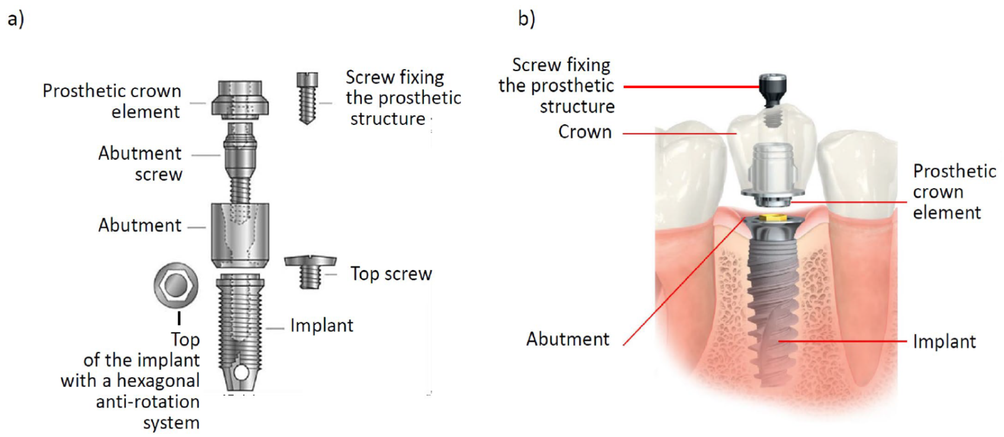
Table 1.
The general classification of implants due to applications in various organs.
| No. | Type of Implant or Function | Therapeutic Functions |
|---|---|---|
| 1. | Implants of the stomatognathic system | to replace, improve, or increase any soft or hard tissue in the mouth of a jaw, mandible, or temporomandibular joint |
| 2. | Dental implants | replacement of lost teeth and restoration of the possibility of prosthetic reconstruction in this area |
| 3. | Craniofacial implants | to improve or replace hard tissues in the craniofacial area except for the brain, eyes, and inner ear |
| 4. | Orthopedic implants | to support, substitute, or supplement permanently or temporarily bones, cartilage, ligaments, tendons, or related tissues |
| 5. | Implants of the cardiovascular system | in the treatment of disorders of the heart, its valves, and the rest of the circulatory system |
| 6. | Sensory and neurological implants | used in disorders affecting the primary senses and the brain, in the treatment of visual impairment, with hearing loss and diseases, neurological diseases |
| 7. | Electrical implants | used to relieve pain and suffering from rheumatoid arthritis |
| 8. | Cosmetic implants | restoring some body parts to an acceptable asthetic standard |
| 9. | Contraceptive implants | used to prevent unintended pregnancy and to treat conditions, such as non-pathological bleeding |
| 10. | Other implants | removal of dysfunctions of different organs of the body, including those in the digestive, respiratory, and urological systems |
Table 2.
Selected epidemiological data on the causes of dental extractions.
| Country | Number of Teeth Examined | Reason for Dental Extraction, % | Literature | ||
|---|---|---|---|---|---|
| Caries | Periodontal Disease | Remaining | |||
| Brazil | 404 | 70.3 | 15.1 | 14.6 | [95] |
| Italy | 1056 | 33.4 | 33.1 | 33.5 | [13] |
| Japan | 11175 | 55.4 | 38 | 6.6 | [96] |
| Kuwait | 2783 | 43.7 | 37.4 | 18.9 | [97] |
| Scotland | 917 | 51 | 21 | 28 | [98] |
Table 3.
Selected data on the occurrence of caries in the population of developmental age.
| Data on Tooth Decay | Age of Children, Years | Literature | |||
|---|---|---|---|---|---|
| 2–3 | 3–4 | 6–7 | 12 | ||
| Share of caries in the population, % | 35–50 | 56–60 | 85–100 | 90 | [102,111,113] |
| Share in the caries-free population, % | no data | no data | 14 | 19.3 | [111,112] |
Table 4.
Selected epidemiological data regarding the time of endodontic treatment of teeth in the oral cavity.
Table 4.
Selected epidemiological data regarding the time of endodontic treatment of teeth in the oral cavity.
| Country | Time After Which the Test Was Performed, Years | Number of Teeth Covered by the Test | Share of Teeth Retained in the Mouth, % | Share of Removed Teeth, % | Literature |
|---|---|---|---|---|---|
| USA | 3.5 | 110,766 | 94.44 | 5.56 | [121] |
| Taiwan | 5 | 1,446,199 | 92.9 | 7.1 | [123] |
| USA | 8 | 1,462,936 | 97 | 3 | [120] |
| Dania | 17 | 845 | 55 | 45 | [122] |
Table 5.
Selected epidemiological data on the number of teeth treated endodontically with periapical lesions revealed in pantomographic images.
Table 5.
Selected epidemiological data on the number of teeth treated endodontically with periapical lesions revealed in pantomographic images.
| Country | Total Number of Teeth Assessed Radiologically | The Number of Teeth Treated Endodontically | The Share of Teeth with Revealed Endodontic Treatment, % | The Share of Teeth with Revealed Periapical Lesion, % | Literature |
|---|---|---|---|---|---|
| Australia | 5647 | 499 | 8.84 | 21.43 | [131] |
| Belgium | 4617 | 314 | 6.8 | 40.4 | [133] |
| Brazil | 5008 | 553 | 11.04 | 42.5 | [130] |
| Denmark | 15,984 | 773 | 4.84 | 52.2 | [124] |
| Greece | 7664 | 680 | 8.87 | 60 | [128] |
| Iran | 28,942 | 1013 | 3.5 | 52 | [126] |
| Ireland | 7427 | 148 | 1.99 | 25 | [129] |
| Japan | 16,232 | 3408 | 21 | 40 | [125] |
| Lithuania | 2186 | 283 | 12.95 | 43.1 | [132] |
| Spain | 4453 | 93 | 2.09 | 64.5 | [127] |
| Turkey | 8863 | 470 | 5.3 | 53.5 | [119] |
Table 6.
The general classification of dental prostheses and prosthetic restorations.
| Classification Criterion | Mounting Method | Type of Prosthesis/Prosthetic Restoration |
|---|---|---|
| Way deposition | On implants | Solid cement |
| Permanent with the option of periodic withdrawal by the patient | ||
| Permanent with periodic download capability only by a dentist | ||
| Movable on own teeth based on retention elements and the mucosa | ||
| On own teeth | Permanently cemented on own teeth | |
| Fixed on own teeth with the possibility of periodic extraction | ||
| Movable on own teeth based on retention elements | ||
| On the mucosa | Mobile acrylic prostheses | |
| On the mucosa and own teeth | Mucosal (settling) | |
| The method of transferring chewing force to the substrate | On implants | Implantology |
| Implantology-mucosal | ||
| On own teeth | Periodontal (non-settling) | |
| Periodontal-mucosa | ||
| On the mucous | Mucosa (settling) | |
| Period of using | Short-term use | |
| Long-term use |
Table 7.
Classification of dental implants.
| Clinical Conditions | Type | Surgical Interventions | Geometric Features |
|---|---|---|---|
| Partial lack of dentition/ Toothlessness | Intraosseous | Single-phase immediate | Individual implants |
| Single-phase | Cylindrical with the smooth surface | ||
| Two-phase immediate | |||
| Single-phase | Cylindrical with the helical surface | ||
| Two-phase | |||
| Single-phase | Cylindrical with the helical surface with porous zones | ||
| Two-phase | |||
| Single-phase immediate | Needle | ||
| Bicortical screws | |||
| Two-phase | Flat (razor blade) | ||
| Through the bone | Two-phase | Yoke | |
| Toothlessness | For periosteal | Single-phase | Individual |
| Two-phase |
Table 8.
General guidelines for the design of the implant.
| Design Stages | Basic Conditions |
|---|---|
| Determining the geometrical features of the implant | Anatomical and physiological conditions |
| Surgical techniques envisaged for usage | |
| Stress and displacement analysis in the implant-tissue system | |
| Analysis of the dimensional characteristics of the implants corresponding to the anthropometric characteristics of the patient population | |
| Selection of implant material | Ensuring implant bio tolerance |
| Obtaining the required mechanical properties of the implant | |
| Ensuring the desired technological properties during implant manufacturing | |
| Development of documentation | Construction |
| Technology | |
| Acceptance control |
Table 9.
Examples of materials used in dental prosthetics in the case of the authors’ own experience.
Table 9.
Examples of materials used in dental prosthetics in the case of the authors’ own experience.
| l | Type | Detailed Information |
|---|---|---|
| 1. | Metals and their alloys | Co, Ti, and Mg alloys and their powders, including Co25Cr5W5MoSi, Ti6Al4V, Ti6Al7Nb, Ti24Nb4Zr8Sn, Ti7.5Mo and Ti40Nb, Mg8Al, Mg3Al1ZnMn, Mg9AlZnMn, Mg4Y3NdZr, Mg3Nd1GdZrZn, and pure Ti |
| 2. | Ceramic materials | Sintered ZrO2 and Al2O3 |
| 3. | Precursors for the preparation of coatings inside the pores | TiCl4, AlCl3, Zr[N(CH3)∙(C2H5)]4, and ZrD-04, i.e., (CpMe)2Zr(OMe)Me, where Cp=C5H5−x Rx is cyclopentadienyl ligands and heteroleptic metallocenes [Zr(Cp)4−n(L)n], L = Me and OMe using water and ozone as reagents and triethylphosphine (6,6,7,7,8,8,8-heptafluoro-2,2-dimethyl-3,5-octanedionate)silver(I) or Ag(fod)(PEt3),triethylphosphine (6,6,7,7,8,8,8-heptafluoro-2,2-dimethyl-3,5-octanedionate) silver(I) connected with dimethyl amineborane (BH3(NHMe2) or (hfac)Ag(1,5-COD) ((hexafluoroacetylace-tonato)silver(I)(1,5-cyclooctadiene)) as a metal source and tertiary butyl hydrazine (TBH) as a reactant for TiO2, Al2O3, ZrO2, and nanosilver coatings, respectively |
| 4. | Semi-finished products for applying porcelain coatings | Pastes or powders with thinners, including, e.g., DeguDent or Willi Geller, layers connecting the metal substrate with porcelain, including bonder metallic for porcelain and paste opaque or Crea Alloy Bond and paste opaque, cervical layer Power porcelain Chrome or Opaque Dentine and subsequent layers of porcelain Flu Inside, Detine, Incisale, Incisale opalescente, Transpa with properly selected shades or Opaque Dentine, Dentine, Enamel, Clear mixed with Universal Mixing Liquide by Harvest Dental and outside Glaze with Stain dyes mixed sequentially with Ducera Liquid or Crea Color |
| Form: 98 mm diameter discs for milling work, laser sintering powders, liquid and gas precursors | ||
Table 10.
Examples of how to meet the requirements contained in the technological platform of the augmented holistic Industry 4.0 model in the case of the authors’ own projects regarding the application of the augmented holistic Industry 4.0 concept at the dental prosthetic restoration center according to idea Dentistry 4.0.
| No. | Model Module | Type of Device or Element |
|---|---|---|
| 1. | Technological processes | Imaging of the patient’s teeth |
| Additive manufacturing models using stereolithography (SLA) | ||
| Milling of metal, ceramic, and polymer discs and precision machining of elements manufactured by SLS method | ||
| Applying atomic layers deposition (ALD) on the surface of solid bridges and crowns and inside the pores of porous structures | ||
| Opaque and porcelain deposition and firing | ||
| 2. | Machines and devices | 3D progress oral scanner |
| Scanner for dental models 3Shape E3, Dental Wings 7 | ||
| CNC milling machining center Fanuc Robodrill, RS-team/Kimia micro | ||
| The machine for the additive manufacturing of 3D Systems PeoJet 6000 HD polymer elements | ||
| Selective laser sintering machine (SLS) Orlas Creator | ||
| Machine for applying atomic layers ALD Picosun 200 | ||
| 3. | Cyber-IT system | Servers, databases, set of computers, network connections, sensors, two-dimensional QR codes (quick response), and cloud computing |
| Computer software for the design of geometric forms and porous structures | ||
| Computer software for imaging and designing prosthetic restorations | ||
| Computer software for coating design | ||
| Software for production management systems, supply and purchasing planning, logistics, and accounting |
© 2020 by the authors. Licensee MDPI, Basel, Switzerland. This article is an open access article distributed under the terms and conditions of the Creative Commons Attribution (CC BY) license (http://creativecommons.org/licenses/by/4.0/).
Share and Cite
MDPI and ACS Style
Dobrzański, L.A.; Dobrzański, L.B. Dentistry 4.0 Concept in the Design and Manufacturing of Prosthetic Dental Restorations. Processes 2020, 8, 525. https://0-doi-org.brum.beds.ac.uk/10.3390/pr8050525
AMA Style
Dobrzański LA, Dobrzański LB. Dentistry 4.0 Concept in the Design and Manufacturing of Prosthetic Dental Restorations. Processes. 2020; 8(5):525. https://0-doi-org.brum.beds.ac.uk/10.3390/pr8050525
Chicago/Turabian StyleDobrzański, Leszek A., and Lech B. Dobrzański. 2020. "Dentistry 4.0 Concept in the Design and Manufacturing of Prosthetic Dental Restorations" Processes 8, no. 5: 525. https://0-doi-org.brum.beds.ac.uk/10.3390/pr8050525
Note that from the first issue of 2016, this journal uses article numbers instead of page numbers. See further details here.