
Poly[3-methyl-1,3-oxazolidin-2-iminium[µ3-cyanido-tri-µ2-cyanido-κ9C:N-tricuprate(I)]]


Chemistry Department, Fordham University, 441 East Fordham Road, Bronx, New York, NY 10458, USA
*
Author to whom correspondence should be addressed.
Molbank 2021, 2021(3), M1259; https://0-doi-org.brum.beds.ac.uk/10.3390/M1259
Submission received: 15 June 2021
/
Revised: 16 July 2021
/
Accepted: 16 July 2021
/
Published: 26 July 2021
(This article belongs to the Collection Molecules from Side Reactions)
Abstract
:The unexpected formation of an oxazole ring has occurred during synthesis of a copper(I) cyanide network polymer as part of our ongoing studies of the structural chemistry of these networks. Crystals of the title compound were formed during the synthesis of a previously reported CuCN network solid containing protonated N-methylethanolamine and have been characterized by single crystal X-ray structure analysis. The structure shows well-defined oxazole-2-iminium cations sitting in continuous channels along the short a-axis of the crystal in a new three-dimensional copper(I) cyanide polymeric network. Evidently, a reaction has occurred between the cyanide ion and the protonated N-methylethanolamine base.

1. Introduction
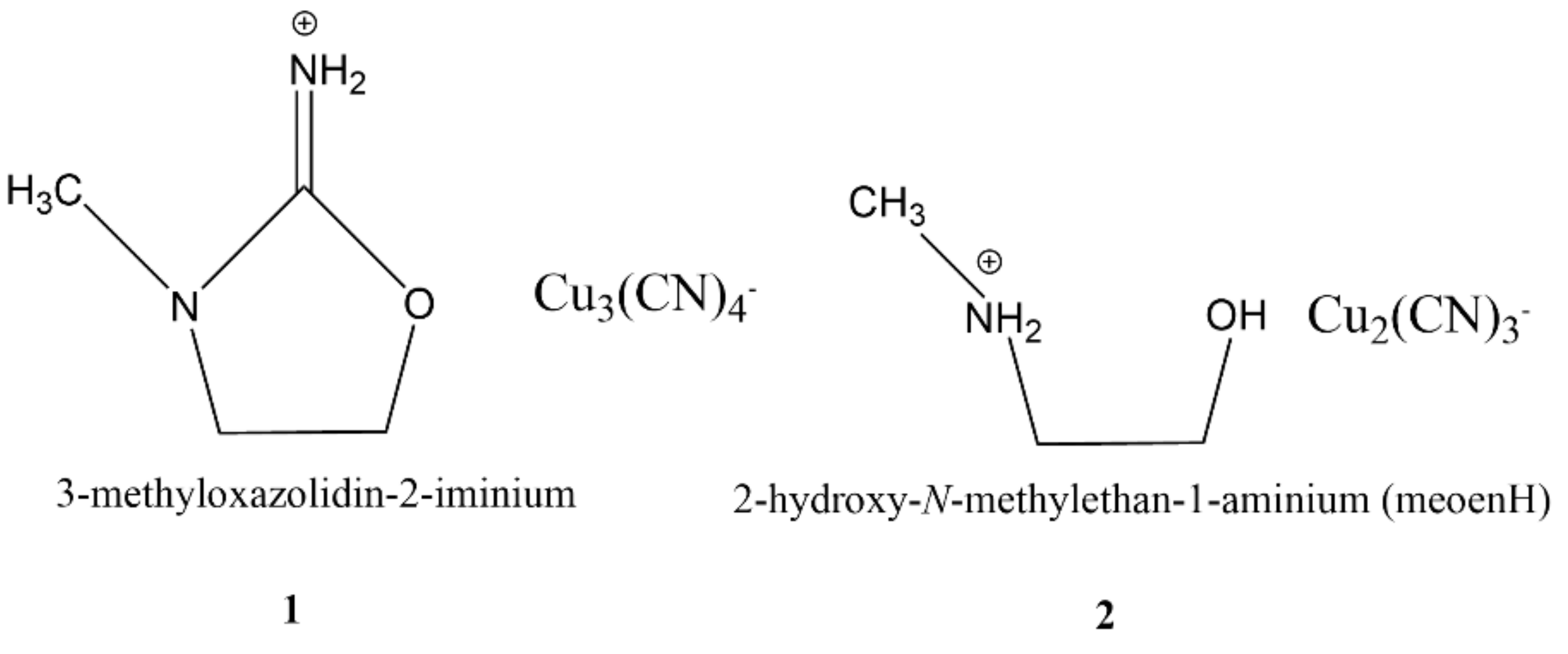
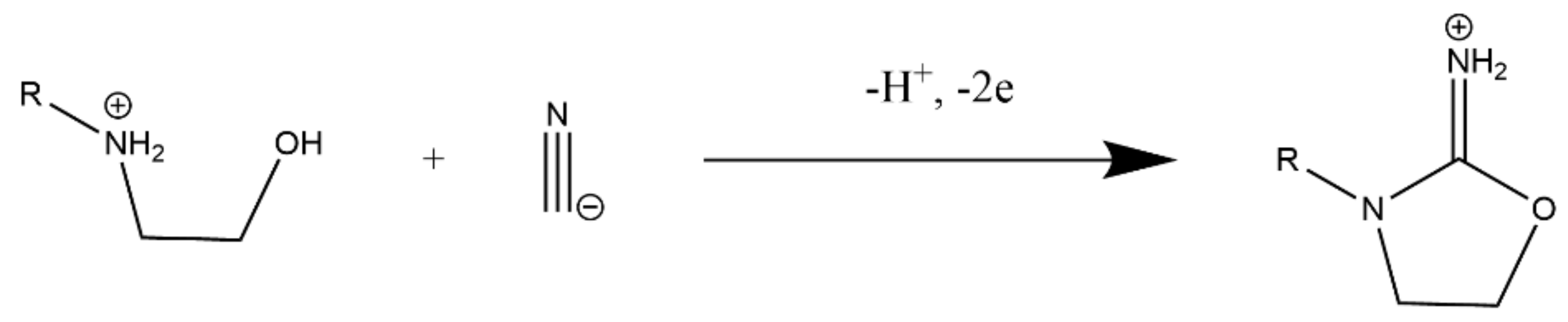
We report here the unexpected formation of poly[3-methyl-1,3-oxazolidin-2-iminium[µ3-cyanido-tri-µ2-cyanido-κ9C:N-tricuprate(I)]], (1, Scheme 1) during preparative studies of NaCN/CuCN/N-methylethanolamine (meoen) mixtures as part of our ongoing studies of the structural chemistry of copper(I) cyanide networks. The usual product was [meoenH][Cu2(CN)3], 2, (Scheme 1) obtained as tetragonal crystals after one or two weeks, with a structure that contains meoenH cations embedded in an anionic three-dimensional CuCN network [1]. In efforts to obtain more product, we allowed further fractions to crystallize from the filtrates. The title compound was obtained from such fractions as crystals with a platelike morphology rather than the rods usually obtained for the expected compound 2. The crystal structure analysis reported here shows that a reaction between the cyanide ion and the ethanolamine cation has occurred (Scheme 2) to cause ring closure and the formation of an N-methylated 1,3-oxazole ring with a 2-iminium group, which acts as cation guest in a 3D anionic copper(I) cyanide network. Oxazole chemistry is a mature field [2,3,4,5] and oxazoles play an important role in pharmaceutical chemistry [6]. Oxazole synthesis has been achieved in at least fifteen different types of reactions [4], but we are not aware of a reaction similar to that reported here. There is less literature on the 2-iminium derivatives, and a search of the Cambridge Structural Database (CSD) [7] indicated only three crystallographic studies of their 2-iminium salts [8,9,10]. Reference 8 gives the structure of an oxaolidin-2-iminium ring with a seven-membered ring fused at C4 and C5, whereas the other references refer to the simple ring with various substituents.
2. Results
2.1. Synthesis
The title compound, 1, is almost always obtained from the second or third crystal fraction from aqueous mixtures of the neutralized N-methylethanolamine and CuCN/NaCN solutions after two or more weeks. Inevitably the first fraction obtained contains crystals of 2, sometimes mixed with a green powder. Although we have varied concentrations and the pH, we have not yet developed a procedure that will reliably give 1 as the first fraction. A typical synthesis involved mixing 0.90 g (10 mmol) of CuCN with 0.78 g (16 mmol) of NaCN in 60 mL of water until dissolved and adding a solution with volume 95 mL containing 3.00 g (40 mmol) meoen base neutralized with HCl(aq) to a pH of 2. The first precipitated fraction was filtered off after two weeks, and a second fraction filtered three weeks later gave 0.17 g of 1 as colorless plates, from which the crystal used for the X-ray analysis was selected. When air was excluded, no solid precipitated, suggesting that dioxygen from the air was the oxidant.
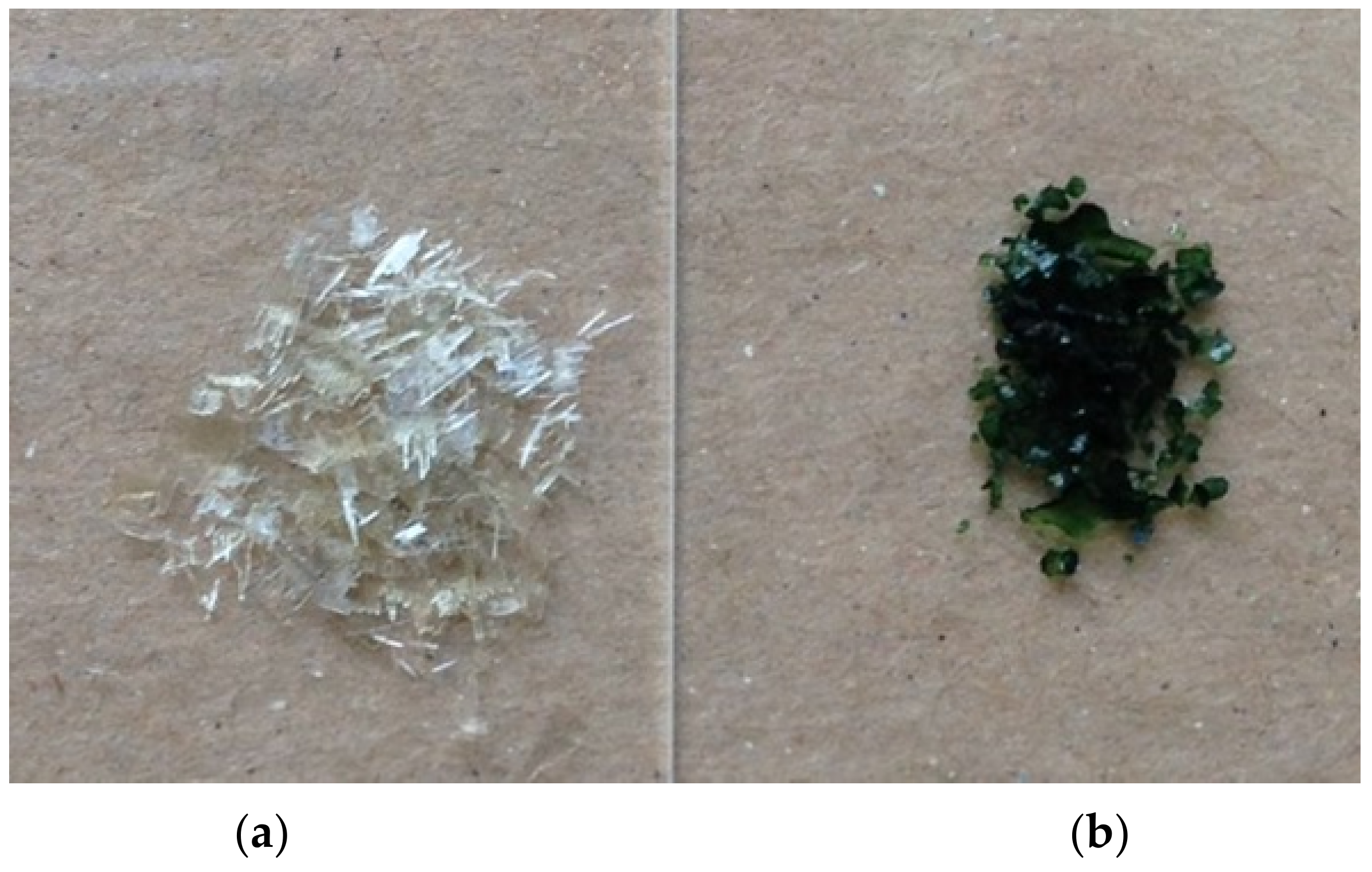
Sometimes pale or dark green crystals of 1 were obtained, shown by X-ray analysis to have the same structure as reported here. The dark green “crystals” shown in Figure 1 were very irregular in shape, yet diffracted well, and showed no extra diffraction pattern due to a separate phase. Examination optically at higher magnification (Figure S2) and with a scanning electron microscope (Figure S3) confirmed the presence of what looks like an amorphous phase deposited on crystalline material. Some of the sample was ground and examined by electron spin resonance—separate green and white particles were now visible. The esr spectrum, recorded as a first derivative absorption curve, (Figure S4) clearly shows the presence of Cu(II). Cu(II)-N bonding is indicated by the sharp secondary hyperfine lines that correspond the interaction of 14N with the unpaired electron associated with the Cu(II) ion. We conclude that the “green” crystals are composed of colorless crystalline plates of 1 capped with deposits of a green amorphous phase that contains Cu(II), probably in the form of a mixed-valence copper cyanide complex. A similar phenomenon was reported in the analysis of the meoenH complex, 2 [1].
2.2. X-Ray Structure
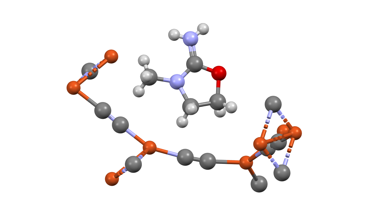
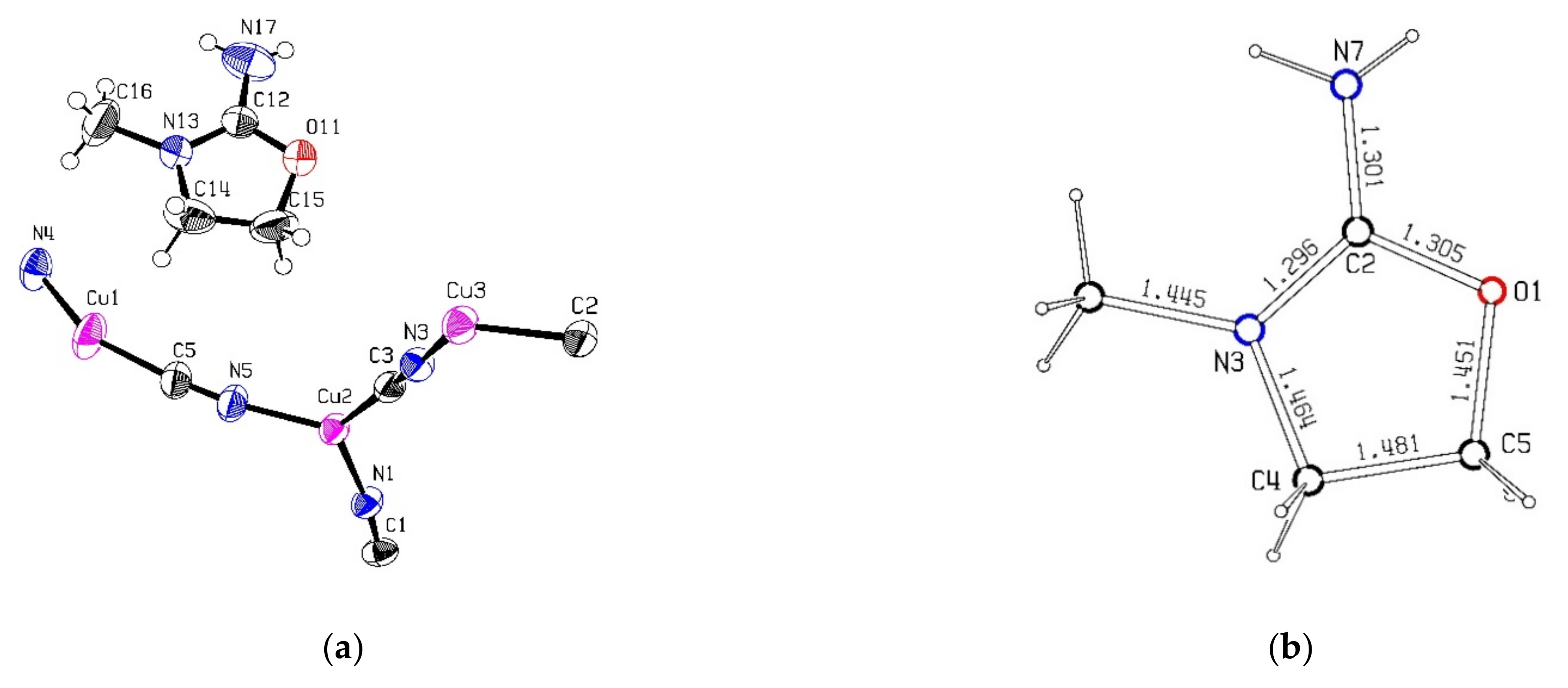
The crystal structure is built up from the oxazolidin-2-iminium cations in channels in a three-dimensional Cu(I)CN network with overall stoichiometry [LH][Cu3(CN)4], where LH represents the cation. Figure 2a presents the crystallographic asymmetric unit, showing the thermal ellipsoids and atom numbering. Figure 2b shows geometrical parameters for the oxazolidin-2-iminium cation. Atom numbering follows the typical convention for these rings; in the crystal structure, they are O11, C12, N13, C14, etc., to avoid confusion with the cyanide atom numbers. The bond distances around C2 are essentially equal at 1.30 Å, indicating resonance between three hybrid structures with the positive charge spread over O1, N3, and N7. The four atoms O1, C2, N3, N7 are strictly coplanar, but the constraints of the five-membered ring and its asymmetry force significant deviations from the idealized 120° angles: the internal angle is reduced to 114.1(3)°, while the external angle N7-C2-N3 at 127.2(4)° is much larger than the angle N7-C2-O1, at 118.8(4)°. A similar geometry for this portion of the cation is seen in the three X-ray structures of similar iminium cations mentioned earlier [8,9,10], even in reference [8], where there is no substituent on N3. Other bond lengths and angles seem normal.
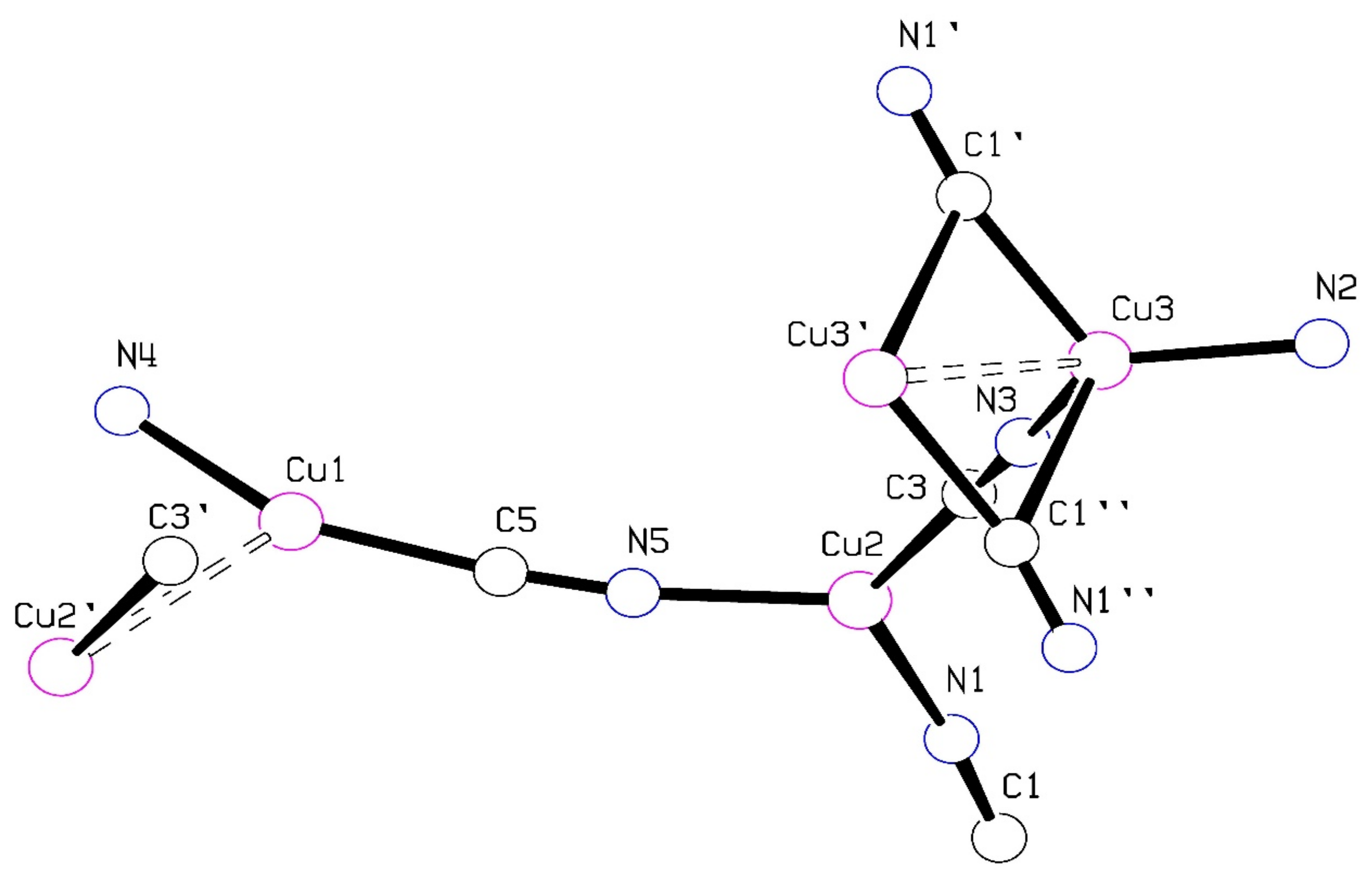
Each of the three Cu atoms in the CuCN network is involved in a cuprophilic interaction, with Cu1 close to Cu2(x−1,y,z), and Cu3 close to Cu3(2−x,−y,1−z). Such weak d10-d10 interactions are well known in CuI complexes, and have been explored in a recent review [11]. They are common in our CuCN network studies. Each Cu atom has a different geometry, as seen in Figure 3. Cu1 has a distorted digonal geometry, with average Cu-CN distance of 1.864(3) Å and angle at the Cu atom of 154.5(2)° and is 2.632(1) Å from Cu2(x−1,y,z). This cuprophilic interaction is without the bridging group(s) usually found in such structures, that help bring the Cu atoms close together—in the case of the CuCN structures, a µ3 CN bridge. The present structure is unusual in this regard, as the cyanide group C3≡N3 clearly seems to act as a µ2 bridging ligand, not as a µ3 bridge. It could possibly be regarded as forming a very asymmetric µ3 bridge, with C3-Cu1(1+x,y,z) and C3-Cu2 distances of 2.631(3) Å and 1.929(3) Å, but 2.631 Å is longer than any Cu-CN distances seen in a series of 63 µ3CN-Cu2 bridging bonds found in the Cambridge Structural Database [7], and the extreme asymmetry would lead one to expect a much greater Cu … Cu distance than that found in the present structure [12]. Apart from its cuprophilic interaction with Cu1, atom Cu2 is trigonally coordinated with average Cu-CN distance of 1.934(3) Å and angles varying from 116.1(1)° to 125.6(1)°. Cu3 is in a rough tetrahedral coordination to cyanide groups, with also a close interaction with Cu3(2−x,−y,1−z), related by an inversion center. The Cu3 atoms are 2.730(1) Å apart and are bridged asymmetrically by a pair of symmetry related µ3 C1≡N1 groups, with Cu-CN distances of 2.013(3) Å and 2.397(3) Å. The remaining two Cu-CN distances are shorter, with Cu3-N2 =1.938(3) Å and Cu3-N3 = 1.964(3) Å.
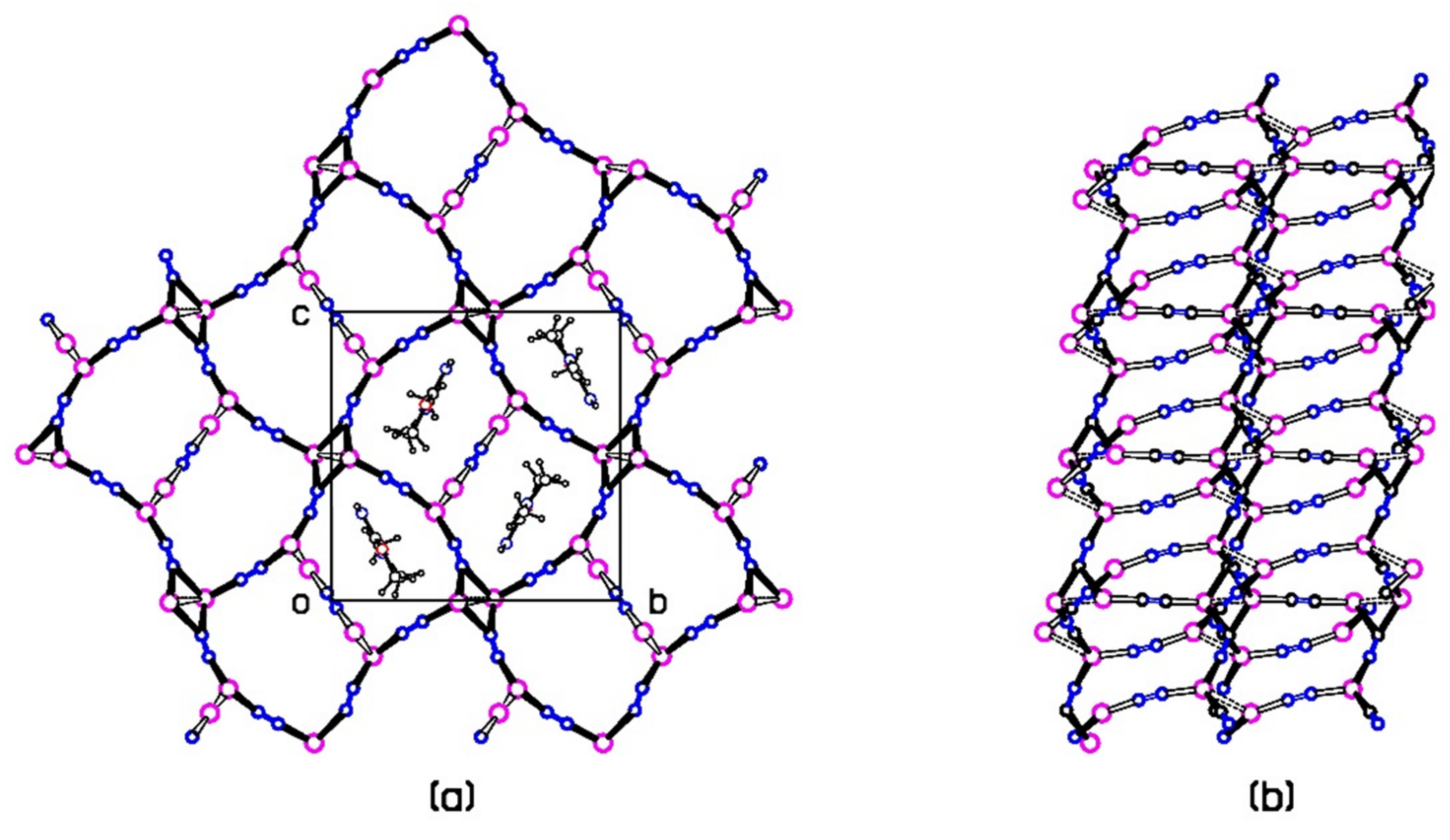
The CuCN 3D network can be considered as made up of roughly planar networks connected by approximately vertical Cu-CN-Cu linkages which form walls to channels along the a-axis direction that contain the oxazalone-iminium cations, Figure 4. The planar networks are made up of 24-membered rings composed of repeated -Cu2-C3N3-Cu3-C1N1-Cu2-linkages with the Cu3 cuprophilic pairs acting as nodes where four rings meet. A Cu1-C4≡N4-Cu1 group forms a belt across the center of each 24-membered ring via the cuprophilic Cu1 … Cu2 interactions. As seen in Figure 4a, the cations pack neatly into channels in the CuCN network. There are no obvious H-bonding interactions between the cations and the anionic CuCN network.
2.3. Spectroscopy and Analyses
The infrared spectrum of 1 (Figure S5) shows strong absorptions due to C≡N stretching at 2082, 2106, and 2138 cm−1. The absorption at 2106 cm−1 is probably due to µ2 bridging CN groups 2, 3, and 5, as we see single absorptions close to this value in our CuCN structures that contain only this type of CN group [13,14] and other unpublished results]. The absorption at 2082 cm−1 will be due to the µ3 bridging CN group as in Reference 1. The absorption at the higher energy, 2138 cm−1, is likely due to C4≡N4, which bridges two digonal low-coordinate Cu1 atoms.
Strong absorptions due to the iminium N-H bonds occur at 3357 and 3461 cm−1 which may be compared with the absorptions at 3406 and 3497 cm−1 seen in the guanidinium cation in a CuCN network [14]. There are two other weak bands at 3182 and 3290 cm−1. Other features of the ir spectrum include very strong absorbances at 1303, 1519, 1596, and 1705 cm−1.
The elemental analysis of 1 gave 24.36%C, 2.12%H and 21.16%N, which are comparable with the values calculated for the new product, 24.27%C, 2.29%H and 21.24%N. The %Cu of 46.55% found was somewhat lower than the expected value of 48.16%.
3. Experimental Section
Crystal data for C8H9Cu3N6O (M = 395.84 g/mol): crystal was a plate with dimensions 0.36 × 0.20 × 0.09 mm, monoclinic, space group P21/c (no. 14), a = 6.9393(1) Å, b = 13.4468(3) Å, c = 13.4972(3) Å, β = 95.071(1)°, V = 1254.55(4) Å3, Z = 4, T = 300(2) K, µ(MoKα) = 5.03 mm−1, Dcalc = 2.096 g/cm3, Dmeas = 2.08(1) g/cm3, 19,265 reflections measured (1.0° ≤ Θ ≤ 27.48°, 2867 unique (Rint = 0.046), which were used in all calculations. The final R1 was 0.0344 (I > 2σ(I)) and wR2 was 0.0790 (all data) for 175 variables. The structure was solved by the heavy atom method from a Patterson map calculated with SHELXS and refined with SHELXL Version 2017 [15]. All CN groups were modeled with disorder in their orientation. CN groups 2 and 4 were given 50% C/N occupancies, as they bridged an inversion center, and CN group 1 because the refined occupancies did not differ significantly from 50%. Occupancies for the major orientation for disordered CN groups 3 and 5 refined to 81(3)% and 78(4)%, respectively. Hydrogen atoms on C were constrained at idealized positions, riding on the carbon atoms, with C–H distances of 0.96 Å for methyl groups and 0.97 Å for methylene groups, and isotropic temperature factors 50% larger than the Ueq of the bonded C atoms. The iminium H atoms were refined independently, with only a minimum restraint on distance. The cif file for this structure is included in the Supplementary Materials for this article and has also been deposited with the Cambridge Crystallographic Data Center as CCDC 2088682. These data can be obtained free of charge via http://www.ccdc.cam.ac.uk/conts/retrieving.html (accessed date: 15 June 2021) or from the CCDC, 12 Union Road, Cambridge CB2 1EZ, UK; Fax: +44 1223 336033; E-mail: [email protected]).
4. Discussion
This work has shown the formation of a 3-methyl-1,3-oxazolidin-2-iminium cation via the reaction of a cyanide ion and protonated N-methylethanolamine in aqueous solution in the presence of CuCN/NaCN. In ongoing work on this system, we are attempting to find reaction conditions and pH levels that favor the formation of the title compound, to isolate salts of the cation that are independent of the copper cyanide network, and to develop an understanding of the reaction mechanism.
5. Materials and Methods
Chemicals were used as obtained from suppliers without further purification. The X-ray structure was determined with diffraction data collected with a Nonius Kappa CCD system (Bruker Axis LLC, Madison, WI, USA) using graphite monochromated MoKα radiation with λ = 0.7107 Å. Chemical analysis was performed by Robertson Microlit (Ledgewood, NJ, USA). FTIR data were obtained with a Nicolet iS50 FT-IR spectrometer (Thermo Fisher Scientific, Waltham, MA, USA. The SEM image was collected with a Zeiss Evo MA10 Scanning Electron Microscope and the esr spectrum was obtained with a Bruker EMXNano spectrometer.
Supplementary Materials
Figure S1: Checkcif Report, Figure S2: Optical photo of green “crystal” at high magnification; Figure S3: SEM picture of a different green “crystal”; Figure S4: esr spectrum of green sample; Figure S5: Infrared spectrum of title compound. MOL File, cif file.
Author Contributions
P.W.R.C. designed the experiments, carried out the X-ray analysis, analyzed the results and wrote the manuscript. L.N.R. carried out the syntheses and ir spectra. All authors have read and agreed to the published version of the manuscript.
Funding
This research received no external funding.
Institutional Review Board Statement
Not Applicable.
Informed Consent Statement
Not Applicable.
Data Availability Statement
The data presented in this study are available on request from the corresponding author.
Acknowledgments
We gratefully acknowledge support from the chemistry department at Fordham University, and we thank colleagues C. Koenigsmann for assistance with the electron microscopy and C. Bender for the esr measurements. Students A. Felix Varona, T. DaCunha and N. Eisha assisted with follow-up syntheses and density measurements.
Conflicts of Interest
The authors declare no conflict of interest.
References
- Koenigsmann, C.; Rachid, L.N.; Sheedy, C.M.; Corfield, P.W.R. Synthesis, decomposition studies and crystal structure of a three-dimensional CuCN network structure with protonated N-methylethanolamine as the guest cation. Acta Crystallogr. Sect. C Struct. Chem. 2020, 76, 405–411. [Google Scholar] [CrossRef] [PubMed]
- Wiley, R.H. The chemistry of the oxazoles. Chem. Rev. 1945, 37, 389–437. [Google Scholar] [CrossRef] [PubMed]
- Lakhan, R.; Ternai, B. Advances in oxazole chemistry. Adv. Heterocycl. Chem. 1974, 17, 99–211. [Google Scholar]
- Turchi, I.J.; Dewar, M.J.S. The Chemistry of Oxazoles. Chem. Rev. 1975, 75, 401–442. [Google Scholar] [CrossRef]
- Turchi, I.J. Oxazole chemistry: A review of recent advances. Ind. Eng. Chem. Prod. Res. Dev. 1981, 20, 32–76. [Google Scholar] [CrossRef]
- Kakkar, S.; Narasimhan, B. A comprehensive review on biological activities of oxazole derivatives. BMC Chem. 2019, 13, 16. [Google Scholar] [CrossRef] [PubMed] [Green Version]
- Groom, C.R.; Bruno, I.J.; Lightfoot, M.P.; Ward, S.C. The Cambridge Structural Database. Acta Cryst. B 2016, 72, 317–325. [Google Scholar] [CrossRef] [PubMed]
- Rynearson, K.D.; Dutta, S.; Tran, K.; Dibrov, S.M.; Hermann, T. Synthesis of Oxazole Analogs of Streptolidine Lactam. Eur. J. Org. Chem. 2013, 2013, 7337–7342. [Google Scholar] [CrossRef]
- Cruz, A.; Padilla-Martínez, I.I.; García-Báez, E.V.; Contreras, R. Reactivity of Chlorodeoxypseudoephedrines with Oxo-, Thio-, and Selenocyanates. Tetrahedron Asymmetry 2007, 18, 123–130. [Google Scholar] [CrossRef]
- Misaiszek, C.; Jarry, C.; Ouhabi, J.; Carpy, Y. Synthesis and Structural Study of 3-(2-propanone) 5-phenoxymethyl 2-iminooxazolidine: C13H16N2O3HCl. Comptes Rendue L’Academie Sci. Ser. II 1988, 307, 1189–1193. [Google Scholar]
- Satyachand Harisomayajula, N.V.; Makovetskyi, S.; Tsai, Y.-C. Cuprophilic Interactions in and between Molecular Entities. Chem. Eur. J. 2019, 25, 8936–8954. [Google Scholar] [CrossRef] [PubMed]
- Stocker, F.B.; Staeva, T.P.; Rienstra, C.M.; Britton, D. Crystal Structures of a Series of Complexes Produced by Reactions of Copper(I) Cyanide with Diamines. Inorg. Chem. 1999, 38, 984–991. [Google Scholar] [CrossRef]
- Corfield, P.W.R.; Stavola, T.J. Poly[diethylammonium [tetra-µ2-cyanido-κ8C:N-tricuprate(I)]], a two-dimensional network solid. IUCrData 2020, 5, x200968. [Google Scholar] [CrossRef]
- Corfield, P.W.R.; Dayrit, J.R. Poly[1,3-Dimethyltetrahydropyrimidin-2(1H)-iminium [tri-µ2-cyanido-κ6C:N-dicuprate(I)]]. Molbank 2020, 4, M1170. [Google Scholar] [CrossRef]
- Sheldrick, G.M. Crystal structure refinement with SHELXL. Acta Crystallogr. Sect. C Struct. Chem. 2015, 71, 3–8. [Google Scholar] [CrossRef]
Scheme 1.
Structures of the title compound, 1, and of the expected product, 2.

Scheme 2.
Reaction of protonated N-methylethanolamine with cyanide ion.

Figure 1.
Photograph of crystalline samples of 1. (a) Colorless plates; (b) plates coated with green amorphous material, as text describes.
Figure 1.
Photograph of crystalline samples of 1. (a) Colorless plates; (b) plates coated with green amorphous material, as text describes.

Figure 2.
(a) Asymmetric unit for 1, showing thermal ellipsoids with 50% probability. Only one orientation of the disordered CN groups is shown. The CN groups C2≡N2 and C4≡N4 are each disordered about one of the inversion centers in the unit cell. (b) Bond lengths in the oxazolidin-2-iminium cation. Estimated standard deviations range from 0.004–0.007 Å.

Figure 3.
Coordination of the three copper atoms. Primed atoms are related by symmetry. Cuprophilic interactions are shown as dashed bonds.
Figure 3.
Coordination of the three copper atoms. Primed atoms are related by symmetry. Cuprophilic interactions are shown as dashed bonds.

Figure 4.
CuCN network and packing. (a) View down a-axis, showing the belted 24-membered rings. Oxazolidin cations shown only in the unit cell outline. (b) View down b-axis, showing the rings edge on linked by cyanide groups into the 3D structure. Cell outline is not included in this view. Magenta: Cu; red: O; blue: N; black: C or H.

Publisher’s Note: MDPI stays neutral with regard to jurisdictional claims in published maps and institutional affiliations. |
© 2021 by the authors. Licensee MDPI, Basel, Switzerland. This article is an open access article distributed under the terms and conditions of the Creative Commons Attribution (CC BY) license (https://creativecommons.org/licenses/by/4.0/).
Share and Cite
MDPI and ACS Style
Rachid, L.N.; Corfield, P.W.R. Poly[3-methyl-1,3-oxazolidin-2-iminium[µ3-cyanido-tri-µ2-cyanido-κ9C:N-tricuprate(I)]]. Molbank 2021, 2021, M1259. https://0-doi-org.brum.beds.ac.uk/10.3390/M1259
AMA Style
Rachid LN, Corfield PWR. Poly[3-methyl-1,3-oxazolidin-2-iminium[µ3-cyanido-tri-µ2-cyanido-κ9C:N-tricuprate(I)]]. Molbank. 2021; 2021(3):M1259. https://0-doi-org.brum.beds.ac.uk/10.3390/M1259
Chicago/Turabian StyleRachid, Leena N., and Peter W. R. Corfield. 2021. "Poly[3-methyl-1,3-oxazolidin-2-iminium[µ3-cyanido-tri-µ2-cyanido-κ9C:N-tricuprate(I)]]" Molbank 2021, no. 3: M1259. https://0-doi-org.brum.beds.ac.uk/10.3390/M1259
Note that from the first issue of 2016, this journal uses article numbers instead of page numbers. See further details here.