Article

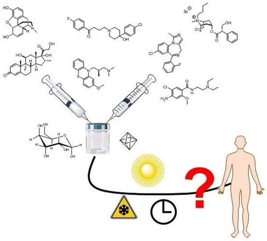
Filling Gaps on Stability Data: Development, Validation and Application of a Multianalyte UHPLC-DAD Method to Determine the Stability of Commonly Administered Drugs in Different Carrier Solutions Used in Palliative Care

1
ETH Zurich, Institute of Pharmaceutical Sciences, Vladimir-Prelog-Weg1-5/10, CH-8093 Zurich, Switzerland
2
Kantonsspitalapotheke Winterthur, Brauerstrasse 17, CH-8400 Winterthur, Switzerland
3
Kantonsspital Baden, Hospital Pharmacy, Im Ergel 1, CH-5404 Baden, Switzerland
*
Author to whom correspondence should be addressed.
Submission received: 27 August 2020 / Revised: 22 September 2020 / Accepted: 5 October 2020 / Published: 13 October 2020

Abstract

:
In palliative care, continuous subcutaneous infusion (CSCI) is common practice for drug administration when oral application of drugs is not feasible or not reliable anymore. However, use of CSCI is limited to chemical stability of drugs and their combination in carrier solution. To determine the stability of different mixtures of commonly used drugs in palliative care, a multi-analyte UHPLC-DAD method controlled by an internal standard was successfully developed. The method was validated in terms of specificity, accuracy, precision, and linearity across the calibration range. Seven analytes could be separated within 10 min by C18-reversed phase chromatography. The method was successfully applied to close gaps in stability data and complete missing data for decision makers in health care units. Our results indicated the stability of binary mixtures and one ternary mixture in 0.9% saline and 5% glucose as carrier solutions. The obtained data will support pharmacists in palliative care for the preparation of parenteral drug solutions in the future.

Graphical Abstract

1. Introduction

In palliative and critical care, drugs are often administered parenterally, since many patients cannot take drugs orally due to persistent nausea and/or vomiting, dysphagia, bowel obstruction or malabsorption, significant tablet burden or reduced level of consciousness such as in the last days of life. In these situations, continuous subcutaneous infusion (CSCI) has become common practice [1]. CSCI is safe in application, well tolerated by the patients, less painful, cheap and ensures a continuous administration of drugs [2]. The subcutaneous (s.c.) application is easier to perform than intravenous (i.v.) application, as it can be applied in any part of the body, where subcutaneous fat tissue is present. Furthermore, i.v. application poses a risk of phlebitis and local and systemic infections, which can be mostly avoided by s.c. application [3]. For the continuous application of drugs, syringe pumps, also known as syringe drivers, are often used [4]. They also allow for the administration of mixtures of drugs. Commonly, either 0.9% saline or 5% glucose are used as carrier solutions. CSCI applications further minimize pricking events which improves the compliance among patients and their relatives [5]. However, there are limited data about the chemical compatibility over at least 24 h of application [6].
Although active pharmaceutical ingredients (API) like dexamethasone sodium phosphate (DexP), haloperidol lactate (Hal), levomepromazine hydrochloride (Lev), metoclopramide hydrochloride (Met) and midazolam hydrochloride (Mid) are not registered for s.c. application, off-label use is common to ensure that patients in palliative care receive the necessary treatment [7]. In 2015, Masman and co-workers published a list indicating aforementioned drugs as most applied drugs used in palliative care [8]. In addition, Lev is reported as an effective, subcutaneous drug offering positive effects to prevent and treat seizures at the end of life [9,10]. When combinations of drugs are administered to multi-morbid palliative patients, stability and compatibility problems between the drugs and the excipients have to be considered [11]. For many drug combinations, data on the chemical stability and compatibility are scarce in the literature or data are incomplete in terms of concentration and carrier solutions [4,12,13,14,15]. Mixtures of drugs are defined as stable if there is no loss greater than 10% of any of the components over a determined period of time [4,16,17]. Several reports confirmed that combinations containing dexamethasone (Dex) are prone to precipitation upon mixing, or are not stable, even though they might not precipitate, such as the combination of Dex with Mid [18,19,20]. However, no data were available on the stability of the binary combinations containing Lev and either Hal, Met or Scopolamine (Sco) [13]. For the combination of Hal with Sco, stability is only ensured in 5% glucose, as there are incompatibilities reported in 0.9% saline at a concentration of 1.25 mg/mL Hal [21]. Additionally, Gomez et al. described a loss of Hal in the presence of Sco in 0.9% saline. Tests were performed over a time period of 15 days, which is much longer than necessary for the intended use [12]. For other combinations, such asLev with Midor the combination of Sco with either Dex or Met, only stability data using 0.9% saline are published. No data in 5% glucose solution are available [13]. For the combination of morphine hydrochloride (Mor) with Lev, stability data are converted from data measured with morphine sulfate [22]. According to the literature, Sco and Mid are stable in 0.9% saline, whereas data on stability in 5% glucose are missing [13]. Data in 5% glucose were only reported for concentrations lower than therapeutic reference values [12]. Combining Met and Mor is verified for 0.9% saline, as there are no data available on this combination in 5% glucose [13]. Schrijvers et al. reported that this combination is stable at several different concentrations of Mor [23]. However, drugs were not diluted in carrier and only UV spectrophotometry for the quantification of drugs was used without any chromatography [23]. Further examination of this combination in 0.9% saline and 5% glucose is necessary by a more stable method such as HPLC-DAD and UHPLC-DAD, respectively.
Although numerous stability reports are available in the literature, data points regarding the clinically interesting combination of API and carrier are lacking (Figure 1).
In this study, we focused on closing these gaps and providing information on chemical stability of binary or ternary drug mixtures at upper therapeutic concentrations which are frequently used in palliative care. Accordingly, a fully validated UHPLC-DAD method was developed to simultaneously detect and quantify seven target analytes. The method was applied to complete data through a targeted analysis. These data will help healthcare professionals in decision-making for producing CSCI preparations in the future.

2. Materials and Methods

Analytical reference standards of Dex, Met, Hal, Sco, Mor, Mid, Lev and formic acid were obtained from Sigma Aldrich (Buchs, Switzerland). Acetonitrile (ACN) was purchased from Merck (Darmstadt, Germany) and was of LCMS grade. 3-Aminobenzoic acid was obtained from Fluka (Buchs, Switzerland) and was of the highest analytical grade. HPLC vials, screwcaps and inlets were purchased from BGB (Boeckten, Switzerland). NaCl 0.9% and glucose 5% were obtained from Braun (Sempach, Switzerland). Pure water was generated from an in-house water purification system from Labtec (Villmergen, Switzerland). For all experiments, Gilson pipettes and Gilson DIAMOND tips were used (Mettmenstetten, Switzerland). Drug preparations for injection were provided by Kantonsspital Baden (KSB; Baden, Switzerland).

2.1. Method Development

All samples were analyzed on a HITACHI ChromasterUltra UHPLC system (Darmstadt, Germany) equipped with an autosampler (6270), a binary pump (6170), a column oven (6310) and a diode array detector (6430). The wavelengths used for analysis were λ = 210 nm and λ = 254 nm, respectively. For all separations, pure water with 0.1% FA (mobile phase A) and ACN with 0.1% FA (mobile phase B) were used. Three different columns were tested for the best separation performance. Kinetex C18 150 × 2.1 mm, 1.7 μm, 100 Å (Phenomenex, Torrance, CA, USA), Acquity BEH C18, 50 × 2.1 mm, 1.7 μm (Waters, Milford, MA, USA) and ACE® Excel C18-AR, 100 × 2.1 mm, 1.7 μm (VWR, Dietlikon, Switzerland) were investigated in detail. Flow rate was examined at 0.3–0.4 mL/min. Column oven temperature varied between 30 and 40 °C. Starting content of the mobile phase A was set to 95% or 98%. Three different gradients were applied which can be found in the supporting information (Table S1). For stability testing, the following conditions were used: 0.3 mL/min; 0–1.0 min 98% A, 1.0–7.1 min 30% A, 7.1–7.3 min 0% A, 7.3–8.3 min 0% A (flow set to 0.5 mL/min), 8.3–8.5 min 98% A, 8.5–9.5 min 98% A (flow set to 0.3 mL/min).

2.2. Preparation of Calibration and Quality Control (QC) Samples

Stock solutions in MeOH (Mid, Hal, Lev, Dex) or pure water (Met, Mor, Sco) were prepared using the reference substances. Six different concentrations and three quality controls (QC High, QC Med and QC Low) were chosen within the calibration range for each analyte. The upper limit of the range was equal or above the highest therapeutically used concentration for each analyte and was defined as 100%. The other concentrations were chosen at 80%, 40%, 16%, 14% and 10% of the highest concentration. The QC High was chosen at 90%, QC Med at 45% and QC Low at 11.25%. Absolute concentrations (mg/mL) can be found in the supporting information (Table S2). Aliquots of 25 μL were stored in the freezer at −80 °C.

2.3. Preparation of Therapeutic Solutions

Finished products for CSCI preparation were obtained from different Swiss suppliers and from Runge Pharma Germany (Sco and Met, Sanofi Aventis; Mor, Sintetica; Mid, Roche; Lev, Neuraxpharm; Hal, Janssen-Cilag; Dex, Mepha). Final concentrations of the respective samples corresponded to the concentrations used clinically for CSCI and are listed in Table S2 (supporting information). Final concentrations were obtained by diluting selected drugs in 0.9% saline or 5% glucose solution. All measurements were carried out in triplicates. Individual aliquots were stored either at room temperature (25 ± 3 °C) or in the fridge (4 °C) and were analyzed at different time points (0 h, 4 h, 8 h, 24 h, 48 h).

2.4. Sample Preparation

For analysis, 25 µL, Cal, QC or authentic samples were mixed with 975 µL internal standard mix. 3-Amine benzoic acid was used as internal standard (0.08 mg/mL in mobile phase A).

2.5. Method Validation

On eight different days, each concentration level was analyzed according to the aforementioned procedure. The regression lines were calculated using a non-weighted or weighted [1/x2] least-squares regression model. Daily regression lines were used to back-calculate the concentration of each calibrant. The back-calculated concentrations of all calibration samples were compared to their corresponding theoretical values. Quantitative accuracy was limited to be within 20% of target. 3-Aminobenzoic acid was applied as internal standard for all target analytes. QC samples (Low, Med, High) were prepared and analyzed in duplicate on each of the eight days. Accuracy was determined in terms of bias as the percent deviation of the mean calculated concentration at each QC level from their respective nominal concentration. Intra-day and inter-day precision were calculated as relative standard deviation (RSD) according to Peters and coworkers [24]. Different storage conditions (25 °C, 4 °C, −20 °C and −80 °C) were analyzed on days 0, 1, 2, 5, 7, 14 and 28. Seven freeze–thaw cycles were performed within 28 days. Between each freeze–thaw cycle, samples were kept for at least 24 h in the freezer.

2.6. Data Analysis

Peak integration was performed in Agilent EZChrom Elite (Version 3.3.2 SP2, Santa Clare, CA, USA) software. GraphPad Prism 7 (GraphPad Software, San Diego, CA, USA) was used for regression and statistical analysis, respectively. In addition, all illustrations were done with GraphPad Prism 7.

3. Results

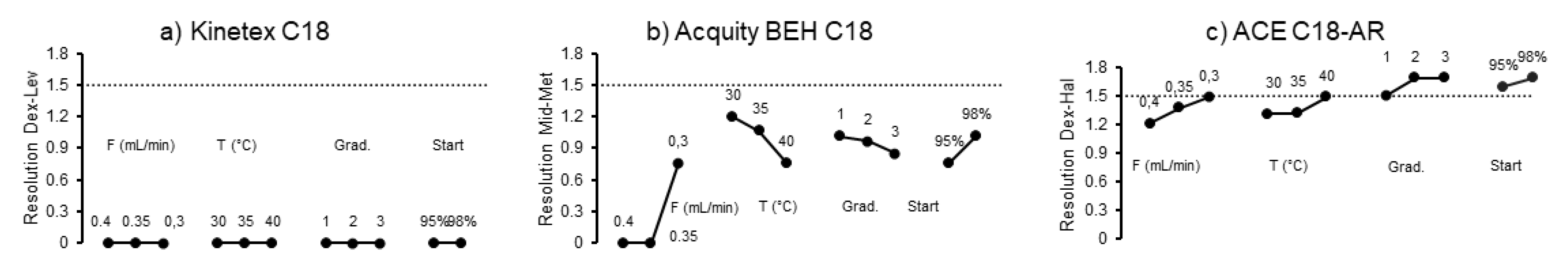
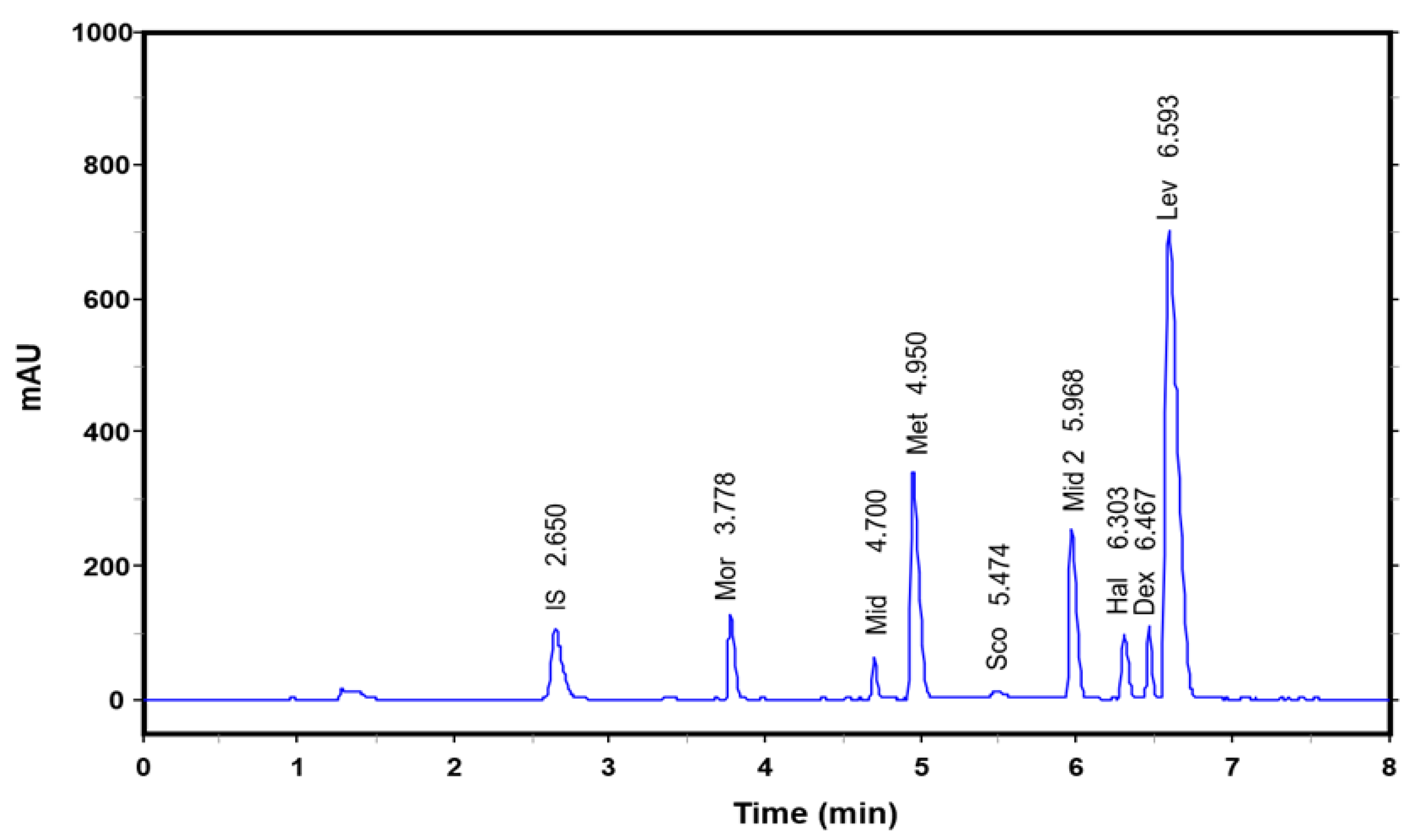
3.1. Method Developement

In general, there are several approaches available for method development of reversed phase based UHPLC methods [25,26]. In the beginning, we decided to use the one-variable-at-time approach because of simplicity and limited number of chromatographic variables. The principle of surrogate matrix and surrogate analytes is well established in clinical and forensic chemistry approaches [27,28,29]. For chromatographic separations and quantification, Dex was used here as a surrogate analyte for DexP. At the same molar concentration, the surrogate analyte showed the same peak intensity as DexP which was used for method validation. Three different UPLC C18 columns were used to separate all target analytes (Figure 2).
For a quantitative approach, baseline separation with an Rs of 1.5 is required. Each column showed different separation behavior with one unresolved analyte pairs. Subsequently, five different methods were applied using different flow rates (0.3–0.4 mL/min), temperatures (35–40 °C) and start condition (95–98% mobile phase A) to resolve the aforementioned analyte pair, respectively (Figure 2). Furthermore, three different gradient modes were tested in detail (supporting information, Table S1). On Kinetex C18 column Dex and Lev could not be separated under any tested chromatographic condition. With Acquity BEH C18, the analyte pair of Mid and Met was identified to be critical for the separation since a resolution of 1.5 was not achieved under any combination of tested chromatographic conditions. Finally, separation was performed on ACE C18-AR column. Dex and Hal turn out to be the critical analyte pair. Decreasing the flow rate from 0.4 to 0.3 mL/min and increasing column temperature from 35 to 40 °C improved the separation of Dex and Hal. When the starting content of mobile phase A was changed from 95 to 98%, further improvement was observed. Applying the optimized gradient, target analytes could finally be separated within 10 min. Neither Dex nor DexP coeluted with any other target analyte (Figure 3).
Method Validation
The developed method was validated in terms of accuracy, repeatability, carry-over, stability and linearity. Calibrations models were chosen according to best accuracy of back-calculated concentrations of all calibration samples. Calibration models, accuracy as the bias from the expected value and the repeatability as the relative standard deviation (RSD) for each analyte are summarized in Table 1.
Blank runs were performed both after the highest calibrator (K1) and QC High samples and were evaluated for detectable peaks. No carry-over was observed. The accuracy and precision were satisfying for all the analytes except for the QC Low of morphine. For Dex, highest bias was reported at QC Med and QC Low levels. A one-point calibration using second highest (K2) calibrator was also considered. The accuracy was calculated over eight days, using K2 as single calibrant. Accuracy of one-point calibration with K2 to the six-point calibration was controlled by analyzing QC High and QC Med. Differences in accuracy were always below 6% (Figure 4).
In order to save further analysis time in daily routine work, the usefulness of saved calibration curves was evaluated using the method validation data. Accuracy and precision data were calculated again, using the calibration curve of validation day 1 against freshly prepared QC samples. Eight validation days correspond to four weeks (Figure S1, Supporting information). At all QC levels, no drift was observed for all target analytes. QC samples were stable at −80 °C for at least four weeks and after submission to three freeze–thaw cycles (Figure S2, Supporting information). At −21 °C a decline of Sco was observed starting at day 3. All other analytes were stable over the indicated time period.

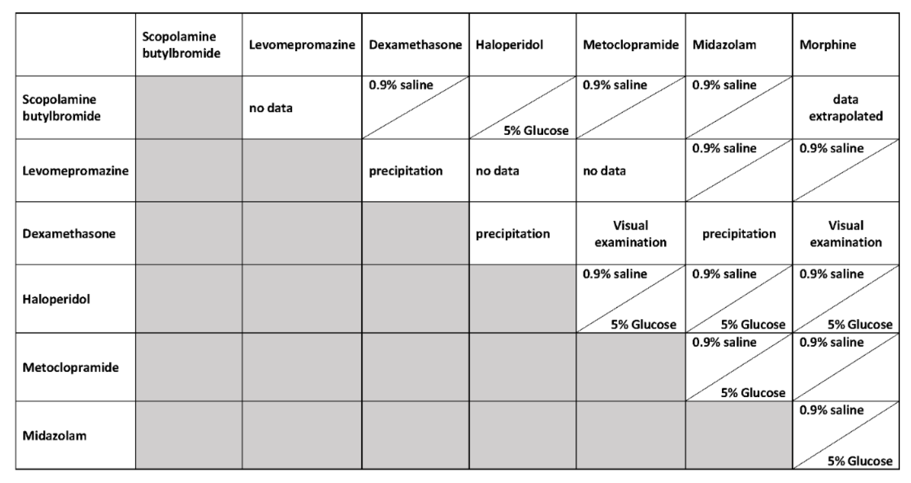
3.2. Application of the Method

In the presented study, 18 different drug-carrier combinations were tested at different storage conditions after preparation. Their stability was always assessed at the higher therapeutic range over a time period of 48 h. For verification of our study concept, binary mixtures containing DexP were tested. In general, incompatibility of DexP with Mid, Lev and Hal was confirmed, respectively (Table S3, supporting information) [4,13].

3.2.1. Binary Mixtures

According to our knowledge, no stability data are reported for combinations of Lev–Hal, Lev–Met and Lev–Sco. In general, light protection seems to be crucial for Lev stability. Without light protection, a new peak developed reproducibly in each combination (data not shown). This is in line with previous reports [21]. Most likely, this peak corresponds to the sulfoxide of levomepromazine. The combination of Lev with Hal was stable under all conditions in both carrier solutions. Final concentrations were above 90% under all conditions. In addition, Lev–Sco and Lev–Met were tested in 0.9% saline and in 5% glucose, respectively. No loss of target analyte was observed. Furthermore, Lev–Mid was tested in 5% glucose as a carrier solution. Again, no loss of analyte was detected while solution was protected from light. According to Figure 1, data for scopolamine butylbromide containing mixtures are incomplete. In 5% glucose, Sco–Met and Sco–Mid were stable over a time period of 48 h. Additionally, in 0.9% NaCl, no reduced concentration of Sco and Hal was determined (Table 2).

3.2.2. Ternary Mixture of Morphine—Midazolam—Scopolamine Butylbromide

This mixture was assessed separately in 0.9% saline at room temperature and at 0 °C. Furthermore, stability of APIs was tested in 5% glucose at room temperature. As shown in Table 3, no decrease in content of target analytes was observed.

4. Discussion

Dex and DexP were often used in palliative care for the same indication. However, they differ in their solubility in aqueous environment. Since both steroids showed the same response at the detector, we decided to use only Dex in our method and also as surrogate analyte for quantification of DexP. Since both dexamethasone species were baseline separated from all other peaks, this method is also applicable for mixtures containing dexamethasone. Under selected chromatographic conditions on ACE C-18AR, a clear separation with Rs > 1.5 was achieved. As all compounds eluted within seven minutes, we started the washing and the reequilibration of the column with an increased solvent flow at 7.1 min to finish a single chromatographic run in less than 10 min (Figure 3).
The method was validated according to international guidelines in terms of accuracy and repeatability. However, LoD and LoQ were not investigated in detail since the linear range was adapted to therapeutic used concentrations. Based on accuracy and repeatability data, LoQ was set as equal to the lowest calibrant. In Figure 2, the robustness of the method is shown. It is obvious that changes in the temperature, starting conditions and flow rate affected the performance of the method. The method showed a high accuracy for all analytes (<12%) at QC Low and QC Med level. However, as the expected results of the later measurements were rather in the concentration ranges of QC High to QC Med and not as low as QC Low, the cause for this deviation was not further investigated. The bias was accepted since accuracies at QC High level were important. As a consequence, single-point calibration at the second highest calibration level was checked against six-point calibration. It is evident that for all analytes, single-point calibration is sufficient for concentration determination in the upper concentration range. Furthermore, stability of calibration is provided for at least four weeks.
Results on DexP stability were used as verification of our stability studies. Similar results were obtained in comparison to the current literature (Figure 1). Therefore, the developed protocol and chromatographic method is suitable for conducting stability studies in our laboratories. In general, no significant decline could be identified for all tested binary and ternary API mixtures. Until now, no data were available on the combination of Lev with either Hal, Sco or Met. These combinations were tested in 0.9% saline and in 5% glucose. Without light protection, a new and time-dependent increasing peak was observed. Most likely, it was the sulfoxide of Lev, as this API is prone to oxidation when exposed to light. However, concentration of Lev was always above 90% after 48 h. Oxidation was avoided by light protection, since in 5% glucose with light protection, an additional peak for the oxidation product was not observed. For the combination of DexP with Sco, only data on the stability in 0.9% saline, but not in 5% glucose were available [13]. Therefore, this combination was tested using 5% glucose as a carrier solution. This mixture was stable since the concentration of both analytes was above 90% of the nominal values. Since information on binary mixtures of Sco is incomplete, combination with Hal in 0.9% NaCl and combination with Met and Mid in 5% glucose were investigated in detail. Each combination showed a high stability in saline and glucose solution, respectively. Therefor these API-excipient combinations are suitable for further clinical use.

5. Limitations

This study shows some limitations. Although several combinations and conditions were investigated in detail, only a limited number of independently prepared samples were analyzed (n = 3). However, the full-validated chromatographic procedure provides a reliable basis for further investigations.

6. Conclusions

In the present study, we reported the successful development and full validation of a multi-analyte UHPLC-DAD method to determine the stability of seven commonly used drugs in parenteral solutions in palliative and critical care. The fast and accurate UHPLC method allows further in-depth studies of multi analyte mixtures—e.g., other concentrations of API or carrier. The developed method was applied to 15 different binary mixtures and one ternary mixture to assess the stability of selected analytes in parenteral solutions over 48 h. Missing data on carrier compatibility could be completed and limitations on the choice of these carrier solutions may be eliminated. The results of this study support clinical pharmacists and healthcare practitioners in decision-making for selecting drug combinations for CSCI administration.

Supplementary Materials

The following are available online at https://0-www-mdpi-com.brum.beds.ac.uk/2673-4532/1/1/5/s1, Figure S1: Stability of calibration was assessed over 28 days; freshly prepared QC samples were calculated against calibration curve of day 1; Figure S2: Stability data of QC med under different storage conditions; Table S1: Overview of applied gradients; Table S2: Overview of calibrator, QC and test concentrations. All concentration are given in mg/mL; Table S3: Stability data of DexP in various combinations.

Author Contributions

Conceptualization, C.S. and U.M.; methodology, C.S. and F.H.; software, F.H.; validation, C.S. and F.H.; formal analysis, C.S., U.M. and P.W.; investigation, F.H. and C.S.; resources, C.S. and P.W.; data curation, C.S., U.M., F.H., P.W.; writing—original draft preparation, C.S.; writing—review and editing, C.S.; visualization, C.S. and F.H.; supervision, C.S.; project administration, C.S. All authors have read and agreed to the published version of the manuscript.

Funding

This research received no external funding.

Acknowledgments

We thank Ina Schmidt for valuable analytical advice and fruitful discussions.

Conflicts of Interest

The authors declare no conflict of interest.

References

  1. Guter, A. Standard zur subkutanen Applikation von Parenteralia in der Palliativmedizin. Krankenhauspharmazie 2011, 32, 484–489. [Google Scholar]
  2. Thomas, T.; Barclay, S. Continuous subcutaneous infusion in palliative care: A review of current practice. Int. J. Palliat. Nurs. 2015, 21, 62–64. [Google Scholar] [CrossRef] [PubMed]
  3. Jain, S.; Mansfield, B.; Wilcox, M.H. Subcutaneous fluid administration—Better than the intravenous approach? J. Hosp. Infect. 1999, 41, 269–272. [Google Scholar] [CrossRef] [PubMed]
  4. Dickman, A.; Schneider, J. The Syringe Driver—Continuous Subcutaneous Infusions in Palliative Care, 4th ed.; Oxford University Press: Oxford, UK, 2016. [Google Scholar]
  5. Frühwald, T. Ist die subkutane Infusion eine praktische Alternative in der Geriatrie? J. Für Ernährungsmedizin 2001, 3, 16–18. [Google Scholar]
  6. Baker, J.; Dickman, A.; Mason, S.; Ellershaw, J. The current evidence base for the feasibility of 48-hour continuous subcutaneous infusions (CSCIs): A systematically-structured review. PLoS ONE 2018, 13, e0194236. [Google Scholar] [CrossRef] [PubMed] [Green Version]
  7. Bausewein, C.; Rémi, C. Besonderheiten der Pharmakotherapie in der Palliative Care, Bigorio. 2012. Available online: https://www.palliative.ch/fileadmin/user_upload/palliative/fachwelt/E_Standards/Besonderheiten_der_Pharmakotherapie_in_der_Palliative_Care.pdf (accessed on 5 August 2020).
  8. Masman, A.D.; van Dijk, M.; Tibboel, D.; Baar, F.P.; Mathot, R.A. Medication use during end-of-life care in a palliative care centre. Int. J. Clin. Pharm. 2015, 37, 767–775. [Google Scholar] [CrossRef] [Green Version]
  9. Sutherland, A.E.; Curtin, J.; Bradley, V.; Bush, O.; Presswood, M.; Hedges, V.; Naessens, K. Subcutaneous levetiracetam for the management of seizures at the end of life. BMJ Support Palliat Care 2018, 8, 129–135. [Google Scholar] [CrossRef]
  10. Dickman, A.; Bickerstaff, M.; Jackson, R.; Schneider, J.; Mason, S.; Ellershaw, J. Identification of drug combinations administered by continuous subcutaneous infusion that require analysis for compatibility and stability. BMC Palliat. Care 2017, 16, 22. [Google Scholar] [CrossRef] [Green Version]
  11. Aeberhard, C.; Steuer, C.; Saxer, C.; Huber, A.; Stanga, Z.; Muhlebach, S. Physicochemical stability and compatibility testing of levetiracetam in all-in-one parenteral nutrition admixtures in daily practice. Eur. J. Pharm. Sci. 2017, 96, 449–455. [Google Scholar] [CrossRef]
  12. Gomez, M.A.; Arenas, V.J.; Sanjuan, M.M.; Hernandez, M.J.; Almenar, C.B.; Torres, V.J. Stability studies of binary mixtures of haloperidol and/or midazolam with other drugs for parenteral administration. J. Palliat. Med. 2007, 10, 1306–1311. [Google Scholar] [CrossRef]
  13. IBM. IBM Micromedex (Trissel online); IBM: Armonk, NY, USA, 2018. [Google Scholar]
  14. Scholes, S.; William, L.; Halliwell, D.; Sherwen, P.; Whelan, T.; Clifton, C. Syringe Driver Drug Compatibilities—Guide to Palliative Care Practice. East. Metrop. Reg. Palliat. Care Consort. 2016. Available online: https://www.emrpcc.org.au/uploads/136/Syringe-Driver-Drug-Compatibilities-May-2016.pdf (accessed on 5 August 2020).
  15. Rossignol, E.; Sorrieul, J.; Beaussart, H.; Kieffer, H.; Folliard, C.; Dupoiron, D.; Devys, C. Validation Study of UPLC Method for Determination of Morphine, Ropivacaïne and Ziconotide in Combination for Intrathecal Analgesia. J. Anal. Bioanal. Tech. 2016, 7, 1000310. [Google Scholar]
  16. Destro, M.; Ottolini, L.; Vicentini, L.; Boschetti, S. Physical compatibility of binary and ternary mixtures of morphine and methadone with other drugs for parenteral administration in palliative care. Support Care Cancer 2012, 20, 2501–2509. [Google Scholar] [CrossRef] [PubMed]
  17. Negro, S.; Reyes, R.; Azuara, M.L.; Sanchez, Y.; Barcia, E. Morphine, haloperidol and hyoscine N-butyl bromide combined in s.c. infusion solutions: Compatibility and stability. Evaluation in terminal oncology patients. Int. J. Pharm. 2006, 307, 278–284. [Google Scholar] [CrossRef]
  18. Good, P.D.; Schneider, J.J.; Ravenscroft, P.J. The compatibility and stability of midazolam and dexamethasone in infusion solutions. J. Pain Symptom Manag. 2004, 27, 471–475. [Google Scholar] [CrossRef]
  19. Negro, S.; Azuara, M.L.; Sanchez, Y.; Reyes, R.; Barcia, E. Physical compatibility and in vivo evaluation of drug mixtures for subcutaneous infusion to cancer patients in palliative care. Support Care Cancer 2002, 10, 65–70. [Google Scholar] [CrossRef]
  20. Wong, H.A.; Law, S.; Walker, S.E.; Bowles, S.K. Concentration-Dependent Compatibility and Stability of Dexamethasone and Midazolam. Can. J. Hosp. Pharm. 2000, 53, 24–31. [Google Scholar]
  21. Barcia, E.; Reyes, R.; Azuara, M.L.; Sanchez, Y.; Negro, S. Stability and compatibility of binary mixtures of morphine hydrochloride with hyoscine-n-butyl bromide. Support Care Cancer 2005, 13, 239–245. [Google Scholar] [CrossRef]
  22. Al-Tannak, N.F.; Cable, C.G.; McArthur, D.A.; Watson, D.G. A stability indicating assay for a combination of morphine sulphate with levomepromazine hydrochloride used in palliative care. J. Clin. Pharm. Ther. 2012, 37, 71–73. [Google Scholar] [CrossRef]
  23. Schrijvers, D.; Tai-Apin, C.; De Smet, M.C.; Cornil, P.; Vermorken, J.B.; Bruyneel, P. Determination of compatibility and stability of drugs used in palliative care. J Clin Pharm Ther. 1998, 23, 311–314. [Google Scholar] [CrossRef]
  24. Peters, F.T.; Hartung, M.; Herbold, M.; Schmitt, G.; Daldrup, T.; Mußhoff, F. APPENDIX B Requirements for the validation of analytical methods. Toxichem. Krimtech. 2009, 76, 185–208. [Google Scholar]
  25. Oliva, A.; Monzon, C.; Santovena, A.; Farina, J.B.; Llabres, M. Development of an ultra high performance liquid chromatography method for determining triamcinolone acetonide in hydrogels using the design of experiments/design space strategy in combination with process capability index. J. Sep. Sci. 2016, 39, 2689–2701. [Google Scholar] [CrossRef] [PubMed]
  26. Leardi, R. Experimental design in chemistry: A tutorial. Anal. Chim. Acta. 2009, 652, 161–172. [Google Scholar] [CrossRef] [PubMed]
  27. Binz, T.M.; Braun, U.; Baumgartner, M.R.; Kraemer, T. Development of an LC-MS/MS method for the determination of endogenous cortisol in hair using (13)C3-labeled cortisol as surrogate analyte. J. Chromatogr. B Anal. Technol. Biomed. Life Sci. 2016, 1033–1034, 65–72. [Google Scholar] [CrossRef] [PubMed]
  28. Van de Merbel, N.C. Quantitative determination of endogenous compounds in biological samples using chromatographic techniques. Trac-Trend Anal. Chem. 2008, 27, 924–933. [Google Scholar] [CrossRef]
  29. Steuer, C.; Schuetz, P.; Bernasconi, L.; Huber, A.R. Quantification of Quaternary Ammonium Compounds Against Surrogate Matrix by HILIC-MS/MS. CHIMIA Int. J. Chem. 2017, 71, 601. [Google Scholar] [CrossRef] [Green Version]
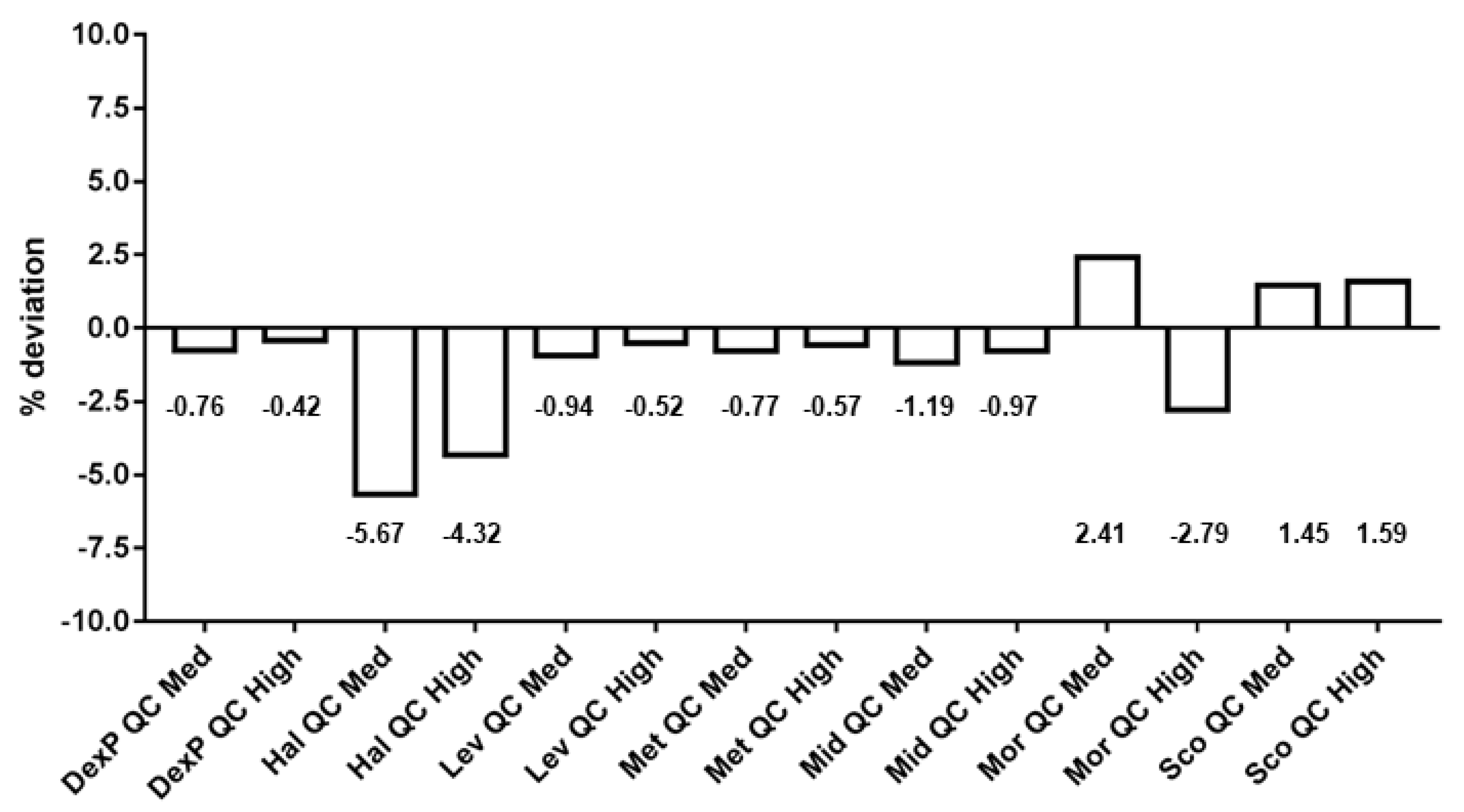
Figure 1. Available data on compatibilities of pharmaceuticals used in palliative care for parenteral/subcutaneous (s.c.) applications [4,7,13].
Analytica 01 00005 g001
Figure 2. The influence of chromatographic parameters on the resolution of critical analyte pairs applied to different columns (ac). The dotted line at R = 1.5 represents baseline separation.
Analytica 01 00005 g002
Figure 3. Example chromatogram on the ACE® C18-AR detected at a wavelength of λ = 254 nm. All the peaks were baseline separated.
Analytica 01 00005 g003
Figure 4. Comparison of single-point calibration versus six-point calibration controlled by QC Med und QC High of each analyte.
Analytica 01 00005 g004
Table 1. Method validation data; relative standard deviation (RSD)R intraday repeatability; RSDT interday repeatability.
Range mg/mLCal. ModelQC HighQC MedQC Low
Bias [%]RSDR [%]RSDT [%]Bias [%]RSDR [%]RSDT [%]Bias [%]RSDR [%]RSDT [%]
Dex0.04–0.411/x2−8.93.33.8−11.21.93.5−12.43.56.2
Hal0.05–0.521/x20.73.03.0−1.41.43.05.53.94.8
Lev0.32–3.241/x2−7.53.43.4−9.82.13.0−8.92.34.7
Met0.33–3.301/x2−0.63.43.4−3.02.02.9−2.02.54.4
Mid0.12–1.21/x22.23.23.1−0.61.83.10.42.64.5
Mor0.52–5.181/x2−4.32.62.44.42.02.4−6.53.36.1
Sco0.73–7.321/x25.13.13.53.32.13.31.42.85.0
Table 2. Stability data of different binary mixtures under selected carrier and storage conditions.
MetLevHalLevScoLevScoMorScoHal
Time [h]Mean [%]SDMean [%]SDMean [%]SDMean [%]SDMean [%]SDMean [%]SDMean [%]SDMean [%]SDMean [%]SDMean [%]SD
0.9% NaCl RT0100.02.9100.02.4100.01.6100.01.9100.05.8100.02.8100.00.5100.02.699.81.2100.01.3
493.72.393.01.894.71.394.61.995.10.295.30.795.92.192.62.398.80.299.30.4
895.71.194.90.993.91.494.40.992.80.292.60.296.03.292.23.196.84.696.53.1
2497.00.496.20.495.80.895.80.492.33.892.23.299.10.995.10.999.60.399.20.4
4897.10.595.10.395.00.894.70.997.33.597.41.399.40.194.10.3100.00.198.80.6
MetLevHalLevScoLevScoMorScoHal
Time [h]Mean [%]SDMean [%]SDMean [%]SDMean [%]SDMean [%]SDMean [%]SDMean [%]SDMean [%]SDMean [%]SDMean [%]SD
0.9% NaCl 0 °C0100.03.1100.03.0100.03.7100.03.499.13.299.33.399.92.198.62.0100.01.5100.01.7
494.70.394.80.496.61.896.31.495.95.097.95.098.71.096.70.897.41.098.32.0
895.21.094.51.297.71.397.60.8100.00.9100.00.897.70.496.00.699.00.8100.01.3
2495.91.794.81.793.62.494.31.898.22.997.62.899.00.499.30.896.90.296.00.7
4895.20.695.00.791.05.191.95.296.62.296.22.0100.00.3100.00.599.62.098.62.3
MidLevScoMidScoMetScoMorScoDexP
Time [h]Mean [%]SDMean [%]SDMean [%]SDMean [%]SDMean [%]SDMean [%]SDMean [%]SDMean [%]SDMean [%]SDMean [%]SD
5% Glucose RT0100.02.2100.02.4100.01.4100.01.1100.02.899.53.2100.00.3100.00.3100.01.6100.02.7
495.50.395.90.197.70.297.60.398.60.998.80.696.70.296.10.499.50.297.40.4
896.21.397.01.197.10.396.00.698.60.999.41.097.70.496.60.197.60.794.90.7
2495.60.096.10.198.40.497.70.399.70.7100.00.298.50.297.40.396.60.994.40.9
4895.10.296.90.599.80.198.00.398.60.699.50.499.20.399.40.597.90.795.70.5
Table 3. Stability data of ternary mixture under selected carrier and storage conditions.
MorMidSco
Time [h]Mean [%]SDMean [%]SDMean [%]SD
0.9% NaCl RT0100.00.7100.00.7100.00.5
496.70.198.70.497.60.4
893.12.795.43.294.42.9
2494.33.996.73.995.73.9
4895.22.998.62.796.42.7
MorMidSco
Time [h]Mean [%]SDMean [%]SDMean [%]SD
0.9% NaCl 0 °C0100.02.2100.02.2100.01.9
495.72.696.12.495.52.5
898.01.298.21.098.01.1
2499.90.499.70.998.90.2
4896.64.497.04.496.64.0
MorMidSco
Time [h]Mean [%]SDMean [%]SDMean [%]SD
5% Glucose RT0100.01.0100.01.1100.01.9
497.60.697.70.297.00.6
898.00.498.10.397.90.8
2497.80.699.00.298.70.4
4898.30.899.20.498.10.4

