Article

Myostatin and Related Factors Are Involved in Skeletal Muscle Protein Breakdown in Growing Broilers Exposed to Constant Heat Stress

State Key Laboratory of Animal Nutrition, Institute of Animal Sciences, Chinese Academy of Agricultural Sciences, Beijing 100193, China
*
Author to whom correspondence should be addressed.
Submission received: 26 March 2021 / Revised: 11 May 2021 / Accepted: 12 May 2021 / Published: 20 May 2021
(This article belongs to the Special Issue Recent Advances in Poultry Nutrition and Production)

Abstract

:

Simple Summary

Our results showed that constant heat stress could affect the expression of myostatin and related factors involved in skeletal muscle protein breakdown in growing broilers, resulting in a decrease in muscle protein deposition. These findings suggest a new strategy for regulating muscle protein breakdown in growing broilers, which could benefit the modern broiler industry in combating constant heat stress.

Abstract

Heat stress has an adverse effect on the development of poultry farming, which has always aroused great concern. This study was carried out to investigate the protein breakdown mechanism responsible for the suppressive effect of constant heat stress on muscle growth in growing broilers. A total of 96, 29-day-old, Arbor Acres male broilers were randomly divided into two groups, a thermoneutral control (21 ± 1 °C, TC) and a heat stress (31 ± 1 °C, HS) group, with six replicates in each group and eight birds in each replicate. The trial period lasted for 14 d, and the trial was performed at 60 ± 7% relative humidity, a wind speed of <0.5 m/s and an ammonia level of <5 ppm. The results showed that the average daily feed intake and average daily gain in the HS group were distinctly lower than those in the TC group (p < 0.05), whereas the HS group showed a significantly increased feed conversion ratio, nitrogen excretion per weight gain and nitrogen excretion per feed intake compared to the TC group (p < 0.05). In addition, the HS group showed a significantly reduced breast muscle yield and nitrogen utilization in the broilers (p < 0.05). The HS group showed an increase in the serum corticosterone level (p < 0.05) and a decrease in the thyroxine levels in the broiler chickens (p < 0.05) compared to the TC group, whereas the HS group showed no significant changes in the serum 3,5,3′-triiodothyronine levels compared to the TC group (p > 0.05). Moreover, the HS group showed increased mRNA expression levels of myostatin, Smad3, forkhead box O 4, muscle atrophy F-box and muscle ring-finger 1, but reduced mRNA expression levels of the mammalian target of rapamycin, the protein kinase B and the myogenic determination factor 1 (p < 0.05). In conclusion, the poor growth performance of birds under constant heat stress may be due to an increased protein breakdown via an mRNA expression of myostatin and related factors.

1. Introduction

Heat stress (HS) is a recognized long-term problem in the poultry farming industry, and its effect on poultry production performance is a common challenge in tropical and subtropical climatic conditions and even during the summer in temperate regions [1]. It is well known that HS can decrease the production of birds [2,3,4,5]. According to the United States Department of Agriculture’s statistics, global chicken consumption reached 101.486 million tons in 2018. Poultry meat has seen a rise in its global demand because of its relatively high nutritional value, its low price and the lack of cultural or religious obstacles for its consumption [6]. Almost 50% of a broiler’s total body mass is skeletal muscle, which exhibits a strong metabolic activity as the largest protein source in the body [7]. Heat stress has a significant effect on the growth of breast muscle, which can greatly reduce the breast muscle yield [8,9,10,11,12] and result in a substantial economic loss.
The essence of muscle growth is the accumulation of protein, and the balance between the rates of protein synthesis and protein breakdown influences the muscle mass [13]. At present, studies on protein synthesis are mainly focused on the insulin-like growth factor-1 (IGF-1)/protein kinase B (Akt) pathway in mammals and birds [14,15,16]. One of the principal protein degradation systems in skeletal muscle is the ubiquitin–proteasome pathway [17]. Forkhead box O (FoxO) transcription factors play an important role in muscle wastage through the regulation of ubiquitin E3 ligases, muscle atrophy F-box (MAFbx) and muscle ring-finger 1 (MuRF1) [14].
Myostatin, a transforming growth factor-beta family member, is a potent negative regulator of skeletal muscle growth [18]. Many previous in vitro studies have revealed that myostatin affects mammalian muscle growth. For example, blocking the myostatin activity in mice has applications in the promotion of muscle growth [19], and myostatin can inhibit the Akt activation in human skeletal muscle cells [20]. In addition, McFarlane et al. (2006) reported that in cultured C2C12 muscle cells, myostatin treatment blocked the IGF-1/phosphatidylinositol 3-kinase/Akt pathway and activated FoxO1, leading to an increased expression of MAFbx and MuRF-1. Smad3 transcription factors are found downstream of myostatin type II receptors and can be activated by the interaction of myostatin with its receptors [21].
In birds, a recent study revealed that myostatin significantly increased the phosphorylation rate of Smad2 and the mRNA levels of MAFbx in a chick’s embryonic myotubes cultured at 37 °C for 2 h in vitro [22]. Several previous studies of broilers have shown that HS affected muscle growth via the expression of several genes. Acute (24 h) HS decreased the expression of the IGF-1 and phosphatidylinositol 3-kinase R1 genes in the liver and increased the cathepsin L2 and MAFbx gene expression in male broilers (Cobb 500) [23]. Chronic HS decreased the muscle protein synthesis in Arbor Acres male broilers by downregulating the IGF-mammalian target of rapamycin (mTOR) signaling pathway [16]. Furukawa et al. (2016) [24] reported that short-term (0, 0.5 and 3 d) HS treatment induced a superoxide production in the muscle mitochondrial of Ross male broilers as well as a MAFbx gene expression and affected the signaling pathway governing the FoxO3 activity and expression. Zuo et al. (2015) [1] reported that constant HS reduced the skeletal muscle protein deposition in broilers by decreasing the IGF-1, phosphatidylinositol 3-kinase and p70S6 kinase expression and increasing the MuRF1 and MAFbx expression. However, whether the myostatin and related factors involved in skeletal muscle protein breakdown in growing broilers are affected by constant HS remains unclear. Therefore, the present study aimed to investigate the effects of constant HS on the growth performance, breast muscle yield, nitrogen utilization and mRNA expression of myostatin and related factors in growing broilers.

2. Materials and Methods

2.1. Birds and Treatments

One-day-old male Arbor Acres broilers were kept in one-tier cages and were maintained with administrative procedures and a standard corn–soybean-meal diet consistent with the Nutrient Requirements of Poultry (1994) for Arbor Acres broilers. At 29 d old, a total of 96 healthy Arbor Acres male chicks with similar BWs (1000 ± 70 g) were selected and randomly divided into two groups, the thermoneutral control (TC) and the HS group, which were raised in two environmentally controlled chambers. There were 6 cages (one-tier, 0.80 m × 0.80 m × 0.40 m) with 8 birds per cage in each environmentally controlled chamber (each cage served as a replicate). From the age of 29 to 42 d, the birds in the TC group were reared at a constant temperature of 21 ± 1 °C, whereas those in the HS group were reared at a constant temperature of 31 ± 1 °C. The two chambers were maintained at 60 ± 7% RH with a wind velocity of <0.5 m/s, an ammonia level of <5 ppm and a 24-h light. All broilers had ad libitum access to feed and water.

2.2. Sampling Collection and Chemical Analysis

Growth performance and breast muscle yield. At 42 d of age, 12 broilers from each group (two sampled birds per replicate) were randomly selected and euthanized by cervical dislocation. Their feed intake was recorded daily to calculate the average daily feed intake (ADFI). Their average daily gain (ADG) was calculated as the difference between the values of the body weight of all the birds at the beginning (29 d) and the end (42 d) of the experiment. The feed conversion ratio (FCR) was calculated as the ratio of the ADG to the ADFI. The breast muscle yield was expressed as the ratio of the breast muscle mass to the eviscerated carcass weight.

2.2.1. Nitrogen Utilization and Nitrogen Excretion

The randomly selected feed from the two groups was mixed, reduced to 200 g using the quartering method and crushed. Then, the feed was placed in a sealed bag prior to being tested to detect its nitrogen intake. The excreta of all the birds in each cage was collected (each cage was a replicate) on the 14th day of the experiment (every 4 h for a total of 6 times) to detect nitrogen in the excreta. Sundries in the excreta were removed, and the excreta was weighed and then nitrogen-fixed with 10 mL of 10% H2SO4 per 100 g excreta sample. Finally, the excreta collected from one cage was mixed and 200 g of the excreta was selected using the quartering method, dried at 65–70 °C until it reached a constant weight, collected and crushed. The content of nitrogen in the feed and the excreta was determined using the Kjeldahl method [25]. The following equations were used for the calculation:
Nitrogen utilization (%) = [(nitrogen intake-nitrogen excretion)/nitrogen intake] × 100
Nitrogen excretion per weight gain (%) = (nitrogen excretion/daily gain) × 100
Nitrogen excretion per feed intake (%) = (nitrogen excretion/daily feed intake) × 100

2.2.2. Blood Measurements

Serum samples were obtained by blood centrifugation at 3000× g for 20 min at 4 °C, and then stored at −20 °C until the analysis took place. The serum corticosterone, 3,5,3′-triiodothyronine (T3) and thyroxine (T4) levels were measured by radioimmunoassay using a gamma radioimmunoassay counter (GC-2010, Anhui Ustczonkia Scientific Instruments Co., Ltd., Anhui, China). All procedures were conducted by following the manufacturer’s instructions.

2.2.3. Regulatory Factors Gene Expression

Breast muscle samples were collected and stored at −80 °C for further analysis. Total RNA was isolated from each breast muscle sample using a TRIzol reagent (Invitrogen, Carlsbad, CA, USA) as described in the manufacturer’s instructions. A real-time quantitative PCR was carried out using a LightCycler 96 system (LightCycler 96 system, Roche, Basel, Switzerland) according to a common real-time quantitative PCR method. The mRNA levels of myostatin, Smad3, MAFbx, MuRF1, mTOR, FoxO4, myogenic determination factor 1 (MyoD) and Akt in the breast muscle were examined. The primers for the target genes were designed and confirmed based on the sequences described in GenBank, which are listed in Table 1. The β-actin gene was used as an internal control for normalization. The mRNA expression data were analyzed using the 2−ΔΔCt method [26].

2.3. Statistical Analysis

The data from the present study were analyzed using a one-way ANOVA by SAS 9.2 (SAS Institute Inc., Cary, NC, USA). The ADG, ADFI, FCR, nitrogen utilization, nitrogen excretion per weight gain and nitrogen excretion per feed intake were analyzed using the cage as the experimental unit, and other indexes were analyzed by determining the mean of two sampled birds per replicate as the experimental unit (n = 6). The data are expressed as the means ± SD. Statistical significance was indicated at p < 0.05.

3. Results

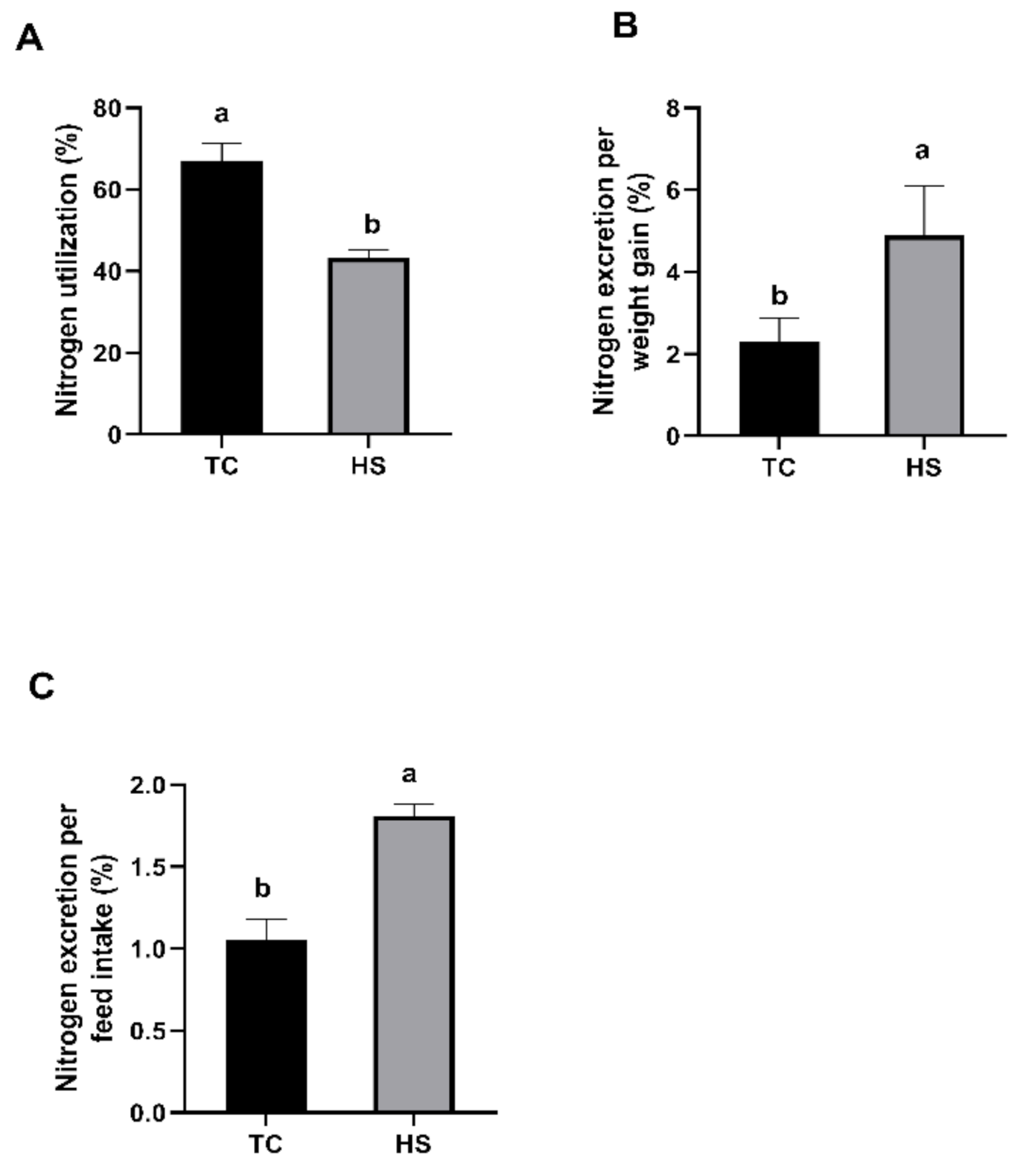
3.1. Growth Performance, Breast Muscle Yield, Nitrogen Utilization and Nitrogen Excretion

During the trial period, none of the birds suffered from clinical diseases, and there was no mortality. As shown in Table 2, the ADFI and ADG in the HS group were significantly lower than those in the TC group (p < 0.05), and the HS group had a significantly increased FCR compared to the TC group (p < 0.05). As shown in Figure 1A, the nitrogen utilization in the HS group was significantly lower than that in the TC group (p < 0.05), whereas the nitrogen excretion per weight gain (Figure 1B) and the nitrogen excretion per feed intake (Figure 1C) in the HS group were significantly higher than those in the TC group (p < 0.05). As shown in Figure 2, compared to those in the TC group, the broilers in the HS group showed a significantly reduced breast muscle yield (p < 0.05).

3.2. Blood Biochemical Indexes

We tested the effects of HS on the blood biochemical indexes of broilers, and the results of the serum corticosterone, T4 and T3 levels in broilers are presented in Figure 3. As shown in Figure 3A, the serum corticosterone levels in the broilers in the HS group were significantly higher (p < 0.05) than the serum corticosterone levels in the broilers in the TC group, whereas the levels of T4 in the serum from the broiler chickens in the HS group were significantly less than the serum T4 levels in the TC group (p < 0.05, Figure 3B). No significant differences in the levels of T3 were observed between the TC and HS groups (p > 0.05, Figure 3C).

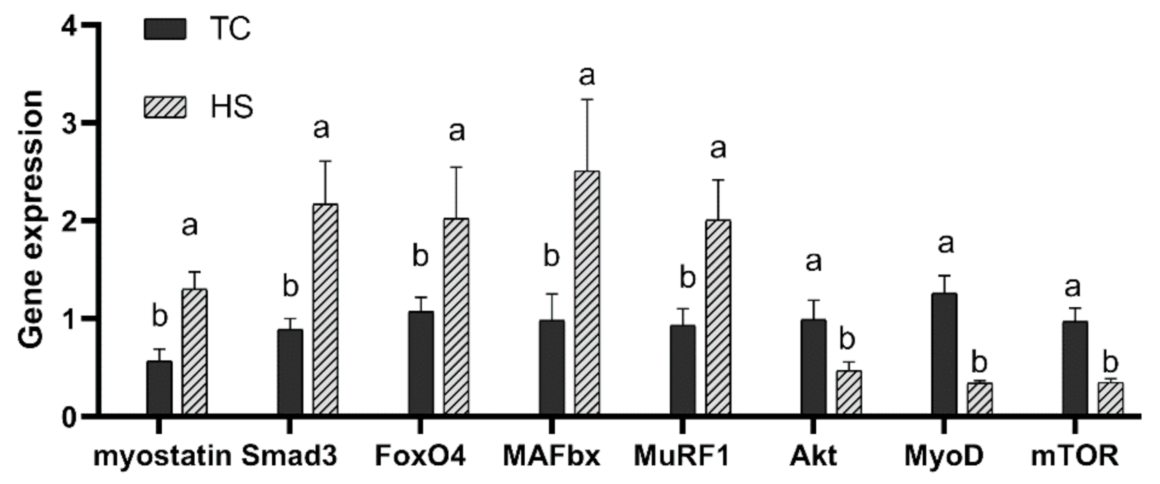
3.3. Regulatory Factors Gene Expression

The gene expression of breast muscle growth-related regulatory factors in broilers exposed to constant HS was determined, and the results of the myostatin, Smad3, FoxO4, MAFbx, MuRF1, Akt, MyoD and mTOR mRNA expression levels are presented in Figure 4. As shown in Figure 4, the HS group showed significantly increased mRNA expression levels of myostatin compared to the TC group (p < 0.05), and the HS group showed significantly increased mRNA expression levels of Smad3, FoxO4, MAFbx and MuRF1 as well, whereas the HS group showed significantly reduced mRNA expression levels of Akt, MyoD and mTOR (p < 0.05).

4. Discussion

Previous studies indicated that HS could affect productivity, reduce the body weight and feed intake, and increase the FCR expression of broilers [2,9,27,28]. The data in the present study revealed that the HS group showed significantly reduced ADFI and ADG values and increased FCR values compared to the TC group. These results were consistent with the findings of previous studies. HS significantly decreased nitrogen utilization and increased the nitrogen excretion per weight gain and nitrogen excretion per feed intake in this study, which revealed that HS reduced protein utilization. Kumar et al. (2017) [29] reported that a reduced nitrogen excretion is a result of an increased digestibility of protein and an increased deposition of protein in broilers. A significant increase was observed in the nitrogen excretion per weight gain and the nitrogen excretion per feed intake, showing that HS directly led to muscle protein degradation. The breast muscle is a main part of the total body, and HS directly impairs broiler production. It has been reported that HS decreased the proportion of breast muscle [10,30,31]; as expected, our results were the same as those of previous studies and indicated that HS significantly reduced the breast muscle yield of broilers. Muscle growth is the result of a rate of protein synthesis greater than the rate of protein breakdown; in heat-stressed broilers, both protein synthesis and breakdown are affected by heat exposure [32]. Based on the decreases in the ADFI, ADG, breast muscle yield and nitrogen utilization and the increases in the FCR, nitrogen excretion per weight gain and nitrogen excretion per feed intake, HS reduced protein deposition and promoted protein breakdown in the broilers. Lin et al. (2004) [33] pointed out that the reduced growth rate was related to the proteolysis that was induced by corticosterone, which could cause a reduction in animal body weight gain. T3 and T4 are the major hormones that are required to support normal growth and are known to impact almost every physiological process in chickens [34]. Studies have shown that HS reduced the plasma concentrations of T4 and T3 [35]; based on the present results, HS significantly increased serum corticosterone levels but reduced the levels of T4, as observed in previous studies [36,37,38]. HS has no effect on serum T3 levels, probably because the effect of HS on thyroid hormones is determined by many factors [39]. These endocrinological changes, which are consistent with the changes in growth performance caused by HS, could accelerate protein hydrolysis in vivo.
Many studies have reported that a high ambient temperature decreases muscle protein content [10,16,40], and Yunianto et al. (1997) [8] demonstrated that HS decreases muscle protein synthesis and accelerates protein breakdown. In the past few years, the IGF-1-phosphatidylinositol 3-kinase-mTOR signaling pathway, which is responsible for regulating the protein synthesis pathways, has been defined and studied [15,41]. Ma et al. (2018) [16] reported that chronic heat stress decreased muscle protein synthesis by downregulating the IGF–mTOR signaling pathway. Akt can phosphorylate a series of protein substrates to activate its downstream mTOR channels once it is activated [1]. The mammalian target of rapamycin can mediate protein synthesis through its downstream targets, and the mTOR pathway is known as a key signaling pathway that regulates the muscular hypertrophy process in vivo [42]. It has been reported that a member of the transforming growth factor-beta superfamily, myostatin, has a dramatically negative effect on muscle growth by binding to Akt in order to elicit its biological effects [18]. Forbes et al. (2006) [43] indicated that the R-Smads, Smad2 and Smad3, could be activated by myostatin to transduce signaling, which could cause the formation of complexes in the nucleus to regulate the expression of target genes through interactions with transcription coactivators or repressors. In normal chicken myotubes cultured in vitro, Smad controlled the myostatin expression [22]. The present study showed an increase in Smad3 mRNA levels in growing broilers under constant heat stress, indicating that there is a relationship between Smad3 and myostatin expression changes under constant HS, but the specific molecular changes remain to be further studied. Intracellular protein degradation occurs mainly through the ubiquitin–protein enzyme (proteasome) process, which is associated with FoxO transcription factors and two muscle-specific ubiquitin ligases (E3s), MAFbx and MuRF1 [1,44]. The FoxO transcription factors play an important role in muscle atrophy. Sandri et al. (2004) [14] showed that the decreased activity of the Akt signaling pathway seemed to lead to an increase in the hypophosphorylated active forms of the FoxO transcription factors, and the FoxO transcription factors resulted in skeletal muscle atrophy by regulating atrophy-related genes, including MAFbx. In the current study, the HS group showed increased mRNA expression levels of myostatin, FoxO4, MAFbx and MuRF1, and reduced mRNA expression levels of mTOR and Akt. These data implied that HS may decrease broiler breast muscle growth by increasing the activity of myostatin and related factors, thus promoting muscle protein degradation. The myogenic determination factor 1 is involved in myoblast differentiation and is required for fast fiber formation [45,46]. Moreover, it has been reported that myostatin negatively regulates MyoD expression in muscle [47]. In the present study, we found that the mRNA expression levels of MyoD in breast muscle were reduced in the HS group, which indicated that HS affected breast muscle differentiation and hypertrophy, and thereby inhibited muscle growth.

5. Conclusions

The change in myostatin mRNA expression in growing broilers exposed to constant HS was preliminarily studied for the first time in this experiment. Therefore, we speculated that in growing broilers under high temperature conditions, protein deposition may be affected not only by the IGF-1-Akt signaling pathway, but also by myostatin and related factors involved in skeletal muscle protein breakdown. Moreover, further studies are required to clarify the molecular mechanism of myostatin and related factors in growing broilers under constant HS.

Author Contributions

Conceptualization, M.Z., X.L. and J.F.; methodology, X.L. and Y.Z.; software, X.L.; validation, M.Z.; formal analysis, X.L.; investigation, X.L., J.F. and Y.Z.; resources, M.Z.; data curation, X.L.; writing—original draft preparation, X.L.; writing—review and editing, X.L., M.Z., J.F. and Y.Z.; funding acquisition, M.Z. All authors have read and agreed to the published version of the manuscript.

Funding

This research was supported by the National Key Research and Development Program of China (2016YFD0500509). This work was also supported by the Independent Research Program of State Key Laboratory of Animal Nutrition (2004DA125184G1609) and the Science and Technology Innovation Project of the Chinese Academy of Agricultural Sciences (ASTIP-IAS09).

Institutional Review Board Statement

The study was conducted according to the guidelines of the Declaration of Helsinki and approved by the Animal Experimental Welfare and Ethical Inspection Committee of Institute of Animal Science, Chinese Academy of Agricultural Sciences (permit number: IAS2019-43).

Data Availability Statement

Not Applicable.

Conflicts of Interest

The authors declare no conflict of interest.

References

  1. Zuo, J.; Xu, M.; Abdullahi, Y.A.; Ma, L.; Zhang, Z.; Feng, D. Constant heat stress reduces skeletal muscle protein deposition in broilers. J. Sci. Food Agric. 2015, 95, 429–436. [Google Scholar] [CrossRef] [PubMed]
  2. Sahin, K.; Orhan, C.; Tuzcu, M.; Sahin, N.; Hayirli, A.; Bilgili, S.; Kucuk, O. Lycopene activates antioxidant enzymes and nuclear transcription factor systems in heat-stressed broilers. Poult. Sci. 2016, 95, 1088–1095. [Google Scholar] [CrossRef] [PubMed]
  3. Zhai, W.; Corzo, A.; Mejia, L.; Zumwalt, C.D.; Peebles, E.D. Effects of dietary amino acid density and metabolizable energy level on the growth and meat yield of summer-reared broilers. J. Appl. Poult. Res. 2014, 23, 501–515. [Google Scholar] [CrossRef]
  4. Awad, A.E.; Idrus, Z.; Farjam, A.S.; Bello, A. Growth performance, duodenal morphology and the caecal microbial population in female broiler chickens fed glycine-fortified low protein diets under heat stress conditions. Br. Poult. Sci. 2018, 59, 340–348. [Google Scholar] [CrossRef]
  5. Sifa, D.; Bai, X.; Zhang, D.; Hu, H.; Wu, X.; Wen, A.; He, S.; Zhao, L. Dietary glutamine improves meat quality, skeletal muscle antioxidant capacity and glutamine metabolism in broilers under acute heat stress. J. Appl. Anim. Res. 2018, 46, 1412–1417. [Google Scholar] [CrossRef]
  6. Petracci, M.; Cavani, C. Muscle Growth and Poultry Meat Quality Issues. Nutrients 2012, 4, 1–12. [Google Scholar] [CrossRef]
  7. Matsakas, A.; Patel, K. Skeletal muscle fibre plasticity in response to selected environmental and physiological stimuli. Histol. Histopathol. 2009, 24, 611–629. [Google Scholar]
  8. Yunianto, V.D.; Hayashit, K.; Kaiwda, S.; Ohtsuka, A.; Tomita, Y. Effect of environmental temperature on muscle protein turnover and heat production in tube-fed broiler chickens. Br. J. Nutr. 1997, 77, 897–909. [Google Scholar] [CrossRef]
  9. Dai, S.F.; Gao, F.; Zhang, W.H.; Song, S.X.; Xu, X.L.; Zhou, G.H. Effects of dietary glutamine and gamma-aminobutyric acid on performance, carcass characteristics and serum parameters in broilers under circular heat stress. Anim. Feed Sci. Technol. 2011, 168, 51–60. [Google Scholar] [CrossRef]
  10. Zhang, Z.; Jia, G.; Zuo, J.; Zhang, Y.; Lei, J.; Ren, L.; Feng, D. Effects of constant and cyclic heat stress on muscle metabolism and meat quality of broiler breast fillet and thigh meat. Poult. Sci. 2012, 91, 2931–2937. [Google Scholar] [CrossRef]
  11. Akşit, M.; Yalçin, S.; Ozkan, S.; Metin, K.; Ozdemir, D. Effects of Temperature During Rearing and Crating on Stress Parameters and Meat Quality of Broilers. Poult. Sci. 2006, 85, 1867. [Google Scholar] [CrossRef]
  12. Rosa, P.; Filho, D.F.; Dahlke, F.; Vieira, B.; Macari, M.; Furlan, R. Performance and carcass characteristics of broiler chickens with different growth potential and submitted to heat stress. Rev. Bras. De Ciência Avícola 2007, 9, 181–186. [Google Scholar] [CrossRef]
  13. Li, Y.; Li, F.; Duan, Y.; Guo, Q.; Wang, W.; Wen, C.; Huang, X.; Yin, Y. The Protein and Energy Metabolic Response of Skeletal Muscle to the Low-Protein Diets in Growing Pigs. J. Agric. Food Chem. 2017, 65, 8544–8551. [Google Scholar] [CrossRef]
  14. Sandri, M.; Sandri, C.; Gilbert, A.; Skurk, C.; Calabria, E.; Picard, A.; Walsh, K.; Schiaffino, S.; Lecker, S.H.; Goldberg, A.L. Foxo transcription factors induce the atrophy-related ubiquitin ligase atrogin-1 and cause skeletal muscle atrophy. Cell 2004, 117, 399–412. [Google Scholar] [CrossRef] [Green Version]
  15. Lozier, N.R.; Kopchick, J.J.; Lacalle, S.D. Relative Contributions of Myostatin and the GH/IGF-1 Axis in Body Composition and Muscle Strength. Front. Physiol. 2018, 9, 1418. [Google Scholar] [CrossRef] [Green Version]
  16. Ma, B.; He, X.; Lu, Z.; Lin, Z.; Li, J.; Yun, J.; Zhou, G.; Gao, F. Chronic heat stress affects muscle hypertrophy, muscle protein synthesis and uptake of amino acid in broilers via insulin like growth factor-mammalian target of rapamycin signal pathway. Poult. Sci. 2018, 97, 4150–4158. [Google Scholar] [CrossRef]
  17. Gomes, M.D.; Lecker, S.H.; Jagoe, R.T.; Navon, A.; Goldberg, A.L. Atrogin-1, a muscle-specific F-box protein highly expressed during muscle atrophy. Proc. Natl. Acad. Sci. USA 2001, 98, 14440–14445. [Google Scholar] [CrossRef] [Green Version]
  18. Rebbapragada, A.; Benchabane, H.; Wrana, J.; Celeste, A.; Attisano, L. Myostatin signals through a transforming growth factor β-like signaling pathway to block adipogenesis. Mol. Cell. Biol. 2003, 23, 7230–7242. [Google Scholar] [CrossRef] [Green Version]
  19. Lee, S.J.; Mcpherron, A.C. Regulation of myostatin activity and muscle growth. Proc. Natl. Acad. Sci. USA 2001, 98, 9306–9311. [Google Scholar] [CrossRef] [Green Version]
  20. Trendelenburg, A.U.; Meyer, A.; Rohner, D.; Boyle, J.; Hatakeyama, S.; Glass, D.J. Myostatin reduces Akt/TORC1/p70S6K signaling, inhibiting myoblast differentiation and myotube size. Am. J. Physiol. Cell Physiol. 2009, 296, C1258–C1270. [Google Scholar] [CrossRef] [Green Version]
  21. Gao, L.; Yang, M.; Wei, Z.; Gu, M.; Li, G. MSTN Mutant Promotes Myogenic Differentiation by Increasing Demethylase TET1 Expression via the SMAD2/SMAD3 Pathway. Int. J. Biol. Sci. 2020, 16, 1324–1334. [Google Scholar] [CrossRef] [Green Version]
  22. Saneyasu, T.; Honda, K.; Kamisoyama, H. Myostatin Increases Smad2 Phosphorylation and Atrogin-1 Expression in Chick Embryonic Myotubes. J. Poult. Sci. 2019, 56, 224–230. [Google Scholar] [CrossRef] [Green Version]
  23. Del Vesco, A.P.; Eliane, G.; de Oliveira Grieser, D.; Zancanela, V.; Soares, M.A.M.; de Oliveira Neto, A.R. Effects of methionine supplementation on the expression of oxidative stress-related genes in acute heat stress-exposed broilers. Br. J. Nutr. 2015, 113, 549–559. [Google Scholar] [CrossRef]
  24. Furukawa, K.; Kikusato, M.; Kamizono, T.; Toyomizu, M. Time-course changes in muscle protein degradation in heat-stressed chickens: Possible involvement of corticosterone and mitochondrial reactive oxygen species generation in induction of the ubiquitin-proteasome system. Gen. Comp. Endocrinol. 2016, 228, 105–110. [Google Scholar] [CrossRef]
  25. Helrich, K. Official Methods of Analysis of the Association of Official Analytical Chemists; Association of Official Analytical Chemists: Washington, DC, USA, 1990. [Google Scholar]
  26. Livak, K.J.; Schmittgen, T.D. Analysis of Relative Gene Expression Data using Real-Time Quantitative PCR. Methods 2002, 25, 402–408. [Google Scholar] [CrossRef] [PubMed]
  27. Zulkifili, I.; Akmal, A.F.; Soleimani, A.F.; Hossain, M.A.; Awad, E.A. Effects of low-protein diets on acute phase proteins and heat shock protein 70 responses, and growth performance in broiler chickens under heat stress condition. Poult. Sci. 2018, 97, 1306–1314. [Google Scholar] [CrossRef]
  28. Luo, J.; Song, J.; Liu, L.; Xue, B.; Tian, G.; Yang, Y. Effect of epigallocatechin gallate on growth performance and serum biochemical metabolites in heat-stressed broilers. Poult. Sci. 2018, 97, 599–606. [Google Scholar] [CrossRef]
  29. Kumar, P.; Patra, A.K.; Mandal, G.P.; Samanta, I.; Pradhan, S. Effect of black cumin seeds on growth performance, nutrient utilization, immunity, gut health and nitrogen excretion in broiler chickens. J. Sci. Food Agric. 2017, 97, 3742–3751. [Google Scholar] [CrossRef]
  30. Hadad, Y.; Cahaner, A.; Halevy, O. Featherless and feathered broilers under control versus hot conditions. 2. Breast muscle development and growth in pre-and posthatch periods. Poult. Sci. 2014, 93, 1076–1088. [Google Scholar] [CrossRef]
  31. Piestun, Y.; Patael, T.; Yahav, S.; Velleman, S.G.; Halevy, O. Early posthatch thermal stress affects breast muscle development and satellite cell growth and characteristics in broilers. Poult. Sci. 2017, 96, 2877–2888. [Google Scholar] [CrossRef] [PubMed]
  32. Lin, H.; Sui, S.J.; Jiao, H.C.; Buyse, J.; Decuypere, E. Impaired development of broiler chickens by stress mimicked by corticosterone exposure. Comp. Biochem. Physiol. Part. A Mol. Integr. Physiol. 2006, 143, 400–405. [Google Scholar] [CrossRef] [PubMed]
  33. Lin, H.; Decuypere, E.; Buyse, J. Oxidative stress induced by corticosterone administration in broiler chickens (Gallus gallus domesticus) 1. Chronic exposure. Comp. Biochem. Physiol. Part. B Biochem. Mol. Biol. 2004, 139, 737–744. [Google Scholar] [CrossRef]
  34. Xiao, Y.; Wu, C.; Li, K.; Gui, G.; Zhang, G.; Yang, H. Association of growth rate with hormone levels and myogenic gene expression profile in broilers. J. Anim. Sci. Biotechnol. 2017, 8, 43. [Google Scholar] [CrossRef] [PubMed] [Green Version]
  35. Kataria, N.; Kataria, A.K.; Gahlot, A.K. Ambient temperature associated variations in serum hormones and interrelated analytes of broiler chickens in arid tract. Slov. Vet. Res. 2008, 45, 127–34616612. [Google Scholar]
  36. Sohail, M.U.; Hume, M.E.; Byrd, J.A.; Nisbet, D.J.; Ijaz, A.; Sohail, A.; Shabbir, M.Z.; Rehman, H. Effect of supplementation of prebiotic mannan-oligosaccharides and probiotic mixture on growth performance of broilers subjected to chronic heat stress. Poult. Sci. 2012, 91, 2235. [Google Scholar] [CrossRef]
  37. Lara, L.; Rostagno, M. Impact of Heat Stress on Poultry Production. Animals 2013, 3, 356–369. [Google Scholar] [CrossRef]
  38. Loyau, T.; Bedrani, L.; Berri, C.; Métayer-Coustard, S.; Praud, C.; Coustham, V.; Mignon-Grasteau, S.; Duclos, M.J.; Tesseraud, S.; Rideau, N. Cyclic variations in incubation conditions induce adaptive responses to later heat exposure in chickens: A review. Animal 2015, 9, 76–85. [Google Scholar] [CrossRef]
  39. Jiang, S.; Mohammed, A.A.; Jacobs, J.A.; Cramer, T.A.; Cheng, H.W. Effect of synbiotics on thyroid hormones, intestinal histomorphology, and heat shock protein 70 expression in broiler chickens reared under cyclic heat stress-ScienceDirect. Poult. Sci. 2020, 99, 142–150. [Google Scholar] [CrossRef]
  40. Geraert, P.A.; Padilha, J.; Guillaumin, S. Metabolic and endocrine changes induced by chronic heatexposure in broiler chickens: Growth performance, body composition and energy retention. Br. J. Nutr. 1996, 75, 195–204. [Google Scholar] [CrossRef] [Green Version]
  41. Mcfarlane, C.; Plummer, E.; Thomas, M.; Hennebry, A.; Ashby, M.; Ling, N.; Smith, H.; Sharma, M.; Kambadur, R. Myostatin induces cachexia by activating the ubiquitin proteolytic system through an NF-κB-independent, FoxO1-dependent mechanism. J. Cell. Physiol. 2006, 209, 501–514. [Google Scholar] [CrossRef]
  42. Bodine, S.C.; Stitt, T.N.; Gonzalez, M.; Kline, W.O.; Yancopoulos, G.D. Akt/mTOR pathway is a crucial regulator of skeletal muscle hypertrophy and can prevent muscle atrophy in vivo. Nat. Cell Biol. 2001, 3, 1014–1019. [Google Scholar] [CrossRef] [PubMed]
  43. Forbes, D.; Jackman, M.; Bishop, A.; Thomas, M.; Kambadur, R.; Sharma, M. Myostatin auto-regulates its expression by feedback loop through Smad7 dependent mechanism. J. Cell. Physiol. 2006, 206, 264–272. [Google Scholar] [CrossRef] [PubMed]
  44. Wen, C.; Yueping, C.; Ping, W.; Wang, T.; Zhou, Y. MSTN, mTOR and FoxO4 are involved in the enhancement of breast muscle growth by methionine in broilers with lower hatching weight. PLoS ONE 2014, 9, e114236. [Google Scholar] [CrossRef] [PubMed] [Green Version]
  45. Kitamura, T.; Kitamura, Y.I.; Funahashi, Y.; Shawber, C.J.; Castrillon, D.H.; Kollipara, R.; De Pinho, R.A.; Kitajewski, J.; Accili, D. A Foxo/Notch pathway controls myogenic differentiation and fiber type specification. J. Clin. Investig. 2007, 117, 2477–2485. [Google Scholar] [CrossRef] [Green Version]
  46. Yi, C.; Yao, Z.; Sarkar, D.; Lawrence, M.; Sanchez, G.J.; Parker, M.H.; Macquarrie, K.L.; Davison, J.; Morgan, M.T.; Ruzzo, W.L. Genome-wide MyoD Binding in Skeletal Muscle Cells: A Potential for Broad Cellular Reprogramming. Dev. Cell 2010, 18, 662–674. [Google Scholar]
  47. Hennebry, A.; Berry, C.; Siriett, V.; O’Callaghan, P.; Chau, L.; Watson, T.; Sharma, M.; Kambadur, R. Myostatin regulates fiber-type composition of skeletal muscle by regulating MEF2 and MyoD gene expression. Am. J. Physiol. Cell Physiol. 2009, 296, C525–C534. [Google Scholar] [CrossRef]
Figure 1. Effects of heat stress on the nitrogen utilization (A), nitrogen excretion per weight gain (B) and nitrogen excretion per feed intake (C) of broilers. TC, thermoneutral control group; HS, heat stress group. Each bar presents the means ± SD (n = 6). a,b Values with different superscripts differ significantly at p < 0.05.
Animals 11 01467 g001
Figure 2. Effects of heat stress on the breast muscle yield of broilers. TC, thermoneutral control group; HS, heat stress group. Each bar presents the means ± SD (n = 6). a,b Values with different superscripts differ significantly at p < 0.05.
Animals 11 01467 g002
Figure 3. Effects of heat stress on the serum corticosterone (A), thyroxine (T4) (B) and 3,5,3′-triiodothyronine (T3) (C) levels in broilers. TC, thermoneutral control group; HS, heat stress group. Each bar presents the means ± SD (n = 6). a,b Values with different superscripts differ significantly at p < 0.05.
Animals 11 01467 g003
Figure 4. Effects of heat stress on the mRNA expression of breast muscle growth-related regulatory factors in broilers. TC, thermoneutral control group; HS, heat stress group. Each bar presents the means ± SD (n = 6). a,b Values with different superscripts differ significantly at p < 0.05. FoxO4 = forkhead box O 4; MAFbx = muscle atrophy F-box; MuRF1 = muscle ring-finger 1; Akt = protein kinase B; MyoD = myogenic determination factor 1; mTOR = mammalian target of rapamycin.
Animals 11 01467 g004
Table 1. Primers used for quantitative RT-PCR.
Primer Name 1Primer Sequence 2 5′-3′Product Size (bp)GenBank Accession Number
β-actinF: TGCTGTGTTCCCATCTATCG150NM_205518
R: TTGGTGACAATACCGTGTTCA
mTORF: AAGGATGCTGACAAACGCTATGGA225XM_417614
R: ACTGACTGACTGGCTGAGTAGGAG
myostatinF: TACCCGCTGACAGTGGATTTC153NM_001001461
R: GCCTCTGGGATTTGCTTGG
MyoDF: GGAGAGGATTTCCACAGACAACTC113NM_204214
R: CTCCACTGTCACTCAGGTTTCCT
Akt F: GCTGGCATTGTTTGGCAAGATGT 215NM_205055
R: GCGGTTCCACTGGCTGAATAGG
FoxO4 F: GCTCTTCTCACACCTGGCTCTC 186XM_015278657
R: TGGTTCTGCCTGCTGCTCTG
Smad3 F: GCGTTCTGGTGCTCCATATCCTAC 192NM_204475
R: TCCTCTTCCGATGTGCCGTCTC
MAFbx F: CAGTGAGCCAGCCTCTTGTGATG 114NM_001030956
R: TTCAGCCAGTGTGACAGTCTCAGT
MuRF1 F: GCGAGCAGGAGGACAAGACAAG 240XM_424369
R: CAAGACTGACTGTGAAGGCATCCA
1β-actin, beta-actin; mTOR, mammalian target of rapamycin; MyoD, myogenic determination factor 1; Akt, protein kinase B; FoxO4, forkhead box O 4; MAFbx, muscle atrophy F-box; MuRF1, muscle ring-finger 1. 2 F, forward; R, reverse.
Table 2. Effects of heat stress on the growth performance of broilers 1.
ItemsTC 2HS 2p-Value
ADFI (g)153.30 a ± 1.17127.30 b ± 1.760.02
ADG (g)82.08 a ± 0.5362.07 b ± 1.01<0.01
FCR1.87 b ± 0.012.05 a ± 0.01<0.01
ADFI = average daily feed intake; ADG = average daily gain; FCR = ADFI/ADG. 1 All the means are reported as the means ± SD (n = 6). 2 TC, thermoneutral control group; HS, heat stress group. a,b Values with different superscripts differ significantly at p < 0.05.
Publisher’s Note: MDPI stays neutral with regard to jurisdictional claims in published maps and institutional affiliations.

