Article

Trichomonosis in Austrian Songbirds—Geographic Distribution, Pathological Lesions and Genetic Characterization over Nine Years

by
René Brunthaler
1,
Norbert Teufelbauer
2,
Benjamin Seaman
2,
Nora Nedorost
1,
Karin Bittermann
1,
Julia Matt
1,
Christiane Weissenbacher-Lang
1 and
Herbert Weissenböck
1,*
1
Department for Pathobiology, Institute of Pathology, University of Veterinary Medicine Vienna, Veterinärplatz 1, 1210 Vienna, Austria
2
BirdLife Austria, Museumsplatz 1/10/8, 1070 Vienna, Austria
*
Author to whom correspondence should be addressed.
Submission received: 20 April 2022 / Revised: 12 May 2022 / Accepted: 17 May 2022 / Published: 19 May 2022
(This article belongs to the Section Wildlife)

Abstract

:

Simple Summary

There are many causes of mortality in free-living songbirds, and, usually, the general public is not particularly interested in these issues. Only when large numbers of dead birds are observed within small geographic areas and within a short period of time is some public attention focused on these phenomena. In this study, we investigated episodes of mass mortality of songbirds, especially in greenfinches all over Austria, which occurred between 2012 and 2020. We noticed that all of these losses were caused by a particular protozoal parasite (Trichomonas (T.) gallinae) which induced severe inflammation of the upper digestive tract and subsequently death due to starvation. We were able to establish clear links with almost identical disease outbreaks in other countries and thus trace the path of the pathogen through Europe. It could be shown that T. gallinae has spread over the whole of Austria in recent years and occurred in many different songbird species, with a preference for greenfinches, leading to a significant decline in the population of this bird species. Since most birds are presumably infected at feeding sites, hygiene conditions and the correct selection of feeders play an important role in the prevention of disease.

Abstract

In the early summer of 2012, sudden mass mortality among songbirds, particularly in greenfinches (Chloris chloris, syn: Carduelis chloris) was observed in Austria, which was caused by the protozoan parasite Trichomonas gallinae. This pathogen induced fibrinonecrotic ingluvitis and/or esophagitis, leading to impairment of food intake and ultimately death due to starvation. The pathogen was successfully detected within the lesions by polymerase chain reaction (PCR) and chromogenic in situ hybridization. The epizootic resulted in a significant decline in the Austrian greenfinch population. Continuing passive surveillance in the subsequent years (2013–2020) revealed that the condition occurred each year and was present in the entire country. Genetic characterization of the pathogen showed the presence of an identical strain irrespective of geographical location, bird species, and year.

1. Introduction

Greenfinches (Chloris chloris, syn.: Carduelis chloris) were among the 15 most common species of songbirds in Austria [1]. In 2012, the conservation organization BirdLife Austria estimated a nationwide population of around 235,000 breeding pairs. In the last years, however, this population has decreased by more than 50%. Mass mortality of wild Passeriformes is not an unusual event in wildlife, and, often, infectious diseases are considered to be the main cause [2]. A massive population decline in greenfinches was observed in parts of Europe and has been associated with outbreaks of infections with the protozoan parasite Trichomonas (T.) gallinae [3,4,5,6]. The highly infectious trichomonads are usually taken up via drinking water, where they can survive for about 24 h. Further sources of infection may be partner feeding in old birds or feeding of young animals in the nest. The infection chain is further maintained by small numbers of parasites, which may reside in the crop of subclinically infected reservoir birds [7]. Immune suppressive events, such as stress situations such as low food availability, anthropogenic pressures such as changes in land use and pollution, or other concurring diseases, may be a trigger of increased mortality rates in infected birds [7]. Mass deaths of passerine birds, especially finches, due to trichomonosis, sometimes in association with a marked decline in population, have already been observed in the British Isles, Canadian Maritime provinces, Southern Fennoscandia (Norway, Sweden, Finland), Germany, The Netherlands, France, and Slovenia [5,8,9,10,11,12,13,14,15,16,17,18]. In all these reports, as well as in our present study, macroscopic or histological findings provided no evidence for any other additional infectious causes of the high mortality rate.
A typical sign of infestation with trichomonads is vomiting of feed mixed with sticky mucus. Some birds do not eject mucus or feed but only perform “dry” chewing movements. Frequent sneezing may also be observed. In addition, due to the accumulation of fibrinonecrotic masses in the mucosa, severe swelling of the crop may occur. Some birds are also dyspneic. From the beak cavity, they may emit a foul to fishy smell. Generally, diseased birds are dull, apathetic, and show a ruffled plumage. Due to the nodular masses, which may entirely obstruct the lumen of the upper digestive tract, feed intake is severely compromised, and the birds subsequently lose weight and finally die.
The flagellated protozoan parasites colonize the mucous membranes of the pharynx, crop, and esophagus, where they induce a fibrinonecrotic inflammation. In very severe cases, the inflammation invades the mandibular bone or causes severe periesophageal fasciitis, cellulitis, and/or myositis [14]. Another site of inflammation associated with the presence of T. gallinae in finch birds is the eyelid [19].
In this study, we report on the first severe outbreak with mass mortality of greenfinches and other wild songbird species in Austria due to trichomonosis and the further course of this disease over the subsequent eight years. The emphasis was placed on the geographic location of mortality events, associated pathological changes, diagnostic investigations, and genetic characterization of the causative parasite strain.

2. Materials and Methods

2.1. Sample Collection and Laboratory Investigations

In the early summer months (May, June, and July) of 2012, an episode of mass mortality among songbirds in different parts of Austria was registered. These observations were made by the attentive general public as well as by ornithologists. The dead animals were mainly found near feeding sites. The observers also noticed obviously weak birds sitting on the ground and not flying away when approached. The carcasses were sent to the Institute of Pathology of the University of Veterinary Medicine in Vienna. The animals were subjected to a standardized examination procedure. First, species, age, sex, weight, and exact location of origin were recorded. Subsequently, as far as the state of preservation allowed, necropsy was performed, and tissue samples of heart, lung, liver, spleen, kidneys, crop, proventriculus, stomach, and intestine were taken for histological examination. The samples were fixed in 4% buffered formaldehyde solution (10% formalin), dehydrated in graded series of ethanol, and embedded in paraffin wax. Thereafter, approximately 3 μm thick sections were stained with hematoxylin and eosin (H&E). Additional sections were also subjected to Grocott’s methenamine silver nitrate (GMS) staining of the digestive tract when there was histologic evidence of additional fungal disease. Selective isolation of Salmonella spp. was attempted from the crop lesions of the first four dead birds submitted in 2012. In all animals, a previously described PCR assay for the detection of trichomonads [20], and in all animals, a previously described chromogenic in situ hybridization (ISH) [21] was carried out. After the mass die-off of songbirds in 2012, increased attention was paid to songbird mortality events, and over the next eight years (2013–2020), dead birds were repeatedly collected, examined, and subjected to the investigations already described above.

2.2. DNA Extraction for Sequence Analysis

DNA was extracted from 31 native crop tissue samples and 3 pooled samples, each consisting of lung and liver tissue, using the Nexttec clean column kit (NexttecTM Biotechnologie GmbH, Hilgertshausen, Germany) according to the manufacturer’s instructions.

2.3. Trichomonad Partial Genome Sequencing

The genetic heterogeneity of the trichomonads present in the birds was investigated based on the partial sequence identity of the ITS1-5.8S-ITS2 region [15,22] and the Fe-hydrogenase gene [23]. Both protocols were modified. The PCR reaction master mixture consisted of 12.5 µL of KAPA2G Fast HotStart ready mix with dye (Sigma-Aldrich, Vienna, Austria), 0.4 µM of each primer, 2 µL of template DNA, and distilled water to a total volume of 25 µL per reaction. The cycling parameters consisted of an initial denaturation at 95 °C for 3 min, followed by 40 cycles at 95 °C for 15 s, 55 °C for 15 s, and 72 °C for 25 s with a final extension step at 72 °C for 1 min. The successful amplification was checked by gel electrophoresis by analyzing an aliquot of 10 μL of each PCR product on a 2% Tris acetate–EDTA–agarose gel. The agarose gel was stained (ROTI® GelStain; Lactan, Graz, Austria), and bands were detected (Molecular Imager, GEL DOCTM XR+, BioRad Laboratories, Vienna, Austria). PCR products of the expected sizes (ITS1-5,8S-ITS2 region: 526 bp, Fe-hydrogenase gene: 993 bp) were submitted for Sanger DNA sequencing (Microsynth, Vienna, Austria). The obtained nucleotide sequences were assembled using the software BioEdit Sequence Alignment Editor 7.0.5.3 and analyzed using a BLAST search of the NCBI GenBank database [24].

2.4. Bird Monitoring Counts

The Austrian “Brutvogel-Monitoring” is a monitoring scheme of common breeding birds coordinated by BirdLife Austria since 1998 [25]. The counts are conducted by experienced volunteers throughout the country twice annually during the breeding season. Each monitoring “site” (comprised of approx. 10–15 count points) is visited by the same volunteers every year, at the same time of day, and using the same survey method. Volunteers spend exactly five minutes at each count point and register the number of individuals of each species detected. Since the start of the program in 1998, the number of monitoring sites has increased from approx. 150 to almost 300 in 2020. Annual numbers are pooled for each site using the maximum number of individuals registered at each count point during that year’s two survey periods. Population trends are then calculated using log-linear Poisson regressions [26]. The first year of the program is set as the 100%-mark, and a population index value with corresponding confidence intervals is calculated for every year since then. The width of the confidence intervals is largely dependent on sample size, which has improved for most species with the increasing number of monitoring sites.

3. Results

3.1. Dead Bird Reports

During the first epidemic in the summer of 2012, 16 entire animal carcasses, as well as one tissue sample of the crop of a deceased bird, were submitted by private individuals or via BirdLife Austria for clarification of the cause of death. Most of these dead songbirds had been found in the immediate vicinity of feeders or at places where drinking water was offered. Attentive birdwatchers reported dozens of dead birds or birds that were unable to fly away, showing markedly fluffed plumage and beak cavities appearing to be congested with food remains. The places where dead birds were found were distributed over several provinces of Austria (Vorarlberg, Upper- and Lower Austria, Carinthia, Burgenland, and Styria) (Figure 1). The majority (15) of the birds were greenfinches, and two birds were great tits (Parus major) (Table S1). All were adults, of which eight greenfinches could be identified as female and four as male. In the remaining five birds, the sex could not be determined.
In 2013, no exceptionally increased mortality in the wild-living songbird population of Austria was recorded, and no songbirds were submitted for necropsy. Between 2014 and 2020, 71 songbirds were submitted to determine the cause of disease or death. These birds were rarely in an excellent or good preservation status; some had been dead for several days or had been frozen after collection. Again, the majority of submitted birds were greenfinches, with 44 individuals. Furthermore, carcasses of six chaffinches (Fringilla coelebs), five goldfinches (Carduelis carduelis), three bullfinches (Pyrrhula pyrrhula), three great tits, three hawfinches (Coccothraustes coccothraustes), three Eurasian siskins (Spinus spinus, syn.: Carduelis spinus), two bramblings (Fringilla montifringilla) and two yellowhammers (Emberiza citrinella) were received (Table S1). These wild birds had also been found in sick or moribund conditions, mainly in gardens at year-round feeding sites. Over the years from 2014 to 2020, increased deaths of finch birds were observed in all nine federal states of Austria. All were adults, of which 28 greenfinches, two chaffinches, four goldfinches, one bullfinch, two siskins and two yellowhammers were male, and three greenfinches, one great tit, and one hawfinch were female. In the remaining 27 birds, the sex could not be determined.

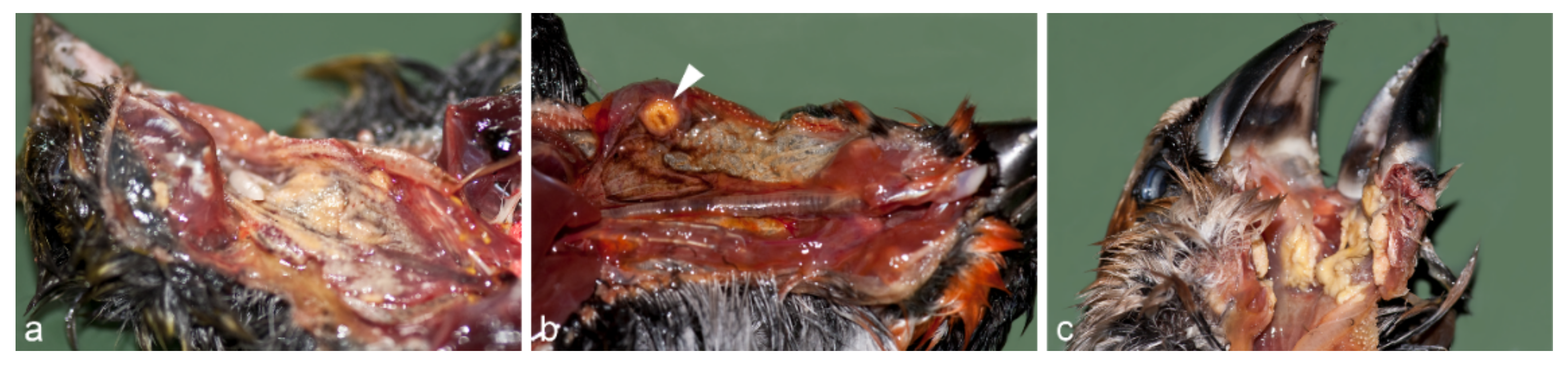
3.2. Macroscopic Findings

The most striking change at macroscopic examination was a very bad nutritional condition with considerable atrophy of the pectoral muscles and complete loss of fat reserves. The average body weight of the greenfinches was 19.2 g (compared to a physiological weight of 24–30 g [27]. Similarly, the majority of the other songbird species showed signs of emaciation with moderate to severe atrophy of the pectoral muscles. Only four chaffinches, one goldfinch, and one bullfinch had a physiological weight. In 84 birds, macroscopically, either single nodular, multifocal to coalescing, or diffuse yellowish diphtheritic pseudomembranes of the mucosa were found in the beak cavity, pharynx, cranial part of the esophagus, and crop (Figure 2). The submitted crop sample showed the same changes. Due to these protruding lesions of the mucosa, the lumen was considerably narrowed or even completely obstructed. In most cases, the proventriculus and gizzard were nearly empty, and large amounts of blood were seen in the intestinal lumen of at least 23 birds. No other pathomorphological abnormalities occurred.

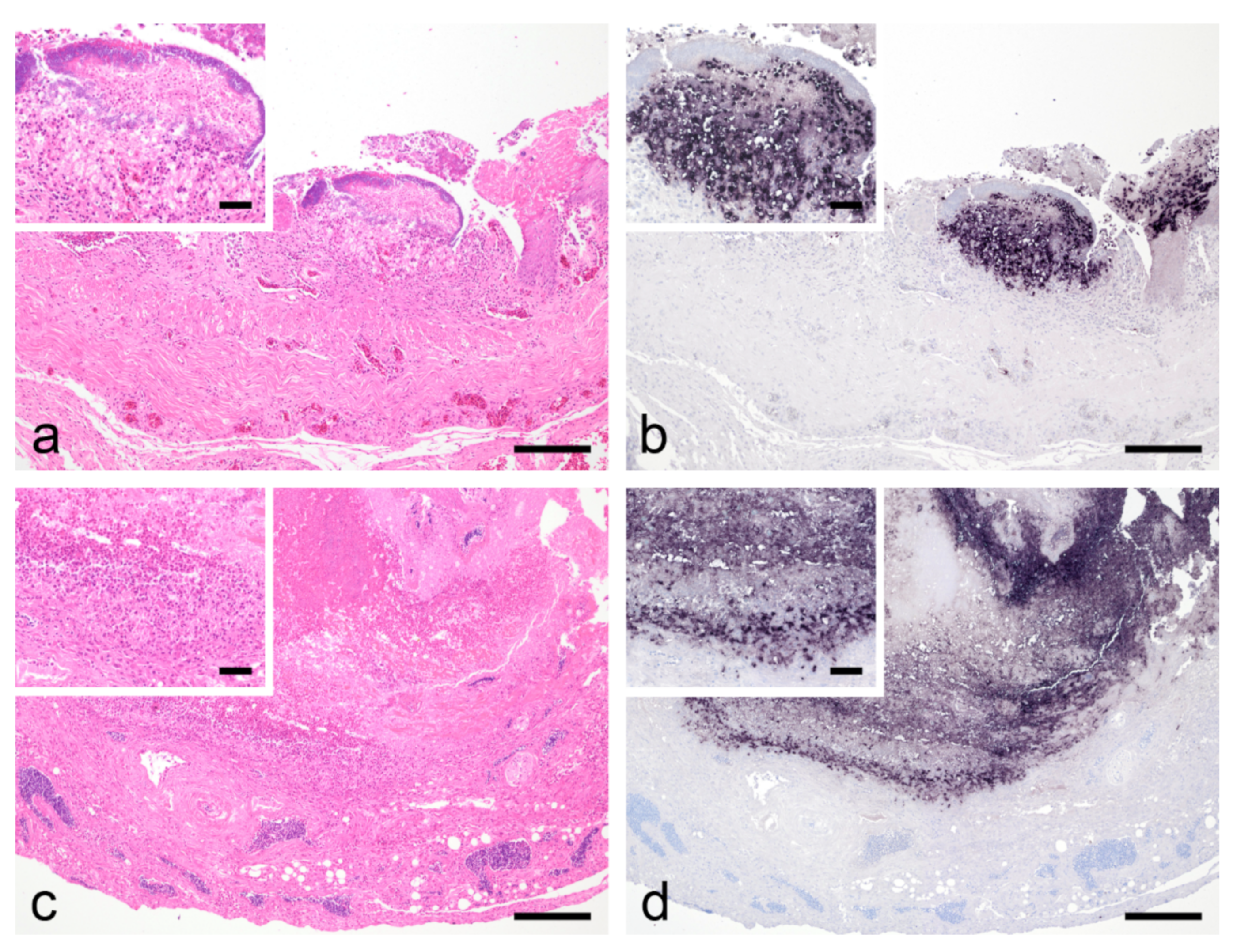
3.3. Histological Findings, PCR, In Situ Hybridization and Bacteriological Examination

Histologically, there was a severe acute, frequently transmural fibrinoheterophilic to necrotizing pharyngitis, ingluvitis, or esophagitis with abundant superficial predominantly coccoid bacterial colonies (Figure 3). Salmonella spp. were not found by bacteriological examination in the first four dead birds. However, ISH with a probe directed against the majority of representatives of the order Trichomonadida localized abundant specific signals in all studied pharynx, crop, or esophagus lesions (Figure 3). PCR for the detection of trichomonads also yielded a positive result in the examined tissue samples. In four birds, small numbers of Macrorhabdus ornithogaster were identified on the mucosal surface at the transition of the proventriculus to gizzard by H&E and GMS staining. There were no associated inflammatory reactions. No other relevant pathologic lesions occurred.
Of the 88 birds examined, only one greenfinch, one great tit, and one Eurasian siskin showed neither macroscopically nor histologically detectable lesions. However, Trichomonas spp. were also detected in the crop tissue of these animals by PCR.

3.4. Sequence Analysis

Thirty-three samples from 2017 to 2020 could be sequenced successfully on the ITS1-5.8S-ITS2 region (NCBI GenBank acc. no. OL678475-OL678507) and 25 on the Fe-hydrogenase gene (NCBI GenBank acc. no. OL654280-OL654304) (Table S1). All acquired sequences were identical and showed 100% identity with published T. gallinae sequences (GenBank acc. no. ITS1-5.8S-ITS2 region: MN385401, Fe- hydrogenase gene: MK172852).

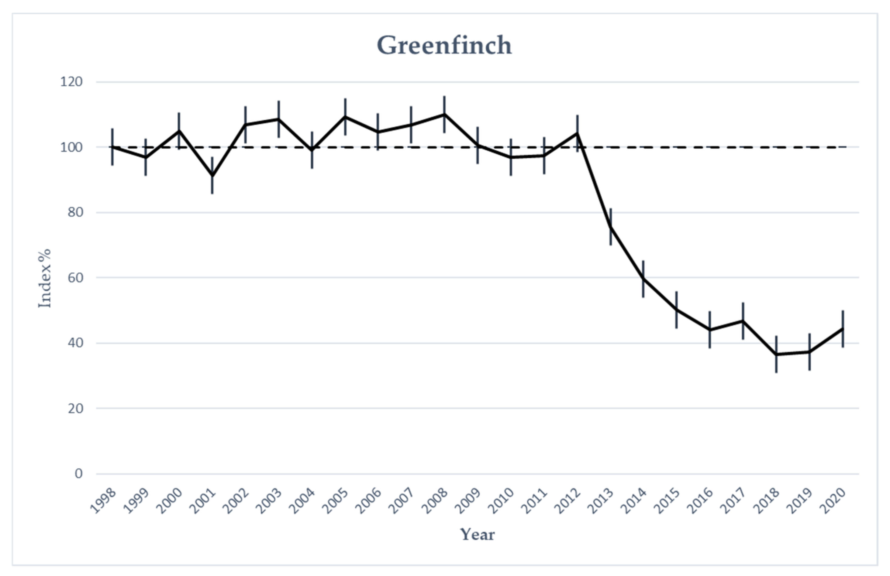
3.5. Greenfinch Monitoring Data

Since the mass mortality of wild songbirds in 2012, a striking gradual decline in Austria’s greenfinch population has been observed. BirdLife Austria’s annual breeding bird monitoring counts showed a reduction of up to 60% of this bird species between 2012 and 2020 (Figure 4).

4. Discussion

The unicellular flagellate T. gallinae is the causative agent of avian trichomonosis, also known as canker in pigeons or as frounce in falcons. This pathogen occurs worldwide and can be found in many other bird species belonging to the orders of Anseriformes [28], Galliformes [29,30], Gruiformes [31], Passeriformes [31,32], Piciformes [33], Psittaciformes [34,35] and Struthioniformes [28]. In general, the typical lesions occur in the beak cavity, infraorbital sinus, pharynx, esophagus, crop, and also in the proventriculus. In addition, the liver or heart and, less frequently, other internal organs may show changes, which usually appear macroscopically as yellowish, cheesy necrotic lesions [36]. Pigeons are very frequently affected, as documented by reports on T. gallinae-associated mass mortalities in free-living mourning doves (Zenaida macroura) in Florida and Alabama, wood pigeons (Columba palumbus) in Spain and Portugal, and band-tailed pigeons (Patagioenas fasciata) in California [37,38,39,40].
In finches, symptomatic trichomonosis was initially only known in experimentally infected, immunosuppressed birds [41]. However, mass mortalities due to natural infections with T. gallinae were described for the first time in the greenfinch population in the UK in 2005 [4,9]. In the following years, a successive spread of trichomonads was detected in finch populations in continental Europe and westward to Canada, with episodes of increased mortality in free-ranging songbirds in Wales, Ireland, southern Scandinavia, the Netherlands, France, Germany, and finally in Austria and Slovenia in 2012 [4,5,14,15,16,17,18,42]. During the first epidemic in the summer of 2012, dead songbirds, in particular, greenfinches, were spotted over almost the entire Austrian territory. After no trichomonads-associated deaths in the finch population were reported to us in Austria in 2013, there was a steady increase in finch trichomonosis from 2014 onwards, with a peak in 2018 and 2020. Interestingly, over the years, the number of bird species found to be infected with T. gallinae increased. Still, greenfinches remained the most affected bird species, but other Fringillidae, such as chaffinches, goldfinches, bullfinches, hawfinches, Eurasian siskins, and bramblings, and also songbirds belonging to the families Paridae (great tits) or Emberizidae (yellowhammers) succumbed to the disease. The large majority, however, were finches. Thus, our observations are consistent with those from other studies indicating the emergence and spread of trichomonosis, particularly in the finch population [3,4,5,6,9,14,15,16,17,18,42,43]. Trichomonosis in passerine birds was unknown in Austria before 2012, and this raises the question of how the pathogen actually was introduced to this country. According to Lawson et al., the most likely reason for the quite rapid transboundary spread of finch trichomonosis in Europe might have been seasonally migrating chaffinches [43], which use flyways directly from England across the North Sea to southern Scandinavia in spring and migrate back in autumn across the English Channel via Denmark, Germany, the Netherlands and Belgium [44]. This is consistent with the timing of the occurrence of finch trichomonosis in Germany and the Netherlands in 2009 and in France in 2010 [5,15,18]. The greenfinch is native to the entire European continent and is actually an annual bird, but migrations to western and south-western Europe have been recorded [45]. Thus, greenfinches breeding in Fennoscandia use overwintering sites in Belgium, The Netherlands, and Luxembourg [46]. This might relate to the spread of the disease southwards to Austria and Slovenia, which could be linked to less well-documented migration routes of finches. The vast majority of affected birds showed fibrinopurulent to fibrinonecrotizing pharyngitis, ingluvitis, or esophagitis with extensive superficial bacterial growth, which may have had a negative influence on the course of the disease due to possible acute to peracute septicemia. The inflammatory lesions presented as multifocal to confluent yellow nodules or as diffuse, yellowish, longitudinal thickenings of the mucosa of the pharynx, crop, and esophagus. As these lesions sometimes significantly obstructed the lumen of the upper digestive tract and the inflammation of the mucous membranes most likely caused severe pain, the birds’ feed intake was considerably impeded. In accordance with the cases reported from Canada and southern Fennoscandia [13,14], the vast majority of finches in our study showed marked emaciation with atrophy of the pectoral muscles and sometimes complete loss of fat reserves. In budgerigars (Melopsittacus undulatus), weight loss and subsequent death may occur due to constant vomiting and regurgitation as well as anorexia [34]. In finches that died of trichomonosis in Austria, findings suggesting regurgitation were also found. The beaks were regularly congested with feed, and frequently also, the pharynx was filled with grain material. According to Kocan et al., infection with T. gallinae can cause lesions large enough to obstruct the esophagus and thus lead to starvation [47]. In addition, the proventriculus and gizzard were often empty, and large amounts of blood could be found in the intestinal tract, suggesting a hemorrhagic diathesis in the intestine, which is indicative of approximately 24 to 36 h of food abstinence [48]. In the case of fibrinopurulent inflammation of the pharyngeal, crop, and esophageal mucosa, infection with Salmonella typhimurium, yeasts or poxviruses must be considered in addition to trichomonosis [5]. In particular, infections with Salmonella spp. in passerine birds are responsible for frequent deaths with almost identical pathomorphological changes of the upper digestive tract with yellowish nodular enlargements in the mucous membranes [49,50,51,52,53,54,55,56,57,58,59,60,61,62,63,64]. Since the lesions in the pharynx, crop, and esophagus are morphologically indistinguishable from those of a trichomonad infection, further etiological investigations must be carried out for differential diagnosis. Microscopic examination of the throat and crop swabs from living or freshly dead birds is well suited for the diagnosis of trichomonads [65,66]. The problem, however, is that these pathogens are extremely sensitive to environmental conditions and require a warm, moist environment to survive but do not form true cysts [16,67]. It is thought that T. gallinae may have a transient cyst-like stage (pseudocyst) and, therefore, may increase the likelihood of out-of-host survival [68]. For example, Neimanis et al. were able to detect viable parasites in only 22 to 38% of samples from autolytic birds in their study. Therefore, it can be stated that the diagnosis of a trichomonad infection by postmortal microscopic examination of swabs is not achieved in all cases since bird carcasses are often received with considerable autolytic changes or after being stored in a freezer for some time. According to our experience, trichomonads are also difficult to detect by light microscopic investigation of tissue sections. Frequently, the affected tissues are so severely necrotic that in conventional H&E stained slides, unequivocal identification of the protozoa is difficult or impossible [14]. Therefore, we relied on ISH for the visualization of the trichomonads. This method not only allowed the localization of the protozoal pathogens directly in the tissue but also to correlate them with the accompanying lesions and estimate the parasitic load, which enabled an assessment of pathogenicity [69]. ISH shows robust, specific labeling of protozoal RNA even in necrotic, autolyzed, or frozen tissue. Another reliable diagnostic method for the diagnosis of trichomonad infections in autolytic and frozen material or after formalin fixation and embedding in paraffin wax was a PCR previously developed for this purpose by our group [20]. The PCR protocols used for the generation of amplification products for subsequent sequencing were less successful because the insufficient preservation conditions of the samples obviously led to DNA fragmentation, not allowing for amplification of the nearly 1000 bp fragment in case of the Fe-hydrogenase gene in some of the samples. In cases of successful amplification and sequencing of the ITS1-5.8S-ITS2 region and the Fe-hydrogenase gene, a 100% sequence identity among all Austrian samples investigated between 2012 and 2020 in our study and by Ganas et al. could be demonstrated [42]. Furthermore, our sequences were 100% identical with sequences derived from trichomonads of Dutch, French and Canadian finches on both genetic loci [15,16,70] and of southern Scandinavian and British finches on the ITS1-5.8S-ITS2 gene locus [6,43]. Our results, therefore, support the findings of Lawson et al. that a single genetically stable clonal strain of T. gallinae is responsible for trichomonosis in finch birds in Europe and also in the Canadian maritime provinces [23].
A major epidemiological factor for the perpetuation of the epidemic is certainly the feeding of songbirds in private gardens throughout the year, where water bowls or water baths are provided [6,14,71,72]. Although the parasite can only survive in the environment for a short period of time, these artificial devices are considered potential sources of infection as water or feed can easily become contaminated with saliva, vomit, or feces [34,47]. The estimated maximum survival time of trichomonads in water is 26 h [73]. According to Zentralanstalt für Meteorologie und Geodynamik (ZAMG), 2012 was one of the warmest summers in the history of measurements in Austria [74], so many different bird species probably increasingly visited water points near feeding areas. Finches tend to stay in feeding areas for prolonged periods of time and eat on the ground. As a result, these birds are more likely to defecate and come in contact with contaminated feed [55]. In contrast, other bird species, e.g., black-capped chickadees (Poecile atricapillus), fly to the feeders only very briefly to grab individual seeds [55]. The time they spend there is, therefore, much shorter, and the risk of infection is lower [55]. According to Refsum et al., finches are very gregarious and prefer to feed in larger groups on the ground, which easily results in infections via contaminated seeds [57]. Greenfinches, in particular, often feed each other grains from their beaks [5]. In addition, finches usually hold several grains in their beaks and peel them one after the other [57]. This could be another factor for the increased incidence of the disease in greenfinches.
As mentioned above, T. gallinae is also found worldwide in other bird species. Columbiformes, and particularly Columba livia are considered primary reservoirs of this protozoan pathogen, with broods usually being infected with these flagellates via crop milk after hatching or transmission taking place between mating partners via the oral route [75,76,77]. Interestingly, in the ITS1-5.8S-ITS2 region, there is 100% sequence homology of the T. gallinae strains detected in the songbirds of our study with strains from rock pigeons (Columba livia) from Spain, China, and in several dove species from different regions of western and southern Europe [78,79,80]. Ganas et al. also detected the same strain in a feral pigeon (Columba livia domestica) from Austria. According to Marx et al., turtle doves (Streptopelia turtur), collared doves (Streptopelia decaocto), stock doves (Columba oenas), and wood pigeons (Columba palumbus) in particular have a high prevalence of T. gallinae. Similar results were found in the United Kingdom, where Trichomonas was detected in adult wood pigeons, collared doves, stock doves, turtle doves, and some of their nestlings from Essex, Norfolk, and Suffolk [81]. When comparing these sequences, especially from turtle doves, with our sequences from Austrian songbirds, a 100% match in the ITS1-5.8S-ITS2 region is found. These data support the assumption that transmission of trichomonads between pigeons and finches occurs under natural conditions, probably at jointly used feeding and watering places.
In Slovenia, a T. gallinae-associated disease with several deaths occurred in a flock of canaries [82]. It is very likely that this event was directly related to the finch trichomonosis already circulating in Slovenia at that time because the acquired sequences show 100% identity to the strains of a free-living greenfinch and are also completely identical to the sequences in our study [42].
Birds of prey can also contract trichomonosis in case their prey [71,75,76,78,83,84,85,86,87], pigeons or small songbirds, are infected with T. gallinae, and they develop granulomas in the beak cavity and pharyngeal mucosa [76,78]. Interestingly, sequences of trichomonads infecting a barn owl (Tyto alba), a Bonelli’s eagle (Aquila fasciata) and a kestrel (Falco tinnunculus) from the Valencia area, and sparrow hawks (Accipiter nisus) from the Czech Republic showed complete identity in the ITS1-5.8S-ITS2 with sequences of Austrian finch trichomonosis cases [78,88]. In the course of our investigations, however, we did not become aware of increased trichomonad infections in other bird species, except songbirds, in Austria.
One clear commonality recorded by all authors is the time of year when deaths due to finch trichomonosis occur. T. gallinae infections were predominantly observed in the warmer season, from spring to early autumn [3,4,6,13,14,15,16,17,18]. In the vast majority of cases, our observations confirmed these findings. Only occasionally did T. gallinae-related deaths occur in winter. This can be explained by the dependency of trichomonads on a warm, humid environment and their sensitivity to environmental conditions [67]. In contrast, morphologically indistinguishable inflammations of the crop mucosa due to Salmonella typhimurium mainly occur in the winter months [64].
Co-infections with other pathogens as possible predisposing factors for the widespread emergence of trichomonosis have not been described previously and seem to have not been in the focus of former reports. Furthermore, the present work does not contribute much to this issue. We only were able to exclude Salmonella in a part of the samples and to confirm M. ornithogaster in several investigated birds. In a separate publication by our group, frequent co-infections with haemosporidian parasites were reported in cases with finch trichomonosis [89].
Robinson et al. and Lehikoinen et al. described a significant decline in the finch populations of the United Kingdom and Finland, especially in greenfinches and chaffinches [3,6]. Both studies suggest that finch trichomonosis is largely responsible for this decline in bird numbers. The authors of a Dutch study also assume that trichomonosis is similarly lethal in their country as in the UK, but the impact on the greenfinch population in the available bird census data was probably not yet conclusive enough [15]. However, a continuous decline, especially in the greenfinch population, has also been observed in Austria over the last nine years, which is probably closely related to cases of trichomonosis. Data on the development of the Chaffinch population in Austria showed a moderate decline of approximately 11% since 1998, though the majority of this decline (8%) occurred between 2019 and 2020, the year with the last available data [BirdLife Austria, unpublished].

5. Conclusions

Our investigations showed that a single genetically stable clonal strain of T. gallinae was responsible for increased mortality in Austria’s free-living finches between 2012 and 2020 and was also a significant cause of the decline in the greenfinch population. Furthermore, with the help of molecular investigations, it was possible to demonstrate that there is a high probability of a direct connection with the mass deaths of finches in other parts of Europe and that trichomonosis has spread strongly in the wild songbird population of Austria in recent years.

Supplementary Materials

The following supporting information can be downloaded at: https://0-www-mdpi-com.brum.beds.ac.uk/article/10.3390/ani12101306/s1, Table S1: Postmortem findings and results of laboratory examinations of 88 adult wild birds of the order Passeriformes, from 2012 to 2020 in Austria.

Author Contributions

Conceptualization, R.B. and H.W.; methodology, R.B., H.W., N.T, B.S., N.N., K.B., J.M. and C.W.-L.; software, R.B.; validation, R.B., H.W. and C.W.-L.; investigation, R.B., H.W., N.T., B.S., N.N., K.B., J.M. and C.W.-L.; writing—original draft preparation, R.B.; writing—review and editing, H.W.; visualization, R.B.; supervision, H.W.; project administration, H.W. All authors have read and agreed to the published version of the manuscript.

Funding

This research received no external funding.

Institutional Review Board Statement

Not applicable.

Informed Consent Statement

Not applicable.

Data Availability Statement

The data will be available with the corresponding author upon request.

Acknowledgments

We are grateful to everyone, who helped in the field, especially to Katharina Loupal and the Team of BirdLife Austria. Joachim Spergser from the Institute of Microbiology is acknowledged for the bacteriological examinations. Furthermore, a special thank goes to Katharina Brugger, who was extremely helpful in creating Figure 1.

Conflicts of Interest

The authors declare no conflict of interest.

References

  1. Ausarbeitung des Österreichischen Berichts Gemäß Artikel 12 der Vogelschutzrichtlinie, 2009/147/EG Berichtszeitraum 2008 bis 2012. Available online: https://www.noe.gv.at/noe/Naturschutz/Endbericht_VS_Art12_2014_hp.pdf (accessed on 24 March 2022).
  2. Massey, J.G. Diseases and medical management of wild passeriformes. Semin. Avian Exot. Pet Med. 2003, 12, 29–36. [Google Scholar] [CrossRef]
  3. Lehikoinen, A.; Lehikoinen, E.; Valkama, J.; Väisänen, R.A.; Isomursu, M. Impacts of trichomonosis epidemics on Greenfinch Chloris chloris and Chaffinch Fringilla coelebs populations in Finland. Ibis 2013, 155, 357–366. [Google Scholar]
  4. Lawson, B.; Robinson, R.A.; Colvile, K.M.; Peck, K.M.; Chantrey, J.; Pennycott, T.W.; Simpson, V.R.; Toms, M.P.; Cunningham, A.A. The emergence and spread of finch trichomonosis in the British Isles. Philos. Trans. R. Soc. B Biol. Sci. 2012, 367, 2852–2863. [Google Scholar] [CrossRef] [Green Version]
  5. Peters, M.; Kilwinski, J.; Reckling, D.; Henning, K. Gehäufte Todesfälle von wild lebenden Grünfinken an Futterstellen infolge Trichomonas gallinae-Infektionen—Ein aktuelles Problem in Norddeutschland. Kleintierprax 2009, 54, 433–438. [Google Scholar]
  6. Robinson, R.A.; Lawson, B.; Toms, M.P.; Peck, K.M.; Kirkwood, J.K.; Chantrey, J.; Clatworthy, I.R.; Evans, A.D.; Hughes, L.A.; Hutchinson, O.C.; et al. Emerging Infectious Disease Leads to Rapid Population Declines of Common British Birds. PLoS ONE 2010, 5, e12215. [Google Scholar] [CrossRef] [Green Version]
  7. Quillfeldt, P.; Schumm, Y.R.; Marek, C.; Mader, V.; Fischer, D.; Marx, M. Prevalence and genotyping of Trichomonas infections in wild birds in central Germany. PLoS ONE 2018, 13, e0200798. [Google Scholar] [CrossRef] [Green Version]
  8. Cousquer, G. Ingluvitis and oesophagitis in wild finches. Vet. Rec. 2005, 157, 455. [Google Scholar] [CrossRef]
  9. Holmes, P.; Duff, P. Ingluvitis and oesophagitis in wild finches. Vet. Rec. 2005, 157, 455. [Google Scholar] [CrossRef]
  10. Pennycott, T.; Lawson, B.; Cunningham, A.; Simpson, V.; Chantrey, J. Necrotic ingluvitis in wild finches. Vet. Rec. 2005, 157, 360. [Google Scholar] [CrossRef]
  11. Lawson, B.; Cunningham, A.; Chantrey, J.; Hughes, L.; Kirkwood, J.; Pennycott, T.; Simpson, V. Epidemic finch mortality. Vet. Rec. 2006, 159, 367. [Google Scholar] [CrossRef] [Green Version]
  12. Simpson, V.; Molenaar, F. Increase in trichomonosis in finches. Vet. Rec. 2006, 159, 606. [Google Scholar] [CrossRef]
  13. Forzán, M.J.; Vanderstichel, R.; Melekhovets, Y.F.; McBurney, S. Trichomoniasis in finches from the Canadian Maritime provinces—An emerging disease. Can. Vet. J. Rev. Vet. Can. 2010, 51, 391–396. [Google Scholar]
  14. Neimanis, A.S.; Handeland, K.; Isomursu, M.; Ågren, E.; Mattsson, R.; Hamnes, I.S.; Bergsjø, B.; Hirvelä-Koski, V. First Report of Epizootic Trichomoniasis in Wild Finches (Family Fringillidae) in Southern Fennoscandia. Avian Dis. 2010, 54, 136–141. [Google Scholar] [CrossRef]
  15. Rijks, J.M.; Laumen, A.A.G.; Slaterus, R.; Stahl, J.; Gröne, A.; Kik, M.L. Trichomonosis in Greenfinches (Chloris chloris) in the Netherlands 2009–2017: A Concealed Threat. Front. Vet. Sci. 2019, 6, 425. [Google Scholar] [CrossRef]
  16. Chavatte, J.-M.; Giraud, P.; Esperet, D.; Place, G.; Cavalier, F.; Landau, I. An outbreak of trichomonosis in European green-finches Chloris chloris and European goldfinches Carduelis carduelis wintering in Northern France. Parasite 2019, 26, 21. [Google Scholar]
  17. Zadravec, M.; Marhold, C.; Slavec, B.; Rojs, O.Z.; Răcnik, J. Trichomonosis in finches in Slovenia. Vet. Rec. 2012, 171, 253–254. [Google Scholar] [CrossRef]
  18. Gourlay, P.; Decors, A.; Jouet, D.; Treilles, D.; Lemberger, K.; Faure, E.; Moinet, M.; Chi, J.; Tyler, K.; Cunningham, A.; et al. Finch trichomonosis spreads to France. Eur. Sect. Wildl. Dis. Assoc. Bull. 2011, 2, 9–10. [Google Scholar]
  19. Anderson, N.L.; Johnson, C.K.; Fender, S.; Heckly, S.; Metzler, M.; Nave, P.; Yim, J. Clinical Signs and Histopathologic Findings Associated with a Newly Recognized Protozoal Disease (Trichomonas gallinae) in Free-Ranging House Finches (Carpodacus mexicanus). J. Zoo Wildl. Med. 2010, 41, 249–254. [Google Scholar] [CrossRef]
  20. Mostegl, M.M.; Richter, B.; Nedorost, N.; Lang, C.; Maderner, A.; Dinhopl, N.; Weissenböck, H. First evidence of previously undescribed trichomonad species in the intestine of pigs? Vet. Parasitol. 2012, 185, 86–90. [Google Scholar]
  21. Mostegl, M.M.; Wetscher, A.; Richter, B.; Nedorost, N.; Dinhopl, N.; Weissenböck, H. Detection of Tritrichomonas foetus and Pentatrichomonas hominis in intestinal tissue specimens of cats by chromogenic in situ hybridization. Vet. Parasitol. 2012, 183, 209–214. [Google Scholar] [CrossRef] [Green Version]
  22. Felleisen, R.S. Comparative sequence analysis of 5.8S rRNA genes and internal transcribed spacer (ITS) regions of tricho-monadid protozoa. Parasitology 1997, 115 Pt 2, 111–119. [Google Scholar]
  23. Lawson, B.; Cunningham, A.A.; Chantrey, J.; Hughes, L.A.; John, S.K.; Bunbury, N.; Bell, D.J.; Tyler, K.M. A clonal strain of Trichomonas gallinae is the aetiologic agent of an emerging avian epidemic disease. Infect. Genet. Evol. 2011, 11, 1638–1645. [Google Scholar] [CrossRef]
  24. Hall, T. BioEdit: A user-friendly biological sequence alignment editor and analysis program for Windows 95/98/NT. Nucleic Acids Symp. Ser. 1999, 41, 95–98. [Google Scholar]
  25. Teufelbauer, N.; Seaman, B.S.; Dvorak, M. Population changes of common Austrian breeding birds in the period 1998-2016—Results of the breeding bird monitoring scheme. Egretta 2017, 55, 43–76. [Google Scholar]
  26. Van Strien, A.; Pannekoek, J.; Hagemeijer, W.; Verstrael, T. A loglinear Poisson regression method to analyse bird monitoring data. Bird Census News 2004, 13, 33–39. [Google Scholar]
  27. Grzimek, B.; Meise, W.; Niethammer, G.; Steinbacher, J. Die Finkenfamilie. In Grzimeks Tierleben Vögel 3, 1st ed.; Grzimek, B., Ed.; Deutscher Taschenbuch Verlag GmbH & Co.KG: München, Germany, 1993; p. 396. [Google Scholar]
  28. Gordo, F.P.; Herrera, S.; Castro, A.; Durán, B.G.; Díaz, R.M. Parasites from farmed ostriches (Struthio camelus) and rheas (Rhea americana) in Europe. Vet. Parasitol. 2002, 107, 137–160. [Google Scholar] [CrossRef]
  29. Mirzaei, M.; Ghashghaei, O.; Khedri, J. First report of an outbreak trichomoniasis in turkey in Sistan, Iran. J. Parasit. Dis. 2014, 40, 61–64. [Google Scholar] [CrossRef] [Green Version]
  30. Pennycott, T.W. Carriage of trichomonads, Hexamita species and Blastocystis species by adult pheasants. Vet. Rec. 1998, 143, 142–143. [Google Scholar] [CrossRef]
  31. Forrester, D.J.; Foster, G.W. Trichomonosis. In Parasitic Diseases of Wild Birds; Atkinson, C.T., Thomas, N.J., Hunter, D.B., Thomas, N.J., Eds.; Wiley-Blackwell: Ames, IA, USA, 2008; pp. 120–153. [Google Scholar]
  32. Chalmers, A.; Alberta, G. Trichomoniasis in finches. Can. Vet. J. Rev. Vet. Can. 1992, 33, 616–617. [Google Scholar]
  33. Ecco, R.; Preis, I.S.; Vilela, D.A.; Luppi, M.M.; Malta, M.C.; Beckstead, R.B.; Stimmelmayer, R.; Gerhold, R. Molecular confirmation of Trichomonas gallinae and other parabasalids from Brazil using the 5.8S and ITS-1 rRNA regions. Vet. Parasitol. 2012, 190, 36–42. [Google Scholar] [CrossRef] [Green Version]
  34. Baker, J.R. Trichomoniasis, a major cause of vomiting in budgerigars. Vet. Rec. 1986, 118, 447–449. [Google Scholar] [CrossRef]
  35. McKeon, T.; Dunsmore, J.; Raidal, S. Trichomonas gallinae in budgerigars and columbid birds in Perth, Western Australia. Aust. Vet. J. 1997, 75, 652–655. [Google Scholar] [CrossRef]
  36. Deplazes, P.; Joachim, A.; Mathis, A.; Strube, C.; Taubert, A.; von Samson-Himmelstjerna, G.; Zahner, H. Parasitologie für die Tiermedizin, 4th ed.; Georg Thieme Verlag: Stuttgart, Germany; New York, NY, USA, 2021; pp. 57–58. [Google Scholar]
  37. Haugen, A.O. Trichomoniasis in Alabama Mourning Doves. J. Wildl. Manag. 1952, 16, 164. [Google Scholar] [CrossRef]
  38. Gerhold, R.W.; Tate, C.M.; Gibbs, S.E.; Mead, D.G.; Allison, A.B.; Fischer, J.R. Necropsy Findings and Arbovirus Surveillance in Mourning Doves from the Southeastern United States. J. Wildl. Dis. 2007, 43, 129–135. [Google Scholar] [CrossRef] [Green Version]
  39. Rogers, K.H.; Girard, Y.A.; Koenig, W.D.; Johnson, C.K. Ecologic Drivers and Population Impacts of Avian Trichomonosis Mortality Events in Band-Tailed Pigeons (Patagioenas Fasciata) in California, USA. J. Wildl. Dis. 2016, 52, 484–494. [Google Scholar] [CrossRef] [Green Version]
  40. Höfle, U.; Gortázar, C.; Ortiz, J.A.; Knispel, B.; Kaleta, E.F. Outbreak of trichomoniasis in a woodpigeon (Columba palumbus) wintering roost. Eur. J. Wildl. Res. 2004, 50, 73–77. [Google Scholar] [CrossRef]
  41. Reisen, W.K.; Chiles, R.E.; Green, E.N.; Fang, Y.; Mahmood, F.; Martinez, V.M.; Laver, T. Effects of immunosuppression on encephalitis virus infection in the house finch, Carpodacus mexicanus. J. Med Èntomol. 2003, 40, 206–214. [Google Scholar] [CrossRef] [Green Version]
  42. Ganas, P.; Jaskulska, B.; Lawson, B.; Zadravec, M.; Hess, M.; Bilic, I. Multi-locus sequence typing confirms the clonality of Trichomonas gallinae isolates circulating in European finches. Parasitology 2014, 141, 652–661. [Google Scholar]
  43. Lawson, B.; Robinson, R.A.; Neimanis, A.; Handeland, K.; Isomursu, M.; Ågren, E.O.; Hamnes, I.S.; Tyler, K.M.; Chantrey, J.; Hughes, L.A.; et al. Evidence of Spread of the Emerging Infectious Disease, Finch Trichomonosis, by Migrating birds. EcoHealth 2011, 8, 143–153. [Google Scholar] [CrossRef]
  44. Normann, D. Chaffinch Fringilla coelebs. In The Migration Atlas: Movements of the Birds of Britain and Ireland; Wenham, C.V., Toms, M.P., Marchant, J.H., Clark, J.A., Siriwardena, G.M., Baille, S.R., Eds.; T. & A.D. Poyser: London, UK, 2002; pp. 637–640. [Google Scholar]
  45. Svensson, L.; Grant, P.J.; Mullarney, K.; Zetterström, D. Sperlingsvögel. In Der Kosmos Vogelführer: Alle Arten Europas, Nordafrikas und Vorderasiens, 2nd ed.; Svensson, L., Grant, P.J., Mullarney, K., Zetterström, D., Eds.; Franckh-Kosmos Ver-lags-GmbH & Co: Stuttgart, Germany, 2018; p. 350. [Google Scholar]
  46. Bakken, V.; Runde, O.; Tjørve, E. [Norsk ringmerkingsatlas] The Norwegian Bird Ringing Atlas. Stavanger Museum: Stavanger, Norway, 2006; Volume 2, p. 446. [Google Scholar]
  47. Kocan, R.M.; Herman, C.M. Trichomoniasis. In Infectious and Parasitic Diseases of Wild Birds, 1st ed.; Davis, J.W., Anderson, R.C., Karstad, L., Trainer, D.O., Eds.; Iowa State University Press: Ames, IA, USA, 1971; pp. 282–290. [Google Scholar]
  48. Dorrestein, G.M.; Kummerfeld, N. Singvögel. In Krankheiten der Heimtiere, 8th ed.; Fehr, M., Sassenburg, L., Zwart, P., Eds.; Schlütersche Verlagsgesellschaft mbH & Co. KG: Hannover, Germany, 2014; pp. 425–492. [Google Scholar]
  49. Hudson, C.B.; Tudor, D.C. Salmonella typhimurium infection in feral birds. Cornell Vet. 1957, 47, 394–395. [Google Scholar]
  50. Wilson, J.; Macdonald, J. Salmonella Infection in Wild Birds. Br. Vet. J. 1967, 123, 212–219. [Google Scholar] [CrossRef]
  51. Schaal, E.; Ernst, H. Enzootic occurrence of salmonella infections in free-living, domestic bird species. Berl. Munch. Tierarztl. Wochenschr. 1967, 80, 13–16. [Google Scholar]
  52. Locke, L.N.; Shillinger, R.B.; Jareed, T. Salmonellosis in Passerine Birds in Maryland and West Virginia. J. Wildl. Dis. 1973, 9, 144–145. [Google Scholar] [CrossRef]
  53. Hurvell, B.; Jevring, J. Infections with Salmonella typhimurium in passerine birds in Sweden (author’s transl). Nord. Vet. Med. 1974, 26, 392–399. [Google Scholar]
  54. Kapperud, G.; Stenwig, H.; Lassen, J. Epidemiology of Salmonella typhimurium O: 4-12 Infection in Norway: Evidence of Transmission from an Avian Wildlife Reservoir. Am. J. Epidemiol. 1998, 147, 774–782. [Google Scholar] [CrossRef] [Green Version]
  55. Daoust, P.Y.; Busby, D.G.; Ferns, L.; Goltz, J.; McBurney, S.; Poppe, C.; Whitney, H. Salmonellosis in songbirds in the Canadian Atlantic provinces during winter-summer 1997–1998. Can. Vet. J. Rev. Vet. Can. 2000, 41, 54–59. [Google Scholar]
  56. Alley, M.R.; Connolly, J.; Fenwick, S.; Mackereth, G.; Leyland, M.; Rogers, L.; Haycock, M.; Nicol, C.; Reed, C. An epidemic of salmonellosis caused by Salmonella Typhimurium DT160 in wild birds and humans in New Zealand. N. Z. Vet. J. 2002, 50, 170–176. [Google Scholar] [CrossRef]
  57. Refsum, T.; Vikøren, T.; Handeland, K.; Kapperud, G.; Holstad, G. Epidemiologic and Pathologic Aspects of Salmonella Typhimurium Infection in Passerine Birds in Norway. J. Wildl. Dis. 2003, 39, 64–72. [Google Scholar] [CrossRef]
  58. Hall, A.J.; Saito, E.K. Avian Wildlife Mortality Events Due to Salmonellosis in the United States, 1985–2004. J. Wildl. Dis. 2008, 44, 585–593. [Google Scholar] [CrossRef] [Green Version]
  59. Une, Y.; Sanbe, A.; Suzuki, S.; Niwa, T.; Kawakami, K.; Kurosawa, R.; Izumiya, H.; Watanabe, H.; Kato, Y. Salmonella enterica serotype Typhimurium infection causing mortality in eurasian tree sparrows (Passer montanus) in Hokkaido. Jpn. J. Infect. Dis. 2008, 61, 166–167. [Google Scholar]
  60. Lawson, B.; Howard, T.; Kirkwood, J.K.; Macgregor, S.K.; Perkins, M.; Robinson, R.A.; Ward, L.R.; Cunningham, A.A. Epi-demiology of salmonellosis in garden birds in England and Wales, 1993 to 2003. Ecohealth 2010, 7, 294–306. [Google Scholar]
  61. Pennycott, T.W.; Mather, H.A.; Bennett, G.; Foster, G. Salmonellosis in garden birds in Scotland, 1995 to 2008: Geographic region, Salmonella enterica phage type and bird species. Vet. Rec. 2010, 166, 419–421. [Google Scholar] [CrossRef]
  62. Hernandez, S.M.; Keel, K.; Sanchez, S.; Trees, E.; Gerner-Smidt, P.; Adams, J.K.; Cheng, Y.; Ray, A.; Martin, G.; Presotto, A.; et al. Epidemiology of a Salmonella enterica subsp. enterica Serovar Typhimurium Strain Associated with a Songbird Outbreak. Appl. Environ. Microbiol. 2012, 78, 7290–7298. [Google Scholar] [CrossRef] [Green Version]
  63. Krawiec, M.; Pietkiewicz, M.; Wieliczko, A. Salmonella spp. as a cause of mortality and clinical symptoms in free-living garden bird species in Poland. Pol. J. Vet. Sci. 2014, 17, 729–731. [Google Scholar] [CrossRef] [Green Version]
  64. Brunthaler, R.; Spergser, J.; Weissenböck, H. Multiple Epidemics in Austrian Fringillidae Caused by A Single Variant of Salmonella Typhimurium. J. Wildl. Dis. 2021, 57, 891–899. [Google Scholar] [CrossRef]
  65. Knispel, B.H.M. Morphologische und molekularbiologische Untersuchungen (PCR und REA der 5,8S rRNA-Region) an Trichomonas gallinae und Tetratrichomonas gallinarum verschiedener Vogelarten. Ph.D. Thesis, Justus-Liebig-Universität, Gießen, Germany, 2005. [Google Scholar]
  66. Anderson, N.L.; Grahn, R.A.; Van Hoosear, K.; BonDurant, R.H. Studies of trichomonad protozoa in free ranging songbirds: Prevalence of Trichomonas gallinae in house finches (Carpodacus mexicanus) and corvids and a novel trichomonad in mockingbirds (Mimus polyglottos). Vet. Parasitol. 2009, 161, 178–186. [Google Scholar] [CrossRef]
  67. Greiner, E.C.; Ritchie, B.W. Parasites. In Avian Medicine: Principles and Application; Ritchie, B.W., Harrison, G.J., Harrison, L.R., Eds.; Wingers Publishing: Lake Worth, FL, USA, 1994; pp. 1007–1029. [Google Scholar]
  68. Mehlhorn, H.; Al-Quraishy, S.; Aziza, A.; Hess, M. Fine structure of the bird parasites Trichomonas gallinae and Tetra-trichomonas gallinarum from cultures. Parasitol. Res. 2009, 105, 751–756. [Google Scholar]
  69. Mostegl, M.M.; Richter, B.; Nedorost, N.; Maderner, A.; Dinhopl, N.; Kulda, J.; Liebhart, D.; Hess, M.; Weissenböck, H. Design and validation of an oligonucleotide probe for the detection of protozoa from the order Trichomonadida using chromogenic in situ hybridization. Vet. Parasitol. 2010, 171, 1–6. [Google Scholar] [CrossRef] [Green Version]
  70. Mcburney, S.; Kelly-Clark, W.K.; Forzán, M.J.; Lawson, B.; Tyler, K.M.; Greenwood, S.J. Molecular characterization of Trichomonas gallinae isolates recovered from the Canadian Maritime provinces’ wild avifauna reveals the presence of the genotype responsible for the European finch trichomonosis epidemic and additional strains. Parasitology 2015, 142, 1053–1062. [Google Scholar] [CrossRef] [Green Version]
  71. Boal, C.W.; Mannan, R.W. Cooper’s Hawks in Urban and Exurban Areas: A Reply. J. Wildl. Manag. 2000, 64, 601–604. [Google Scholar]
  72. Gerhold, R.W.; Maestas, L.P.; Harnage, P.M. Persistence of Two Trichomonas gallinae Isolates in Chlorinated and Distilled Water with or without Organic Material. Avian Dis. 2013, 57, 681–683. [Google Scholar] [CrossRef]
  73. Kaleta, E.F.; Tschimmel, S.; Kraus, S.; Rullof, R. Beobachtungen bei der spontanen (natürlichen) Infektion von Brieftauben mit Trichomonas sp. Proc. VII Tag. Vogelkrankheiten DVG Hann. 1990, 171–176. [Google Scholar]
  74. Einer der wärmsten Sommer der Messgeschichte. Available online: https://www.zamg.ac.at/cms/de/klima/news/einer-der-waermsten-sommer-der-messgeschichte (accessed on 22 February 2022).
  75. Stabler, R.M. Trichomonas gallinae: A review. Exp. Parasitol. 1954, 3, 368–402. [Google Scholar] [CrossRef]
  76. Cole, R.A. Trichomoniasis. In Field Manual of Wildlife Diseases: General Field Procedures and Diseases of Birds; Friend, M., Franson, J.C., Eds.; U.S. Dept. of the Interior U.S. Geological Survey: Washington, DC, USA, 1999; pp. 201–206. [Google Scholar]
  77. Amin, A.; Bilic, I.; Liebhart, D.; Hess, M. Trichomonads in birds—A review. Parasitology 2014, 141, 733–747. [Google Scholar]
  78. Sansano-Maestre, J.; Garijo-Toledo, M.M.; Gómez-Muñoz, M.T. Prevalence and genotyping of Trichomonas gallinae in pigeons and birds of prey. Avian Pathol. 2009, 38, 201–207. [Google Scholar]
  79. Marx, M.; Reiner, G.; Willems, H.; Rocha, G.; Hillerich, K.; Masello, J.F.; Mayr, S.L.; Moussa, S.; Dunn, J.C.; Thomas, R.C.; et al. High prevalence of Trichomonas gallinae in wild columbids across western and southern Europe. Parasites Vectors 2017, 10, 242. [Google Scholar] [CrossRef] [Green Version]
  80. Jiang, X.; Sun, J.; Wang, F.; Li, H.; Zhao, X. Prevalence of Trichomonas spp. in domestic pigeons in Shandong Province, China, and genotyping by restriction fragment length polymorphism. Vet. J. 2016, 211, 88–93. [Google Scholar] [CrossRef]
  81. Lennon, R.J.; Dunn, J.C.; Stockdale, J.E.; Goodman, S.J.; Morris, A.J.; Hamer, K.C. Trichomonad parasite infection in four species of Columbidae in the UK. Parasitology 2013, 140, 1368–1376. [Google Scholar] [CrossRef] [Green Version]
  82. Zadravec, M.; Slavec, B.; Krapež, U.; Gombač, M.; Švara, T.; Poljšak-Prijatelj, M.; Gruntar, I.; Račnik, J. Trichomonosis outbreak in a flock of canaries (Serinus canaria f. domestica) caused by a finch epidemic strain of Trichomonas gallinae. Vet. Parasitol. 2017, 239, 90–93. [Google Scholar] [CrossRef]
  83. Cooper, J.E.; Petty, S.J. Trichomoniasis in Free-Living Goshawks (Accipiter Gentilis Gentilis) from Great Britain. J. Wildl. Dis. 1988, 24, 80–87. [Google Scholar] [CrossRef] [Green Version]
  84. Keymer, I. Diseases of birds of prey. Vet. Rec. 1972, 90, 579–594. [Google Scholar] [CrossRef]
  85. Krone, O.; Altenkamp, R.; Kenntner, N. Prevalence of Trichomonas Gallinae in Northern Goshawks from the Berlin Area of Northeastern Germany. J. Wildl. Dis. 2005, 41, 304–309. [Google Scholar] [CrossRef] [Green Version]
  86. Real, J.; Mañosa, S.; Muñoz, E. Trichomoniasis in a Bonelli’s eagle population in Spain. J. Wildl. Dis. 2000, 36, 64–70. [Google Scholar]
  87. Samour, J.H.; Bailey, T.A.; Cooper, J.E. Trichomoniasis in birds of prey (order Falconiformes) in Bahrain. Vet. Rec. 1995, 136, 358–362. [Google Scholar] [CrossRef]
  88. Kunca, T.; Šmejkalová, P.; Cepicka, I.; Èepièka, I. Trichomonosis in Eurasian sparrowhawks in the Czech Republic. Folia Parasitol. 2015, 62, 035. [Google Scholar] [CrossRef] [Green Version]
  89. Himmel, T.; Harl, J.; Matt, J.; Weissenböck, H. A citizen science-based survey of avian mortality focusing on haemosporidian infections in wild passerine birds. Malar. J. 2021, 20, 417. [Google Scholar]
Figure 1. Map of Austria showing locations of detected cases of trichomonosis in songbirds from 2012 to 2020. Each color represents a particular year; the number in the circle determines the bird species: (1) Greenfinch; (2) Chaffinch; (3) Goldfinch; (4) Bullfinch; (5) Hawfinch; (6) Eurasian siskin; (7) Great tit; (8) Yellowhammer; (9) Brambling.
Animals 12 01306 g001
Figure 2. (a) Diphtheritic pseudomembrane affecting the esophagus, crop and pharynx of a greenfinch as multifocal to coalescing nodules in the crop obstructing the lumen; (b) Focal diphtheritic pseudomembrane of a bullfinch as a single nodule in the crop mucosa; (c) Hawfinch with extensive diphtheritic pseudomembrane affecting the pharynx.
Animals 12 01306 g002
Figure 3. (a) Greenfinch, esophagus with predominantly superficial heterophilic inflammation, mucosal ulceration and bacterial growth (H&E staining, bar = 160). Insert shows fibrin formation, infiltration of heterophils, intralesional and superficial coccoid bacteria (H&E staining, bar = 40 µm); (b) Esophageal wall of the same bird; ISH reveals abundant colonization with T. gallinae appearing as purple to black stained objects (bar = 160). Insert shows higher magnification of positive ISH (bar = 40 µm); (c) Bullfinch, esophagus with severe transmural heterophilic inflammation and necrosis (H&E staining, bar = 160). Insert shows high-grade diffuse infiltration with heterophils (bar = 40 µm); (d) Esophageal wall of the same bird; ISH reveals large amounts of purple to black stained protozoal objects corresponding to T. gallinae within the necrotic and inflamed tissue sites (bar = 160). Insert shows higher magnification of positive ISH (bar = 40 µm).
Animals 12 01306 g003
Figure 4. Graphical representation of population trend (index values and 95%-confidence limits) of the Austrian greenfinch population for the period 1998–2020 (1998 = 100%).
Animals 12 01306 g004
Publisher’s Note: MDPI stays neutral with regard to jurisdictional claims in published maps and institutional affiliations.

