Article

Taxonomic Identification of Different Species of the Genus Aeromonas by Whole-Genome Sequencing and Use of Their Species-Specific β-Lactamases as Phylogenetic Markers

1
Institut d’Investigació Biomèdica Sant Pau, 08026 Barcelona, Spain
2
Department of Microbiology, Hospital de la Santa Creu i Sant Pau, 08041 Barcelona, Spain
*
Authors to whom correspondence should be addressed.
These authors contributed equally to this work.
Submission received: 2 January 2021 / Revised: 10 March 2021 / Accepted: 18 March 2021 / Published: 28 March 2021

Abstract

:
Some Aeromonas species, potentially pathogenic for humans, are known to express up to three different classes of chromosomal β-lactamases, which may become hyperproduced and cause treatment failure. The aim of this study was to assess the utility of these species-specific β-lactamase genes as phylogenetic markers using whole-genome sequencing data. Core-genome alignments were generated for 36 Aeromonas genomes from seven different species and scanned for antimicrobial resistance genes. Core-genome alignment confirmed the MALDI-TOF identification of most of the isolates and re-identified an A. hydrophila isolate as A. dhakensis. Three (B, C and D) of the four Ambler classes of β-lactamase genes were found in A. sobria, A. allosacharophila, A. hydrophila and A. dhakensis (blaCphA, blaAmpC and blaOXA). A. veronii only showed class-B- and class-D-like matches (blaCphA and blaOXA), whereas those for A. media, A. rivipollensis and A. caviae were class C and D (blaCMY, blaMOX and blaOXA427). The phylogenetic tree derived from concatenated sequences of β-lactamase genes successfully clustered each species. Some isolates also had resistance to sulfonamides, quinolones and aminoglycosides. Whole-genome sequencing proved to be a useful method to identify Aeromonas at the species level, which led to the unexpected identification of A. dhakensis and A.rivipollensis and revealed the resistome of each isolate.

1. Introduction

The taxonomy of the genus Aeromonas is quite complex. The history of this genus of Gammaproteobacteria has been in constant change, as described by Fernandez-Bravo and Figueras [1]. The members of the Aeromonas genus are Gram-negative rods, oxidase- and catalase-positive, and glucose fermenters. They are capable of degrading nitrates to nitrites and are resistant to the vibriostatic factor O/129 (2,4-diamino-6,7-di-iso-propylpteridine phosphate) [1]. Despite these established biochemical characteristics, identifying species within the Aeromonas genus has always been a convoluted task. Included in the Vibrionaceae in 1965, the Aeromonas were subsequently classified in their own family, the Aeromonadaceae, in 1976 [2]. Since then, 36 different species have been included in the genus, up to 19 of which are considered to be potential human pathogens related to gastrointestinal, as well as blood and wound, infections in both immunocompromised and immunocompetent patients [2]. The most pervasive species are A. caviae, A. hydrophila, A. veronii and A. dhakensis [2,3,4,5].
Regarding their phenotypical identification, Abbot et al. [6] tested 63 biochemical traits in 193 strains representing 14 different Aeromonas species and found that only 15% (9/63) returned consistent results for each species. More elaborate identification methods rely on amplifying housekeeping genes, with rpoB, rpoD and gyrB being the most used, as their interspecies mean sequence similarity in aeromonads is 89% to 92% [2,4,7,8,9]. The analyses can be complemented by other conserved genes, such as recA, dnaJ, gyrA, dnaX and atpD [7,10], with their intraspecific and interspecific nucleotide differences of <2% and >3%, respectively, serving as baseline values for identifying Aeromonas isolates. However, not all the genotypic methods are infallible. De Melo et al. [11] found that an Aeromonas isolate was misidentified by multi-locus phylogenetic analysis (MLPA) (gyrB, rpoD, recA, dnaJ, gyrA, and dnaX), whereas clustering with reference genomes allowed species identification.
Faster methods such as MALDI-TOF can identify Aeromonas to the species level with a relatively low error rate (<10%) [2,12,13], although most databases need curating, as errors are still being reported in clinical practice [14] and many new taxa have been discovered in recent years [15,16]. Accordingly, identification becomes more accurate as new spectra are added to the working databases [15,17]. Nevertheless, identification by MALDI-TOF is not always conclusive: for example, A. dhakensis has historically been confused with other clinically relevant aeromonads (A. veronii, A. hydrophila and A. caviae) [2,8,9,10,11,12,13,14,15,16,17,18,19,20]. The phylogenetic study that led to the creation of this taxon was based on the genes gyrB and rpoD, which were later complemented by recA, dnaJ and gyrA in a MLPA [18]. The latest method used to identify different Aeromonas strains in clinical settings is whole-genome sequencing (WGS), which has emerged as a promising and highly discriminative tool, not only for epidemiology, but also in taxonomy [21].
Aeromonas spp. are traditionally associated with resistance to β-lactams such as ampicillin (with the exception of A. trota), as well as other penicillins, and first- and second-generation cephalosporins, whereas they are generally susceptible to third- and fourth-generation cephalosporins, monobactams and carbapenems, as well as aminoglycosides and fluoroquinolones [2,10]. β-lactam resistance in the genus is attributed to the production of two to three chromosomal β-lactamases: an Ambler class B metallo-β-lactamase (MBL), a class C cephalosporinase and a class D penicillinase. In general, A. allosaccharophila, A. dhakensis, A. hydrophila and A. sobria strains produce all three types of β-lactamase, A. caviae, class C and D and A. veronii, class B and D [18,20,22,23,24].
Class B MBLs produced by Aeromonas mostly belong to the blaCphA group and specifically hydrolyse carbapenems. The detection of CphA-like carbapenemases is challenging, requiring a double-disk method combining imipenem or ceftazidime with 500 mM EDTA [4]. Interestingly, the presence of an MBL in aeromonads often does not translate into sufficient carbapenemase activity, even in the presence of inducers [25]. Class C cephalosporinases found in Aeromonas belong to the AmpC family and produce a resistance phenotype against cephamycins (cefoxitin and cefotetan), third-generation cephalosporins (cefotaxime, ceftazidime) and β-lactam inhibitors such as clavulanate, tazobactam and sulbactam, but they cannot hydrolyse penicillins or carbapenems. Lastly, Class D penicillinases related to the oxacillinase (OXA) family show high hydrolysis rates for penicillins, low rates for first- and second-generation cephalosporins and do not hydrolyse carbapenems [4,23].
In this study, we used WGS to look for the chromosomal β-lactamase genes carried by different Aeromonas spp. and assessed their potential role as species-specific identification genes with core-genome alignment. Additionally, we aimed to elucidate the origin of some CMY, FOX and MOX cephalosporinases described in Enterobacteriaceae as an acquired β-lactam resistance mechanism.

2. Results

2.1. Identification by MALDI-TOF

The MALDI-TOF analysis confirmed the previous routine laboratory identification of 13 Aeromonas spp. isolates (Table 1). Both methods correctly identified all the isolates, except for two: the A. allosaccharophila reference strain (ATCC 51208), which was identified as A. veronii by MALDI-TOF, and the A. caviae D547 strain, which was identified as A. hydrophila by MALDI-TOF.

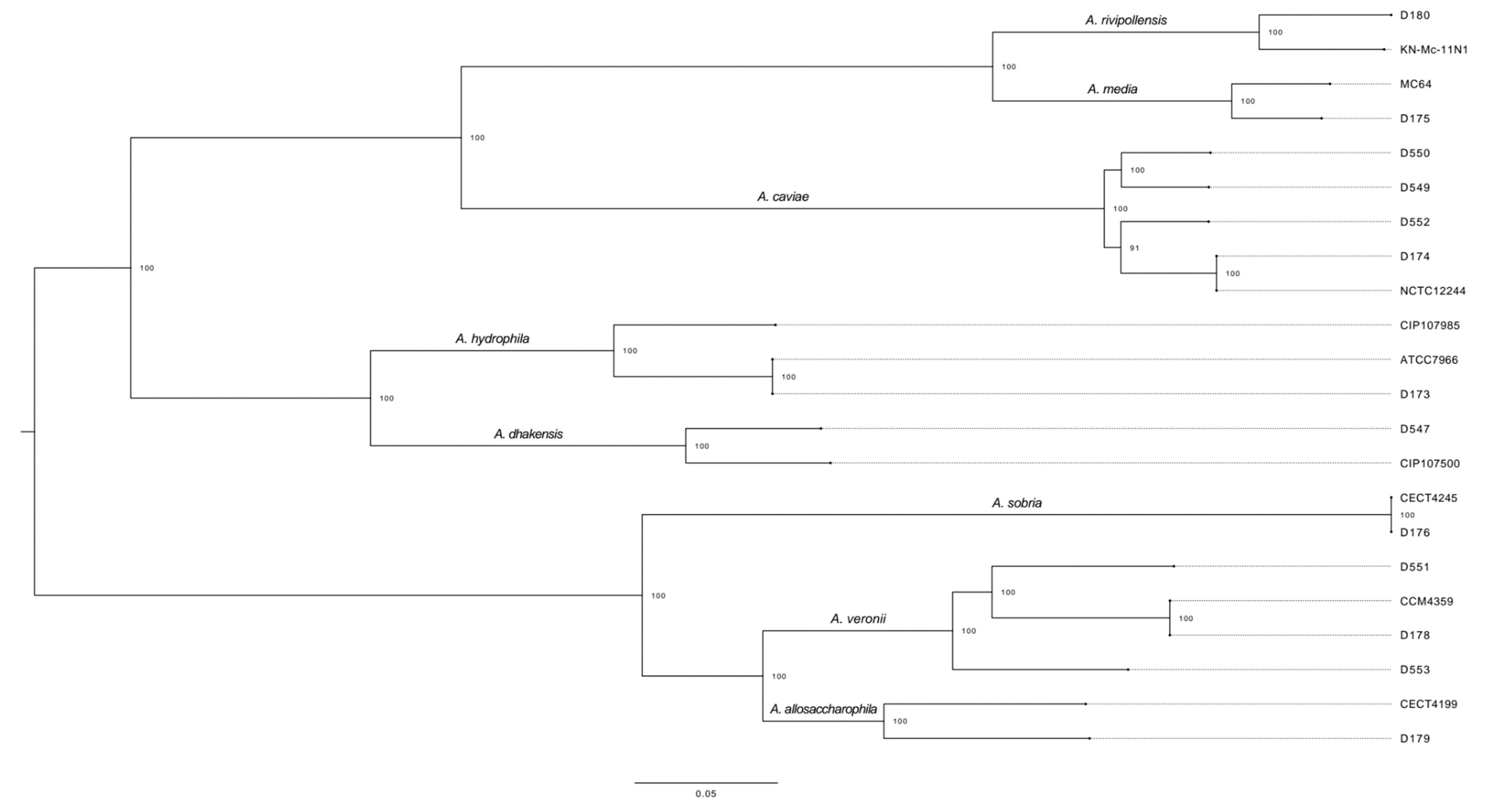
2.2. WGS Analysis: Core-Genome Alignment

The Roary pangenome analysis of the 22 isolates, 9 type strain genomes and 13 de novo sequenced isolates produced a core-genome alignment of 760,946 bp. The Roary analysis showed 745 core genes (found in > 99% genomes), 44 soft core genes (found in 95–99% genomes), 8011 shell genes (found in 15–95% genomes), and 14,312 cloud genes (found in < 15% genomes) from a total of 23,112 genes.
The core-genome alignment produced eight different clusters representing each of the following species: A. rivipollensis (the D180 isolate clustered with the A. rivipollensis type strain genome, so we renamed it as a A. rivipollensis strain), A. media (D175), A. caviae (D174, D549, D550 and D552), A. hydrophila (D173), A. dhakensis (D547, first identified as A. caviae by phenotypic methods, and re-identified as A. hydrophila by MALDI-TOF), A. sobria (D176), A. veronii (D178, D551 and D553) and A. allosaccharophila (D179) (Figure 1).
The core-genome sequence analysis allowed us to correctly identify all clinical isolates to the species level. WGS analysis also revealed which Aeromonas species share the highest core-genome similarities, thus producing three main phyllogenetic groups: one group with A. media, A. rivipollensis and A. caviae, a second one with A. dhakensis and A. hydrophila and a third one with A. sobria, A. allosaccharophila and A. veronii.
The ANI genome comparison supports the core-genome analysis, and is shown in the File S1. Additionally, type strain information is provided in Table S1.

2.3. Antimicrobial Susceptibility Testing
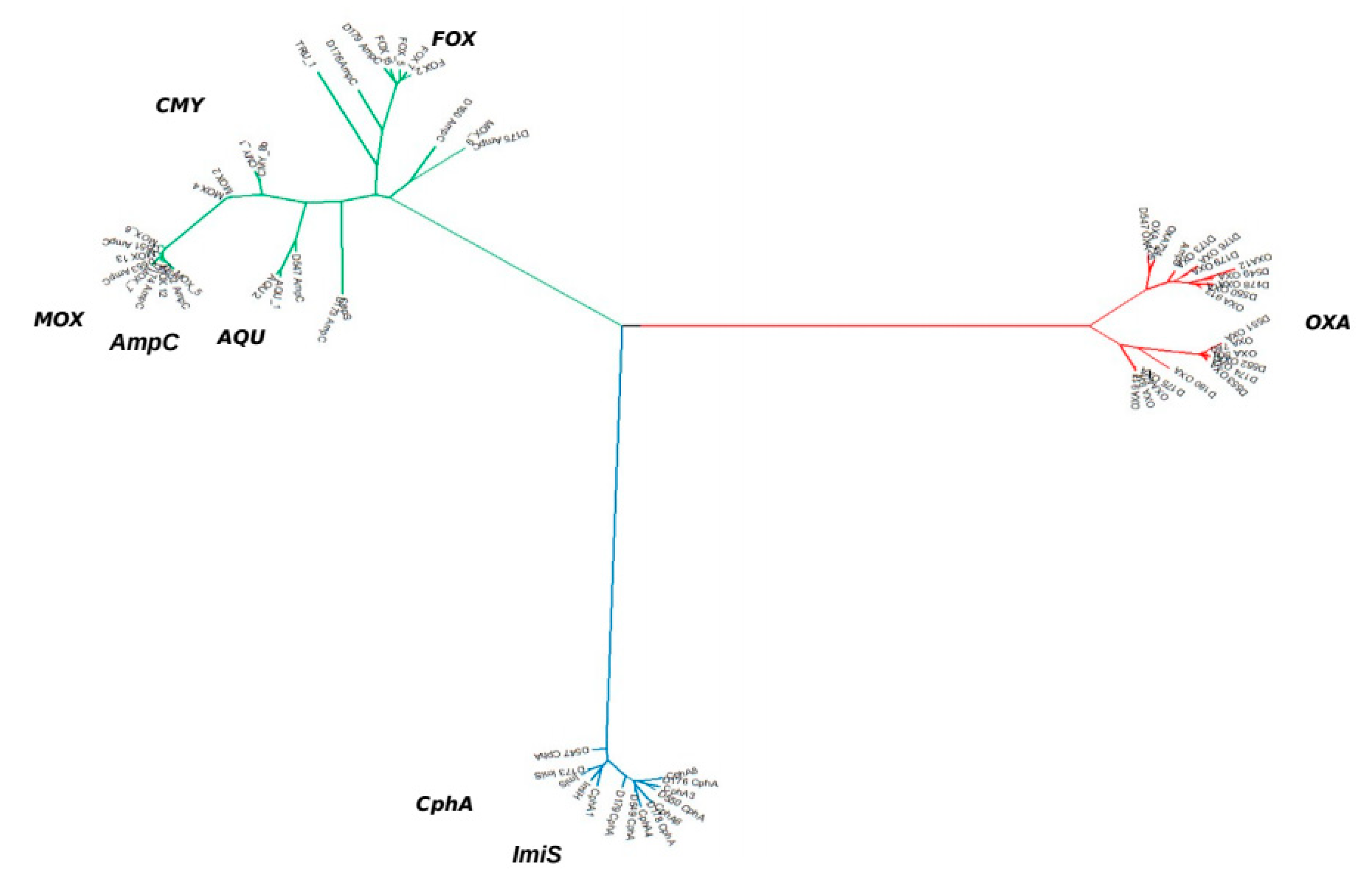
The Antimicrobial Susceptibility Testing (AST) was performed in twelve of the thirteen clinical isolates. A. sobria D176 was removed from the AST because it did not match the growth standards for the test. Results of AST are shown in Table 2.
Practically all Aeromonas isolates studied were resistant to ampicillin, amoxicillin-clavulanate and cefazolin, with three exceptions: A. hydrophila D173 isolate, which was only resistant to first- and second-generation cephalosporins and A. veronii (D551), susceptible to amoxicillin-clavulanate (Table 1). Only two strains showed resistance to third-generation cephalosporins and all of them were resistant to carbapenems. We also found resistance to nalidixic acid in three A. caviae and two A. veronii strains (one of them was also resistant to cotrimoxazol). No species-specific resistance patterns were observed.

2.4. β-Lactamases

Given that WGS is a technique not yet available for all clinical microbiology laboratories, and because various β-lactamases have been described in the genus Aeromonas, we hypothesized that these enzymes could help achieve a correct identification when standard laboratory techniques are inconclusive.
The assembled genomes obtained from the WGS analysis and the genomes obtained from Genbank were analysed to look for molecular mechanisms associated with β-lactam resistance using the public ResFinder and CARD databases (Table 3). As expected, different classes of β-lactamase genes in the Aeromonas genus were found.
The Ambler class B β-lactamase genes detected were mainly of the blaCphA family, including those of the blaImi-type, and showed very little genetic diversity (Figure 2): blaCphA2, blaCphA3, blaCphA4, blaCphA6, blaCphA8, blaImiH and blaImiS. Ambler class C β-lactamase genes were the most diverse, with up to five different families being detected: blaMOX, (blaMOX-5,-6,-7,-9), blaFOX, (blaFOX-2,-7), blaAQU-2 and blaCMY (blaCMY-1, blaCMY-8b). Finally, Ambler class D β-lactamase genes belonged to the blaOXA family, including the blaAmp hits (blaOXA-12,-427,-724 and blaAmpH, blaAmpS).
Thus, the phylogenetic analysis of the β-lactamase genes was carried out by UPGMA and revealed the greatest diversity in the AmpC group (Figure 2), separating the FOX and MOX clusters. This group also possessed the highest species specificity. Following ResFinder nomenclature, all A. caviae strains were clustered in the MOX group along with the A. hydrophila strains. Nevertheless, A. dhakensis, which is phylogenetically related to A. hydrophila, showed a blaCMY-8b gene that has 84% identity with blaMOX genes. The A. dhakensis D547 and the A. hydrophila D173 strains expressed the carbapenemases CphA or Imi, which share a similarity of 94.49% and are classified in the same β-lactamase group (Figure 2).
According to the clusters defined by WGS, the first one was composed of A. sobria, A. allosaccharophila, and A. veronii, which, despite their proximity did not share the same types of β-lactamase genes. While the blaCphA and blaAMPS genes were present in all three species, blaFOX was not detected among the A. veronii strains.
Finally, the A. media and A. caviae strains expressed the cephalosporinases blaCMY-8b or blaMOX-9 (both with a 96% identity) and the blaOXA-427 gene, which have between 96.48% and 98.36% similarity with AmpS (Figure 2).
Therefore, although each cluster was characterized by particular β-lactamase genes, the results did not reveal a sufficiently clear pattern to establish an algorithm for the identification of Aeromonas spp. It would also be important to unify a single nomenclature between the different databases consulted.

2.5. Characterisation of Other Mechanisms of Resistance to Antimicrobials

Aside from the expected β-lactam resistances, five of the 13 isolates (two A. veronii: D551 and D553, and three A. caviae D549, D550 and D553) also showed visible resistance to nalidixic acid. The nalidixic acid resistance was associated with substitutions identified in the gyrA gene in strains D550 and D551 (Ser83Ile) and strain D553 (Ser83Arg). In strains D549 and D552, a substitution was detected in the gyrA gene (Ser83Ile) and in the parC gene (Ser80Ile). No mutations producing resistance were found in gyrB or parE, and the mutations detected were not associated with an increased quinolone resistance.
Regarding resistances to aminoglycosides, although this drug family was not included in the AST, two of the isolates expressed aminoglycoside-modifying enzymes (AME): strain D549 carried the genes aph(3′′)-1b and aph(3′′)-1d, which have the highest affinity for streptomycin, whereas strain D552 possessed aadA2, which hydrolyses streptomycin and spectinomycin.
Only one A. veronii strain (D551) resistant to trimethoprim-sulfamethoxazole carried the sul1 and dfrA12 genes. Query covers and identities for both genes were 100%.

3. Discussion

At present, identification of Aeromonas isolates to the species level remains a complex task [6,11,14]. Nevertheless, we obtained results of high accuracy using a WGS method and core-genome alignment, as have other studies [21,26,27]. This promising technique, however, is out of reach for routine use in the laboratory, and the most commonly used method, MALDI-TOF, may misidentify samples without a sufficiently comprehensive database [13,16], as occurred in our A. allosaccharophila, A. dhakensis and A. rivipollensis strains. A. rivipollensis was described for the first time in 2015, also in Catalonia, from the Ter river and, in agreement with our results, the authors describe it as a new species related with A. media [28]. To improve identification in Aeromonas, we investigated the potential of AmpC β-lactamase genes to serve as species-specific markers and, in our experience, the PCR amplification and Sanger sequencing analysis of the AmpC β-lactamase class can improve the correct identification to the species level in the genus Aeromonas.
Aeromonas spp. are known to carry several chromosomal β-lactam resistance genes belonging to Ambler classes B, C and D [18,20,22,23,24]. By obtaining the genetic sequences of these β-lactamase genes through WGS, we hoped to shed light on whether the class C cephalosporinases MOX and FOX, now pervasive among the Enterobacteriaceae, originate from the Aeromonas genus. Ebmeyer et al. [29] detected different MOX-type enzymes in the genome of various species of Aeromonas, including A. sanaralli (MOX-1), A. caviae (MOX-2) and A. media (MOX-9). The same authors have also reported that the origin of FOX enzymes could be in the chromosome of A. allosaccharophila [30].
The high-resolution method of WGS allowed almost every Aeromonas species to be matched with a sequence type genome. The phylogenetic tree confirmed that A. sobria and A. allosaccharophila differ considerably from the genetically closest species A. veronii in the core-genome aligment, as previously described by MLPA or by gyrB and rpoB phylogenetic analysis [7,8]. The WGS approach also led to the unexpected identification of an A. dhakensis isolate, originally identified as A. hydrophila by MALDI-TOF, a species not previously reported as a clinical strain in Spain. WGS also identified an A. rivipollensis isolate previously identified as A. media.
Using WGS analysis, we were able to determine the resistance genes carried by the Aeromonas isolates. Three types of β-lactamase genes (blaCphA, blaAmpC and blaOXA) were found in the isolates, in accordance with the species-specific patterns described in the literature. A. sobria, A. allosacharophila, A hydrophila and A. dhakensis carried all three types, A. veronii was the only clinical strain to possess only blaCphA and blaOXA, and both A. media, A. caviae and A. rivipollensis carried a blaAmpC and a blaOXA gene [18,20,22,23,24].
It has already been reported that there is no agreement between the presence of so many genes encoding different β-lactamases (which should confer resistance to most β-lactam antibiotics) with the actual resistance profile. A two-component regulator (TCR), closely related to the CreBC of Escherichia coli, has been described in Aeromonas. This TCR includes a putative mutant form of a transcription factor, the BlrA protein (related to the extended family of phosphorylation-dependent response regulators), whose gene was found immediately upstream from the blrB gene, encoding a predicted sensor kinase [25,31]. The presence of this operon prevents the expression of β-lactamase genes, and mutations in this system confer a high level of resistance to β-lactams. In some Aeromonas species (A. veronii, A. hydrophila and A. caviae), a frequency range of blrAB de-repression between 107 and 109 has been described, which increases the MICs of the β-lactams tested by 16 for ampicillin, 4 for imipenem, up to 16 for cephaloridine and up to 129 for cefotaxime [32].
Notably, the Ambler class C β-lactamases showed a pattern of high species-specificity, indicating that each Aeromonas species has a characteristic family of cephalosporinase genes (if any): blaFOX, in A. sobria, A. allosacharophila and A. hydrophila, blaAQU in A. dhakensis, blaCMY/MOX-9 in A. media and A. rivipollensis, and blaMOX in A. caviae. Therefore, plasmid-mediated AmpC genes are derived from the chromosomal AmpC genes of several members of the family Enterobacteriaceae, including Enterobacter cloacae (MIR o ACT group), Morganella morganii (DHA group), and Hafnia alvei (ACC group) [33]. As reported by Jacoby et al. [34], CMY is represented twice, as it has two quite different origins. Six current varieties (CMY-1,-8,-9,-10,-11, and -19) are related to chromosomally determined AmpC genes in Aeromonas spp., while the remainder are related to AmpC β-lactamases of Citrobacter freundii.
The chromosomal β-lactamase genes of A. caviae thus seem to be the progenitors of plasmid-mediated MOX-2 and MOX-4 and of some CMY-1-related enzymes. The plasmid-mediated FOX enzymes seem to have their origin in the AmpC CAV-1 of A. media (first identified as A. punctata) [35]. Finally, A. jandaei and A. enteropelogenes also have their own chromosomal AmpC β-lactamase genes (AsbA1 and TRU-1, respectively) [4,36]. These data support that each Aeromonas species has its own chromosomal β-lactamase genes and is a potential progenitor of new emerging AmpC enzymes.
As it is mandatory to use more than one public resistance database, we found that, using the ResFinder and CARD, some entries were exclusive to either one or the other: blaAmpS and blaAmpH only appeared in ResFinder and blaAQU-1, blaAQU-2, blaMOX-9 and blaCepS were exclusive to CARD. These specific entries yielded matches with higher identities for our query sequences, indicating that completeness is essential for proper diagnosis and that more than one curated database for antimicrobial resistance genes should be used when screening isolates [37]. Nevertheless, regarding Aeromonas resistance genes, both ResFinder and CARD are good tools for screening, but we found that the percentage of identity for many species-specific resistance genes, such as blaAmpC of A. hydrophila and blaOXA-like of A. caviae, was too low.
Cases of fluoroquinolone resistance mediated by genetic mobile elements have been previously described in Aeromonas [4,10,27,38,39]. However, we did not find any qnr-like genes among the resistant isolates D549-D553, correlating with the findings of Ghatak et al. [27]. Additionally, we found D549-D552 to have point mutations in gyrA resulting in amino acid substitution (Ser83Ile) and parC (Ser80Ile), which are typically associated with quinolone-resistant aeromonads [40,41], as well as a mutation in gyrA: Ser83Arg in strain D553 not previously reported in Aeromonas spp.
The results for trimetoprim-sulfamethoxazole AST correlate with findings by Kadlec et al. [42], who reported that 100% (33/33) of Aeromonas spp. carrying sul1 exhibited increased MICs for sulfamethoxazole treatments. Aminoglycoside resistance genes found in strain D549 [aph(3′′)1b and aph(3′′)1d)] match previous reports on Aeromonas [24,40], whereas we were unable to find publications mentioning aadA2, as we found in strain D552.
Overall, WGS allowed the successful identification of all the Aeromonas isolates to the species level. β-lactamase genes, naturally associated with each species of the genus (a class B carbapenemase, a class C cephalosporinase and a class D penicillinase), were present in our isolates in different combinations: A. sobria, A. allosacharophila, A hydrophila and A. dhakensis carried all three types, A. veronii was the sole clinical strain to possess only a blaCphA-type and a blaOXA-type gene, whereas A. media, A. rivipollensis and A. caviae carried a blaAmpC and a blaOXA gene. To our knowledge, this is the first report in Spain of an A. dhakensis isolate, previously identified as A. hydrophila by MALDI-TOF.
Based on the results obtained, we can conclude that WGS is the most appropriate technique both to identify Aeromonas at the species level and to describe the different resistance mechanisms present. In addition, this method produces objective results that are comparable with those from any other laboratory anywhere. The only drawback for its laboratory application, at least at present, is the high cost.

4. Materials and Methods

4.1. Identification and Antimicrobial Susceptibility Testing

Thirteen strains were included in this study. Seven (D173–D176, D178–D180) were from the Colección Española de Cultivos Tipo (CECT) and six (547, 549–553) were isolated in our laboratory from faeces of patients with gastroenteritis. Strains were stored at −20 °C until use, and one copy at −80 °C.
Strains were grown in blood agar medium and underwent MALDI-TOF MS Autoflex II (Bruker Daltonics, Barcelona, Spain) identification from single colonies. From the same cultures, 2–3 colonies were used to prepare a 0.5 McFarland suspension [43,44], which was used in disk-diffusion susceptibility tests for Gram-negative Enterobacteriaceae in Mueller-Hinton agar. Cultures were incubated at 37 °C for 18–24 h. The antimicrobial drugs used were: ampicillin (10 μg), amoxicillin-clavulanic acid (30 μg), piperacillin (30 μg), cefazolin (30 μg), piperacillin-tazobactam (36 μg), cefoxitin (30 μg), cefotaxime (5 μg), ceftazidime (10 μg), aztreonam (30 μg), cefepime (30 μg), ertapenem (10 μg), imipenem (10 μg), cefuroxime (30 μg), ciprofloxacin (5 μg), nalidixic acid (30 μg) and trimethoprim/sulfamethoxazole (25 μg). Cut-off thresholds were established according to the EUCAST criteria [43], as EUCAST defines clinically relevant Aeromonas spp. (A. hydrophila, A. veronii, A. dhakensis and A. caviae) as intrinsically resistant to ampicillin, amoxicillin, amoxicillin-clavulanic acid, ampicillin-sulbactam and cefoxitin [45].

4.2. DNA Extraction and Whole-Genome Sequencing

From single colonies grown in blood agar plates, the thirteen samples were transferred to an enriched growth broth (5 mL Luria Bertani) and cultured overnight at 37 °C. DNA extraction and purification was done according to the DNeasy® UltraClean® Microbial Kit protocol (Qiagen, Frederick, MD, USA).
The quality screening for DNA extraction was done by Invitrogen QUBIT® 3.0 fluorometer, which determines the amount of extracted DNA (ng/μL) per sample. Concentrations that we deemed too low were concentrated using the SpeedVac at 45 °C for 10 min, thus lowering the final volume from 50 μL to 30 μL, and analyzed again. Spectrophotometry at 260 and 280 nm was also calculated to perform a quality control for DNA concentrations and purity using Nanodrop 2000/2000c(Thermofisher Scientific, Waltham, MA, USA).
Sequencing was performed by Sequentia Biotech S.L. (Barcelona, Spain) using Illumina technologies. The sequencing libraries were prepared using a Nextera XT kit (Illumina, Madrid, Spain) and sequenced on an Illumina MiSeq sequencer which generated 2 × 250 bp paired-end reads.

4.3. Computational Analysis

Trim_galore v0.6.5 (available at https://github.com/FelixKrueger/TrimGalore/tree/ accessed on 1 January 2021, Babraham Bioinformatics, Cambridge, UK, 2019) was used to trim Illumina adaptors, and filter low-quality reads and reads under 50 bp. Then Unicycler v0.4.9b (available at github.com/rrwick/Unicycler, Ryan Wick, Victoria, Australia, 2019) was used as a SPAdes v3.14.0 (available at https://cab.spbu.ru/software/spades/ accessed on 1 January 2021, Center for Algorithmic Biotechnology, St Petersburg, Russia, 2019) optimizer in order to generate the best possible assemblies with the Illumina reads. Afterwards, we annotated the genome assemblies using Prokka v1.14.6 (available at https://github.com/tseemann/prokka accessed on 1 January 2021, Torsten Seemann, Victoria, Australia, 2020) [46]. Prokka gff files output was used as input in Roary v1.7.7 (available at https://github.com/sanger-pathogens/Roary accessed on 1 January 2021, Sanger-Pathogens, Cambridge, UK, 2019) [47]. Gubbins v2.4.1 (available at https://github.com/sanger-pathogens/gubbins accessed on 1 January 2021, Sanger-Pathogens, Cambridge, UK, 2020) [48] was used to remove possible recombination events in the core genome alignment. RaxML v8.2.12 (available at https://cme.h-its.org/exelixis/web/software/raxml/ accessed on 1 January 2021, The Exelixis Lab, Heidelberg, Germany, 2018) was used with the core-genome alignment file to infer phylogeny, applying a General Time Reversible (GTRACAT) model with 99 bootstraps.

4.3.1. Whole-Genome Comparison

Complete genomes of Aeromonas were downloaded from GenBank (Table 3). Type strain genomes for ANI genome comparison and core genome analysis were downloaded from the NCBI Assembly database (available at https://0-www-ncbi-nlm-nih-gov.brum.beds.ac.uk/assembly/) (accessed on 9 February 2021). We used OrthoANIu standalone tool for ANI genome comparison (available at https://www.ezbiocloud.net/tools/orthoaniu) Not applicable) [49].

4.3.2. Detection of Antimicrobial Resistance Genes

The antimicrobial resistance genes in the strains were detected using the CARD [50], ResFinder [51] and complete NCBI (Genbank) [52] databases with the software Abricate (available at https://github.com/tseemann/abricate) (accessed on 17 April 2020). Hits below 60% identity were automatically discarded.

4.3.3. Antimicrobial Resistance Genes Phylogenetic Tree

Nucleotide sequences of the genes listed in Table 2 searched against the CARD and ResFinder databases were downloaded as FASTA files. We used BLAST to extract the β-lactamase genes from the genomes of our strains. MEGA X [53] was used for FASTA alignment and UPGMA tree calculation and visualization. All genes used for this analysis are indicated in Supplementary Table S2.

Supplementary Materials

The following are available online at https://0-www-mdpi-com.brum.beds.ac.uk/article/10.3390/antibiotics10040354/s1: Table S1: Type_strains_information.csv. File S1: ANI_genome_comparison.

Author Contributions

Conceptualization, E.M., F.N. and C.M.; Methodology, X.B., L.G. and T.L.; Software, M.R. and X.B.; Validation, E.M. and F.N.; Writing—Original Draft Preparation, X.B., M.R. and E.M.; Writing—Review & Editing, E.M., F.N. and C.M. All authors have read and agreed to the published version of the manuscript.

Funding

This research received no external funding.

Institutional Review Board Statement

Not applicable.

Informed Consent Statement

Not applicable.

Data Availability Statement

All WGS data was uploaded to the SRA under the PRJNA685948 Bioproject. Assembled and annotated genome data are available at genbank under accession numbers: JAGDEM000000000, JAGDEN000000000, JAGDEO000000000, JAGDEP000000000, JAGDEQ000000000, JAGDER000000000, JAGDES000000000, JAGDET000000000, JAGDEU000000000, JAGDEV000000000, JAGDEW000000000, JAGDEX000000000, and JAGDEY000000000.

Acknowledgments

This study was supported by the Microbiology Department of Sant Pau Hospital and the Sant Pau Research Institute.

Conflicts of Interest

The authors declare no competing financial interest.

Sample Availability

Strains are available from the authors.

References

  1. Martin-Carnahan, A.; Joseph, S.W. Order XII. Aeromonadales ord. nov. In Bergey’s Manual of Systematic Bacteriology, 2nd ed.; Brenner, D.J., Krieg, N.R., Staley, J.T., Garrity, G.M., Eds.; Williams & Wilkins: Philadelphia, PA, USA, 2005; pp. 556–578. [Google Scholar]
  2. Fernández-Bravo, A.; Figueras, M.J. An update on the genus Aeromonas: Taxonomy, epidemiology, and pathogenicity. Microorganisms 2020, 8, 129. [Google Scholar] [CrossRef] [Green Version]
  3. Parker, J.L.; Shaw, J.G. Aeromonas spp. clinical microbiology and disease. J. Infect. 2011, 62, 109–118. [Google Scholar] [CrossRef] [PubMed]
  4. Janda, J.M.; Abbott, S.L. The genus Aeromonas: Taxonomy, pathogenicity, and infection. Clin. Microbiol. 2010, 23, 35–73. [Google Scholar] [CrossRef] [Green Version]
  5. Gonçalves Pessoa, R.B.; de Oliveira, W.F.; Marques, D.S.C.; dos Santos Correia, M.T.; de Carvalho, E.V.M.M.; Coelho, L.C.B.B. The genus Aeromonas: A general approach. Microb. Pathog. 2019, 130, 81–94. [Google Scholar] [CrossRef]
  6. Abbott, S.L.; Cheung, W.K.W.; Janda, J.M. The genus Aeromonas: Biochemical characteristics, atypical reactions, and phenotypic identification schemes. J. Clin. Microbiol. 2003, 4, 2348–2357. [Google Scholar] [CrossRef] [PubMed] [Green Version]
  7. Navarro, A.; Martínez-Murcia, A. Phylogenetic analyses of the genus Aeromonas based on housekeeping gene sequencing and its influence on systematics. J. Appl. Microbiol. 2018, 125, 622–631. [Google Scholar] [CrossRef] [Green Version]
  8. Küpfer, M.; Kuhnert, P.; Korczak, B.M.; Peduzzi, R.; Demarta, A. Genetic relationships of Aeromonas strains inferred from 16S rRNA, gyrB and rpoB gene sequences. Int. J. Syst. Evol. Microbiol. 2006, 56, 2743–2751. [Google Scholar] [CrossRef] [Green Version]
  9. Yáñez, M.A.; Catalán, V.; Apráiz, D.; Figueras, M.J.; Martínez-Murcia, A.J. Phylogenetic analysis of members of the genus Aeromonas based on gyrB gene sequences. Int. J. Syst. Evol. Microbiol. 2003, 53, 875–883. [Google Scholar] [CrossRef] [Green Version]
  10. Zhou, Y.; Yu, L.; Nan, Z.; Zhang, P.; Kan, B.; Yan, D.; Su, J. Taxonomy, virulence genes and antimicrobial resistance of Aeromonas isolated from extra-intestinal and intestinal infections. BMC Infect. Dis. 2019, 9, 158. [Google Scholar] [CrossRef] [Green Version]
  11. De Melo, B.S.T.; Mendes-Marques, C.L.; de Lima Campos, T.; de Almeida, A.M.P.; Leal, N.C.; Xavier, D.E. High-resolution genome-wide analysis is essential for the identification of ambiguous Aeromonas strains. FEMS Microbiol. Lett. 2019, 366, fnz245. [Google Scholar] [CrossRef] [PubMed]
  12. Schuetz, A.N. Emerging agents of gastroenteritis: Aeromonas, plesiomonas, and the diarrheagenic pathotypes of Escherichia coli. Semin. Diagn. Pathol. 2019, 36, 187–192. [Google Scholar] [CrossRef]
  13. Shin, H.B.; Yoon, J.; Lee, Y.; Kim, M.S.; Lee, K. Comparison of MALDI-TOF MS, Housekeeping gene sequencing, and 16s rRNA gene sequencing for identification of Aeromonas clinical isolates. Yonsei Med. J. 2015, 56, 550–555. [Google Scholar] [CrossRef] [PubMed] [Green Version]
  14. Ruiz de Alegría-Puig, C.; Aguirre-Quiñonero, A.; Agüero-Balbín, J.; Roiz-Mesones, M.P.; Martínez-Martínez, L. Correlation between MALDI-TOF Vitek-MSTM system and conventional identification methods of gastrointestinal infection causing bacteria. Rev. Esp. Quimioter. 2016, 29, 265–268. [Google Scholar] [PubMed]
  15. Vávrová, A.; Balážová, T.; Sedláček, I.; Tvrzová, L.; Šedo, O. Evaluation of the MALDI-TOF MS profiling for identification of newly described Aeromonas spp. Folia Microbiol. 2015, 60, 375–383. [Google Scholar] [CrossRef]
  16. Sinclair, H.A.; Heney, C.; Sidjabat, H.E.; George, N.M.; Bergh, H.; Anuj, S.N.; Nimmo, G.R.; Paterson, D.L. Genotypic and phenotypic identification of Aeromonas species and CphA-mediated carbapenem resistance in Queensland, Australia. Diagn. Microbiol. Infect. Dis. 2016, 85, 98–101. [Google Scholar] [CrossRef]
  17. Benagli, C.; Demarta, A.; Caminada, A.P.; Ziegler, D.; Petrini, O.; Tonolla, M. A Rapid MALDI-TOF MS Identification Database at Genospecies Level for Clinical and Environmental Aeromonas Strains. PLoS ONE 2012, 7, e48441. [Google Scholar] [CrossRef] [Green Version]
  18. Chen, P.L.; Lamy, B.; Ko, W.C. Aeromonas dhakensis, an increasingly recognized human pathogen. Front. Microbiol. 2016, 7, 793. [Google Scholar] [CrossRef]
  19. Wu, C.J.; Chen, P.L.; Hsueh, P.R.; Chang, M.C.; Tsai, P.J.; Shih, H.I.; Wang, H.C.; Chou, P.H.; Ko, W.C. Clinical implications of species identification in monomicrobial Aeromonas bacteremia. PLoS ONE 2015, 10, e0117821. [Google Scholar] [CrossRef]
  20. Kitagawa, H.; Ohge, H.; Yu, L.; Kayama, S.; Hara, T.; Kashiyama, S.; Kajihara, T.; Hisatsune, J.; Sueda, T.; Sugai, M. Aeromonas dhakensis is not a rare cause of Aeromonas bacteremia in Hiroshima, Japan. J. Infect. Chemother. 2020, 26, 316–320. [Google Scholar] [CrossRef] [PubMed]
  21. Borriss, R.; Rueckert, C.; Blom, J.; Bezuidt, O.; Reva, O.; Klenk, H.P. Whole Genome Sequence Comparisons in Taxonomy. Methods Microbiol. 2011, 38, 409–436. [Google Scholar]
  22. Fosse, T.; Giraud-Morin, C.; Madinier, I. Phénotypes de résistance aux β-lactamines dans le genre Aeromonas. Pathol. Biol. 2003, 51, 290–296. [Google Scholar] [CrossRef]
  23. Chen, P.L.; Ko, W.C.; Wu, C.J. Complexity of β-lactamases among clinical Aeromonas isolates and its clinical implications. J. Microbio. Immunol. Infect. 2012, 45, 398–403. [Google Scholar] [CrossRef] [PubMed] [Green Version]
  24. Walsh, T.R.; Hall, L.; Macgowan, A.P.; Bennett, P.M. Sequence analysis of two chromosomally mediated inducible β-lactamases from Aeromonas sobria, strain 163a, one class d penicillinase, the other an AmpC cephalosporinase. J. Antimicrob. Chemother. 1995, 36, 41–52. [Google Scholar] [CrossRef]
  25. Juan, C.; Torrens, G.; González-Nicolau, M.; Oliver, A. Diversity and regulation of intrinsic β-lactamases from non-fermenting and other Gram-negative opportunistic pathogens. FEMS Microbiol. Rev. 2017, 41, 780–814. [Google Scholar] [CrossRef]
  26. Hughes, H.Y.; Conlan, S.P.; Lau, A.F.; Dekker, J.P.; Michelin, A.V.; Youn, J.H.; Henderson, D.K.; Frank, K.M.; Segre, J.A.; Palmore, T.N. Detection and whole-genome sequencing of carbapenemase- producing Aeromonas hydrophila isolates from routine perirectal surveillance culture. J. Clin. Microbiol. 2016, 54, 1167–1170. [Google Scholar] [CrossRef] [Green Version]
  27. Ghatak, S.; Blom, J.; Das, S.; Sanjukta, R.; Puro, K.; Mawlong, M.; Shakuntala, I.; Sen, A.; Goesmann, A.; Kumar, A.; et al. Pan-genome analysis of Aeromonas hydrophila, Aeromonas veronii and Aeromonas caviae indicates phylogenomic diversity and greater pathogenic potential for Aeromonas hydrophila. Antonie van Leeuwenhoek Int. J. Gen. Mol. Microbiol. 2016, 109, 945–956. [Google Scholar] [CrossRef] [PubMed]
  28. Marti, E.; Balcazar, J.L. Aeromonas rivipollensis sp. Nov.; A novel species isolated from aquatic samples. J. Basic Microbiol. 2015, 55, 1435–1439. [Google Scholar] [CrossRef]
  29. Ebmeyer, S.; Kristiansson, E.; Joakim Larsson, D.G. The mobile FOX AmpC beta-lactamases originated in Aeromonas allosaccharophila. Int. J. Antimicrob. Agents 2019, 54, 798–802. [Google Scholar] [CrossRef]
  30. Ebmeyer, S.; Kristiansson, E.; Joakim Larsson, D.G. CMY-1/MOX-family AmpC β-lactamases MOX-1, MOX-2 and MOX-9 were mobilized independently from three Aeromonas species. J. Antimicrob. Chemother. 2019, 74, 1202–1206. [Google Scholar] [CrossRef]
  31. Niumsup, P.; Simm, A.M.; Nurmahomed, K.; Walsh, T.R.; Bennett, P.M.; Avison, M.B. Genetic linkage of the penicillinase gene, amp, and blrAB, encoding the regulator of β-lactamase expression in Aeromonas spp. J. Antimicrob. Chemother. 2003, 51, 1351–1358. [Google Scholar] [CrossRef] [Green Version]
  32. Walsh, T.R.; Stunt, R.A.; Nabi, J.A.; MacGowan, A.P.; Bennett, P.M. Distribution and expression of beta-lactamase genes among Aeromonas spp. J. Antimicrob. Chemother. 1997, 40, 171–178. [Google Scholar] [CrossRef]
  33. Perez-Perez, F.J.; Hanson, N.D. Detection of plasmid-mediated AmpC β-lactamase genes in clinical isolates by using multiplex PCR. J. Clin. Microbiol. 2002, 40, 2153–2162. [Google Scholar] [CrossRef] [PubMed] [Green Version]
  34. Jacoby, G.A. AmpC β-lactamases. Antimicrob Agents Chemother. 2009, 22, 161–182. [Google Scholar] [CrossRef] [PubMed] [Green Version]
  35. De Luca, F.; Giraud-Morin, C.; Rossolini, G.M.; Docquier, J.D.; Fosse, T. Genetic and biochemical characterization of TRU-1, the endogenous class C β-lactamase from Aeromonas enteropelogenes. Antimicrob. Agents Chemother. 2010, 54, 1547–1554. [Google Scholar] [CrossRef] [Green Version]
  36. Lamy, B.; Laurent, F.; Kodjo, A. Validation of a partial rpoB gene sequence as a tool for phylogenetic identification of aeromonads isolated from environmental sources. Can. J. Microbiol. 2010, 56, 217–228. [Google Scholar] [CrossRef] [PubMed]
  37. Mahfouz, N.; Ferreira, I.; Beisken, S.; von Haeseler, A.; Posch, A.E. Large-scale assessment of antimicrobial resistance marker databases for genetic phenotype prediction: A systematic review. J. Antimicrob. Chemother. 2020, 75, 3099–3108. [Google Scholar] [CrossRef]
  38. Moura, Q.; Fernandes, M.R.; Cerdeira, L.; Santos, A.C.M.; de Souza, T.A.; Ienne, S.; Pignatari, A.C.C.; Gales, A.C.; Silva, R.M.; Lincopan, N. Draft genome sequence of a multidrug-resistant Aeromonas hydrophila ST508 strain carrying rmtD and bla CTX-M-131 isolated from a bloodstream infection. J. Glob. Antimicrob. Resist. 2017, 10, 289–290. [Google Scholar] [CrossRef] [PubMed]
  39. Lukkana, M.; Wongtavatchai, J.; Chuanchuen, R. Class 1 integrons in Aeromonas hydrophila isolates from farmed nile tilapia (Oreochromis nilotica). J. Vet. Med. Sci. 2012, 74, 435–440. [Google Scholar] [CrossRef] [Green Version]
  40. Adesoji, A.T.; Ogunjobi, A.A.; Olatoye, I.O. Genotypic Characterization of Aminoglycoside Resistance Genes from Bacteria Isolates in Selected Municipal Drinking Water Distribution Sources in Southwestern Nigeria. Ethiop. J. Health Sci. 2019, 29, 321. [Google Scholar]
  41. Machuca, J.; Agüero, J.; Miró, E.; del Carmen Conejo, M.; Oteo, J.; Bou, G.; González-López, J.J.; Oliver, A.; Navarro, F.; Pascual, Á.; et al. Prevalence of quinolone resistance mechanisms in Enterobacteriaceae producing acquired AmpC β-lactamases and/or carbapenemases in Spain. Enferm. Infecc. Microbiol. Clin. 2017, 38, 485–490. [Google Scholar] [CrossRef]
  42. Kadlec, K.; von Czapiewski, E.; Kaspar, H.; Wallmann, J.; Michael, G.B.; Steinacker, U.; Schwarz, S. Molecular basis of sulfonamide and trimethoprim resistance in fish-pathogenic Aeromonas isolates. Appl. Environ. Microbiol. 2011, 77, 7147–7150. [Google Scholar] [CrossRef] [Green Version]
  43. EUCAST. Breakpoint Tables for Interpretation of MICs and Zone Diameters, Version 10.0. 2020. Available online: http://www.eucast.org (accessed on 18 March 2021).
  44. Clinical and Laboratory Standards Institute (CLSI). Performance Standards for Antimicrobial Susceptibility Testing, 29th ed.; CLSI supplement M100; Clinical and Laboratory Standards Institute: Wayne, PA, USA, 2019. [Google Scholar]
  45. EUCAST Advice on Intrinsic Resistance and Exceptional Phenotypes v 3.2 (February, 2020, typographical errors corrected May, 2020). Available online: https://www.eucast.org/fileadmin/src/media/PDFs/EUCAST_files/Expert_Rules/2020/Intrinsic_Resistance_and_Unusual_Phenotypes_Tables_v3.2_20200225.pdf (accessed on 18 March 2021).
  46. Seemann, T. Prokka: Rapid prokaryotic genome annotation. Bioinformatics 2014, 30, 2068–2069. [Google Scholar] [CrossRef]
  47. Page, A.J.; Cummins, C.A.; Hunt, M.; Wong, V.K.; Reuter, S.; Holden, M.T.G.; Fookes, M.; Falush, D.; Keane, J.A.; Parkhill, J. Roary: Rapid large-scale prokaryote pan genome analysis. Bioinformatics 2015, 31, 3691–3693. [Google Scholar] [CrossRef]
  48. Croucher, N.J.; Page, A.J.; Connor, T.R.; Delaney, A.J.; Keane, J.A.; Bentley, S.D.; Parkhill, J.; Harris, S.R. Rapid phylogenetic analysis of large samples of recombinant bacterial whole genome sequences using Gubbins. Nucleic Acids Res. 2014. [Google Scholar] [CrossRef] [PubMed] [Green Version]
  49. Yoon, S.H.; Ha, S.M.; Lim, J.M.; Kwon, S.J.; Chun, J. A large-scale evaluation of algorithms to calculate average nucleotide identity. Antonie van Leeuwenhoek. 2017, 110, 1281–1286. [Google Scholar] [CrossRef] [PubMed]
  50. Alcock, B.P.; Raphenya, A.R.; Lau, T.T.Y.; Tsang, K.K.; Bouchard, M.; Edalatmand, A.; Huynh, W.; Nguyen, A.L.V.; Cheng, A.A.; Liu, S.; et al. CARD 2020: Antibiotic resistome surveillance with the comprehensive antibiotic resistance database. Nucleic Acids Res. 2020, 48, 517–525. [Google Scholar] [CrossRef]
  51. Bortolaia, V.; Kaas, R.F.; Ruppe, E.; Roberts, M.C.; Schwarz, S.; Cattoir, V.; Philippon, A.; Allesoe, R.L.; Rebelo, A.R.; Florensa, A.R.; et al. ResFinder 4.0 for predictions of phenotypes from genotypes. J. Antimicrob. Chemother. 2020, 75, 3491–3500. [Google Scholar] [CrossRef] [PubMed]
  52. Clark, K.; Karsch-Mizrachi, I.; Lipman, D.J.; Ostell, J.; Sayers, E.W. GenBank. Nucleic Acids Res. 2016, 4, D67–D72. [Google Scholar] [CrossRef] [Green Version]
  53. Kumar, S.; Stecher, G.; Li, M.; Knyaz, C.; Tamura, K. MEGA X: Molecular Evolutionary Genetics Analysis across Computing Platforms. Mol. Biol Evol. 2018, 35, 1547–1549. [Google Scholar] [CrossRef]
Figure 1. Maximum likelihood phylogenetic tree derived from the core-genome alignment of the Aeromonas spp. isolates and type strain genomes. All strains with names beginning with D are isolates from the laboratory. Bootstrap values above 80% are shown in branch nodes (premutation = 100). The different clusters within the phylogenetic tree contain previously identified sequences posted on GenBank. Type strain information and accession numbers are presented in Table S1.
Antibiotics 10 00354 g001
Figure 2. UPGMA tree containing all β-lactamase resistance genes found in Aeromonas isolates. Class B β-lactamase branch is coloured in blue, Class C β-lactamase branch is coloured in green, and Class D β-lactamase branch is coloured in red. Analyses of the 65 nucleotide sequences were performed using MEGA X.
Antibiotics 10 00354 g002
Table 1. MALDI-TOF identification of the 13 Aeromonas isolates previously identified in routine laboratory work.
LAB IDRoutine Laboratory IdentificationMALDI-TOF IdentificationScore
D180 **A. mediaA. media DSM 4881T HAM2.22
D175A. media ATCC 33907 (CECT4232)A. media DSM 4881T HAM2.38
D174A. caviae ATCC 15468 (CECT838)A. caviae CECT838T DSM2.40
D549A. caviaeA. caviae CECT838T DSM2.43
D550A. caviaeA. caviae CECT838T DSM2.41
D552A. caviaeA. caviae CECT838T DSM2.31
D547 *A. caviaeA. hydrophila CECT 839T DSM2.34
D173A. hydrophila ATCC 7966 (CECT839)A. hydrophila CECT 839T DSM2.47
D176A. sobria ATCC 43979 (CECT4245)A. sobria CECT 4245T DSM2.45
D178A. veronii ATCC 35624 (CECT4257)A. veronii CECT5761T DSM2.26
D551A. veroniiA. veronii DSM 7386T HAM2.38
D553A. veroniiA. veronii CECT4257T DSM2.30
D179A. allosaccharophila ATCC 51208 (CECT4911)A. veronii DSM 11576THAM2.28
Identified as * A. dhakensis and ** A. rivipollensis by WGS (see below).
Table 2. Antimicrobial susceptibility test results for the 13 Aeromonas strains.
LAB IDWGS IdentificationAMPAMCPRLTPZCFZFOXCXMCTXCAZATMFEPIMPETPNALCIPSXT
D180A. rivipollensis672424143338303746423232333629
D175A. media6122225716263029S362327313419
D174A. caviae612222691728353140362324313722
D549A. caviae616253119333836344540273063429
D550A. caviae6172726283438393542402126626S
D552A. caviae61228287172432264236302262722
D173A. hydrophila2730293261130292836343432323028
D547A. dhakensis61123316625163152402527343934
D178A. veronii6122023152730303334321822313722
D551A. veronii62632321329363636444035S6296
D553A. veronii61525288223438324240282263122
D179A. allosaccharophila61620236203263244362123344021
AMP: Ampicillin; AMC: Amoxicillin-Clavulanate; PRL: Piperacillin; CFZ: Cefazolin; TPZ: Tazobactam-Piperacillin; FOX: Cefoxitin; CXM: Cefuroxime; CTX: Ceftazidime; CAZ: Cefazoline; ATM: Aztreonam; FEP: Cefepime; IMP: Imipinem; ETP: Ertapenem; CIP: Ciprofloxacin; NAL: Nalidixic Acid; SXT: Cotrimoxazol. The numbers represent the diameter of the halos in millimeters. Resistances are in bold. Strain D176 did not grow sufficiently to have readable halos.
Table 3. Comparison of ResFinder and CARD databases to screen for β-lactamase genes.
SpeciesResFinder (% ID)CARD (% ID)
Class BClass CClass DClass BClass CClass D
A. rivipollensis (n = 1) blaCMY-1 (95.21)
blaCMY-8b (94.87)
blaOXA-427 (100) blaMOX-9 (99.74–99.48)blaOXA-427 (100)
A. media (n = 2) blaCMY-8b (96.26)blaOXA-427 (100–99.12) blaMOX-9 (100)blaOXA-427 (100–99.12)
A. caviae (n = 5) blaMOX-5 (99.91)
blaMOX-6 (99.91)
blaMOX-7 (100) (n = 3)
blaOXA-427 (100–98.87) blaMOX-5 (99.91)
blaMOX-6 (99.91)
blaMOX-7 (100) (n = 3)
blaOXA-427 (100–98.87)
A. hydrophila (n = 2)blaImiS (99.61)blaMOX-5 (91.67)blaAmpS (100)blaImiS (99.61)blaCepS (100)blaOXA-12 (98.74)
A. dhakensis (n = 2)blaCphA2 (100)
blaImiH (99.74)
blaCMY-8b (91.99–98.35)blaAmpH (100)blaCphA2 (100)
blaImiH (99.74)
blaAQU-2 (100-93.7)blaOXA-724 (100)
A. sobria (n = 2)blaCphA8 (100)blaFOX-2 (98.09)blaAmpS (98.87)blaCphA8 (100)blaFOX-2 (98.09)blaOXA-12 (97.48)
A. veronii (n = 4)blaCphA3 (100)
blaCphA4 (99.48)
blaCphA6 (99.87) (n = 2)
blaAmpS (100)blaCphA3 (100)
blaCphA4 (99.48)
blaCphA6 (99.87) (n = 2)
blaOXA-12 (99.12)
A. allosacharophila (n = 2)blaCphA4 (99.48)blaFOX-7 (100)
blaFOX-2 (100)
blaAmpS (98.87)blaCphA4 (99.48)blaFOX-7 (100)
blaFOX-2 (100)
blaOXA-12 (98.99–98.74)
Publisher’s Note: MDPI stays neutral with regard to jurisdictional claims in published maps and institutional affiliations.

