Article

Molecular Epidemiology of Extensively Drug-Resistant mcr Encoded Colistin-Resistant Bacterial Strains Co-Expressing Multifarious β-Lactamases

by
Hasan Ejaz
1,*,
Sonia Younas
2,
Muhammad Usman Qamar
3,
Kashaf Junaid
1,
Abualgasim Elgaili Abdalla
1,
Khalid Omer Abdalla Abosalif
1,
Ayman Ali Mohammed Alameen
1,
Mohammed Yagoub Mohammed Elamir
1,
Naveed Ahmad
4,
Sanaa Samir Mohamed Hamam
5,
Eman Hosney Mohammed Salem
5 and
Syed Nasir Abbas Bukhari
6
1
Department of Clinical Laboratory Sciences, College of Applied Medical Sciences, Jouf University, Al Jouf 72388, Saudi Arabia
2
Department of Pathology, Tehsil Headquarter Hospital Kamoke, District Gujranwala, Kamoke 50661, Pakistan
3
Department of Microbiology, Faculty of Life Sciences Government College University, Faisalabad 38000, Pakistan
4
Department of Pharmaceutics, College of Pharmacy, Jouf University, Sakaka, Al Jouf 72388, Saudi Arabia
5
Department of Medical Microbiology and Immunology, Faculty of Medicine, Menoufia University, Shebin Elkoum 32511, Egypt
6
Department of Pharmaceutical Chemistry, College of Pharmacy, Jouf University, Sakaka, Al Jouf 72388, Saudi Arabia
*
Author to whom correspondence should be addressed.
Submission received: 13 March 2021 / Revised: 15 April 2021 / Accepted: 17 April 2021 / Published: 20 April 2021

Abstract

:
Plasmid-mediated colistin resistance (Col-R) conferred by mcr genes endangers the last therapeutic option for multifarious β-lactamase-producing bacteria. The current study aimed to explore the mcr gene molecular epidemiology in extensively drug-resistant (XDR) bacteria. Col-R gram-negative bacterial strains were screened using a minimum inhibitory concentration (MIC) breakpoint ≥4 µg/mL. Resistant isolates were examined for mcr variants, extended-spectrum β-lactamase, AmpC, and carbapenemase genes using polymerase chain reaction (PCR). The MIC breakpoints for mcr-positive strains were determined using broth microdilution and E-test strips. Overall, 19/718 (2.6%) gram-negative rods (GNRs) harboring mcr were identified, particularly in pus (p = 0.01) and tracheal secretions (p = 0.03). Molecular epidemiology data confirmed 18/19 (95%) mcr-1 and 1/19 (5%) mcr-2 genes. Integron detection revealed 15/17 (88%) Int-1 and 2/17 (12%) Int-2. Common co-expressing drug-resistant β-lactamase genes included 8/16 (50%) blaCTM-1, 3/16 (19%) blaCTM-15, 3/3 (100%) blaCMY-2, 2/8 (25%) blaNDM-1, and 2/8 (25%) blaNDM-5. The MIC50 and MIC90 values (µg/mL) were as follows: Escherichia coli, 12 and 24; Klebsiella pneumoniae, 12 and 32; Acinetobacter baumannii, 8 and 12; and Pseudomonas aeruginosa, 32 and 64, respectively. Treatment of XDR strains has become challenging owing to the co-expression of mcr-1, mcr-2, multifarious β-lactamase genes, and integrons.

1. Introduction

Multidrug-resistant (MDR) and extensively drug-resistant (XDR) gram-negative bacteria represent significant global public health threats [1]. The contemporary emergence of carbapenem-resistant enterobacteria has dramatically increased the cationic peptide colistin’s reliance, which is commonly considered the last-resort antibiotic [2]. The reinstatement of the older and less user-friendly antibiotic (nephrotoxic and neurotoxic) colistin has occurred as a response to the limited alternative treatment options available against MDR bacteria [3]. However, as an ultimate antimicrobial drug, colistin is impaired by the emergence of mobile colistin resistance (mcr) genes [1].
In 2016, mcr-1 was initially reported among Enterobacteriaceae isolated from humans and food-producing animals in approximately thirty territories over five continents [1,4]. The unbridled use of colistin in agriculture and the increased demand for colistin in clinical practice resulted in the rapid propagation of resistance [2]. Acquired colistin resistance (Col-R) has been observed in Klebsiella pneumoniae, Acinetobacter baumannii, Pseudomonas aeruginosa, and some other bacterial genera. Inherent resistance to this class of antibiotics has been identified in some strains of Proteus, Serratia, Neisseria, Burkholderia, and Providencia [5].
Col-R relies on a reduction in the electrostatic attraction between colistin and the outer membrane of gram-negative bacteria [2]. The mcr genes diminish bacterial affinity toward colistin by encoding phosphorylethanolamine transferase, which reduces the negative charge of the microbial outer membrane, resulting in the development of microbial resistance [6]. The increased detection of mcr in bacterial strains has shifted the paradigm of MDR to XDR for several bacterial strains, and the dissemination of mcr appears to be associated with the rapid horizontal transmission of plasmids [7]. Several mcr genes (mcr-1 to mcr-9) have been described during the last four years from different countries [6].
The occurrence of the most commonly isolated mcr gene, mcr-1, has been reported in species of Escherichia coli and K. pneumoniae isolated from poultry, meat, and humans [1]. Poultry is the most frequent source of mcr-1-harboring bacteria, which has been isolated from various phases of poultry development [8]. Resistance mediated by mcr-1 has also been reported in bacteria isolated from humans, poultry, retail meat, pigs, pigeons, ducks, and geese in China [1]. Extended-spectrum β-lactamase (ESBL)-producing bacteria represent a major health concern worldwide. The most common ESBL variants include blaTEM, blaSHV, and blaCTX-M, which are considered major causes of hospital and community infections [9]. AmpC β-lactamase (AmpC) is another class of β-lactamase, which differs from ESBLs owing to their ability to hydrolyze cephamycins, and AmpCs are not affected by available β-lactamase inhibitors [10]. The most abundant plasmid AmpC found in E. coli is CMY-2, which has been recorded in various geographical areas, including Asia, North America, and Europe. In particular, the occurrence of Col-R in ESBL or carbapenemase-producing bacteria poses a serious health risk owing to the limited therapeutic options available for the treatment of these strains [11]. The plasmids containing mcr-1 have been identified in MDR Enterobacteriaceae isolates, including species that produce carbapenemases, such as K. pneumoniae carbapenemase (KPC), Verona integron-encoded metallo-β-lactamase (MBL)-producing (VIM) species, or New Delhi MBL (NDM)-producing species [12]. Since the discovery of NDM-1 in India, over 24 NDM variants have been identified [13]. The co-existence of mcr and NDM has been reported in specimens isolated from food-producing animals and clinical samples, increasing the public health burden associated with antimicrobial resistance [14].
The emergence of plasmid-mediated mcr bacterial species in animals and humans is an overwhelming and current problem that has jeopardized public health and may lead to the development of virtually untreatable infections. The present study aimed to explore the molecular epidemiology of mcr genes and rule out the co-existence of ESBLs, AmpCs, and carbapenemases in mcr encoded bacterial strains. This study’s findings will help in understanding the co-existence of drug-resistant genes, the menace posed by horizontal gene transfer, and the minimum inhibitory concentrations (MICs) required for antibacterial drugs to treat these bacterial strains.

2. Results

2.1. Demographic Characteristics of Patients Infected with Col-R Strains

A total of 718 gram-negative, non-duplicate strains were evaluated for colistin resistance, which resulted in the identification of 57 (7.9%) Col-R and 661 (92.1%) Col-S strains. Overall, mcr genes were detected in 19 (2.6%) gram-negative strains, which represented 33.3% of the total Col-R strains (Figure 1). The frequency of mcr-encoded strains was higher in women than in men; however, the statistical analysis indicated no significant association (p = 0.78) between the sex of the patients and the collection of mcr-positive isolates. The highest number of mcr genes was detected from the isolates obtained from the medical ward and intensive care unit (ICU), followed by nephrology, the outpatient department (OPD), and the orthopedic ward. However, only specimens obtained from the ICU and surgical wards (p = 0.01) were significantly associated with the presence of mcr genes. Among all sources, mcr-harboring isolates were significantly associated with pus (p = 0.01) and tracheal secretion (p = 0.03) specimens (Table 1).

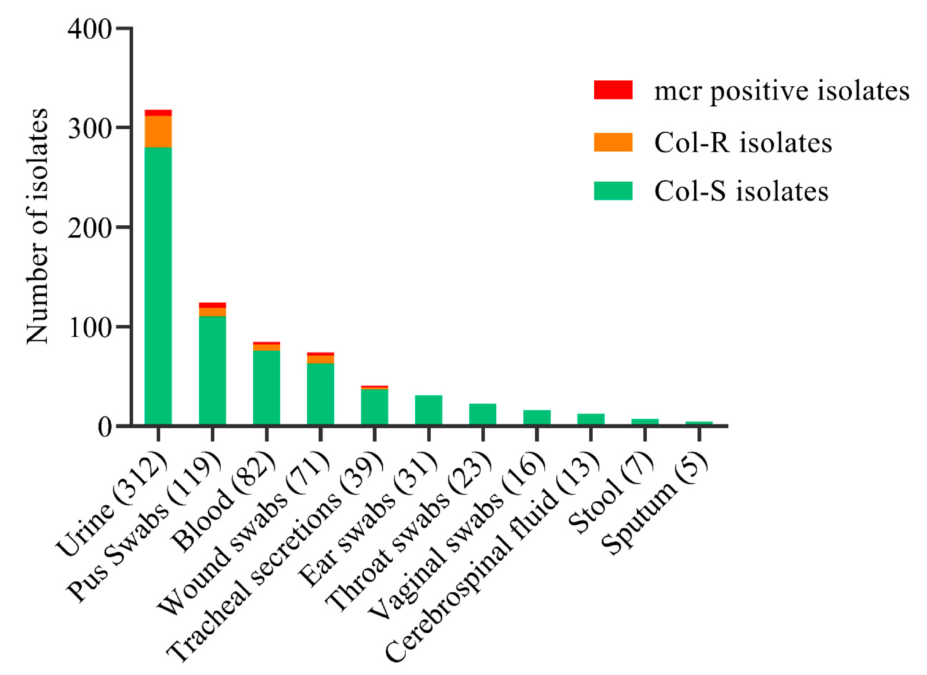
2.2. Distribution of Col-R and Col-S Bacterial Strains

Overall, 17/341 (5%) E. coli, 9/185 (4.9%) K. pneumoniae, 4/52 (7.7%) A. baumannii, 2/90 (2.2%) P. aeruginosa, and 25/25 (100%) P. mirabilis were identified as Col-R species. No significant association was observed between Col-R and mcr gene-harboring strains. We detected mcr genes in 9/17 (52.9%) E. coli (p = 0.43), 5/9 (55.6%) K. pneumoniae (p = 0.78), 3/4 (75%) A. baumannii (p = 0.43), and 2/2 (100%) P. aeruginosa (p = 0.49) Col-R species. None of the P. mirabilis strains were positive for mcr genes; however, these strains were found to be Col-R owing to intrinsic resistance (Table 2). Col-R specimens were obtained from 32/312 (10%) urine samples, 9/119 (7.6%) pus swabs, 6/82 (7.3%) blood samples, 8/71 (11.3%) wound swabs, and 2/39 (5.1%) tracheal secretions. Similarly, mcr sources were observed in 6/312 (1.9%) urine samples, 5/119 (4.2%) pus swabs, 3/82 (3.7%) blood samples, 3/71 (4.2%) wound swabs, and 2/39 (5.1%) tracheal secretions (Figure 2).

2.3. Molecular Detection of mcr and β-Lactam Drug-Resistant Gene Variants

The molecular analysis detected two mcr variants among the Col-R strains. Overall, 18/19 (95%) strains harbored mcr-1, and 1/19 (5%) strain harbored mcr-2 among all Col-R clinical isolates. The source of the only isolate carrying mcr-2 was a tracheal secretion isolated from an ICU case. All mcr-positive strains co-expressed drug-resistant β-lactamase genes. The most common ESBL-producing gene variants were 8/16 (50%) blaCTM-1 and 3/16 (19%) blaCTM-15. The AmpC gene variant blaCMY-2 was detected in 3/3 (100%) strains. Carbapenemase-producing strains included 2/8 (25%) blaNDM-1, 2/8 (25%) blaNDM-5, and 1/8 (13%) each of blaIPM, blaOXA-48, blaOXA-51, and blaVIM, respectively. Notably, 17 bacterial strains co-harbored integrons, of which 15/17 (88%) were Int1 and 2/17 (12%) were Int-2 (Table 3).

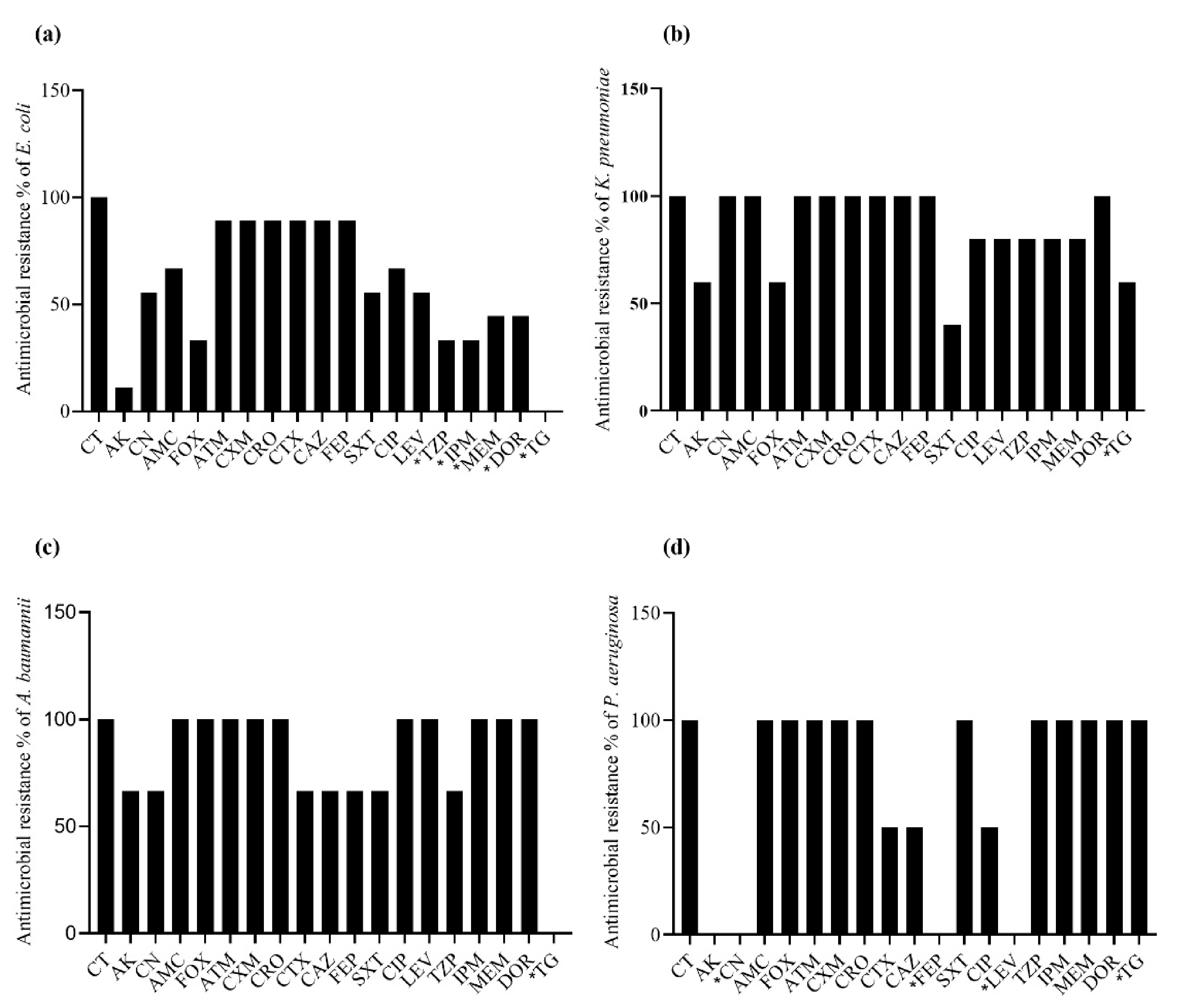
2.4. Antibacterial Resistance Spectrum in mcr-Positive Bacteria

Overall, the spectrum of MCRPBS showed extensive drug resistance against several antibiotics. A total of eight (88.9%) E. coli strains showed resistance to aztreonam, cefuroxime, ceftriaxone, cefotaxime, ceftazidime, and cefepime. Four (44.4%) E. coli isolates were resistant to each of meropenem and doripenem, and three (33.3%) were resistant to piperacillin-tazobactam and imipenem. None of the isolates showed resistance to tigecycline (Figure 3a). All of the K. pneumoniae strains were resistant to cephalosporin, aztreonam, co-amoxiclav, gentamicin, and doripenem. A total of three (60%) strains were resistant to amikacin, cefoxitin, and tigecycline, and only two isolates were resistant to (40%) co-trimoxazole (Figure 3b). Extensive drug resistance was also observed in the A. baumannii isolates; however, none of the A. baumannii isolates were found to be resistant to tigecycline (Figure 3c). All P. aeruginosa isolates were resistant to piperacillin-tazobactam, carbapenems, tigecycline, and several cephalosporin drugs. Despite drug resistance to several antibiotic classes, P. aeruginosa expressed no resistance to gentamicin, cefepime, and levofloxacin (Figure 3d).

2.5. MIC50 and MIC90 in mcr Gene-Harboring Bacterial Strains

The MIC to inhibit 50% growth (MIC50) and the MIC to inhibit 90% growth (MIC90) were observed using colistin (breakpoint ≥ 4 µg/mL) and other antibiotic groups, based on their respective breakpoints. The colistin MIC50 and MIC90 values were as follows: E. coli, 12 and 24 µg/mL; K. pneumoniae 12 and 32 µg/mL; A. baumannii 8 and 12 µg/mL; and P. aeruginosa 32 and 64 µg/mL, respectively. The MIC50 and MIC90 of all the isolates against each tested drug are listed in Table 4.

3. Discussion

The recent appearance of plasmid-mediated Col-R Enterobacteriaceae has drawn remarkable attention globally because this emergence has resulted in the deterioration of the last-resort antimicrobial commonly used to treat XDR bacterial infections, leading to reports of dramatic colistin inefficacy in several cases. This study examined 718 clinical isolates and identified 57 (7.9%) as Col-R and 661 (92.1%) as Col-S. These findings are consistent with a previous report from Colombia, in which (8.7%) Col-R was detected among clinical isolates [15]. The data from various research studies have reported a range from 0% to 31.7% Col-R [16,17] strains, which could be due to differences in factors such as poor infection control practices, the extensive use of colistin, and differences in the methodologies used to detect resistance. The literature suggests a lower prevalence of mcr-positive bacterial strains (MCRPBS) among human sources than among animal sources, which could indicate that plasmid-mediated colistin resistance first evolved in animal strains and then transferred to humans [1].
The prevalence of mcr in our study was 2.6%, similar to an earlier report, which showed a prevalence in Pakistan of 2.8% [18]. The findings of studies performed in other countries correspond well with those of our study, with studies in Iran and India reporting mcr rates among clinical isolates of 3% and 3.2%, respectively [19,20]. The mcr-1 gene is most commonly found in E. coli compared with other bacterial species globally [21,22]. We identified nine E. coli, five K. pneumoniae, three A. baumannii, and two P. aeruginosa isolates harboring mcr genes, which are similar proportions as those reported by a study performed in Korea [23]. Our findings show the wide distribution of mcr-harboring E. coli isolates, primarily recovered from urine, followed by pus swabs, blood, wound swabs, and tracheal secretions, which agrees with previous reports [24,25]. The frequency of mcr genes among clinical isolates was higher among women than men, consistent with an Iranian study [25]. There is no significant relationship between mcr detection and sex, and the only explanation for the higher occurrence in females is associated with the source of MCRPBS. Most of the mcr-harboring isolates were detected from the urinary samples, and urinary tract infections occur more frequently in women.
The molecular analysis showed two mcr variants among the Col-R strains, 95% mcr-1 and 5% mcr-2, consistent with global surveillance reports in which mcr-1 was detected in 75.8% of isolates from 18 countries [20]. Colistin has been used as the last-option antibacterial to treat XDR infections, but its efficacy has reduced since the emergence of mcr-positive strains [26], which has greatly compromised the therapeutic strategy for addressing MDR strains. MCRPBS in our study expressed extensive drug resistance to several classes of antibiotics. E. coli strains were resistant to aztreonam, cefuroxime, ceftriaxone, cefotaxime, ceftazidime, and cefepime and presented a variable spectrum of resistance to other classes of drugs. K. pneumoniae strains emerged as MDR and presented resistance to cephalosporin, aztreonam, co-amoxiclav, gentamicin, and doripenem. The findings of our study are consistent with those of an earlier report [25]. A. baumannii showed extensive drug resistance, and the diverse XDR strains had been reported by other studies [27,28,29].
The expanded drug resistance to β-lactams, aminoglycosides, carbapenems, and other antimicrobial drugs poses a significant global threat [30,31]. We found that high proportions of mcr-harboring isolates were characterized with an MDR profile, with particular resistance against third-generation cephalosporins. The expression of ESBLs and AmpC can explain the expansion of cephalosporin drug resistance, which is consistent with previously reported findings [20]. MCRPBS co-expressed ESBL gene variants (blaCTM-1 and blaCTM-15), one AmpC gene variant (blaCMY-2), and carbapenemase-producing genes (blaNDM-1, blaNDM-5, blaIPM, blaOXA-48, blaOXA-51, and blaVIM). These results agree with a previous report, in which 50% of mcr-harboring E. coli strains were found to be resistant to third-generation cephalosporins owing to the co-expression of blaCTX-M-2, and five isolates also expressed blaNDM and blaKPC [24]. Our study shows the co-existence of mcr-1 and blaNDM in several strains, which agrees with a previous report [32]. This study found one strain of P. aeruginosa co-harboring mcr-1 and blaNDM-5, which was recovered from urine, similar to a previous study in which a strain of E. coli co-expressing mcr-1 with blaNDM-5 was recovered from urine [33]. The most common ESBL-producing gene variants were 8/16 (50%) blaCTM-1 and 3/16 (19%) blaCTM-15, suggesting the possible dissemination between humans and animals owing to selective pressure between the animal and human environment.
Integrons contain a drug-resistance gene cassette that can act against many drug categories and represent a core component of multidrug resistance. We found 17 bacterial strains that co-harbored integrons, of which 88% were Int-1 and 12% were Int-2. Integrons are active in the development and dissemination of antibiotic resistance in gram-negative pathogens [34]. One limitation of the present study was that we focused on the most commonly reported mcr-1 to mcr-5 variants. We were unable to examine all of the mcr variants and sub-variants in the bacterial strains of clinical significance owing to limited resources.

4. Materials and Methods

4.1. Study Design and Ethics Approval

Bacterial strains were collected prospectively from various clinical settings located in Faisalabad and Lahore, Pakistan. The study design followed the ethical principles described by the World Medical Association (WMA) and the Declaration of Helsinki [35]. The study was performed as a collaboration between Government College University Faisalabad and Jouf University Saudi Arabia. The institutional review bodies for both institutions issued ethical approval for the analysis of bacterial strains. No human or animal trials were conducted during the study. Informed consent was not necessary because the strains were collected from human samples, but not associated with any individual patient data.

4.2. Specimen Collection and Processing

A total of 6879 clinical specimens were collected over six months from various sources and examined for bacterial isolation. The patients’ sources include blood, urine, pus, tracheal secretion, cerebrospinal fluid (CSF), stool, and different swabs. No environmental swabs or water samples from the hospital environment were included for analysis. Blood and MacConkey’s agar were used to culture all clinical specimens, except for blood and urine specimens. The blood samples were inoculated first in brain heart infusion broth. After a period of incubation following bacterial growth indicators, these cultures were subcultured on blood and MacConkey’s agar. The urine specimens were processed on cystine–lactose–electrolyte-deficient (CLED) agar. All the cultures were incubated at 35–37 °C overnight in an aerobic incubator.

4.3. Bacterial Growth and Characterization

Bacterial strains were phenotypically (growth characteristics) and biochemically characterized using Gram’s stain, conventional biochemical tests (catalase, oxidase, urease, and indole), and analytical profile index (API) 20E and 20NE (bioMérieux). Gram-negative rods (GNRs) were selected for further identification, and the remaining cultures were excluded from the analysis (Figure 1). Urine cultures showing GNRs with >105 colony-forming units (CFU)/mL were considered significant bacteriuria.

4.4. Resistance Profile and MIC Determination

The Col-R status of the retained GNRs was detected using SensiTest™ Colistin (Liofilchem, Via Scozia, Italy). Bacteria exhibiting MICs ≥4 µg/mL were phenotypically reported as Col-R strains. MCRPBSs were tested for MDR and XDR. MICs were determined against several antibacterial drugs using the broth microdilution method and E-test strips (Liofilchem, Via Scozia, Italy), and the inoculum size was standardized using a 0.5 McFarland standard. The tested antibacterial drugs included cephalosporin, fluoroquinolones, carbapenems, aminoglycosides, and β-lactam combined with colistin to determine co-resistance. The established MIC breakpoints were used to interpret the results as resistant and susceptible bacterial strains [36].

4.5. Screening of ESBLs, AmpC, and Carbapenemases

MCRPBSs were phenotypically characterized to detect the presence of other drug-resistant enzymes (ESBLs, AmpC, and carbapenemases). Phenotypically, ESBLs were identified by the hydrolysis of cefotaxime and ceftazidime and the formation of keyhole effect when using the conventional double-disk synergy technique [37,38]. AmpC was characterized by cefoxitin resistance and the enhancement of the inhibitory zone when using a combination of cefoxitin and boronic acid (inhibitor-based) compared with cefoxitin alone [39]. The isolates were screened for carbapenem resistance and subsequently confirmed for the presence of MBL by the zone enhancement in the presence of ethylenediaminetetraacetic acid (0.5 M EDTA) combined with a carbapenem in the disk-diffusion assay compared with carbapenem alone. Further confirmations were performed using the modified Hodge test [40,41].

4.6. Molecular Characterization of mcr Genes

Col-R strains were selected for the analysis of mcr-positive bacterial strains (MCRPBSs), and the presence of most frequently isolated mcr-1 to mcr-5 variants was detected. We used previously described primers and well-optimized multiplex polymerase chain reaction (PCR) conditions to detect the presence of mcr genes [8,42,43]. MCRPBSs were subcultured on nutrient agar to refresh the bacterial growth, and a few colonies from overnight strains were mixed in 500 µL TE buffer following the 0.5 McFarland standard. The suspension was placed in an Eppendorf tube, the caps were sealed with Parafilm to prevent accidental opening, and the tubes were boiled for 15 min in a preheated (100 °C) water bath. The mixture was centrifuged for five minutes at 14,000 rpm/min, and the supernatant was used to perform gene amplification [42]. A 50 µL reaction mixture was prepared for each specimen using 0.5 µM forward and reverse primers (mcr-1, 2, 3, 4, and 5), 2 µL template, 25 µL master mix, and 1.5 µL dimethyl sulfoxide (DMSO), and the final reaction mixture was brought to 50 µL using Milli-Q water. Amplified gene products were detected on agarose gel electrophoresis using 6× loading dye at 90 V for 50 min. A 100 bp DNA ladder was used to quantify the gene products, and a gel documentation system was used to visualize the genes.

4.7. Molecular Characterization ESBLs, AmpC, Carbapenemases, and Integrons

MCRPBS DNA was extracted as described for mcr sequencing and used to amplify drug-resistant genes. ESBL, AmpC, carbapenemases, and integron genes were separately amplified separately using previously described primers and optimized PCR conditions [13,38,44]. The amplicon was preserved at −20 °C for further analysis. DNA sequencing was performed using Big Dye v.3.1 (Life Technologies, Carlsbad, CA, USA) and ABI (Applied Biosystems, Waltham, MA, USA) DNA analyzer to identify the gene variants. FinchTV, NCBI (BlastN and BlastP), ExPASy, and ClustalW2 programs were used to analyze the gene variants.

4.8. Quality Control (QC) Analysis

The following QC strains were obtained from the American Type Culture Collection (ATCC): colistin-susceptible (Col-S) E. coli (25922) and Col-R Proteus mirabilis (25933); ESBL-positive K. pneumoniae (700603) and ESBL-negative E. coli (25922); and E. coli (BAA-2469) was used as an NDM-1.

4.9. Data Analysis

Data analysis was performed using the GraphPad Prism 8.0.2, IBM SPSS v.26, and BioVinci 3.0.0. A p-value of <0.05 was considered significant and descriptive statistics were used for the variables.

5. Conclusions

The study reports the emergence of mcr-1 and mcr-2 in several clinical isolates, including a high number of isolates that co-expressed blaCTM-1, blaCTM-15, blaCMY-2, blaNDM-1, blaNDM-5, and a few other β-lactamases. The detection of the mcr-2 gene variant in K. pneumoniae is a rarely reported finding. The co-expression of diverse gene variants among β-lactamase classes was well-supported by the simultaneous occurrence of Int-1 and Int-2, which can carry several drug-resistant gene cassettes. The molecular epidemiology of the co-expression of mcr and β-lactamases accentuates the increasing emergence of XDR clinical strains, which are difficult to treat and pose the massive threat of the clonal dissemination of these genes. Although we were able to identify therapeutic alternatives for each of the strains isolated in this study, our findings raise the question of how much time remains before a strain develops resistance against every available antimicrobial option. This situation represents a real danger to human lives and requires implacable surveillance, infection control, and the development of novel therapeutic regimens.

Author Contributions

Conceptualization, H.E., S.Y., M.U.Q., K.J., S.N.A.B. and K.O.A.A.; methodology, S.Y., A.E.A., A.A.M.A., M.Y.M.E., S.S.M.H., N.A. and E.H.M.S.; formal analysis, H.E., M.U.Q., S.N.A.B., N.A., A.E.A. and S.Y.; investigations, H.E., S.Y., K.O.A.A. and M.Y.M.E.; resources, H.E., S.Y. and K.J.; data curation, H.E., S.Y., K.J. and M.U.Q.; writing—original draft preparation, S.Y., A.E.A., A.A.M.A., M.Y.M.E., S.S.M.H. and E.H.M.S.; writing—review & editing N.A., A.E.A., S.S.M.H. and E.H.M.S.; supervision, H.E. and S.N.A.B.; project administration, H.E.; funding acquisition; H.E., S.Y., K.J. and K.O.A.A. All authors have read and agreed to the published version of the manuscript.

Funding

The authors’ work was supported through grant number “375213500” from the Deputyship for Research and Innovation, Ministry of Education in Saudi Arabia.

Institutional Review Board Statement

The study was conducted according to the guidelines of the Declaration of Helsinki, and approved by the Institutional Review Board of Jouf University, Saudi Arabia (HAP-13-s-001), and GC University Faisalabad, Pakistan (GCUF/ERC/20/17).

Informed Consent Statement

Not applicable.

Data Availability Statement

Data are contained within the article.

Acknowledgments

The authors extend their appreciation to the Deputyship for Research and Innovation, Ministry of Education in Saudi Arabia, and the central laboratory at Jouf University for supporting this study.

Conflicts of Interest

The authors declare no conflict of interest.

References

  1. Liu, Y.Y.; Wang, Y.; Walsh, T.R.; Yi, L.X.; Zhang, R.; Spencer, J.; Doi, Y.; Tian, G.; Dong, B.; Huang, X.; et al. Emergence of plasmid-mediated colistin resistance mechanism MCR-1 in animals and human beings in China: A microbiological and molecular biological study. Lancet Infect. Dis. 2016, 16, 161–168. [Google Scholar] [CrossRef]
  2. MacNair, C.R.; Stokes, J.M.; Carfrae, L.A.; Fiebig-Comyn, A.A.; Coombes, B.K.; Mulvey, M.R.; Brown, E.D. Overcoming mcr-1 mediated colistin resistance with colistin in combination with other antibiotics. Nat. Commun. 2018, 9, 458. [Google Scholar] [CrossRef] [PubMed] [Green Version]
  3. Temkin, E.; Adler, A.; Lerner, A.; Carmeli, Y. Carbapenem-resistant Enterobacteriaceae: Biology, epidemiology, and management. Ann. N. Y. Acad. Sci. 2014, 1323, 22–42. [Google Scholar] [CrossRef] [PubMed]
  4. Schwarz, S.; Johnson, A.P. Transferable resistance to colistin: A new but old threat. J. Antimicrob. Chemother. 2016, 71, 2066–2070. [Google Scholar] [CrossRef]
  5. Olaitan, A.O.; Morand, S.; Rolain, J.M. Mechanisms of polymyxin resistance: Acquired and intrinsic resistance in bacteria. Front. Microbiol. 2014, 5, 643. [Google Scholar] [CrossRef] [Green Version]
  6. Ling, Z.; Yin, W.; Shen, Z.; Wang, Y.; Shen, J.; Walsh, T.R. Epidemiology of mobile colistin resistance genes mcr-1 to mcr-9. J. Antimicrob. Chemother. 2020. [Google Scholar] [CrossRef]
  7. Malhotra-Kumar, S.; Xavier, B.B.; Das, A.J.; Lammens, C.; Butaye, P.; Goossens, H. Colistin resistance gene mcr-1 harboured on a multidrug resistant plasmid. Lancet Infect. Dis. 2016, 16, 283–284. [Google Scholar] [CrossRef] [Green Version]
  8. Javed, H.; Saleem, S.; Zafar, A.; Ghafoor, A.; Shahzad, A.B.; Ejaz, H.; Junaid, K.; Jahan, S. Emergence of plasmid-mediated mcr genes from Gram-negative bacteria at the human-animal interface. Gut Pathog. 2020, 12, 54. [Google Scholar] [CrossRef]
  9. Ejaz, H. Dissemination of SHV, TEM and CTX-M Genotypes in Pseudomonas aeruginosa: A Pre-eminent Reason for Therapeutic Failure in Pediatrics. Ann. Clin. Lab. Sci. 2020, 50, 797–805. [Google Scholar]
  10. Manchanda, V.; Singh, N.P. Occurrence and detection of AmpC beta-lactamases among Gram-negative clinical isolates using a modified three-dimensional test at Guru Tegh Bahadur Hospital, Delhi, India. J. Antimicrob. Chemother. 2003, 51, 415–418. [Google Scholar] [CrossRef]
  11. van Duin, D.; Doi, Y. Outbreak of Colistin-Resistant, Carbapenemase-Producing Klebsiella pneumoniae: Are We at the End of the Road? J. Clin. Microbiol. 2015, 53, 3116–3117. [Google Scholar] [CrossRef] [Green Version]
  12. Li, X.; Mu, X.; Zhang, P.; Zhao, D.; Ji, J.; Quan, J.; Zhu, Y.; Yu, Y. Detection and characterization of a clinical Escherichia coli ST3204 strain coproducing NDM-16 and MCR-1. Infect. Drug Resist. 2018, 11, 1189–1195. [Google Scholar] [CrossRef]
  13. Ejaz, H.; Alzahrani, B.; Hamad, M.F.S.; Abosalif, K.O.A.; Junaid, K.; Abdalla, A.E.; Elamir, M.Y.M.; Aljaber, N.J.; Hamam, S.S.M.; Younas, S. Molecular Analysis of the Antibiotic Resistant NDM-1 Gene in Clinical Isolates of Enterobacteriaceae. Clin. Lab. 2020, 66. [Google Scholar] [CrossRef]
  14. Wang, Y.; Tian, G.B.; Zhang, R.; Shen, Y.; Tyrrell, J.M.; Huang, X.; Zhou, H.; Lei, L.; Li, H.Y.; Doi, Y.; et al. Prevalence, risk factors, outcomes, and molecular epidemiology of mcr-1-positive Enterobacteriaceae in patients and healthy adults from China: An epidemiological and clinical study. Lancet Infect. Dis. 2017, 17, 390–399. [Google Scholar] [CrossRef] [Green Version]
  15. Saavedra, S.Y.; Diaz, L.; Wiesner, M.; Correa, A.; Arévalo, S.A.; Reyes, J.; Hidalgo, A.M.; de la Cadena, E.; Perenguez, M.; Montaño, L.A.; et al. Genomic and Molecular Characterization of Clinical Isolates of Enterobacteriaceae Harboring mcr-1 in Colombia, 2002 to 2016. Antimicrob. Agents Chemother. 2017, 61. [Google Scholar] [CrossRef] [Green Version]
  16. Bayram, Y.; Parlak, M.; Aypak, C.; Bayram, I. Three-year review of bacteriological profile and antibiogram of burn wound isolates in Van, Turkey. Int. J. Med. Sci. 2013, 10, 19–23. [Google Scholar] [CrossRef] [Green Version]
  17. Gill, M.M.; Usman, J.; Kaleem, F.; Hassan, A.; Khalid, A.; Anjum, R.; Fahim, Q. Frequency and antibiogram of multi-drug resistant Pseudomonas aeruginosa. J. Coll. Physicians Surg. Pak. 2011, 21, 531–534. [Google Scholar]
  18. Hameed, F.; Khan, M.A.; Muhammad, H.; Sarwar, T.; Bilal, H.; Rehman, T.U. Plasmid-mediated mcr-1 gene in Acinetobacter baumannii and Pseudomonas aeruginosa: First report from Pakistan. Rev. Soc. Bras. Med. Trop. 2019, 52, e20190237. [Google Scholar] [CrossRef] [Green Version]
  19. Aghapour, Z.; Hasani, A.; Aghazadeh, M.; Rezaee, M.A.; Ganbarov, K.; Pourlak, T.; Gholizadeh, P.; Asgharzadeh, M.; Tanomand, A.; Kafil, H. Genes involved in colistin resistance of gram-negative isolates in the northwest of Iran. Gene Rep. 2019, 14, 81–86. [Google Scholar] [CrossRef]
  20. Wise, M.G.; Estabrook, M.A.; Sahm, D.F.; Stone, G.G.; Kazmierczak, K.M. Prevalence of mcr-type genes among colistin-resistant Enterobacteriaceae collected in 2014-2016 as part of the INFORM global surveillance program. PLoS ONE 2018, 13, e0195281. [Google Scholar] [CrossRef] [Green Version]
  21. Elbediwi, M.; Li, Y.; Paudyal, N.; Pan, H.; Li, X.; Xie, S.; Rajkovic, A.; Feng, Y.; Fang, W.; Rankin, S.C.; et al. Global Burden of Colistin-Resistant Bacteria: Mobilized Colistin Resistance Genes Study (1980–2018). Microorganisms 2019, 7, 461. [Google Scholar] [CrossRef] [Green Version]
  22. Wang, R.; van Dorp, L.; Shaw, L.P.; Bradley, P.; Wang, Q.; Wang, X.; Jin, L.; Zhang, Q.; Liu, Y.; Rieux, A.; et al. The global distribution and spread of the mobilized colistin resistance gene mcr-1. Nat. Commun. 2018, 9, 1179. [Google Scholar] [CrossRef] [Green Version]
  23. Yoon, E.J.; Hong, J.S.; Yang, J.W.; Lee, K.J.; Lee, H.; Jeong, S.H. Detection of mcr-1 Plasmids in Enterobacteriaceae Isolates from Human Specimens: Comparison With Those in Escherichia coli Isolates From Livestock in Korea. Ann. Lab. Med. 2018, 38, 555–562. [Google Scholar] [CrossRef] [Green Version]
  24. Faccone, D.; Rapoport, M.; Albornoz, E.; Celaya, F.; De Mendieta, J.; De Belder, D.; Lucero, C.; Gomez, S.; Danze, D.; Pasteran, F.; et al. Plasmidic resistance to colistin mediated by mcr-1 gene in Escherichia coli clinical isolates in Argentina: A retrospective study, 2012–2018. Rev. Panam. Salud Publica 2020, 44, e55. [Google Scholar] [CrossRef]
  25. Moosavian, M.; Emam, N. The first report of emerging mobilized colistin-resistance (mcr) genes and ERIC-PCR typing in Escherichia coli and Klebsiella pneumoniae clinical isolates in southwest Iran. Infect. Drug Resist. 2019, 12, 1001–1010. [Google Scholar] [CrossRef] [Green Version]
  26. Giske, C.G. Contemporary resistance trends and mechanisms for the old antibiotics colistin, temocillin, fosfomycin, mecillinam and nitrofurantoin. Clin. Microbiol. Infect. 2015, 21, 899–905. [Google Scholar] [CrossRef] [Green Version]
  27. Baraka, A.; Traglia, G.M.; Montaña, S.; Tolmasky, M.E.; Ramirez, M.S. An Acinetobacter non-baumannii Population Study: Antimicrobial Resistance Genes (ARGs). Antibiotics 2020, 10, 16. [Google Scholar] [CrossRef]
  28. Lin, D.L.; Traglia, G.M.; Baker, R.; Sherratt, D.J.; Ramirez, M.S.; Tolmasky, M.E. Functional Analysis of the Acinetobacter baumannii XerC and XerD Site-Specific Recombinases: Potential Role in Dissemination of Resistance Genes. Antibiotics 2020, 9, 405. [Google Scholar] [CrossRef]
  29. Thirapanmethee, K.; Srisiri, A.N.T.; Houngsaitong, J.; Montakantikul, P.; Khuntayaporn, P.; Chomnawang, M.T. Prevalence of OXA-Type β-Lactamase Genes among Carbapenem-Resistant Acinetobacter baumannii Clinical Isolates in Thailand. Antibiotics 2020, 9, 864. [Google Scholar] [CrossRef]
  30. El-Mokhtar, M.A.; Daef, E.; Mohamed Hussein, A.A.R.; Hashem, M.K.; Hassan, H.M. Emergence of Nosocomial Pneumonia Caused by Colistin-Resistant Escherichia coli in Patients Admitted to Chest Intensive Care Unit. Antibiotics 2021, 10, 226. [Google Scholar] [CrossRef]
  31. Reeves, C.M.; Magallon, J.; Rocha, K.; Tran, T.; Phan, K.; Vu, P.; Yi, Y.; Oakley-Havens, C.L.; Cedano, J.; Jimenez, V.; et al. Aminoglycoside 6’-N-acetyltransferase Type Ib [AAC(6′)-Ib]-Mediated Aminoglycoside Resistance: Phenotypic Conversion to Susceptibility by Silver Ions. Antibiotics 2020, 10, 29. [Google Scholar] [CrossRef] [PubMed]
  32. Zheng, B.; Dong, H.; Xu, H.; Lv, J.; Zhang, J.; Jiang, X.; Du, Y.; Xiao, Y.; Li, L. Coexistence of MCR-1 and NDM-1 in Clinical Escherichia coli Isolates. Clin. Infect. Dis. 2016, 63, 1393–1395. [Google Scholar] [CrossRef] [PubMed] [Green Version]
  33. Mediavilla, J.R.; Patrawalla, A.; Chen, L.; Chavda, K.D.; Mathema, B.; Vinnard, C.; Dever, L.L.; Kreiswirth, B.N. Colistin- and Carbapenem-Resistant Escherichia coli Harboring mcr-1 and blaNDM-5, Causing a Complicated Urinary Tract Infection in a Patient from the United States. mBio 2016, 7. [Google Scholar] [CrossRef] [PubMed] [Green Version]
  34. Gillings, M.R. Integrons: Past, present, and future. Microbiol. Mol. Biol. Rev. 2014, 78, 257–277. [Google Scholar] [CrossRef] [Green Version]
  35. Association, W.M. WMA Declaration of Helsinki–Ethical Principles for Medical Research Involving Human Subjects. Available online: https://www.wma.net/policies-post/wma-declaration-of-helsinki-ethical-principles-for-medical-research-involving-human-subjects/ (accessed on 2 March 2021).
  36. CLSI. Performance Standards for Antimicrobial Susceptibility Testing, 30th ed.; Clinical and Laboratory Standard Institute (CLSI): Wayne, PA, USA, 2020; Volume CLSI, Supplement M100. [Google Scholar]
  37. Amin, H.; Zafar, A.; Ejaz, H.; Jameel, N.U. Phenotypic characterization of ESBL producing Enterobacter cloacae among children. Pak. J. Med. Sci. 2013, 29, 144–147. [Google Scholar] [CrossRef]
  38. Ejaz, H.; Younas, S.; Abosalif, K.O.A.; Junaid, K.; Alzahrani, B.; Alsrhani, A.; Abdalla, A.E.; Ullah, M.I.; Qamar, M.U.; Hamam, S.S.M. Molecular analysis of blaSHV, blaTEM, and blaCTX-M in extended-spectrum β-lactamase producing Enterobacteriaceae recovered from fecal specimens of animals. PLoS ONE 2021, 16, e0245126. [Google Scholar] [CrossRef]
  39. Younas, S.; Ejaz, H.; Zafar, A.; Ejaz, A.; Saleem, R.; Javed, H. AmpC beta-lactamases in Klebsiella pneumoniae: An emerging threat to the paediatric patients. J. Pak. Med. Assoc. 2018, 68, 893–897. [Google Scholar]
  40. Javed, H.; Ejaz, H.; Zafar, A.; Rathore, A.W. Metallo-beta-lactamase producing Escherichia coli and Klebsiella pneumoniae: A rising threat for hospitalized children. J. Pak. Med. Assoc. 2016, 66, 1068–1072. [Google Scholar]
  41. Qamar, M.U.; Ejaz, H.; Walsh, T.R.; Shah, A.A.; Al Farraj, D.A.; Alkufeidy, R.M.; Alkubaisi, N.A.; Saleem, S.; Jahan, S. Clonal relatedness and plasmid profiling of extensively drug-resistant New Delhi metallo-β-lactamase-producing Klebsiella pneumoniae clinical isolates. Future Microbiol. 2021. [Google Scholar] [CrossRef]
  42. Rebelo, A.R.; Bortolaia, V.; Kjeldgaard, J.S.; Pedersen, S.K.; Leekitcharoenphon, P.; Hansen, I.M.; Guerra, B.; Malorny, B.; Borowiak, M.; Hammerl, J.A.; et al. Multiplex PCR for detection of plasmid-mediated colistin resistance determinants, mcr-1, mcr-2, mcr-3, mcr-4 and mcr-5 for surveillance purposes. Euro Surveill 2018, 23. [Google Scholar] [CrossRef]
  43. Lv, D.; Duan, R.; Fan, R.; Mu, H.; Liang, J.; Xiao, M.; He, Z.; Qin, S.; Yang, J.; Jing, H.; et al. bla(NDM) and mcr-1 to mcr-5 Gene Distribution Characteristics in Gut Specimens from Different Regions of China. Antibiotics 2021, 10, 233. [Google Scholar] [CrossRef]
  44. Mohd Khari, F.I.; Karunakaran, R.; Rosli, R.; Tee Tay, S. Genotypic and Phenotypic Detection of AmpC β-lactamases in Enterobacter spp. Isolated from a Teaching Hospital in Malaysia. PLoS ONE 2016, 11, e0150643. [Google Scholar] [CrossRef] [Green Version]
Figure 1. Flow diagram showing the collection of clinical specimens, the isolation of bacterial strains, and the inclusion and exclusion of bacterial strains in the study. ESBL, extended-spectrum β-lactamase; MIC, minimum inhibitory concentration.
Antibiotics 10 00467 g001
Figure 2. Distribution of colistin-sensitive (Col-S), colistin-resistant (Col-R), and mcr genes among the gram-negative bacterial strains isolated from various sources (n = 718).
Antibiotics 10 00467 g002
Figure 3. Resistance profile of mcr-harboring gram-negative bacterial isolates (n = 19). Figure 3a–d show the resistance spectrum of E. coli, K. pneumoniae, A. baumannii and P. aeruginosa, respectively. CT, colistin; AK, amikacin; CN, gentamicin; AMC, co-amoxiclav; FOX, cefoxitin; ATM, aztreonam; CXM, cefuroxime; CRP, ceftriaxone; CTX, cefotaxime; CAZ, ceftazidime; FEP, cefepime; SXT, co-trimoxazole; CIP, ciprofloxacin; LEV, levofloxacin; TZP, piperacillin-tazobactam; IPM, imipenem; MEM, meropenem; DOR, doripenem; TG, tigecycline. * Significant p-values < 0.05.
Antibiotics 10 00467 g003
Table 1. Characteristics of cases infected with colistin-resistant (Col-R) and mcr-positive gram-negative bacterial strains.
CharacteristicCol-R (n = 57)
n (%)
mcr Detected (n = 19)
n (%)
p-Value
Sex
Male19 (33.3)7 (36.8)0.78
Female38 (66.7)12 (63.2)
Wards
Medical ward18 (31.6)5 (26.3)0.16
ICU7 (12.3)5 (26.3)0.01
Surgery6 (10.5)4 (21.1)0.01
Nephrology16 (28.1)3 (15.8)0.74
OPD9 (15.8)1 (5.3)0.71
Orthopedic1 (1.8)1 (5.3)0.12
Sources
Urine32 (56.1)6 (31.6)0.55
Pus9 (15.8)5 (26.3)0.01
Wound swab10 (17.5)4 (21.1)0.06
Blood4 (7)2 (10.5)0.38
Tracheal secretions2 (3.5)2 (10.5)0.03
ICU, intensive care unit; OPD, outpatient department; Chi-square test was used to obtain p-values.
Table 2. Association between mcr genes and colistin-resistant (Col-R) bacterial strains (n = 57).
Organismmcr Positive n (%)mcr Negative n (%)p-Value
Escherichia coli (n = 17)9 (52.9)8 (47.1)0.43
Klebsiella pneumoniae (n = 9)5 (55.6)4 (44.4)0.78
Acinetobacter baumannii (n = 4)3 (75)1 (25)0.49
Pseudomonas aeruginosa (n = 2)2 (100)0 (0)-
Proteus mirabilis (n = 25)0 (0)25 (100)-
Chi-square test was used to obtain p-values.
Table 3. Demographic and clinical characteristics and the co-existence of antibiotic-resistant genes in each gram-negative mcr-positive bacterial.
StrainSourceWardSexAge (years)OrganismDetected Genes
mcr ST-1UrineMedicalMale48E. coliCTX-M-1, mcr-1
mcr ST-2PusOPDFemale30K. pneumoniaeCTX-M-1, NDM-1, mcr-1, Int-I
mcr ST-3PusNephrologyFemale60E. coliCTX-M-15, mcr-1, Int-I
mcr ST-4UrineICUFemale53E. coliCTX-M-1, mcr-1, Int-I
mcr ST-5PusSurgeryFemale49K. pneumoniaeCTX-M-15, IMP, mcr-1, Int-I
mcr ST-6UrineMedicalMale45E. coliCMY-2, mcr-1, Int-I
mcr ST-7UrineNephrologyFemale53E. coliVIM, mcr-1, Int-I
mcr ST-8PusOrthopedicFemale30A. baumanniiNDM-5, mcr-1, Int-I
mcr ST-9WoundSurgeryMale25P. aeruginosaSHV-28, OXA-51, mcr-1, Int-I
mcr ST-10Tracheal secretionsICUMale71K. pneumoniaeCTX-M-1, NDM-1, mcr-2, Int-2
mcr ST-11WoundICUFemale65E. coliCTX-M-11, mcr-1, Int-I
mcr ST-12WoundICUFemale46A. baumanniiCTX-M-1, OXA-48, mcr-1, Int-I
mcr ST-13BloodNephrologyMale43E. coliCMY-2, mcr-1
mcr ST-14BloodMedicalFemale20K. pneumoniaeCTX-M-10, mcr-1, Int-I
mcr ST-15UrineMedicalFemale46E. coliTEM-52, CTX-M-15, mcr-1, Int-I
mcr ST-16WoundSurgeryMale43K. pneumoniaeCTX-M-1, mcr-1, Int-I
mcr ST-17Tracheal secretionsICUFemale54A. baumanniiCTX-M-1, mcr-1, Int-I
mcr ST-18UrineMedicalMale36P. aeruginosaCTX-M-1, CMY-2, NDM-5, mcr-1, Int-2
mcr ST-19PusSurgeryFemale52E. coliSHV-12, mcr-1, Int-I
Table 4. Comparison of the MIC50 and MIC90 (µg/mL) values among the bacterial strains harboring mcr genes.
AntibioticE. coli (n = 9)K. pneumoniae (n = 5)A. baumannii (n = 3)P. aeruginosa (n = 2)
BreakpointsMIC50MIC90BreakpointsMIC50MIC90BreakpointsMIC50MIC90BreakpointsMIC50MIC90
Colistin≥41224≥41232≥4812≥43264
Amikacin≥64264≥646464≥64128128≥6422
Gentamicin≥161632≥163232≥163232≥1622
Co-amoxiclav≥32/1632/1664/32≥32/1632/1664/32≥32/16≥128/64≥128/64≥32/16≥128/64≥128/64
Cefoxitin≥324≥128≥326464≥32≥128≥128≥32≥128≥128
Aztreonam≥1664≥128≥16≥128≥128≥32≥128≥128≥32≥128≥128
Cefuroxime≥32≥128≥128≥32≥128≥128≥32≥128≥128≥32≥128≥128
Ceftriaxone≥464≥128≥4≥128≥128≥64≥128≥128≥64≥128≥128
Cefotaxime≥464≥128≥4≥128≥128≥64≥128≥128≥64≥128≥128
Ceftazidime≥1664≥128≥16≥128≥128≥32≥128≥128≥32232
Cefepime≥163264≥16≥128≥128≥32≥128≥128≥3228
Co-trimoxazole≥4/764/768/152≥4/762/384/76≥4/764/7616/304≥4/7632/60864/1216
Ciprofloxacin≥116≥64≥13232≥4≥64≥64≥20.254
Levofloxacin≥2832≥23232≥8≥64≥64≥40.50.5
Piperacillin-tazobactam≥128/48/4512/4≥128/4128/4256/4≥128/4256/4512/4≥128/4256/4512/4
Imipenem≥4116≥46464≥8≥128≥128≥864≥128
Meropenem≥4132≥46464≥8≥128≥128≥8≥128≥128
Doripenem≥4132≥46464≥8≥128≥128≥8≥128≥128
Tigecycline≥20.51≥2816≥811≥8≥128≥128
Publisher’s Note: MDPI stays neutral with regard to jurisdictional claims in published maps and institutional affiliations.

