
Antimicrobial Activity of Selected Essential Oils against Selected Pathogenic Bacteria: In Vitro Study

 ,
,  ,
,  ,
,  and
and
Abstract
:1. Introduction
2. Results and Discussion
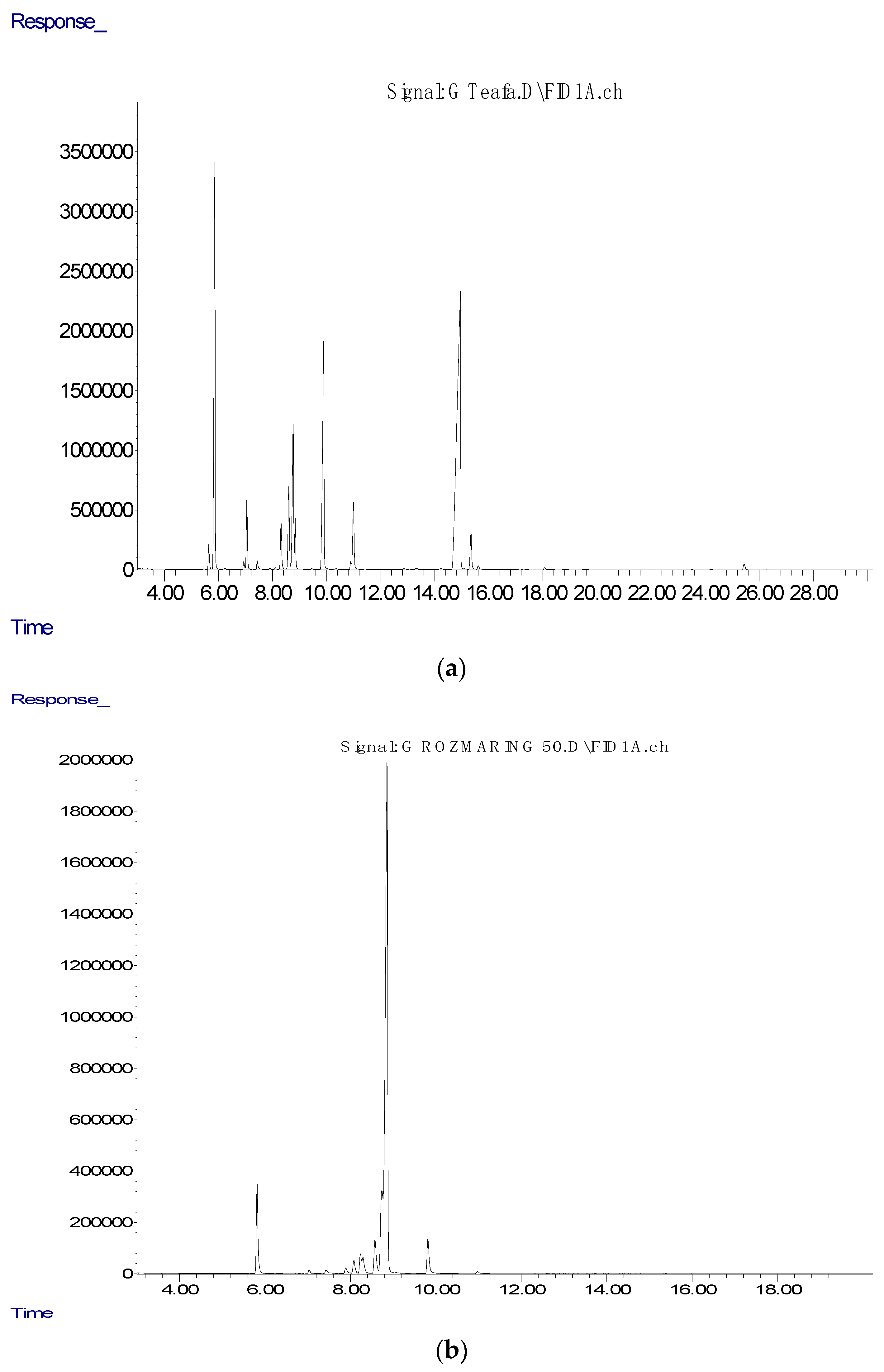
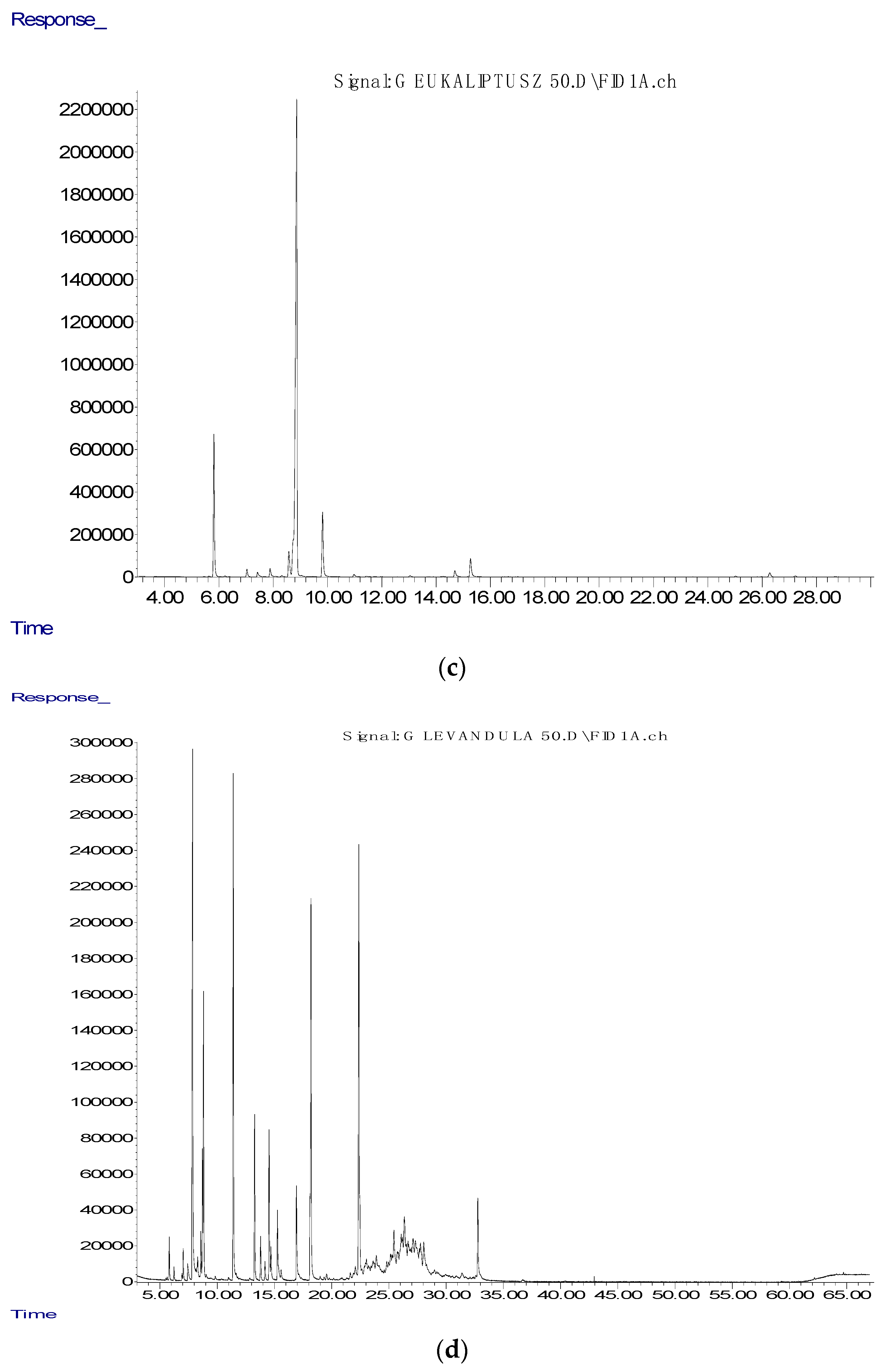
3. Material and Methods
4. Conclusions
Author Contributions
Funding
Data Availability Statement
Acknowledgments
Conflicts of Interest
References
- Puvača, N.; de Llanos Frutos, R. Antimicrobial Resistance in Escherichia coli Strains Isolated from Humans and Pet Animals. Antibiotics 2021, 10, 69. [Google Scholar] [CrossRef]
- Toombs-Ruane, L.J.; Benschop, J.; French, N.P.; Biggs, P.J.; Midwinter, A.C.; Marshall, J.C.; Chan, M.; Drinković, D.; Fayaz, A.; Baker, M.G.; et al. Carriage of Extended-Spectrum-Beta-Lactamase- and AmpC Beta-Lactamase-Producing Escherichia coli Strains from Humans and Pets in the Same Households. Appl. Environ. Microbiol. 2020, 86. [Google Scholar] [CrossRef] [PubMed]
- Friedrich, A.W. Control of Hospital Acquired Infections and Antimicrobial Resistance in Europe: The Way to Go. Wien. Med. Wochenschr. 2019, 169, 25–30. [Google Scholar] [CrossRef] [PubMed] [Green Version]
- Ljubojević, D.; Pelić, M.; Puvača, N.; Milanov, D. Resistance to Tetracycline in Escherichia coli Isolates from Poultry Meat: Epidemiology, Policy and Perspective. World’s Poult. Sci. J. 2017, 73, 409–417. [Google Scholar] [CrossRef]
- Ljubojević, D.; Velhner, M.; Todorović, D.; Pajić, M.; Milanov, D. Tetracycline Resistance in Escherichia coli Isolates from Poultry. Arch. Vet. Med. 2016, 9, 61–81. [Google Scholar] [CrossRef]
- Puvača, N.; Lika, E.; Tufarelli, V.; Bursić, V.; Pelić, D.L.; Nikolova, N.; Petrović, A.; Prodanović, R.; Vuković, G.; Lević, J.; et al. Influence of Different Tetracycline Antimicrobial Therapy of Mycoplasma (Mycoplasma synoviae) in Laying Hens Compared to Tea Tree Essential Oil on Table Egg Quality and Antibiotic Residues. Foods 2020, 9, 612. [Google Scholar] [CrossRef]
- Cosentino, S.; Tuberoso, C.I.G.; Pisano, B.; Satta, M.; Mascia, V.; Arzedi, E.; Palmas, F. In-Vitro Antimicrobial Activity and Chemical Composition of Sardinian Thymus Essential Oils. Lett. Appl. Microbiol. 1999, 29, 130–135. [Google Scholar] [CrossRef]
- Canter, P.H.; Thomas, H.; Ernst, E. Bringing Medicinal Plants into Cultivation: Opportunities and Challenges for Biotechnology. Trends Biotechnol. 2005, 23, 180–185. [Google Scholar] [CrossRef]
- Joana Gil-Chávez, G.; Villa, J.A.; Fernando Ayala-Zavala, J.; Basilio Heredia, J.; Sepulveda, D.; Yahia, E.M.; González-Aguilar, G.A. Technologies for Extraction and Production of Bioactive Compounds to Be Used as Nutraceuticals and Food Ingredients: An overview. Compr. Rev. Food Sci. Food Saf. 2013, 12, 5–23. [Google Scholar] [CrossRef]
- Ebani, V.V.; Nardoni, S.; Bertelloni, F.; Pistelli, L.; Mancianti, F. Antimicrobial Activity of Five Essential Oils against Bacteria and Fungi Responsible for Urinary Tract Infections. Molecules 2018, 23, 1668. [Google Scholar] [CrossRef] [Green Version]
- Ali, B.; Al-Wabel, N.A.; Shams, S.; Ahamad, A.; Khan, S.A.; Anwar, F. Essential Oils Used in Aromatherapy: A Systemic Review. Asian Pac. J. Trop. Biomed. 2015, 5, 601–611. [Google Scholar] [CrossRef] [Green Version]
- Tanveer, M.; Wagner, C.; ul Haq, M.I.; Ribeiro, N.C.; Rathinasabapathy, T.; Butt, M.S.; Shehzad, A.; Komarnytsky, S. Spicing up Gastrointestinal Health with Dietary Essential Oils. Phytochem. Rev. 2020, 19, 243–263. [Google Scholar] [CrossRef]
- Abu-Al-Basal, M.A. Healing Potential of Rosmarinus officinalis L. on Full-Thickness Excision Cutaneous Wounds in Alloxan-Induced-Diabetic BALB/c Mice. J. Ethnopharmacol. 2010, 131, 443–450. [Google Scholar] [CrossRef]
- Ignat, I.; Volf, I.; Popa, V.I. A Critical Review of Methods for Characterisation of Polyphenolic Compounds in Fruits and Vegetables. Food Chem. 2011, 126, 1821–1835. [Google Scholar] [CrossRef] [PubMed]
- Nhu-Trang, T.-T.; Casabianca, H.; Grenier-Loustalot, M.-F. Deuterium/Hydrogen Ratio Analysis of Thymol, Carvacrol, γ-Terpinene and p-Cymene in Thyme, Savory and Oregano Essential Oils by Gas Chromatography–Pyrolysis–Isotope Ratio Mass Spectrometry. J. Chromatogr. A 2006, 1132, 219–227. [Google Scholar] [CrossRef]
- Misharina, T.A.; Terenina, M.B.; Krikunova, N.I. Antioxidant Properties of Essential Oils. Appl. Biochem. Microbiol. 2009, 45, 642–647. [Google Scholar] [CrossRef]
- Bassolé, I.H.N.; Juliani, H.R. Essential Oils in Combination and Their Antimicrobial Properties. Molecules 2012, 17, 3989–4006. [Google Scholar] [CrossRef] [Green Version]
- Reichling, J.; Schnitzler, P.; Suschke, U.; Saller, R. Essential Oils of Aromatic Plants with Antibacterial, Antifungal, Antiviral, and Cytotoxic properties—An Overview. Complement. Med. Res. 2009, 16, 79–90. [Google Scholar] [CrossRef] [Green Version]
- Formisano, C.; Oliviero, F.; Rigano, D.; Saab, A.M.; Senatore, F. Chemical Composition of Essential Oils and in Vitro Antioxidant Properties of Extracts and Essential Oils of Calamintha Origanifolia and Micromeria Myrtifolia, Two Lamiaceae from the Lebanon Flora. Ind. Crops Prod. 2014, 62, 405–411. [Google Scholar] [CrossRef]
- Wannissorn, B.; Jarikasem, S.; Siriwangchai, T.; Thubthimthed, S. Antibacterial Properties of Essential Oils from Thai Medicinal Plants. Fitoterapia 2005, 76, 233–236. [Google Scholar] [CrossRef]
- Pavela, R. Insecticidal Properties of Several Essential Oils on the House Fly (Musca domestica L.). Phytother. Res. 2008, 22, 274–278. [Google Scholar] [CrossRef] [PubMed]
- Puškárová, A.; Bučková, M.; Kraková, L.; Pangallo, D.; Kozics, K. The Antibacterial and Antifungal Activity of Six Essential Oils and Their Cyto/Genotoxicity to Human HEL 12469 Cells. Sci. Rep. 2017, 7, 8211. [Google Scholar] [CrossRef] [Green Version]
- Romero Rocamora, C.; Ramasamy, K.; Meng Lim, S.; Majeed, A.B.A.; Agatonovic-Kustrin, S. HPTLC Based Approach for Bioassay-Guided Evaluation of Antidiabetic and Neuroprotective Effects of Eight Essential Oils of the Lamiaceae Family Plants. J. Pharm. Biomed. Anal. 2020, 178, 112909. [Google Scholar] [CrossRef] [PubMed]
- Usai, M.; Marchetti, M.; Culeddu, N.; Mulas, M. Chemical Composition of Myrtle (Myrtus communis L.) Berries Essential Oils as Observed in a Collection of Genotypes. Molecules 2018, 23, 2502. [Google Scholar] [CrossRef] [Green Version]
- Jamoussi, B.; Romdhane, M.; Abderraba, A.; Hassine, B.B.; Gadri, A.E. Effect of Harvest Time on the Yield and Composition of Tunisian Myrtle Oils. Flavour Fragr. J. 2005, 20, 274–277. [Google Scholar] [CrossRef]
- Puvača, N.; Stanaćev, V.; Glamočić, D.; Lević, J.; Perić, L.; Stanaćev, V.; Milić, D. Beneficial Effects of Phytoadditives in Broiler Nutrition. World’s Poult. Sci. J. 2013, 69, 27–34. [Google Scholar] [CrossRef]
- Parkhill, J.; Dougan, G.; James, K.D.; Thomson, N.R.; Pickard, D.; Wain, J.; Churcher, C.; Mungall, K.L.; Bentley, S.D.; Holden, M.T.G.; et al. Complete Genome Sequence of a Multiple Drug Resistant Salmonella Enterica Serovar Typhi CT18. Nature 2001, 413, 848–852. [Google Scholar] [CrossRef]
- Townsend, S.M.; Pollack, H.A.; Gonzalez-Gomez, I.; Shimada, H.; Badger, J.L. Citrobacter koseri Brain Abscess in the Neonatal Rat: Survival and Replication within Human and Rat Macrophages. Infect. Immun. 2003, 71, 5871–5880. [Google Scholar] [CrossRef] [Green Version]
- Weaver, C.M. Bioactive Foods and Ingredients for Health. Adv. Nutr. 2014, 5, 306S–311S. [Google Scholar] [CrossRef] [Green Version]
- Singla, R.; Mishra, A.; Joshi, R.; Jha, S.; Sharma, A.R.; Upadhyay, S.; Sarma, P.; Prakash, A.; Medhi, B. Human Animal Interface of SARS-CoV-2 (COVID-19) Transmission: A Critical Appraisal of Scientific Evidence. Vet. Res. Commun. 2020, 44, 119–130. [Google Scholar] [CrossRef]
- Kris-Etherton, P.M.; Hecker, K.D.; Bonanome, A.; Coval, S.M.; Binkoski, A.E.; Hilpert, K.F.; Griel, A.E.; Etherton, T.D. Bioactive Compounds in Foods: Their Role in the Prevention of Cardiovascular Disease and Cancer. Am. J. Med. 2002, 113, 71S–88S. [Google Scholar] [CrossRef]
- Crozier, A.; Jaganath, I.B.; Clifford, M.N. Dietary Phenolics: Chemistry, Bioavailability and Effects on Health. Nat. Prod. Rep. 2009, 26, 1001–1043. [Google Scholar] [CrossRef]
- Stein, S.E.; Mikaia, A.; Linstrom, P.; Mirokhin, Y.; Tchekhovskoi, D.; Yang, X.; Mallard, W.G.; Sparkman, O.D.; Sparkman, J.A. NIST 11. Standard Reference Database 1A, Mass Spectral Database. 2011. Available online: https://www.nist.gov/sites/default/files/documents/srd/NIST1a11Ver2-0Man.pdf (accessed on 8 April 2021).
- Dubnicka, M.; Cromwell, B.; Levine, M. Investigation of the Adulteration of Essential Oils by GC-MS. Curr. Anal. Chem. 2020, 16, 965–969. [Google Scholar] [CrossRef]
- Zhou, J.; Tang, F.; Mao, G.; Bian, R. Effect of Alpha-Pinene on Nuclear Translocation of NF-Kappa B in THP-1 Cells. Acta Pharmacol. Sin. 2004, 25, 480–484. [Google Scholar] [PubMed]
- Gershenzon, J.; Dudareva, N. The Function of Terpene Natural Products in the Natural World. Nat. Chem. Biol. 2007, 3, 408–414. [Google Scholar] [CrossRef]
- Ciriminna, R.; Lomeli-Rodriguez, M.; Demma Carà, P.; Lopez-Sanchez, J.A.; Pagliaro, M. Limonene: A Versatile Chemical of the Bioeconomy. Chem. Commun. 2014, 50, 15288–15296. [Google Scholar] [CrossRef]
- Ramalho, T.; Pacheco de Oliveira, M.; Lima, A.; Bezerra-Santos, C.; Piuvezam, M. Gamma-Terpinene Modulates Acute Inflammatory Response in Mice. Planta Med. 2015, 81, 1248–1254. [Google Scholar] [CrossRef] [Green Version]
- Pateiro, M.; Barba, F.J.; Domínguez, R.; Sant’Ana, A.S.; Mousavi Khaneghah, A.; Gavahian, M.; Gómez, B.; Lorenzo, J.M. Essential Oils as Natural Additives to Prevent Oxidation Reactions in Meat and Meat Products: A Review. Food Res. Int. 2018, 113, 156–166. [Google Scholar] [CrossRef]
- Puvača, N.; Čabarkapa, I.; Petrović, A.; Bursić, V.; Prodanović, R.; Soleša, D.; Lević, J. Tea Tree (Melaleuca alternifolia) and Its Essential Oil: Antimicrobial, Antioxidant and Acaricidal Effects in Poultry Production. World’s Poult. Sci. J. 2019, 75, 235–246. [Google Scholar] [CrossRef]
- Hendel, N.; Napoli, E.; Sarri, M.; Saija, A.; Cristani, M.; Nostro, A.; Ginestra, G.; Ruberto, G. Essential Oil from Aerial Parts of Wild Algerian Rosemary: Screening of Chemical Composition, Antimicrobial and Antioxidant Activities. J. Essent. Oil Bear. Plants 2019, 22, 1–17. [Google Scholar] [CrossRef]
- Pant, M.; Dubey, S.; Patanjali, P.K.; Naik, S.N.; Sharma, S. Insecticidal Activity of Eucalyptus Oil Nanoemulsion with Karanja and Jatropha Aqueous Filtrates. Int. Biodeterior. Biodegrad. 2014, 91, 119–127. [Google Scholar] [CrossRef]
- Reyes, E.I.M.; Farias, E.S.; Silva, E.M.P.; Filomeno, C.A.; Plata, M.A.B.; Picanço, M.C.; Barbosa, L.C.A. Eucalyptus Resinifera Essential Oils Have Fumigant and Repellent Action against Hypothenemus hampei. Crop Prot. 2019, 116, 49–55. [Google Scholar] [CrossRef]
- Yadikar, N.; Bobakulov, K.; Li, G.; Aisa, H.A. Seven New Phenolic Compounds from Lavandula angustifolia. Phytochem. Lett. 2018, 23, 149–154. [Google Scholar] [CrossRef]
- Sen, I.; Shrivastava, D.; Aggarwal, M.; Kumar Khandal, R. Carbitol as Adulterant in Menthol; Analytical Method for Quantitative Analysis of Adulteration. AIMS Agric. Food 2020, 5, 129–136. [Google Scholar] [CrossRef]
- Donadu, M.; Usai, D.; Pinna, A.; Porcu, T.; Mazzarello, V.; Fiamma, M.; Marchetti, M.; Cannas, S.; Delogu, G.; Zanetti, S.; et al. In Vitro Activity of Hybrid Lavender Essential Oils against Multidrug Resistant Strains of Pseudomonas Aeruginosa. J. Infect. Dev. Ctries 2018, 12. [Google Scholar] [CrossRef] [PubMed] [Green Version]
- Puvača, N.; Bursić, V.; Petrović, A.; Prodanović, R.; Kharud, M.M.; Obućinski, D.; Vuković, G.; Marić, M. Influence of tea tree essential oil on the synthesis of mycotoxins: Ochratoxin A. Maced. J. Anim. Sci. 2019, 9, 25–29. [Google Scholar]
- Jasovský, D.; Littmann, J.; Zorzet, A.; Cars, O. Antimicrobial Resistance—A Threat to the World’s Sustainable Development. Upsala J. Med Sci. 2016, 121, 159–164. [Google Scholar] [CrossRef] [Green Version]
- De Oliveira, D.M.P.; Forde, B.M.; Kidd, T.J.; Harris, P.N.A.; Schembri, M.A.; Beatson, S.A.; Paterson, D.L.; Walker, M.J. Antimicrobial Resistance in ESKAPE Pathogens. Clin. Microbiol. Rev. 2020, 33, e00181-19. [Google Scholar] [CrossRef]
- Farzaneh, V.; Carvalho, I.S. A Review of the Health Benefit Potentials of Herbal Plant Infusions and Their Mechanism of Actions. Ind. Crops Prod. 2015, 65, 247–258. [Google Scholar] [CrossRef]
- Poolman, J.T.; Anderson, A.S. Escherichia coli and Staphylococcus aureus: Leading Bacterial Pathogens of Healthcare Associated Infections and Bacteremia in Older-Age Populations. Expert Rev. Vaccines 2018, 17, 607–618. [Google Scholar] [CrossRef]
- Li, W.-R.; Li, H.-L.; Shi, Q.-S.; Sun, T.-L.; Xie, X.-B.; Song, B.; Huang, X.-M. The Dynamics and Mechanism of the Antimicrobial Activity of Tea Tree Oil against Bacteria and Fungi. Appl. Microbiol. Biotechnol. 2016, 100, 8865–8875. [Google Scholar] [CrossRef]
- Jiang, H.; Zheng, H. Efficacy and Adverse Reaction to Different Doses of Atorvastatin in the Treatment of Type II Diabetes Mellitus. Biosci. Rep. 2019, 39, BSR20182371. [Google Scholar] [CrossRef] [Green Version]
- Jafari-Sales, A.; Pashazadeh, M. Study of Chemical Composition and Antimicrobial Properties of Rosemary (Rosmarinus Officinalis) Essential Oil on Staphylococcus aureus and Escherichia coli In Vitro. Int. J. Life Sci. Biotechnol. 2020. [Google Scholar] [CrossRef]
- Bachir, R.G.; Benali, M. Antibacterial Activity of the Essential Oils from the Leaves of Eucalyptus Globulus against Escherichia coli and Staphylococcus aureus. Asian Pac. J. Trop. Biomed. 2012, 2, 739–742. [Google Scholar] [CrossRef] [Green Version]
- Predoi, D.; Iconaru, S.; Buton, N.; Badea, M.; Marutescu, L. Antimicrobial Activity of New Materials Based on Lavender and Basil Essential Oils and Hydroxyapatite. Nanomaterials 2018, 8, 291. [Google Scholar] [CrossRef] [PubMed] [Green Version]
- Saraiva, M. In Vitro Evaluation of Antioxidant, Antimicrobial and Toxicity Properties of Extracts of Schinopsis Brasiliensis Engl. (Anacardiaceae). Afr. J. Pharm. Pharmacol. 2011, 5, 1724–1731. [Google Scholar] [CrossRef] [Green Version]
- Silva, A.C.O.; Santana, E.F.; Saraiva, A.M.; Coutinho, F.N.; Castro, R.H.A.; Pisciottano, M.N.C.; Amorim, E.L.C.; Albuquerque, U.P. Which Approach Is More Effective in the Selection of Plants with Antimicrobial Activity? Evid.-Based Complementary Altern. Med. 2013, 2013, 1–9. [Google Scholar] [CrossRef]
- Christoph, F.; Kaulfers, P.-M.; Stahl-Biskup, E. A Comparative Study of the in Vitro Antimicrobial Activity of Tea Tree Oils s.l. with Special Reference to the Activity of β-Triketones. Planta Med. 2000, 66, 556–560. [Google Scholar] [CrossRef]
- Cox, S.D.; Mann, C.M.; Markham, J.L.; Bell, H.C.; Gustafson, J.E.; Warmington, J.R.; Wyllie, S.G. The Mode of Antimicrobial Action of the Essential Oil of Melaleuca alternifolia (Tea Tree Oil): S.D. COX ET AL. J. Appl. Microbiol. 2001, 88, 170–175. [Google Scholar] [CrossRef] [PubMed]
- Mohsen, E.H.K.; Hossein, J.; Behrooz, A.B.; Mohammad, N. Antimicrobial Activity of Rosemary Essential Oil and Its Interaction with Common Therapeutic Antibiotics on Some Gram Positive and Gram Negative Bacteria. Iran. J. Infect. Dis. Trop. Med. 2020, 24, 25–34. [Google Scholar]
- Elaissi, A.; Rouis, Z.; Mabrouk, S.; Salah, K.B.H.; Aouni, M.; Khouja, M.L.; Farhat, F.; Chemli, R.; Harzallah-Skhiri, F. Correlation Between Chemical Composition and Antibacterial Activity of Essential Oils from Fifteen Eucalyptus Species Growing in the Korbous and Jbel Abderrahman Arboreta (North East Tunisia). Molecules 2012, 17, 3044–3057. [Google Scholar] [CrossRef] [PubMed] [Green Version]
- Vaghasiya, Y.; Nair, R.; Chanda, S. Antibacterial and Preliminary Phytochemical and Physico-Chemical Analysis of Eucalyptus citriodora Hk Leaf. Nat. Prod. Res. 2008, 22, 754–762. [Google Scholar] [CrossRef]
- Adaszyńska-Skwirzyńska, M.; Szczerbińska, D. Use of Essential Oils in Broiler Chicken Production—A Review. Ann. Anim. Sci. 2017, 17, 317–335. [Google Scholar] [CrossRef] [Green Version]
- Kunicka-Styczyńska, A.; Sikora, M.; Kalemba, D. Antimicrobial Activity of Lavender, Tea Tree and Lemon Oils in Cosmetic Preservative Systems: Antimicrobial Action of Oils in Cosmetics. J. Appl. Microbiol. 2009, 107, 1903–1911. [Google Scholar] [CrossRef]
- Bosnić, T.; Softić, D.; Grujić-Vasić, J. Antimicrobial Activity of Some Essential Oils and Major Constituents of Essential Oils. Acta Med. Acad. 2006, 35, 9–14. [Google Scholar]
- Hanamanthagouda, M.S.; Kakkalameli, S.B.; Naik, P.M.; Nagella, P.; Seetharamareddy, H.R.; Murthy, H.N. Essential Oils of Lavandula Bipinnata and Their Antimicrobial Activities. Food Chem. 2010, 118, 836–839. [Google Scholar] [CrossRef]


| Compound | Retention Indices | Retention Indices NIST 1 | Retention Time | Tea Tree | Rosemary | Eucalyptus | Lavender |
|---|---|---|---|---|---|---|---|
| α-Thujene | 922 | 924 | 5.636 | 1.10 ± 0.01 | 0.03 ± 0.00 | 0.06 ± 0.00 | 0.05 ± 0.01 |
| α-Pinene | 930 | 932 | 5.862 | 18.38 ± 0.08 | 8.38 ± 0.02 | 12.60 ± 0.01 | 0.72 ± 0.00 |
| Camphene | 945 | 946 | 6.241 | 0.08 ± 0.00 | 0.03 ± 0.00 | 0.10 ± 0.00 | 0.25 ± 0.01 |
| Thuja-2,4(10)-diene | 950 | 952 | 6.378 | 0.01 ± 0.00 | |||
| Sabinene | 970 | 969 | 6.932 | 0.35 ± 0.01 | 0.12 ± 0.00 | ||
| β-Pinene | 974 | 974 | 7.047 | 3.19 ± 0.01 | 0.38 ± 0.01 | 0.84 ± 0.01 | 0.60 ± 0.02 |
| Myrcene | 988 | 988 | 7.428 | 0.45 ± 0.00 | 0.49 ± 0.00 | 0.58 ± 0.01 | 0.56 ± 0.01 |
| Carbitol | 1003 | 1001 | 7.863 | 13.05 ± 0.04 | |||
| α-Phellandrene | 1004 | 1002 | 7.9 | 0.09 ± 0.00 | 0.68 ± 0.00 | 0.94 ± 0.00 | |
| Δ3-Carene | 1009 | 1008 | 8.098 | 0.09 ± 0.00 | 1.45 ± 0.03 | 0.05 ± 0.01 | |
| Hexyl acetate | 1011 | 1009 | 8.146 | 0.13 ± 0.01 | |||
| 1,4-Cineole | 1013 | 1012 | 8.235 | ||||
| α-Terpinene | 1015 | 1014 | 8.311 | 2.35 ± 0.01 | 2.02 ± 0.01 | 0.15 ± 0.00 | 0.41 ± 0.00 |
| p-Cymene | 1023 | 1020 | 8.598 | 4.30 ± 0.01 | 4.30 ± 0.05 | 3.24 ± 0.00 | 0.87 ± 0.01 |
| Limonene | 1027 | 1024 | 8.758 | 7.55 ± 0.01 | 11.86 ± 0.01 | 3.87 ± 0.01 | 2.23 ± 0.02 |
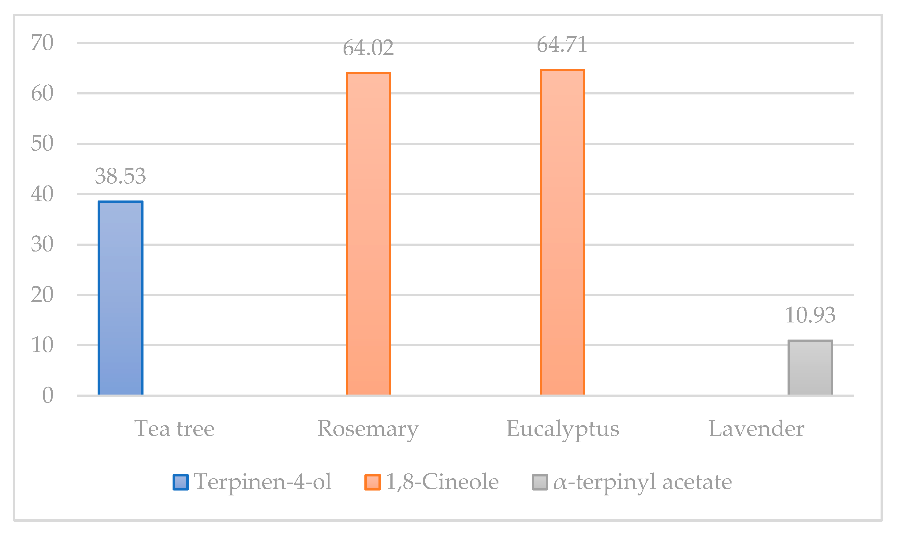
| 1,8-Cineole | 1033 | 1026 | 8.864 | 2.15 ± 0.05 | 64.02 ± 0.04 | 64.71 ± 0.04 | 5.55 ± 0.01 |
| (Z)-β-ocimene | 1035 | 1032 | 9.035 | 0.28 ± 0.00 | 0.06 ± 0.00 | ||
| β-(E)-Ocimene | 1046 | 1046 | 9.45 | 0.08 ± 0.00 | 0.11 ± 0.00 | 0.02 ± 0.00 | |
| γ-Terpinene | 1058 | 1054 | 9.89 | 14.01 ± 0.01 | 4.06 ± 0.00 | 7.37 ± 0.00 | 0.05 ± 0.00 |
| p-Mentha-2,4(8)-diene | 1085 | 1083 | 10.891 | 0.38 ± 0.01 | |||
| Terpinolene | 1088 | 1086 | 10.991 | 3.56 ± 0.02 | 0.31 ± 0.00 | 0.35 ± 0.00 | 0.04 ± 0.00 |
| Linalool | 1099 | 1095 | 11.423 | 0.05 ± 0.00 | 0.10 ± 0.00 | 10.71 ± 0.02 | |
| trans-Sabinol | 1137 | 1137 | 13.036 | 0.06 ± 0.00 | 0.14 ± 0.00 | ||
| Camphor | 1143 | 1141 | 13.267 | 0.12 ± 0.00 | 3.72 ± 0.03 | ||
| Isoborneol | 1154 | 1155 | 13.787 | 1.04 ± 0.02 | |||
| Borneol | 1164 | 1165 | 14.24 | 0.14 ± 0.00 | 0.46 ± 0.01 | ||
| Isononyl acetate | 1171 | 1171 | 14.53 | 3.45 ± 0.01 | |||
| Terpinen-4-ol | 1180 | 1174 | 14.944 | 38.53 ± 0.04 | 0.95 ± 0.00 | 0.90 ± 0.02 | |
| α-Terpineol | 1190 | 1186 | 15.34 | 2.16 ± 0.03 | 2.50 ± 0.01 | 2.00 ± 0.00 | |
| γ-Terpineol | 1196 | 1199 | 15.606 | 0.21 ± 0.00 | |||
| Citronellol | 1226 | 1223 | 16.923 | 2.50 ± 0.00 | |||
| Geraniol | 1254 | 1249 | 18.11 | 1.28 ± 0.00 | |||
| Linalool acetate | 1255 | 1254 | 18.194 | 9.60 ± 0.02 | |||
| Bornyl acetate | 1285 | 1287 | 19.562 | 0.21 ± 0.00 | |||
| α-terpinyl acetate | 1349 | 1346 | 22.374 | 10.93 ± 0.06 | |||
| Neryl acetate | 1364 | 1359 | 23.038 | 0.44 ± 0.00 | |||
| Geranyl acetate | 1384 | 1379 | 23.898 | 0.80 ± 0.02 | |||
| α-Gurjunene | 1409 | 1409 | 25.023 | 0.12 ± 0.00 | |||
| (E)-Caryophyllene | 1420 | 1417 | 25.443 | 0.38 ± 0.01 | 1.80 ± 0.00 | ||
| Aromadendrene | 1439 | 1439 | 26.282 | 0.69 ± 0.01 | |||
| 9-epi-Caryophyllene | 1462 | 1464 | 27.225 | 0.17 ± 0.00 | |||
| Viridiflorene | 1497 | 1496 | 28.693 | 0.07 ± 0.00 | |||
| Total peak area | 564,685,150 | 117,582,225 | 142,637,552 | 98,030,240 | |||
| Total of identified compounds (%) | 99.76 | 98.12 | 99.91 | 74.53 |
| Bacteria | Tea Tree | Rosemary | Eucalyptus | Lavender |
|---|---|---|---|---|
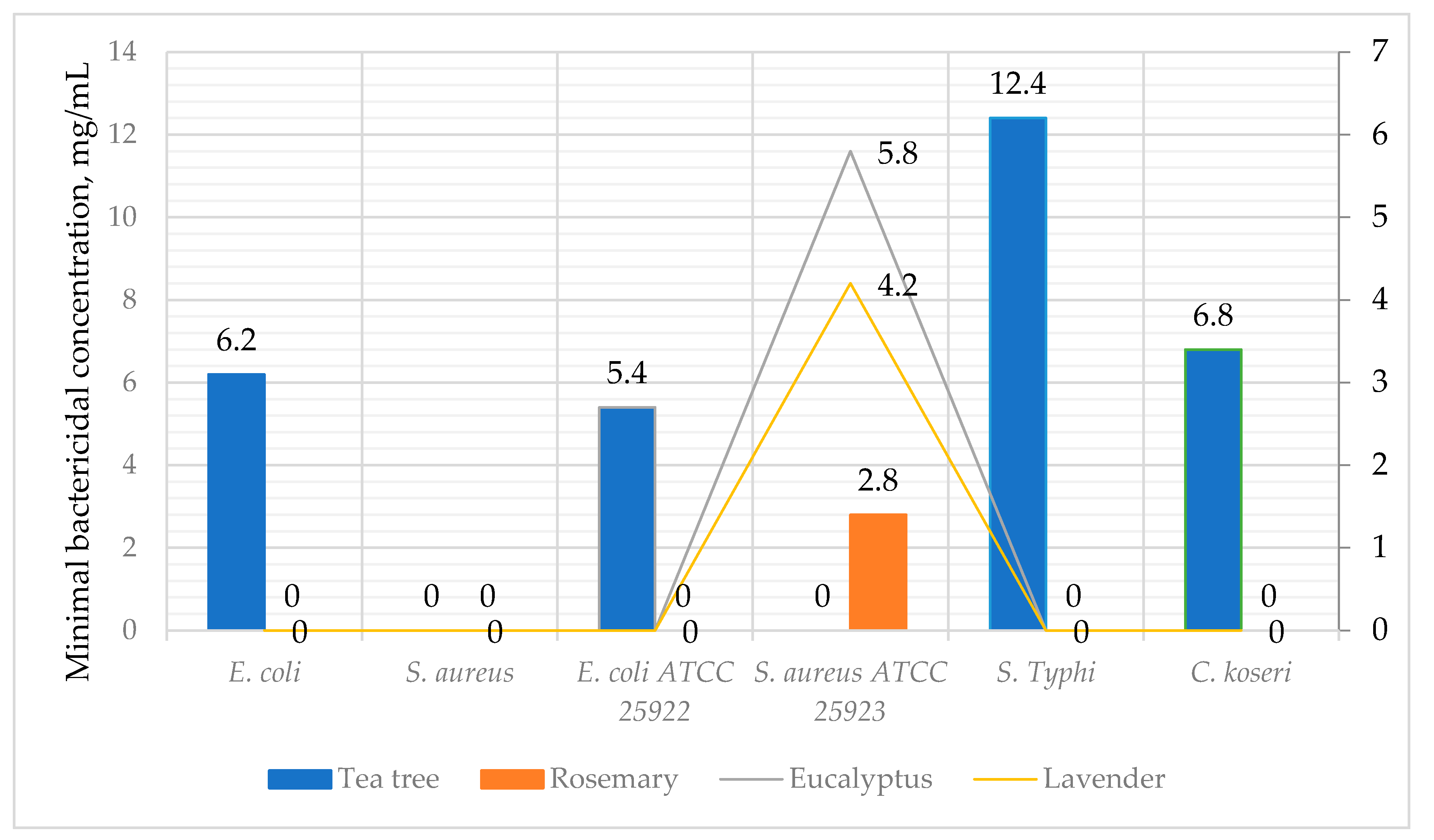
| E. coli | 21 | |||
| S. aureus | 13 | 13 | 13 | |
| E. coli ATCC 25922 | 18 | |||
| S. aureus ATCC 25923 | 13 | 13 | 13 | |
| S. Typhi | 15 | 15 | ||
| C. koseri | 13 |
| Bacteria | Tea Tree | Rosemary | Eucalyptus | Lavender |
|---|---|---|---|---|
| E. coli | 3.1 | |||
| S. aureus | ||||
| E. coli ATCC 25922 | 2.7 | |||
| S. aureus ATCC 25923 | 1.4 | 2.9 | 2.1 | |
| S. Typhi | 6.2 | |||
| C. koseri | 3.4 |
Publisher’s Note: MDPI stays neutral with regard to jurisdictional claims in published maps and institutional affiliations. |
© 2021 by the authors. Licensee MDPI, Basel, Switzerland. This article is an open access article distributed under the terms and conditions of the Creative Commons Attribution (CC BY) license (https://creativecommons.org/licenses/by/4.0/).
Share and Cite
Puvača, N.; Milenković, J.; Galonja Coghill, T.; Bursić, V.; Petrović, A.; Tanasković, S.; Pelić, M.; Ljubojević Pelić, D.; Miljković, T. Antimicrobial Activity of Selected Essential Oils against Selected Pathogenic Bacteria: In Vitro Study. Antibiotics 2021, 10, 546. https://0-doi-org.brum.beds.ac.uk/10.3390/antibiotics10050546
Puvača N, Milenković J, Galonja Coghill T, Bursić V, Petrović A, Tanasković S, Pelić M, Ljubojević Pelić D, Miljković T. Antimicrobial Activity of Selected Essential Oils against Selected Pathogenic Bacteria: In Vitro Study. Antibiotics. 2021; 10(5):546. https://0-doi-org.brum.beds.ac.uk/10.3390/antibiotics10050546
Chicago/Turabian StylePuvača, Nikola, Jovana Milenković, Tamara Galonja Coghill, Vojislava Bursić, Aleksandra Petrović, Snežana Tanasković, Miloš Pelić, Dragana Ljubojević Pelić, and Tatjana Miljković. 2021. "Antimicrobial Activity of Selected Essential Oils against Selected Pathogenic Bacteria: In Vitro Study" Antibiotics 10, no. 5: 546. https://0-doi-org.brum.beds.ac.uk/10.3390/antibiotics10050546