Article

Effect of Titanium Dioxide Nanoparticles on the Expression of Efflux Pump and Quorum-Sensing Genes in MDR Pseudomonas aeruginosa Isolates

by
Fatma Y. Ahmed
1,
Usama Farghaly Aly
2,
Rehab Mahmoud Abd El-Baky
1,3,* and
Nancy G. F. M. Waly
1
1
Department of Microbiology and Immunology, Faculty of Pharmacy, Minia University, Minia 61519, Egypt
2
Department of Pharmaceutics, Faculty of Pharmacy, Minia University, Minia 61519, Egypt
3
Department of Microbiology and Immunology, Faculty of Pharmacy, Deraya University, Minia 11566, Egypt
*
Author to whom correspondence should be addressed.
Submission received: 23 April 2021 / Revised: 19 May 2021 / Accepted: 19 May 2021 / Published: 24 May 2021
(This article belongs to the Special Issue Genomic Analysis of Antibiotics Resistance in Pathogens)

Abstract

:
Most of the infections caused by multi-drug resistant (MDR) P. aeruginosa strains are extremely difficult to be treated with conventional antibiotics. Biofilm formation and efflux pumps are recognized as the major antibiotic resistance mechanisms in MDR P. aeruginosa. Biofilm formation by P. aeruginosa depends mainly on the cell-to-cell communication quorum-sensing (QS) systems. Titanium dioxide nanoparticles (TDN) have been used as antimicrobial agents against several microorganisms but have not been reported as an anti-QS agent. This study aims to evaluate the impact of titanium dioxide nanoparticles (TDN) on QS and efflux pump genes expression in MDR P. aeruginosa isolates. The antimicrobial susceptibility of 25 P. aeruginosa isolates were performed by Kirby–Bauer disc diffusion. Titanium dioxide nanoparticles (TDN) were prepared by the sol gel method and characterized by different techniques (DLS, HR-TEM, XRD, and FTIR). The expression of efflux pumps in the MDR isolates was detected by the determination of MICs of different antibiotics in the presence and absence of carbonyl cyanide m-chlorophenylhydrazone (CCCP). Biofilm formation and the antibiofilm activity of TDN were determined using the tissue culture plate method. The effects of TDN on the expression of QS genes and efflux pump genes were tested using real-time polymerase chain reaction (RT-PCR). The average size of the TDNs was 64.77 nm. It was found that TDN showed a significant reduction in biofilm formation (96%) and represented superior antibacterial activity against P. aeruginosa strains in comparison to titanium dioxide powder. In addition, the use of TDN alone or in combination with antibiotics resulted in significant downregulation of the efflux pump genes (MexY, MexB, MexA) and QS-regulated genes (lasR, lasI, rhll, rhlR, pqsA, pqsR) in comparison to the untreated isolate. TDN can increase the therapeutic efficacy of traditional antibiotics by affecting efflux pump expression and quorum-sensing genes controlling biofilm production.

1. Introduction

According to the World Health Organization (WHO), P. aeruginosa is one of the most virulent and resistant bacteria for which new antimicrobials are urgently needed [1]. P. aeruginosa is characterized by bacterial resistance syndrome, as nearly all recognized antimicrobial resistance pathways can be found in this strain [2].
One of the important mechanisms of antibiotic resistance in P. aeruginosa is the exclusion of antibiotics through multidrug resistance (MDR) efflux systems, especially those that belong to the resistance-nodulation-division (RND) family. MexAB-OprM, MexXY and MexCD-OprJ are considered the main cause of intrinsic and acquired multidrug resistance [3].
Biofilm formation by P. aeruginosa is another mechanism of antibiotic resistance, since biofilm cells are much more resistant to antibiotics than planktonic cells [4]. The quorum-sensing system (QS) regulates the formation of biofilms and the expression of many extracellular virulence factors in many bacterial pathogens such as P. aeruginosa [5]. P. aeruginosa has four types of QS schemes, including rhl, las, PQS, and integrated QS (IQS). The rhl, las and PQS systems have been widely tested, while the IQS system was recently detected in P. aeruginosa [6].
Although numerous antimicrobial drugs are available commercially, they often lack efficacy against multidrug resistant (MDR) microorganisms, which is a great challenge to the health care teams [7]. Nowadays, the substitution of conventional antimicrobials with modern technology to overcome antimicrobial resistance is under trials. Nanotechnology showed promising results in many studies dealing with antibiotic resistance. Many nanostructures involving metallic particles have been created to counteract microbial pathogens [8]. Among them, titanium dioxide nanoparticles have attracted the attention of researchers due to their oxidative and hydrolytic properties [9].
Nanoparticles can exert their antibacterial activity via different mechanisms such as the following: interactions with DNA and/or the bacterial cell wall; inhibition of biofilm development; or the formation of reactive oxygen species (ROS). Nanoparticles, according to previous studies, have different modes of action than antibiotics, which may enhance their effect on MDR bacteria. [10]. This study aims to evaluate the impact of titanium dioxide nanoparticles (TDN) on the activity of the tested antimicrobials, QS and efflux pump genes expression in MDR P. aeruginosa isolates.

2. Results

2.1. Antimicrobial Resistance Pattern of P. aeruginosa Isolates

Twenty-five P. aeruginosa isolates were recovered from different clinical specimens, including three ear discharge specimens, thirteen urine, and nine wound exudate samples. Their resistance patterns were detected by the disc diffusion method against six different antimicrobial agents that are commonly used to treat P. aeruginosa infections.
The antimicrobial susceptibility of P. aeruginosa strains was detected by using the Kirby–Bauer disk diffusion method according to criteria provided by the Clinical & Laboratory Standards Institute (CLSI 2018). The P. aeruginosa strains were multidrug resistant (MDR) as they were resistant to three or more of the tested antibiotics (Table 1).

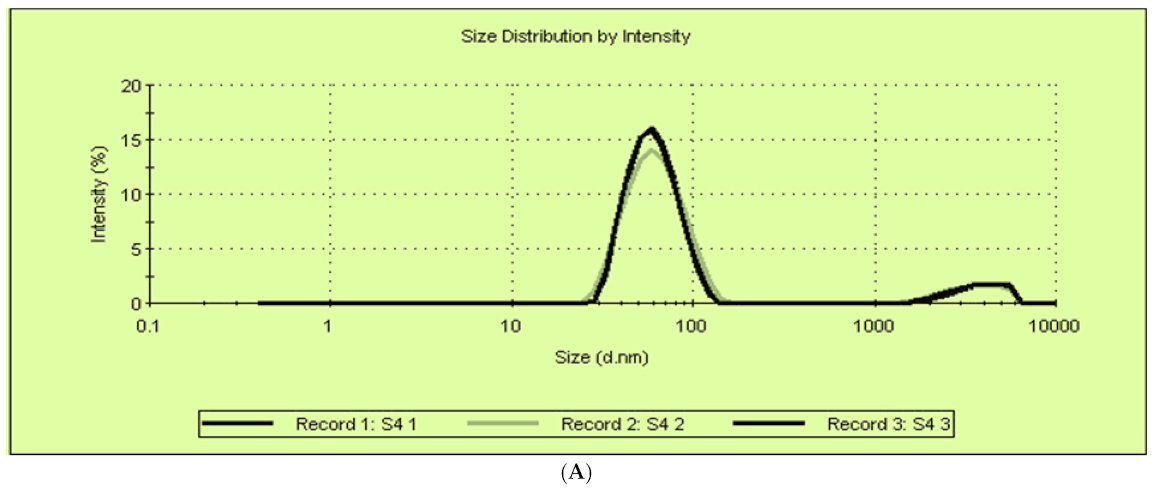
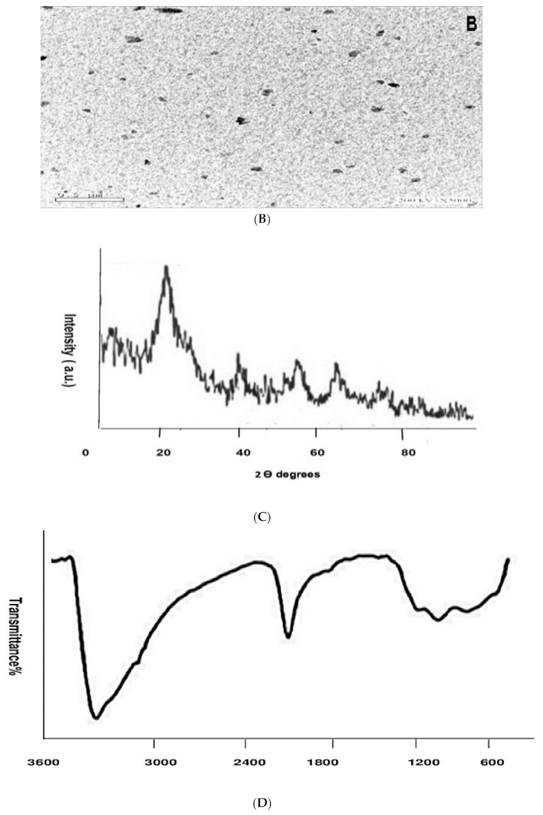
2.2. Synthesis of Titanium Dioxide Nanoparticles and Characterization

Titanium dioxide nanoparticles were prepared by the sol gel method. Dynamic light scattering was used to detect the size distribution of the formed nanoparticles on a scale that ranges from submicron to one nanometer. The average size of the formed TDN was 64.77 ± 0.14 nm (Figure 1A). The HR-TEM of TDN showed that most of the prepared TDN was spheres with asymmetrical edges and slight aggregations (Figure 1B).
X-ray diffraction (XRD) spectrum was performed to detect the phase and crystallinity of the formed TDN (Figure 1C). The XRD patterns showed that TDN was amorphous and had brookite polymorphs that were previously stated by the International Centre for Diffraction Data (ICDD card No. 015-0875).
The chemical composition of the synthesized TDN was analyzed by Fourier transform infrared spectrometry (FTIR). The FTIR spectra of TDN showed a small broad band at 1624 cm−1, which is characteristic to the O–Ti–O bond, a broad band at 3385 cm−1, which is related to O–H stretching, and two small bands observed below 1000 cm−1 representing Ti–O–Ti vibrations (Figure 1D).

2.3. Susceptibility of P. aeruginosa Isolates to TDN, TDP

The susceptibility of 25 MDR P. aeruginosa isolates to titanium dioxide powder (TDP) in bulk form and in nanoform was determined at different concentrations (1 μg/mL–1024 μg/mL) using the agar dilution method.
The P. aeruginosa strains showed high resistance to the titanium dioxide powder (TDP), as 11 isolates (44%) had MICs ≥ 1024 μg/mL and only 2 strains showed MICs ≤ 1 μg/mL. On the other hand, the titanium dioxide nanoparticles (TDN) showed excellent antimicrobial activity in comparison to TDP. As the MICs of TDN were ranged from 8 to 64 μg/mL, and three strains (12%) were sensitive to it (MIC less than 1 μg/mL) (Table 2).

2.4. Study of Efflux Pump System

To detect the expression of the efflux pump in MDR P. aeruginosa isolates, the effect of CCCP on the MICs of the tested antibiotics were determined. Table 3 showed that CCCP caused 4-fold decreases in the MICs of antibiotics in some isolates, which indicates the expression of efflux pumps in the highlighted isolates.

2.5. Effect of TDN on the Antimicrobial Susceptibility of the Tested P. aeruginosa

The addition of TDN resulted a in 2-fold MIC decrease or more in most of the tested isolates. As some of the antibiotics that showed no change in their MICs in the presence of CCCP showed more than a 2-fold decrease in their MICs in the presence of TDN, which suggested that TDN may have anti-efflux activity in addition to its other antimicrobial mechanisms (Table 4).

2.6. Characterization of Biofilm Formation Using Tissue Culture Plate Method (TCP) or Microtitre Plate Test

Out of 25 isolates, 11 (44%) isolates were high biofilm producers, 7 (28%) isolates showed moderate biofilm formation, and 7 (28%) were non or weak biofilm producers (Table 5) according to the decrease in the optical density measured in the presence of TDN in comparison to O.D in the absence of TDN.
We tested the effect of the formed TDN at a sub-inhibitory concentration (4 μg/mL) on the biofilm formation of 25 MDR P. aeruginosa isolates. It was found that TDN showed a high significant inhibitory effect (96%) on biofilm formation.

2.7. Real Time PCR

The isolates (no.2) that showed complete resistance to all tested antibiotics, presented an active efflux pump and strong biofilm producer were selected for testing the effect of TDN on the relative expression of QS genes (lasI, lasR, rhlI, rhlR, pqsA and pqsR) and efflux pump genes (MexY, MexB, MexA and oprM) by using real-time polymerase chain reaction (RT-PCR).
A comparison of the expression ratios of the treated and the untreated isolates was performed for each gene, as shown in Table 6. The results showed overexpression of the efflux pump genes (MexY, MexB, MexA) in the untreated P. aeruginosa isolate, which agreed with the phenotypic results, confirming the importance of an efflux pump as one of the main resistance mechanisms in MDR P. aeruginosa. Furthermore, the expression ratios of the QS-regulated genes (lasI, lasR, rhlI, rhlR, pqsA and pqsR) were also high in the untreated sample.
The addition of TDN resulted in significant downregulation of the efflux pump genes (MexY, MexB, MexA) and the QS-regulated genes (lasR, lasI, rhll, rhlR, pqsA, pqsR) in comparison to the untreated isolate. On other hand, a slight increase in the expression of oprM gene was shown.

3. Discussion

With the emergence of antibiotic-resistant bacteria like P. aeruginosa, the use of conventional antibiotics has contributed to the failure of treatments. Therefore, there is an urgent need for the introduction of new antimicrobial agents or the use of non-antimicrobial agents to increase the therapeutic activity of the current antibiotics [11].
The present study evaluated the antibacterial activity of TDN against MDR P. aeruginosa isolates. Multidrug resistant isolates represent one of the most important challenges in Egypt due to many factors including the misuse of antibiotics and biocides [12], which leads to the accumulation of antibiotic resistance and cross-resistance among antibiotics and the appearance of multidrug resistant (MDR), XRD, PDR P. aeruginosa [13].
The wide spread of MDR strains in Egypt in comparison to other countries warns us that strict antibiotic prescribing policies need to be implemented [14].
In the present study, twenty-five P. aeruginosa isolates recovered from different clinical specimens were considered as multidrug resistant isolates, as these strains were non-susceptible (resistant or intermediate) to one antimicrobial agent in three or more different antimicrobial classes [15].
The activity of TiO2 nanoparticles is of interest to researchers due to the unique characterization of TiO2 nanomaterial involving crystal size, shape, surface stability and structure [16]. Titanium dioxide nanoparticles (TDN) were prepared and characterized by DLS, HR-TEM, XRD, and FTIR. The average size of the titanium dioxide nanoparticles was 64.77 nm. The antimicrobial activities were due to the large surface area and their ability to penetrate the cell wall [17]. The prepared TDN was spheres with asymmetrical edges and slight aggregations. Nanoparticles have a strong tendency to agglomerate due to their large surface area. Typically, the agglomeration occurs due to the Van der Waals attraction forces among the nanoparticles [18]. The size and size distribution have an effect on the nanoparticle’s properties and the possible applications. Similarly, the crystal structure of the NPs and the chemical composition of nanoparticles are extensively studied [19].
X-ray diffraction peaks indicate the small size and the amorphous structure (less crystalline) of the formed TDN. As the size of nanoparticles are inversely proportional to the peak width, increases in the peak width (broad peak) indicate a small size of the formed nanoparticles and the presence of material in nonorange [20], and that supported the data of DLS. FTIR spectroscopy is used to identify the functional groups that exist on nanoparticles. The spectrum represents a fingerprint of the nanoparticles [21].
FTIR confirms the formation of TDN. The characteristic peaks for nano TiO2 were observed around 1624 cm−1, which is characteristic of the O–Ti–O bond, a broad band at 3385 cm−1, which is related to O–H stretching revealing the presence of water [22]; the two small bands for TiO2 nanoparticles observed below 1000 cm−1 were due to Ti–O–Ti vibrations [17]. The absence of any band at 2900 cm−1 indicates complete removing of absolute ethanol from the samples [23].
The MICs of titanium dioxide in bulk and nanoform were detected against P. aeruginosa strains. It was clear that the titanium dioxide nanoparticles had superior antibacterial activity than the bulk form, which may be due to the small size of the nanoparticles and their large surface area [24]. In addition, the antibacterial activity of TiO2 may be enhanced by exposure to UV light or reaction with water, leading to the generation of radical oxygen species (ROS), which have a potent oxidizing power that attacks microbial cells through various processes, such as lipid peroxidation of the cell membrane, inhibition of enzymes, damage of DNA, alteration of proteins, inhibition of enzymes, finally leading to cell death [25]. Many studies reported the antibacterial and antifungal activities of TDN [26]. However, Abdel-Fatah et al. [27] found that TiO2 nanoparticles had no bactericidal activity against both Gram-negative and Gram-positive bacteria isolated in a study performed in Egypt. Also, [28] they noted that titanium dioxide NPs showed very low antibacterial activity against different bacterial species, including P. aeruginosa.
These variations among different studies may depend on the nanoparticles’ size, concentration, particle shape, zeta potential and the tested pathogen [29]. Moreover, many environmental factors, including aeration, pH, and temperature, play a role and influence the bactericidal activity [30].
In this study, the MDR P. aeruginosa isolates showed a 4-fold reduction in the MICs of the tested antibiotics (ceftriaxone, ciprofloxacin, amikacin and meropenem) in the presence of CCCP. These results suggested that efflux pumps are the main mechanism of resistance in the tested isolates. Many studies from Egypt reported that the efflux pump was the main cause of the decrease in ciprofloxacin and meropenem resistance, as the MICs of ciprofloxacin and meropenem significantly reduced after the addition of CCCP [15,31].
Also, Abbas et al. [12] found that all the MDR isolates showed efflux pump activity as reported by using the ethidium bromide cartwheel method and confirmed with the presence of Mex AB-R genes by PCR.
Another study from Iran reported similar results, in which 65%, 71.5%, 60.5% and 66% of P. aeruginosa strains showed a significant decrease in the MICs of imipenem, cefepime, ciprofloxacin, and gentamicin, respectively, in the presence of CCCP. The inhibition of efflux pumps by CCCP in some isolates increased their sensitivity to different antibiotic classes and increased the accumulation of antibiotics that agree with our results [2]. Adabi et al. [32] noted that ciprofloxacin-resistant strains are mediated mainly by an efflux pump.
Titanium dioxide powder combined with antibiotics showed a significant decrease in the MICs of the tested strains by 2-fold or greater, which suggested its activity as an efflux inhibitor according to the method reported by Lamers et al. [33]. In addition, many studies have shown that combining TDN and antibiotics potentiates the antimicrobial action of different classes of antibiotics [34,35]. The combination of TDN with antibiotics increases the concentration of antibiotics at the bacterium–antibiotic interaction site, and the binding of bacteria to antibiotics [36]. Many studies reported that metal nanoparticles can affect proton motif force (PMF), which is essential for efflux pumps [37,38].
Chatterjee et al. [38] reported that copper nanoparticles increase the antimicrobial activity of the tested antibiotics due to the inhibition of the efflux pump of resistant S. aureus and P. aeruginosa because of copper nanoparticles on PMF.
Another significant factor leading to P. aeruginosa pathogenesis in clinical settings is biofilm formation [39]. Regarding biofilm results in the present study, 44%, 28%, and 28% of P. aeruginosa isolates were strong, moderate and weak biofilm producers, respectively.
Numerous previous studies reported various rates of biofilm formation by P. aeruginosa isolates. Elmaraghy et al. [40] from Egypt and Kamali et al. [41] from Iran, reported lower results than ours. While Abbas et al. [42] from Egypt reported higher results than ours.
Numerous experiments have shown the remarkable ability of metal nanoparticles to decompose thick biofilm barriers by different modes of action. The penetrating ability of metallic nanoparticles is often a valuable function for the prevention of biofilm infections [43]. In the current study, the antibiofilm activity of TDN was detected against 25 MDR P. aeruginosa strains. TDN showed a significant reduction in biofilm formation (96%), as TiO2NPs target sulfhydryl (–SH) groups in the cell membrane, resulting in the creation of the S–TiO2 bond, and this reaction suppresses the electron transport chain and enzymes essential for biofilm formation [24].
A recent study using TiO2NPs with polyvinylpyrrolidone polymer (PVP) against pathogenic bacteria on catheters showed that PVP with titanium dioxide nanoparticle films had the capability to prevent biofilm formation by S. aureus (83.97%) and E. coli (65.3%) [44]. However, Polo et al. [45] reported that neither the photocatalytic treatment with TiO2 film nor that with TiO2 nanopowder had any effect on P. aeruginosa biofilms at all the interfaces investigated.
Quorum sensing regulates the expression of different virulence factors and the overall process of biofilm production by pathogenic bacteria, which attracted our interest to detect the anti-QS activity of TDN at sub-inhibitory concentrations (4 μg/mL). Our results showed that TDN not only reduced the biofilm formation of P. aeruginosa strains, but also reduced the expressions of QS-regulated genes (lasR, lasI, rhll, rhlR, pqsA, pqsR).
Many researchers have recently began using nanotechnology for the development of nano-antimicrobials of the next generation, involving QS nano-inhibitors [46]. Silver nanoparticles were evaluated for the inhibition of QS-regulated virulence and biofilm formation in P. aeruginosa. AgNPs were able to decrease LasIR-RhlIR levels, inhibit biofilm formation, and significantly downregulate the expression of QS-regulated genes (lasI, lasR, rhlI, rhlR) [47]. Also, [48] found that glutathione-stabilized silver nanoparticles (GSH-Ag NPs) have antibiofilm activity in P. aeruginosa by reducing the expression the of lasR, lasI genes.
García-Lara et al. [49] found that ZnO nanoparticles decrease pyocyanin, elastase, and biofilm formation in P. aeruginosa strains; this indicates that ZnO nanoparticles may have QS inhibitor activity. It can be considered as an option for treatment of P. aeruginosa infections.
Our results also revealed that efflux pump genes (MexY, MexB, MexA) in an untreated P. aeruginosa sample were significantly overexpressed, especially the MexY gene. Nikaido and Pagès [50] reported that there is a noticeable relationship between MexXY-OprM overproduction in P. aeruginosa strains and the usage of different classes of antibiotics in treatment. It seems that MexXY-OprM has a significant benefit during antibiotic pressure in the medical setting and may play a vital role in the efflux of numerous antibiotics. Also, a direct association between the expression of the MexA and MexB genes and antibiotic resistance was reported by Dashtizadeh et al. [51], which was compatible with our findings.
In the presence of TDN, the efflux pump genes (MexY, MexB, and MexA) were found to be significantly downregulated. TDN may have efflux pump inhibitor activity by suppressing efflux pump genes. Abdolhosseini et al. [52] from Iran found that the efflux pump genes (mexA and mexB) in multidrug resistant P. aeruginosa strains were decreased by 6- and 2.75-fold, respectively, when exposed to Ag–TSC nanocomposite and ciprofloxacin. Also, Dolatabadi et al. [53] noted that both biosynthesized and commercial AgNPs downregulated the expression of the efflux pump gene OxqAB in resistant Klebsiella pneumoniae strains.
The inhibition of MDR efflux pumps by nanoparticles would be helpful in enhancing the therapeutic efficacy of traditional antibiotics by affecting quorum-sensing genes controlling biofilm production and other virulence factors [43].

4. Materials and Methods

4.1. Reagents

Titanium dioxide powder (TiO2, 98%) was obtained from Loba Chemie, Mumbai, India. Meropenem, ceftriaxone, cefepime powder were obtained from Pharco B international, Egypt, ciprofloxacin from Amirya, Egypt and Amikacin from Amount, Egypt.

4.2. Bacterial Strains

In the current study, 25 P. aeruginosa strains were collected from different clinical specimens (urine, ear discharge, wound exudate) of patients attending Minia university hospital. P. aeruginosa strains were confirmed by using the traditional microbiological method and biochemical tests [54].

4.3. Antimicrobial Susceptibility Testing

Antimicrobial susceptibility of P. aeruginosa strains was performed by Kirby–Bauer disc diffusion method according to Clinical & Laboratory Standards Institute guidelines [55]. The used antibiotics were amikacin (AK, 30 μg), cefepime (FEP, 30 μg), levofloxacin (LEV, 10 μg), ceftriaxone (CRO, 30 μg), imipenem (IPM, 10 μg) and ciprofloxacin (CIP, 5 μg).

4.4. Synthesis of Titanium Dioxide Nanoparticles, Characterization

Titanium dioxide nanoparticles (TDN) were synthesized as described before by Ahmed et al. [56]. Dynamic light scattering (DLS) method, high-resolution transmission electron microscopy (HR-TEM), Fourier transform infrared spectroscopy and X-ray diffraction (XRD) were used for the characterization of titanium dioxide nanoparticles (TDN).

4.5. Preparation of TDN Suspension

Titanium dioxide nanoparticle suspensions were prepared by adding 50 mg of TDN to 5 mL of sterile MQ water followed by shaking using ultrasound for 30 min and autoclaving at 121 °C for 20 min [39].

4.6. Determination of Antibacterial Activity of Titanium Dioxide Powder (TDP) and Titanium Dioxide Nanoparticles (TDN)

The antibacterial activity of titanium dioxide powder (TDP) and titanium dioxide nanoparticles (TND) were detected by agar dilution method against 25 P. aeruginosa strains as previously described by [31].
Briefly, overnight cultures of 25 P. aeruginosa strains in a Mueller Hinton Broth (MHB) were adjusted to a cell density of 107 CFU/mL. Then, the bacterial culture spots were applied to the surface of Mueller Hinton Agar containing TDP and TDN with different concentrations (1 to 1024 μg/mL) using a multi-inoculator. The plates were incubated at 37 °C for 18 h and examined for growth. Spots showing no growth were defined as MIC.

4.7. Determination of Efflux Pumps Expression in Resistant Isolates

The MICs of ciprofloxacin, meropenem, amikacin, ceftriaxone were detected for 25 MDR P. aeruginosa isolates by agar dilution method in the presence and absence of efflux pump inhibitor carbonyl cyanide m-chlorophenylhydrazone (CCCP) (Sigma, San Jose, CA, USA) at a final concentration of 10 μM. A four-fold reduction in MIC or more of the tested antibiotics after adding CCCP is an indication for the presence of efflux pumps [31].

4.8. Effect of TDN on the Antimicrobial Susceptibility of the Tested P. aeruginosa

The MICs of ciprofloxacin, meropenem, amikacin, ceftriaxone either alone or in combination with TDN were detected for 25 MDR P. aeruginosa isolates by agar dilution method in the presence of titanium dioxide nanoparticles at sub-inhibitory concentrations. A two-fold or greater change in the MICs of the tested antibiotics in presence of TDN compared to MICs of antibiotics alone was reported as indicating a significant efflux pump inhibitor [33].

4.9. Biofilm Formation Assays

Biofilm formation of 25 P. aeruginosa strains was evaluated by the tissue culture plate assay method (TCP) as previously described by Christensen et al. [57]. About 1 × 10⁷ CFU ml of the tested isolates were incubated in TCP for 24 h at 37 °C. Then, bacterial cultures were removed gently and washed with phosphate buffered saline. Crystal violet (0.1%) was used to stain the adherent cells, followed by washing with PBS. Then, ethyl alcohol (95%) was applied followed by measuring absorbance at 630 nm and the results were interpreted according to Table 7. The assay was performed in triplicates for each isolate.
TDN at sub-MIC concentration was added to the tested isolates suspension in TCP and incubated for 24 h at 37 °C. Bacterial cultures were discarded and tissue culture plate wells were washed gently. The adhered cells were stained by crystal violet (0.1%) for 20 s, followed by washing using PBS. Finally, ethyl alcohol was added, and absorbance was measured as described before.

4.10. Gene Expression Using Real-Time PCR

The impact of TDN (4 µg/mL) on the relative expression of efflux pump genes (MexY, MexB, MexA and OprM) and quorum-sensing genes (rhlR, lasI, lasR, rhlI, pqsA and pqsR) in the chosen isolate (no.2) was evaluated using real-time polymerase chain reaction (RT-PCR).
The tested isolate was grown until the middle of the exponential phase in presence and absence of TDN. Cultures were pelleted, and mRNA was extracted using RNeasy Mini Kit (Qiagen, Hilden, Germany) according to the manufacturer’s instructions. Then, cDNA was performed using High-Capacity cDNA Reverse Transcription Kit (Thermo Fisher, New York, NY, USA).
Real-time polymerase chain reaction (RT-PCR) was performed to determine the expression level of efflux and quorum-sensing genes using Quanti Tect SYBR Green RT-PCR Kit (Qiagen, Germany) according to the manufacturer’s protocol. Synthesized cDNA, primers (Table 8) and master mix were mixed and transferred to Applied Biosystems Step One™ instrument.
The level of gene expression was relatively normalized to the expression of the housekeeping gene rpoD. The expression of genes in P. aeruginosa isolate cultivated with TDN was compared to their expression in the control cultures without TDN. Relative quantities of gene expression were calculated using the 2−ΔΔCt method (Pfaffl, 2001).

5. Conclusions

Titanium dioxide nanoparticles can increase the therapeutic efficacy of traditional antibiotics by affecting efflux pump expression and quorum-sensing genes controlling biofilm production.

Author Contributions

Methodology, R.M.A.E.-B., U.F.A., N.G.F.M.W. and F.Y.A.; formal analysis, R.M.A.E.-B. and F.Y.A.; data curation, N.G.F.M.W.; writing—original draft preparation, R.M.A.E.-B., U.F.A., N.G.F.M.W. and F.Y.A.; writing—review and editing, R.M.A.E.-B. and U.F.A.; visualization, F.Y.A.; supervision, R.M.A.E.-B., U.F.A. and N.G.F.M.W. All authors have read and agreed to the published version of the manuscript.

Funding

This research did not receive any specific grant from funding agencies in the public, commercial, or not-for-profit sectors.

Institutional Review Board Statement

The study was conducted according to the guidelines of the Declaration of Helsinki, and approved by the Ethics committee of faculty of Medicine, Minia university.

Informed Consent Statement

Written informed consent has been obtained from the patient(s) to publish this paper.

Data Availability Statement

Data are applicable from authors.

Acknowledgments

The authors received no financial support for the research or publication of this article.

Conflicts of Interest

The authors declare no conflict of interest.

References

  1. Awan, A.B.; Schiebel, J.; Böhm, A.; Nitschke, J.; Sarwar, Y.; Schierack, P.; Ali, A. Association of Biofilm Formation and Cytotoxic Potential with Multidrug Resistance in Clinical Isolates of Pseudomonas Aeruginosa. EXCLI J. 2019, 18, 79–90. [Google Scholar]
  2. Talebi-Taher, M.; Majidpour, A.; Gholami, A.; Rasouli-Kouhi, S.; Adabi, M. Role of Efflux Pump Inhibitor in Decreasing Antibiotic Cross-Resistance of Pseudomonas Aeruginosa in a Burn Hospital in Iran. J. Infect. Dev. Ctries. 2016, 10, 600–604. [Google Scholar] [CrossRef] [Green Version]
  3. Pourakbari, B.; Yaslianifard, S.; Yaslianifard, S.; Mahmoudi, S.; Keshavarz-Valian, S.; Mamishi, S. Evaluation of Efflux Pumps Gene Expression in Resistant Pseudomonas Aeruginosa Isolates in an Iranian Referral Hospital. Iran. J. Microbiol. 2016, 8, 249–256. [Google Scholar] [PubMed]
  4. Lee, J.H.; Kim, Y.G.; Cho, M.H.; Lee, J. Zno Nanoparticles Inhibit Pseudomonas Aeruginosa Biofilm Formation and Virulence Factor Production. Microbiol. Res. 2014, 169, 888–896. [Google Scholar] [CrossRef] [PubMed]
  5. Ugurlu, A.; Yagci, A.K.; Ulusoy, S.; Aksu, B.; Bosgelmez-Tinaz, G. Phenolic Compounds Affect Production of Pyocyanin, Swarming Motility and Biofilm Formation of Pseudomonas Aeruginosa. Asian Pac. J. Trop. Biomed. 2016, 6, 698–701. [Google Scholar] [CrossRef] [Green Version]
  6. Blach, S.; Zeuzem, S.; Manns, M.; Altraif, I.; Duberg, A.S.; Muljono, D.H.; Waked, I.; Alavian, S.M.; Lee, M.-H.; Negro, F. Global Prevalence and Genotype Distribution of Hepatitis C Virus Infection in 2015: A Modelling Study. Lancet Gastroenterol. Hepatol. 2017, 2, 161–176. [Google Scholar] [CrossRef] [Green Version]
  7. Singh, P.; Garg, A.; Pandit, S.; Mokkapati, V.R.S.S.; Mijakovic, I. Antimicrobial Effects of Biogenic Nanoparticles. Nanomaterials 2018, 8, 1009. [Google Scholar] [CrossRef] [PubMed] [Green Version]
  8. Rudramurthy, G.R.; Swamy, M.K.; Sinniah, U.R.; Ghasemzadeh, A. Nanoparticles: Alternatives against Drug-Resistant Pathogenic Microbes. Molecules 2016, 21, 836. [Google Scholar] [CrossRef] [PubMed]
  9. Arora, B.; Murar, M.; Dhumale, V. Antimicrobial Potential of TiO2 Nanoparticles against MDR Pseudomonas aeruginosa. J. Exp. Nanosci. 2015, 10, 819–827. [Google Scholar] [CrossRef] [Green Version]
  10. Baptista, P.V.; McCusker, M.P.; Carvalho, A.; Ferreira, D.A.; Mohan, N.M.; Martins, M.; Fernandes, A.R. Nano-Strategies to Fight Multidrug Resistant Bacteria—“A Battle of the Titans”. Front. Microbiol. 2018, 9, 1441. [Google Scholar] [CrossRef] [Green Version]
  11. Yang, Y.X.; Xu, Z.H.; Zhang, Y.Q.; Tian, J.; Weng, L.X.; Wang, L.H. A New Quorum-Sensing Inhibitor Attenuates Virulence and Decreases Antibiotic Resistance in Pseudomonas Aeruginosa. J. Microbiol. 2012, 50, 987–993. [Google Scholar] [CrossRef] [PubMed]
  12. Abbas, H.A.; El-Ganiny, A.M.; Kamel, H. Phenotypic and Genotypic Detection of Antibiotic Resistance of Pseudomonas Aeruginosa Isolated from Urinary Tract Infections. Afr. Health Sci. 2018, 18, 11–21. [Google Scholar] [CrossRef] [Green Version]
  13. Yayan, J.; Ghebremedhin, B.; Rasche, K. Antibiotic Resistance of Pseudomonas Aeruginosa in Pneumonia at a Single University Hospital Center in Germany over a 10-Year Period. PLoS ONE 2015, 10, e0139836. [Google Scholar] [CrossRef] [Green Version]
  14. Mohamed, A.; Abdelhamid, F. Antibiotic Susceptibility of Pseudomonas Aeruginosa Isolated from Different Clinical Sources. Zagazig J. Pharm. Sci. 2020, 28, 10–17. [Google Scholar]
  15. Kishk, R.M.; Abdalla, M.O.; Hashish, A.A.; Nemr, N.A.; El Nahhas, N.; Alkahtani, S.; Abdel-Daim, M.M.; Kishk, S.M. Efflux Mexab-Mediated Resistance in P. Aeruginosa Isolated from Patients with Healthcare Associated Infections. Pathogens 2020, 9, 471. [Google Scholar] [CrossRef] [PubMed]
  16. Mahdy, S.A.; Mohammed, W.H.; Emad, H.; Kareem, H.A.; Shamel, R.; Mahdi, S. The Antibacterial Activity of TiO2 Nanoparticles. J. Univ. Babylon 2017, 25, 955–961. [Google Scholar]
  17. Saranya, K.S.; Padil, V.V.T.; Senan, C.; Pilankatta, R.; Saranya, K.; George, B.; Wacławek, S.; Černík, M. Green Synthesis of High Temperature Stable Anatase Titanium Dioxide Nanoparticles Using Gum Kondagogu: Characterization and Solar Driven Photocatalytic Degradation of Organic Dye. Nanomaterials 2018, 8, 1002. [Google Scholar] [CrossRef] [Green Version]
  18. Othman, S.H.; Rashid, S.A.; Ghazi, T.I.M.; Abdullah, N. Dispersion and Stabilization of Photocatalytic TiO2 Nanoparticles in Aqueous Suspension for Coatings Applications. J. Nanomater. 2012, 2012. [Google Scholar] [CrossRef] [Green Version]
  19. Mourdikoudis, S.; Pallares, R.M.; Thanh, N.T. Characterization Techniques for Nanoparticles: Comparison and Complementarity Upon Studying Nanoparticle Properties. Nanoscale 2018, 10, 12871–12934. [Google Scholar] [CrossRef] [PubMed] [Green Version]
  20. Kantheti, P.; Alapati, P. Green Synthesis of TiO2 Nanoparticles Using OcimumBasilicum Extract and Its Characterization. Int. J. Chem. Stud. 2018, 6, 670–674. [Google Scholar]
  21. Patra, J.K.; Baek, K.-H. Green Nanobiotechnology: Factors Affecting Synthesis and Characterization Techniques. J. Nanomater. 2014, 2014, 1–12. [Google Scholar] [CrossRef] [Green Version]
  22. Chatterjee, M.; Anju, C.; Biswas, L.; Kumar, V.A.; Mohan, C.G.; Biswas, R. Antibiotic Resistance in Pseudomonas Aeruginosa and Alternative Therapeutic Options. Int. J. Med. Microbiol. 2016, 306, 48–58. [Google Scholar] [CrossRef]
  23. Devi, R.; Venckatesh, R.; Sivaraj, R. Synthesis of Titanium Dioxide Nanoparticles by Sol-Gel Technique. Int. J. Innov. Res. Sci. Eng. Technol. 2014, 3, 15206–15211. [Google Scholar] [CrossRef]
  24. Abdulazeem, L.; AL-Amiedi, B.H.; Alrubaei, H.A.; AL-Mawlah, Y.H. Titanium Dioxide Nanoparticles as Antibacterial Agents against Some Pathogenic Bacteria. Drug Invent. Today 2019, 12. [Google Scholar]
  25. de Dicastillo, C.L.; Patiño, C.; Galotto, M.J.; Vásquez-Martínez, Y.; Torrent, C.; Alburquenque, D.; Pereira, A.; Escrig, J. Novel Hollow Titanium Dioxide Nanospheres with Antimicrobial Activity against Resistant Bacteria. Beilstein. J. Nanotechnol. 2019, 10, 1716–1725. [Google Scholar] [CrossRef] [Green Version]
  26. Manjunath, K.; Yadav, L.S.R.; Jayalakshmi, T.; Reddy, V.; Rajanaika, H.; Nagaraju, G. Ionic Liquid Assisted Hydrothermal Synthesis of TiO2 Nanoparticles: Photocatalytic and Antibacterial Activity. J. Mater. Res. Technol. 2018, 7, 7–13. [Google Scholar] [CrossRef]
  27. Abdel-Fatah, W.I.; Gobara, M.M.; Mustafa, S.F.; Ali, G.W.; Guirguis, O.W. Role of Silver Nanoparticles in Imparting Antimicrobial Activity of Titanium Dioxide. Mater. Lett. 2016, 179, 190–193. [Google Scholar] [CrossRef]
  28. Kareem, P.A.; Alsammak, E.G.; Abdullah, Y.J.; Bdaiwi, Q.M. Estimation of Antibacterial Activity of Zinc Oxide, Titanium Dioxide, and Silver Nanoparticles against Multidrug-Resistant Bacteria Isolated from Clinical Cases in Amara City, Iraq. Drug Invent. Today 2019, 11, 2887–2890. [Google Scholar]
  29. Skandalis, N.; Dimopoulou, A.; Georgopoulou, A.; Gallios, N.; Papadopoulos, D.; Tsipas, D.; Theologidis, I.; Michailidis, N.; Chatzinikolaidou, M. The Effect of Silver Nanoparticles Size, Produced Using Plant Extract from Arbutus Unedo, on Their Antibacterial Efficacy. Nanomaterials 2017, 7, 178. [Google Scholar] [CrossRef] [Green Version]
  30. Beyth, N.; Houri-Haddad, Y.; Domb, A.; Khan, W.; Hazan, R. Alternative Antimicrobial Approach: Nano-Antimicrobial Materials. Evid. Based Complementary Altern. Med. 2015, 2015. [Google Scholar] [CrossRef] [Green Version]
  31. Gad, G.F.; El-Domany, R.A.; Zaki, S.; Ashour, H.M. Characterization of Pseudomonas Aeruginosa Isolated from Clinical and Environmental Samples in Minia, Egypt: Prevalence, Antibiogram and Resistance Mechanisms. J. Antimicrob. Chemother. 2007, 60, 1010–1017. [Google Scholar] [CrossRef] [PubMed] [Green Version]
  32. Adabi, M.; Talebi-Taher, M.; Arbabi, L.; Afshar, M.; Fathizadeh, S.; Minaeian, S.; Moghadam-Maragheh, N.; Majidpour, A. Spread of Efflux Pump Overexpressing-Mediated Fluoroquinolone Resistance and Multidrug Resistance in Pseudomonas Aeruginosa by Using an Efflux Pump Inhibitor. Infect. Chemother. 2015, 47, 98. [Google Scholar] [CrossRef] [PubMed]
  33. Lamers, R.P.; Cavallari, J.F.; Burrows, L.L. The Efflux Inhibitor Phenylalanine-Arginine Beta-Naphthylamide (Paβn) Permeabilizes the Outer Membrane of Gram-Negative Bacteria. PLoS ONE 2013, 8, e60666. [Google Scholar] [CrossRef] [Green Version]
  34. Abdulrahman, N.B.; Nssaif, Z.M. Antimicrobial Activity of Zinc Oxide, Titanium Dioxide and Silver Nanoparticles against Mithicillin-Resistant Staphylococcus Aureus Isolates. Tikrit J. Pure Sci. 2018, 21, 49–53. [Google Scholar]
  35. Kareem, P.A.; Alsammak, E.G. The Effect of Silver and Titanium Dioxide Nanoparticles on Klebsiella Pneumoniae Isolates Multi Resistant to Antibiotics and Observed by Scanning Electron Microscopy. Cihan. Univ. Sci. J. 2017, 284–297. [Google Scholar] [CrossRef] [Green Version]
  36. Allahverdiyev, A.M.; Kon, K.V.; Abamor, E.S.; Bagirova, M.; Rafailovich, M. Coping with Antibiotic Resistance: Combining Nanoparticles with Antibiotics and Other Antimicrobial Agents. Expert Rev. Anti-Infect. Ther. 2011, 9, 1035–1052. [Google Scholar] [CrossRef]
  37. Dibrov, P.; Dzioba, J.; Gosink, K.K.; Häse, C.C. Chemiosmotic Mechanism of Antimicrobial Activity of Ag+ in Vibrio Cholerae. Antimicrob. Agents Chemother. 2002, 46, 2668–2670. [Google Scholar] [CrossRef] [Green Version]
  38. Chatterjee, A.K.; Chakraborty, R.; Basu, T. Mechanism of Antibacterial Activity of Copper Nanoparticles. Nanotechnology 2014, 25, 135101. [Google Scholar] [CrossRef]
  39. Vincent, M.G.; John, N.P.; Narayanan, P.M.; Vani, C.; Murugan, S. In Vitro Study on the Efficacy of Zinc Oxide and Titanium Dioxide Nanoparticles against Metallo Beta-Lactamase and Biofilm Producing Pseudomonas Aeruginosa. J. Appl. Pharm. Sci. 2014, 4, 41. [Google Scholar]
  40. Elmaraghy, N.; Abbadi, S.; Elhadidi, G.; Hashem, A.; Yousef, A. Virulence Genes in Pseudomonas Aeruginosa Strains Isolated at Suez Canal University Hospitals with Respect to the Site of Infection and Antimicrobial Resistance. Int. J. Clin. Microbiol. Biochem. Technol. 2019, 2, 8–19. [Google Scholar]
  41. Kamali, E.; Jamali, A.; Ardebili, A.; Ezadi, F.; Mohebbi, A. Evaluation of Antimicrobial Resistance, Biofilm Forming Potential, and the Presence of Biofilm-Related Genes among Clinical Isolates of Pseudomonas Aeruginosa. BMC Res. Notes 2020, 13, 1–6. [Google Scholar] [CrossRef] [PubMed]
  42. Abbas, H.A.; Serry, F.M.; EL-Masry, E.M. Combating Pseudomonas Aeruginosa Biofilms by Potential Biofilm Inhibitors. Asian J. Res. Pharm. Sci. 2012, 2, 2012. [Google Scholar]
  43. Gupta, D.; Singh, A.; Khan, A.U. Nanoparticles as Efflux Pump and Biofilm Inhibitor to Rejuvenate Bactericidal Effect of Conventional Antibiotics. Nanoscale Res. Lett. 2017, 12, 1–6. [Google Scholar] [CrossRef]
  44. Salman, J.A.S.; Al-Kadhemy, M.F.H.; Madhloom, S.A. Preparation of Titanium Dioxide Nanoparticles and Polyvinyl Pyrrolidone Polymer Films as Antibacterial, Antibiofilm against Pathogenic Bacteria on Different Surfaces. MJS 2017, 36, 132–144. [Google Scholar] [CrossRef] [Green Version]
  45. Polo, A.; Diamanti, M.V.; Bjarnsholt, T.; Høiby, N.; Villa, F.; Pedeferri, M.P.; Cappitelli, F. Effects of Photoactivated Titanium Dioxide Nanopowders and Coating on Planktonic and Biofilm Growth of Pseudomonas Aeruginosa. Photochem. Photobiol. 2011, 87, 1387–1394. [Google Scholar] [CrossRef] [PubMed]
  46. Singh, B.N.; Prateeksha, P.; Upreti, D.K.; Singh, B.R.; Defoirdt, T.; Gupta, V.K.; De Souza, A.O.; Singh, H.B.; Barreira, J.C.M.; Ferreira, I.C.F.R.; et al. Bactericidal, Quorum Quenching and Anti-Biofilm Nanofactories: A New Niche for Nanotechnologists. Crit. Rev. Biotechnol. 2017, 37, 525–540. [Google Scholar] [CrossRef] [PubMed]
  47. Singh, B.R.; Singh, B.N.; Singh, A.; Khan, W.; Naqvi, A.H.; Singh, H.B. MycofabricatedBiosilver Nanoparticles Interrupt Pseudomonas Aeruginosa Quorum Sensing Systems. Sci. Rep. 2015, 5, 1–14. [Google Scholar] [CrossRef]
  48. Mahnaie, M.P.; Mahmoudi, H. Effect of Glutathione-Stabilized Silver Nanoparticles on Expression of Las I and Las R of the Genes in Pseudomonas Aeruginosa Strains. Eur. J. Med. Res. 2020, 25, 1–11. [Google Scholar]
  49. García-Lara, B.; Saucedo-Mora, M.; Roldán-Sánchez, J.A.; Pérez-Eretza, B.; Ramasamy, M.; Lee, J.; Coria-Jimenez, R.; Tapia, M.; Varela-Guerrero, V.; García-Contreras, R. Inhibition of Quorum-Sensing-Dependent Virulence Factors and Biofilm Formation of Clinical and Environmental P Seudomonas Aeruginosa Strains by Zno Nanoparticles. Lett. Appl. Microbiol. 2015, 61, 299–305. [Google Scholar] [CrossRef] [PubMed]
  50. Nikaido, H.; Pagès, J.-M. Broad-Specificity Efflux Pumps and Their Role in Multidrug Resistance of Gram-Negative Bacteria. FEMS Microbiol. Rev. 2012, 36, 340–363. [Google Scholar] [CrossRef] [Green Version]
  51. Dashtizadeh, Y.; Moattari, A.; Gorzin, A.A. Phenotypic and Genetically Evaluation of the Prevalence of Efflux Pumps and Antibiotic Resistance in Clinical Isolates of Pseudomonas Aeruginosa among Burned Patients Admitted to Ghotbodin Shirazi Hospital. J. Microb. World 2014, 19, 118–127. [Google Scholar]
  52. Abdolhosseini, M.; Zamani, H.; Salehzadeh, A. Synergistic Antimicrobial Potential of Ciprofloxacin with Silver Nanoparticles Conjugated to Thiosemicarbazide against Ciprofloxacin Resistant Pseudomonas Aeruginosa by Attenuation of Mexa-B Efflux Pump Genes. Biologia 2019, 74, 1191–1196. [Google Scholar] [CrossRef]
  53. Dolatabadi, A.; Noorbazargan, H.; Khayam, N.; Moulavi, P.; Zamani, N.; Asghari Lalami, Z.; Ashrafi, F. Ecofriendly Biomolecule-Capped Bifidobacterium Bifidum-Manufactured Silver Nanoparticles and Efflux Pump Genes Expression Alteration in Klebsiella Pneumoniae. Microb. Drug Resist. 2021, 27, 247–257. [Google Scholar] [CrossRef] [PubMed]
  54. Abdelraheem, W.M.; Abdelkader, A.E.; Mohamed, E.S.; Mohammed, M.S. Detection of Biofilm Formation and Assessment of Biofilm Genes Expression in Different Pseudomonas Aeruginosa Clinical Isolates. Meta Gene 2020, 23, 100646. [Google Scholar] [CrossRef]
  55. Wayne, P.A. Clinical and Laboratory Standards Institute: Performance Standards for Antimicrobial Susceptibility Testing: 20th Informational Supplement. CLSI Document M100-S20. Inform Suppl 2010. pp. 100–121. Available online: http://file.qums.ac.ir/repository/mmrc/CLSI2015.pdf (accessed on 20 April 2021).
  56. Ahmed, F.Y.; Aly, U.F.; El-Baky, R.M.A.; Waly, N.G.F.M.; Youssef, F. Comparative Study of Antibacterial Effects of Titanium Dioxide Nanoparticles Alone and in Combination with Antibiotics on Mdr Pseudomonas Aeruginosa Strains. Int. J. Nanomed. 2020, 15, 3393. [Google Scholar]
  57. Christensen, G.D.; A Simpson, W.; Younger, J.J.; Baddour, L.M.; Barrett, F.F.; Melton, D.M.; Beachey, E.H. Adherence of Coagulase-Negative Staphylococci to Plastic Tissue Culture Plates: A Quantitative Model for the Adherence of Staphylococci to Medical Devices. J. Clin. Microbiol. 1985, 22, 996–1006. [Google Scholar] [CrossRef] [PubMed] [Green Version]
Figure 1. Characterization of titanium dioxide nanoparticles. (A) Dynamic light scattering (DLS). (B) High-resolution transmission electron microscopy. (C) X-ray differentiation. (D) Fourier transform infrared spectrometry.
Antibiotics 10 00625 g001aAntibiotics 10 00625 g001b
Table 1. Antibiotic resistance patterns of the tested P. aeruginosa strains.
No.Type *CFIMAKCPLVCT
1WRSRRRR
2WRRRRRR
3WRRRRRR
4URRRRRR
5ERRRRRRR
6URRRRIR
7URRRRRR
8URRRRIR
9URRRRIR
10URSRSIR
11URRRRIR
12WRIRRRR
13WRRRRRR
14WRRRRRR
15URSRSIR
16URIRIIR
17URRRRRR
18WRSRSRR
19WRRIRSR
20URSIRSR
21ERRRRRRR
22WRRRRRR
23URRRSSR
24URRRSSR
25ERRRISSR
* Type of clinical specimen; U: urine specimen; W: wound exudate; ER: ear discharge. CP: ciprofloxacin; CF: cefepime; CT: ceftriaxone; AK: amikacin; IM: imipenem; LV: levofloxacin. R: resistance; I: intermediate; S: sensitive.
Table 2. Distribution of minimum inhibitory concentrations of TDN, TDP against MDR P. aeruginosa isolates (25 isolates).
Number of Isolates with MICs (μg/mL)
<11248163264128256512≥1024
TDN3000161140000
TDP2000002162111
TDN: titanium dioxide nanoparticles; TDP: titanium dioxide powder (bulk form).
Table 3. Effect of addition of CCCP on antibiotic resistance pattern against 25 MDR Pseudomonas aeruginosa isolates.
No.Et BrCPMCTAK
MIC1MIC2MIC1MIC2MIC1MIC2MIC1MIC2MIC1MIC2
1>102425688SND *323251216
2>1024232864<110241651216
35121632<1512<1512<1128<1
4>10245123232512<1>102416>102416
5>1024512322512<1>1024161281
6>1024512888464645128
7>10242564<116812812851216
810242564<116<1256<164<1
9>102451232<1512<1256264<1
10512128SND *SND *256<116<1
11>10245128816<11281651216
12102451216<1168646412816
13>102425632<1168>10248102432
14>10242564<116<1256<1128<1
151024256SND *SND *25612816<1
1610245128<18<132<1512<1
17>1024648<18<12564512<1
18512256SND *SND *16<1512<1
19>10242564<164<1322328
20>10245124<1SND *32<116<1
2110245124<132232864<1
22>10245124<18<132<1512<1
23>1024512SND *16<1641664<1
24>1024256SND *32<16416648
252568SND *32<1256816<1
Et Br: ethidium bromide, CP: ciprofloxacin; CT: ceftriaxone; AK: amikacin; M: meropenem, MIC1: antibiotic alone, MIC2: antibiotic + CCCP, ND *: not determined, S: sensitive.
Table 4. Effect of the addition of TDN on MICs of the tested antibiotics.
No.CPMCTAK
MIC1MIC2MIC1MIC2MIC1MIC2MIC1MIC2
180.5SS3215121
2320.56416102451251264
332151225651211281
4328512256>1024512>10241024
53216512128>1024256128128
680.5846415121
744168128165124
840.5161625616644
932251225625632644
10S (<1)S (<1)SS25632161
1180.51616128165121
12160.516166411281
13320.51616>102425610244
1440.51616256161284
15SSss2568161
1680.5883215121
1780.58825685121
18S (<1)S (<1)S (<1)S (<1)1615124
194S6416321324
204SSS321161
2140.532163216641
2240.58432165121
23S (<1)S (<1)16166416641
24S (<1)S (<1)3286416644
25S (<1)S (<1)321625616164
CP: ciprofloxacin; CT: ceftriaxone; AK: amikacin; M: meropenem, MIC1: antibiotic alone MIC2: antibiotic + TDN, S: sensitive.
Table 5. Degree of biofilm formation in absence and presence of TDN among 25 MDR P. aeruginosa isolates.
TargetDegree of Biofilm Formation
HighModerateNon/Weak Biofilm
No.%No.%No.% *
In absence of TDN1144728728
In presence of TDN14--2496
* Percentages were correlated to MDR P. aeruginosa isolates (25).
Table 6. Relative quantitation of gene expression of treated and control P. aeruginosa isolate.
SampleCtΔCtΔΔCtFold Difference of Gene Expression
Target rpoD ---
(Housekeeping Gene)
Ps *37.203
Ps1 **34.0287
Target lasI −4.131817.607
Ps *27.9−9.253
Ps1 **20.637-13.3911
Target lasR −3.578311.9447
Ps *27.721−9.4821
Ps1 **20.9683−13.0604
Target MexA −5.59948.47
Ps *30.7584−6.4446
Ps1 **21.984−12.0438
Target MexY −7.458175.86
Ps *34.5908−2.6122
Ps1 **25.9582−10.0705
Target Mex B −6.003264.1428
Ps *29.905−7.2525
Ps1 **20.773−13.2557
Target OprM 3.9370.0653
Ps *34.9282−2.2749
Ps1 **35.69171.663
Target pqsA −1.4992.8279
Ps *26.669−10.5335
Ps1 **21.99−12.033
Target pqsR −3.998515.98
Ps *27.9577−9.2453
Ps1 **20.7849−13.243
Target rhlR −3.20549.2242
Ps *27.3754−9.8277
Ps1 **20.9956−13.0331
Target rhll −2.00594.0165
Ps *27.1394−10.0637
Ps1 **21.959−12.0696
PS *: Treated isolate with TDN, PS1 **: control isolate, Ct: cycle threshold, ΔCt: target gene Ct of sample—housekeeping gene Ct of the same sample. ΔΔCt: ΔCt of treated strain−ΔCt of control.
Table 7. Classification of bacterial adherence and biofilm formation by TCP method.
Biofilm FormationAdherenceMean OD Values
Non/WeakNon/Weak<0.120
ModerateModerate0.120–0.240
HighStrong>0.240
Table 8. The sequence of the primers used in this study.
GenePrimer DirectionPrimer SequenceSize of Amplified Product (bp)Reference
rpoDF
R
5-GCGAGAGCCTCAAGGATAC-3
5-CGAACTGCTTGCCGACTT-3
131(El-Shaer et al., 2016)
MexYF
R
5-CCGCTACAACGGCTATCCCT-3
5-AGCGGGATCGACCAGCTTTC-3
246(Yoneda et al., 2005)
MexAF
R
5′ACCTACGAGCCGACTACCAGA-3′
5′GTTGGTCACCAGGGCGCCTTC-3′
179(Pourakbari et al., 2016)
MexBF
R
5-GTGTTCGGCTCGCAGTACTC-3
5-AACCGTCGGGATTGACCTTG-3
244(Pourakbari et al., 2016)
OprMF
R
5-CCATGAGCCGCCAACTGTC-3
5-CCTGGAACGCCGTCTGGAT-3
205(Pourakbari et al., 2016)
Las IF
R
5-CGCACATCTGGGAACTCA-3
5-CGGCACGACGATCATCATCT-3
176(El-Shaer et al., 2016)
Las RF
R
5-CTGTGGATGCTCAAGGACTAC-3
5-AACTGGTCTTGCCGATGG-3
133(El-Shaer et al., 2016)
rhIIF
R
5-GTAGCGGGTTTGCGGATG-3
5-CGGCATCAGGTCTTCATCG-3
101(El-Shaer et al., 2016)
rhIRF
R
5-GCCAGCGTCTTGTTCGG-3
5-CGGTCTGCCTGAGCCATC-3
160(El-Shaer et al., 2016)
Pqs AF
R
5-GACCGGCTGTATTCGATTC-3
5-GCTGAACCAGGGAAAGAAC-3
74(El-Shaer et al., 2016)
pqsRF
R
5-CTGATCTGCCGGTAATTGG-3
5-ATCGACGAGGAACTGAAGA-3
142(El-Shaer et al., 2016)
Publisher’s Note: MDPI stays neutral with regard to jurisdictional claims in published maps and institutional affiliations.

