Article

Antimicrobial Resistance Profiling of Biofilm Forming Non Typhoidal Salmonella enterica Isolates from Poultry and Its Associated Food Products from Pakistan

1
Atta Ur Rahman School of Applied Biosciences (ASAB), National University of Sciences and Technology (NUST), H-12, Islamabad 44000, Pakistan
2
Animal Health Program, Animal Sciences Institute, National Agriculture Research Centre, Park Road, Islamabad 44000, Pakistan
3
Department of Biosciences, Faculty of Sciences, COMSATS University Islamabad, Park Road, Islamabad 44000, Pakistan
*
Author to whom correspondence should be addressed.
Submission received: 19 March 2021 / Revised: 25 May 2021 / Accepted: 25 May 2021 / Published: 28 June 2021
(This article belongs to the Special Issue Antimicrobial Resistance: From Farm to Fork)

Abstract

:
Salmonellosis caused by non-typhoidal Salmonella enterica from poultry products is a major public health concern worldwide. This study aimed at estimating the pathogenicity and antimicrobial resistance in S. enterica isolates obtained from poultry birds and their food products from different areas of Pakistan. In total, 95/370 (25.67%) samples from poultry droppings, organs, eggs, and meat were positive for Salmonella. The isolates were further identified through multiplex PCR (mPCR) as Salmonella Typhimurium 14 (14.7%), Salmonella Enteritidis 12 (12.6%), and other Salmonella spp. 69 (72.6%). The phenotypic virulence properties of 95 Salmonella isolates exhibited swimming and/or swarming motility 95 (100%), DNA degrading activity 93 (97.8%), hemolytic activity 92 (96.8%), lipase activity 87 (91.6%), and protease activity 86 (90.5%). The sopE virulence gene known for conferring zoonotic potential was detected in S. Typhimurium (92.8%), S. Enteritidis (100%), and other Salmonella spp. (69.5%). The isolates were further tested against 23 antibiotics (from 10 different antimicrobial groups) and were found resistant against fifteen to twenty-one antibiotics. All isolates showed multiple drug resistance and were found to exhibit a high multiple antibiotic-resistant (MAR) index of 0.62 to 0.91. The strong biofilm formation at 37 °C reflected their potential adherence to intestinal surfaces. There was a significant correlation between antimicrobial resistance and the biofilm formation potential of isolates. The resistance determinant genes found among the isolated strains were blaTEM-1 (59.3%), blaOxA-1 (18%), blaPSE-1 (9.5%), blaCMY-2 (43%), and ampC (8.3%). The detection of zoonotic potential MDR Salmonella in poultry and its associated food products carrying cephalosporin and quinolone resistance genes presents a major threat to the poultry industry and public health.

1. Introduction

Non-typhoidal Salmonella (NTS) is one of the most important zoonotic foodborne pathogens [1] globallyGharieb, Tartor. About 2600 serovars of Salmonella enterica have been reported, which can cause disease in both animals and humans [2,3]. Gastroenteritis is the most common Salmonella infection in humans, accounting for 94 million cases each year, where 80.3 million are related to foodborne illnesses [4,5]. Major Salmonella outbreaks are caused by consuming contaminated poultry food (meat and eggs). At the same time, poultry farm handlers are also at risk due to direct or indirect contact with poultry birds [6,7]. Biofilm formation is important for the spread of NTS because biofilm-forming bacteria are resistant to drugs, disinfectants, and mechanical stress, making these biofilms a safety risk for the food industry. A variety of virulence factors and biofilm formation potential play an important role in the pathogenesis of Salmonella infection [8]. The irrational use of antibiotics in animal husbandry results in ever-increasing antimicrobial resistance in pathogens, including Salmonella enterica. Multidrug-resistant (MDR) and extended drug-resistant (XDR) Salmonella causes a serious threat to humans via transmission through the food chain [9]. Therefore, it is mandatory to monitor the antibiotic resistance patterns of Salmonella enterica in the food chain. Currently, third- and fourth-generation cephalosporin and fluoroquinolones are widely used to treat salmonellosis in humans and animals. However, the emergence of bacterial resistance to these clinically important antibiotics needs to be monitored [10,11,12]. In the Enterobacteriaceae family, resistance to cephalosporins is mainly linked with the production of large spectrum beta-lactamases such as ESBL (extended-spectrum beta-lactamases) and AmpC beta-lactamase [13]. Quinolone resistance is mainly associated with the mutations in quinolone resistance determining regions (QRDR) of gyrA, gyrB, parC, and parE. Extended-spectrum cephalosporin and fluoroquinolone-resistant Salmonella serovars have been isolated from food-producing animals and their products in many countries [14].
In Pakistan, there are very few reports regarding the pathogenicity and antimicrobial susceptibility pattern of Salmonella serovars from poultry farms. In addition, such studies were have been limited to one geographical region and targeted limited serovars for antimicrobial resistance. Therefore, it necessitates the surveillance of antibiotic resistance and biofilm potential and virulent characteristics of S. enterica serovars from poultry gut and poultry food from major cities across Pakistan. This study is the first report to the best of our knowledge which is based on samples from four major regions (Punjab, Sindh, Khyber Pakhtunkhwa (KPK), and Islamabad (Capital Territory) of Pakistan, where extensive commercial poultry farming is practiced. This study aimed to investigate the incidence, molecular detection of Salmonella enterica serovars, antibiotic resistance pattern, virulence factors, and biofilm potential of Salmonella isolated from poultry droppings, organs, and poultry products from commercial poultry farms and retail markets.

2. Material and Methods

2.1. Sample Collection

Salmonella enterica isolates were isolated from poultry droppings and poultry products, as previously described [14]. A total of 370 samples were collected between 2017 and 2018 from different commercial poultry farms and retail markets from various cities in Punjab, Sindh, KPK, and Islamabad (Capital Territory) of Pakistan. The samples were collected from birds indicating Salmonella infection symptoms, as confirmed by the farm resident veterinarian. Among 370 samples, 180 were from fresh droppings, 70 from poultry organs (liver, spleen, intestine, and ovary), and 60 samples each from meat and eggs. Sterile swabs were used for sampling from eggs and fresh feces, while 10 g meat and organs were collected aseptically. Samples were stored at 4 °C and transported to the lab for subsequent isolation.

2.2. Isolation of Salmonella Isolates

Fecal and egg samples were washed with 0.5 mL phosphate buffer saline (PBS), and 0.1 mL of them was inoculated in selective enrichment broth Selenite F broth (HIMEDIA, IND) and incubated at 37 °C for 24 h. For meat and organ samples, 1 g of each sample was mixed with 5 mL PBS and homogenized using pestle and mortar, and 1 mL of homogenized sample was mixed with 9 mL Selenite F broth and incubated at 37 °C for 24 h. After selective enrichment was completed, a serial dilution of each sample was made up to 10−8. 100 μL of enriched samples were spread on Salmonella Shigella (SS) agar (Oxoid, UK) plates and incubated at 37 °C for 24 h. Two or three suspected Salmonella black colonies on agar plates were picked to obtain purified isolates by further streaking method. Biochemical tests including Triple Sugar Iron (TSI), Citrate utilization, Urease, Sulphate, Indole, and motility tests were performed for preliminary screening of Salmonella enterica identification [15]. Overnight grown bacterial cultures were streaked on Triple Sugar Iron agar (Oxoid, UK.) Simmons Citrate agar (Oxoid, UK), Urease agar (Oxoid, UK), and Sulphate, Indole Motility (SIM) agar (HIMEDIA, IND) and were incubated at 37 °C for 24 h for subsequent biochemical characterization.

2.3. Molecular Detection of Salmonella using Multiplex PCR

The identified isolates were cultivated in Luria broth LB (Merck, Germany) and incubated at 37 °C for 24 h. Bacterial DNA was extracted using a DNA extraction kit (GF-1 Bacterial DNA Extraction Kit, Vivantis, Malaysia) according to the manufacturer’s instructions. For serovar identification, a multiplex PCR was performed. ST11–ST15 primers were selected from a randomly cloned gene and were specific to Salmonella enterica. Sef167-Sef478 primers were chosen from the sefA gene and were specific to S. Enteritidis. Fli15-Tym primers were selected from the fliC gene and were specific to S. Typhimurium. All primers for these genes were purchased from (Eurofins Scientific, France) (Table 1). The PCR reaction was carried out for initial denaturing at 94 °C for 5 min, 35 cycles of 94 °C for 30 s, 56 °C for 1 min 30 s and 72 °C for 30 s, followed by a final extension at 72 °C for 10 min [16]. Amplified PCR products were separated by electrophoresis on 1.5% agarose gel (bio-WORLD, USA). The gel was visualized under UV light, and images were analyzed with the Bio-Rad Gel Doc 1000 imager system.

2.4. Antimicrobial Susceptibility Assay

The antimicrobial susceptibility of Salmonella isolates was performed according to the Kirby-Bauer Disk Diffusion method as previously described [3]. Twenty-three antibiotics were selected based on clinical relevance, veterinary and poultry farm practices, which belong to different antimicrobial groups. The antibiotic discs (Oxoid, UK) used were as follows; amikacin (30 μg), chloramphenicol (30 μg), tetracycline (30 μg), cefixime (5 μg), amoxicillin/clavulanic acid (10 μg), ciprofloxacin (10 μg), gentamicin (30 μg), nalidixic acid (30 μg), cefepime (30 μg), trimethoprim/sulfamethoxazole (25 μg), ampicillin (30 μg), imipenem (10 μg), meropenem (10 μg), vancomycin (30 μg), streptomycin (25 μg), erythromycin (30 μg), linezolid (30 μg), rifampicin (30 μg), enrofloxacin (30 μg), oxacillin (5 μg), clindamycin (10 μg), minocycline (30 μg), and kanamycin (30 μg). Salmonella isolates were grown on Luria broth (LB) (Oxoid, UK) at 37 °C for 18 h. 100 μL of each overnight grown bacterial isolate was spread on 6-inch Muller–Hinton agar (MH) (Oxoid, UK) plates, antibiotic discs were placed on the agar plates and incubated at 37 °C for 18 h. Zones of inhibition were measured and interpreted by comparing with the breakpoints established for each antimicrobial according to the guidelines by the Clinical and Laboratory Standards Institute (CLSI 2017). Any isolate which has acquired non-susceptibility to at least one agent in three or more antimicrobial categories is considered multi-drug-resistant (MDR). The multiple antibiotic resistance index (MAR) was calculated as:
MAR index = No. of antibiotics resistant/No. of antibiotics tested

2.5. Molecular Detection of Antibiotic Resistance and Virulence Genes

Major antibiotic resistance genes in Salmonella isolates were identified using multiplex PCR. Different groups of antibiotics were selected, including beta-lactam, cephalosporins, and carbapenems (bla CMY-2, blaOXA-1, bla PSE-1, bla TEM-1, bla NDM-1, and ampC), and the zoonotic potential virulence gene sopE was targeted. The primer sequences and annealing temperature conditions were showed (Table 2). PCR conditions (except annealing temperature) of all target genes were: initial denaturation at 94 °C for 5 min followed by 30 cycles of denaturation at 94 °C for 30 s, primer annealing at a specific temperature for 45 s and extension at 72 °C for 30 s. The final extension step was done at 72 °C for 8 min [17].

2.6. Biofilm Characterization of Salmonella Isolates

The biofilm-forming potential of Salmonella isolates was determined by using a 96 well microtiter plate method as described previously [22], with slight modifications. Two-hundred microliters of Tryptic Soy Broth (TSB) and 20 μL overnight grown Salmonella culture was poured into 96 well microtiter plate. Plates were incubated at 30 °C and 37 °C separately for 48 h. Each well was washed twice with sterile phosphate-buffered saline (PBS) to remove planktonic cells. The remaining cells were fixed with 200 μL of methanol for 15 min. Wells were allowed to air dry and stained with 200 μL of 2% crystal violet for 30 min. The wells were carefully washed with distilled water to remove the excess stain. Plates were allowed to dry at room temperature. Dye bound to adherent cells were solubilized with 150 μL of 30% acetic acid. 30% acetic acid was taken as a negative control. A microplate reader (Bio-rad, USA) was used to read the plates at 620 nm wavelength. Three standard deviations above the mean OD of the negative control for the microtiter plate test were defined as the cut-off optical density (ODc). Isolates were classified as follows: (4 × ODC) < OD = strongly adherent, (2 × ODC) < OD ≤ (4 × ODC) = moderately adherent, ODC < OD ≤ (2 × ODC) = weakly adherent, and OD ≤ ODC = non-adherent [23].

2.7. Phenotypic Characterization of Extracellular Virulence Factors in Salmonella Isolates

Salmonella isolates were grown in tryptic soy broth (Oxoid, UK) and incubated at 37 °C for 24 h. For hemolytic activity, 100 uL, 0.5 McFarland bacterial suspensions were streaked on blood agar plates supplemented with 7% sheep blood and incubated for 24–48 h at 37 °C. Plates were observed for the formation of any clean (α-hemolysis) or greenish (β-hemolysis) hemolytic zones or no zone (γ-hemolysis). For lipase activity, 100 µL bacterial culture was streaked on tryptic soy agar supplemented with tween 80 and incubated for 24–48 h at 37 °C. Clear halo zones around bacterial colonies were taken as positive. For protease activity, 100 µL inoculum was poured on TSA plates supplemented with 1% casein from bovine milk (Sigma Aldrich, Germany) and incubated for 24–48 h at 37 °C. A clear zone because of casein hydrolysis was considered a positive result. For DNA degrading activity, 100 µL bacterial suspension was inoculated on DNase agar (Oxoid, UK). Plates were incubated for 24–48 h at 37 °C. The clear zone around colonies was considered positive for DNase activity [24].

2.8. Statistical Analysis

Spearman’s correlations between the number of MDR isolates and their biofilm formation were analyzed using SPSS version 20.0 software (IBM Corporation, Armonk, NY, USA).

3. Results

3.1. Prevalence and Isolation of Salmonella Enterica Serovars

Among 370 samples, 26.7% (48/180) from poultry feces, 24.3% (17/70) from poultry organs, and 25% (30/120) from poultry meat and eggs were positive for Salmonella (Table 3). Colorless colonies with black center on SS agar plates were observed. The biochemical reactions on TSI agar slants were typical of Salmonella (alkaline slant and acidic butt and produce H2S). All 95 isolates were citrate and sulfate positive and negative for urease and indole tests, respectively. All Salmonella isolates exhibited swimming and swarming motility. Multiplex PCR of 95 isolates differentiated them into various Salmonella enterica serovars: Salmonella Enteritidis (12/95) 12.6%, Salmonella Typhimurium (14/95) 14.7%, and other Salmonella spp. (69/95) 72.6% (Table 3).

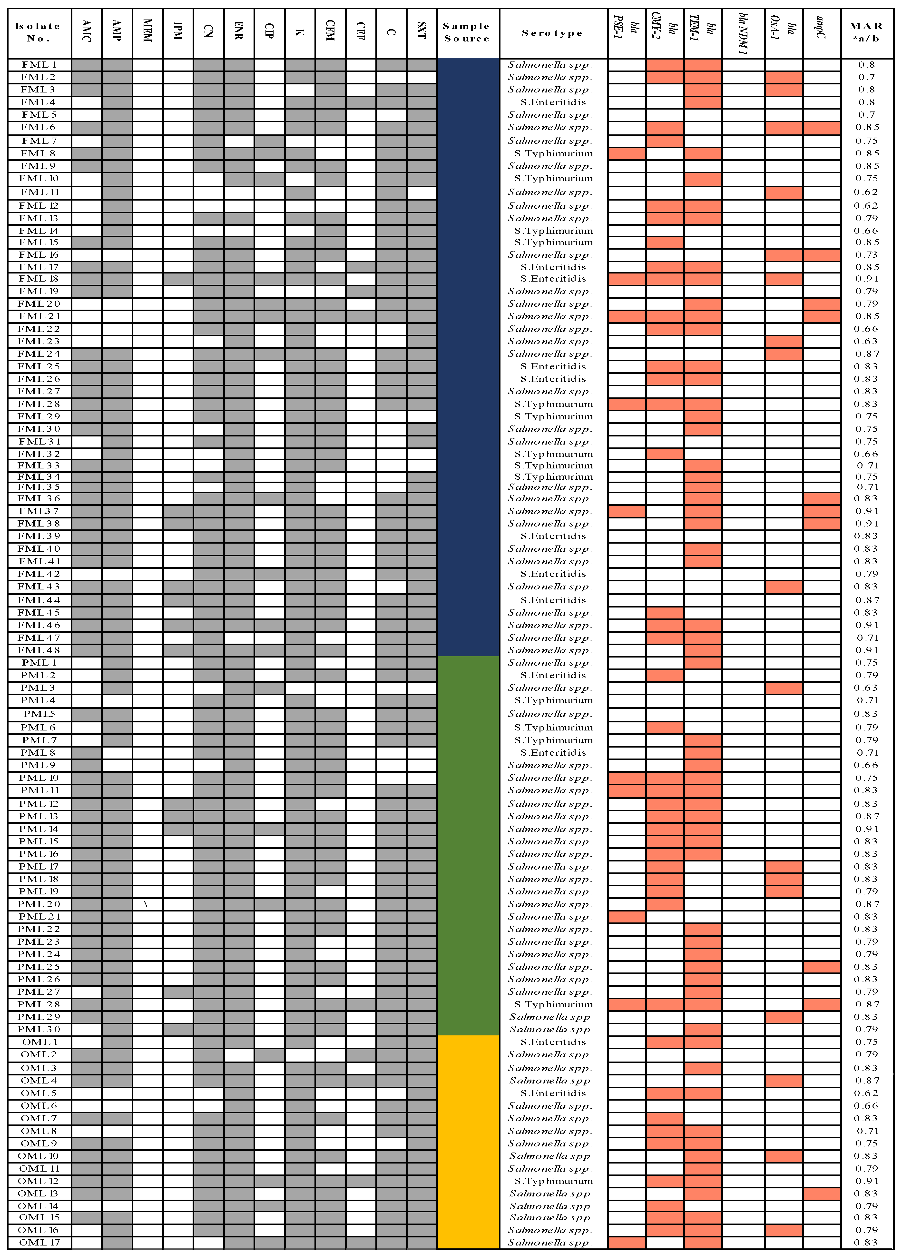
3.2. Antimicrobial Susceptibility Assay

A high incidence of MDR was observed in all Salmonella isolates. Antimicrobial resistance pattern and MAR index of 95 Salmonella isolates were presented (Figure 1). All isolates were resistant against 11 antibiotics (oxacillin, clindamycin, erythromycin, streptomycin, nalidixic acid, fusidic acid, linezolid, rifampicin, tetracycline, minocycline, and vancomycin). Resistance to other antimicrobials was as follows: enrofloxacin, 95%; gentamycin, 93%; kanamycin, 91%; sulphamethoxazole/trimethoprim, 91%; ampicillin, 86%; amoxicillin/clavulanic acid, 81%; chloramphenicol, 81%; cefixime, 76%; ciprofloxacin, 19%; imipenem, 12%; cefepime, 9%; meropenem, 2% (Figure 1). Isolates from associated poultry products (meat and eggs) were highly resistant to different antibiotics, which is a public health concern. A high MAR index (0.62–0.91) was observed in Salmonella. MAR index of different Salmonella serovars was as follows: S. Typhimurium ranged from 0.66 to 0.87, S. Enteritidis ranged from 0.71–0.91, and in non-typeable Salmonella spp., it ranged from 0.62 to 0.91.

3.3. Distribution of Antibiotic Resistance and Virulence Genes in Salmonella Isolates

The presence and absence of antibiotic resistance genes are presented in (Figure 1). The results showed that S. Enteritidis harbored resistance genes for cephalosporin and carbapenems resistance bla CMY-2 7/12 (58.3%), bla TEM-1 8/12 (66.6%), bla NDM-1 0/12 (0%), and ampC 0/12 (0%); penicillin resistance genes bla PSE-1 1/12 (8.3%) and bla OXA-1 1/12 (8.3%); and the virulence gene of zoonotic importance sopE 12/12 (100%). S. Typhimurium exhibited cephalosporins and carbapenems resistance genes blaCMY-2 6/14 (42.9%), bla TEM-19/14 (64.3%) bla NDM-1 0/14 (0%), and ampC 1/14 (7.1%); penicillin resistance genes bla PSE-23/14(21.4%), bla OXA-1 0/14 (0%), and sopE 13/14 (92.8%). Other Salmonella spp. exhibited cephalosporins and carbapenems resistance genes bla CMY-2 28/69 (40.5%), bla TEM-1, 40/69 (57.9%), bla NDM-1 0/69 (0%), and ampC 7/69 (10.1%); penicillin resistance genes bla PSE-1 6/69 (8.6%), bla OXA-1 16/69 (23.1%), and sopE 48/69 (69.5%).

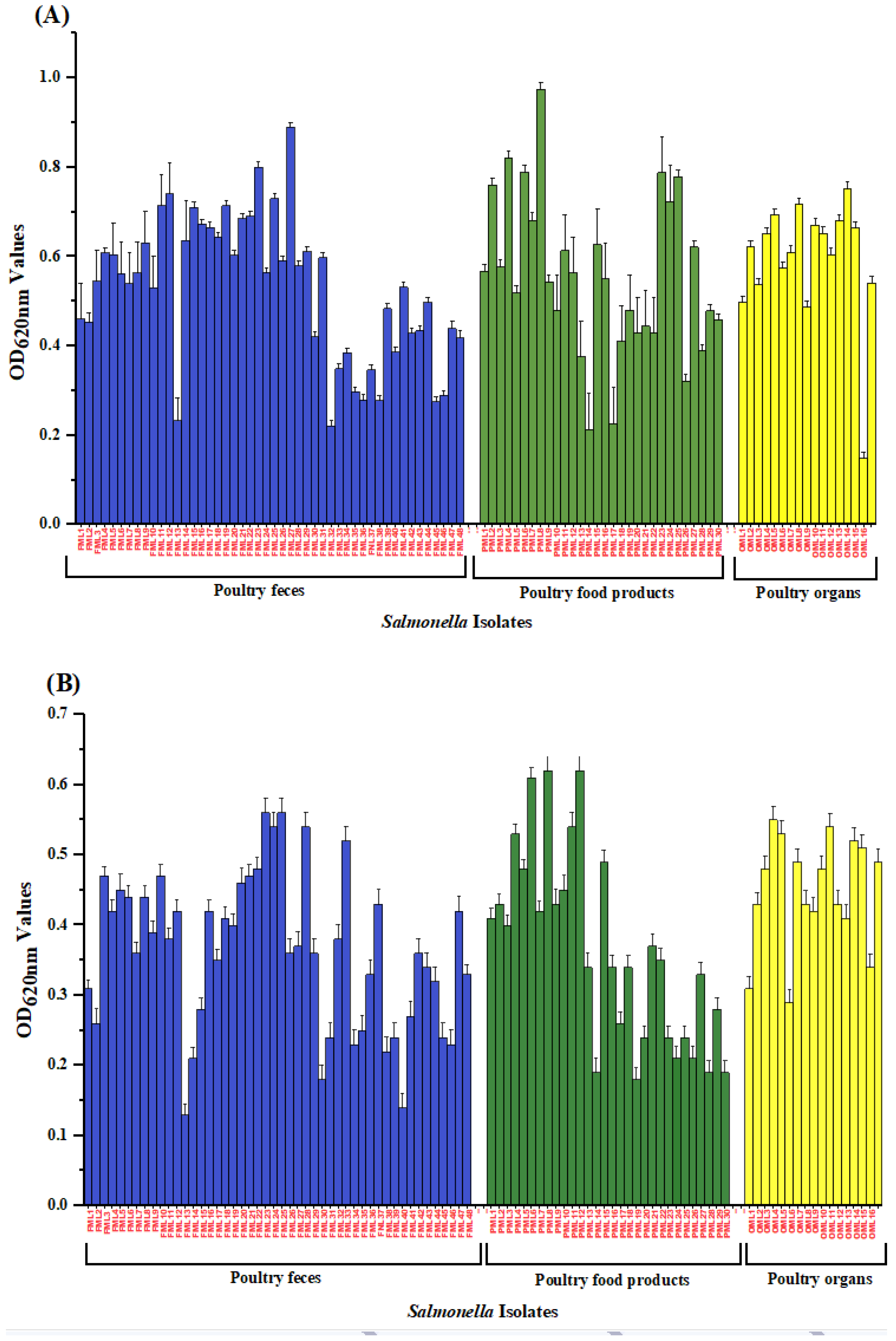
3.4. Biofilm Formation Potential of Salmonella Isolates

The Salmonella isolates’ biofilm formation was significantly influenced by the source of isolation, serotype, and incubation temperature. Biofilm formation of Salmonella isolates from different sources at two temperatures 30 °C and 37 °C is presented (Figure 2). The results revealed that poultry food isolates (meat and eggs) exhibited strong biofilm production at both temperatures. Biofilm potential was assessed at 30 °C and 37 °C for 48 h. Most of the Salmonella isolates of different origins showed strong biofilm at 37 °C for 48 h (Table 4). The data showed that the number of S. Typhimurium 11/14 (78.5%) with strong biofilm potential was almost double that of S. Enteritidis 5/12 (41.7%) at 37 °C for 48 h.

3.5. Correlation between the Number of Isolates Resistant to Antibiotics and the Ability to Produce Biofilms at Different Temperatures

To determine correlation between biofilm formation and the number of MDR Salmonella, Spearman’s rank correlation was used. All Salmonella isolates were biofilm producers at 30 °C, and they exhibited MDR profiles. The Spearman’s correlation coefficient (rs) in this case was 0.591 (p < 0.001). Similarly, all MDR Salmonella produced biofilms at 37 °C. The Spearman’s correlation coefficient (rs) comparison was 0.423 (p < 0.001). We found a significant correlation between biofilm formation and the number of multidrug-resistant isolates at 30 °C (p = 0.0001) and 37 °C (p = 0.00073).

3.6. Phenotypic Characterization of Extracellular Virulence Factors in Salmonella Isolates

Phenotypic characterization of external virulence factors of Salmonella enterica obtained from poultry feces and associated poultry products were shown (Table 5). All Salmonella enterica 95/95 (100%) in our study exhibited swarming and swimming motility. However, for S. Enteritidis isolates, 12/12 (100%) showed hemolytic activity, 11/12 (91.6%) displayed lipase activity, 12/12 (100%) showed DNA degrading activity, and 10/12 (83%) portrayed protease activity. For S. Typhimurium isolates, 13/14 (92.8%) had hemolytic activity, 11/14 (78.6%) showed lipase activity, 14/14 (100%) displayed DNA degrading activity, 10/14 (83%) portrayed protease activity. For non-typeable Salmonella isolates 67/69 (97.1%) exhibited hemolytic activity, 67/69 (97.1%) DNA degrading activity, 66/69 (95.6%) protease activity, and 65/69 (94.2%) had lipase activity.

4. Discussion

Non-typhoidal Salmonella gastrointestinal infections have become a major public health concern. Consumption of undercooked/semi-cooked poultry products is a major source of Salmonella infection in humans [25]. Poultry and poultry food products (meat and eggs) are a cheap source of high-quality protein for human consumption [26].
In our study, the overall incidence of Salmonella was 26% from poultry droppings, organs, and poultry food product samples, which emphasizes the monitoring of NTS at poultry farms as well as retail markets in Pakistan. The incidence of Salmonella in this study was higher than a previous study from Pakistan (12%), in other geographical regions, China (20%), Trinidad, Spain (20.5%), and Japan (7.9%) [27]. However, it was lower compared to those detected in India (33.1%), Canada (40%), Oklahoma (41%), Burkina Faso (55%), and Myanmar (97.8%) [20]. According to our study, we found non-typeable Salmonella spp. (73%), Salmonella Typhimurium (15%), and Salmonella Enteritidis (13%). However, these results are similar to another study in Faisalabad, Punjab, where S. Typhimurium and S. Enteritidis prevalence was found 28.4% and 9.2%, respectively [28]. A study from China revealed that Salmonella Enteritidis was the most abundant serovars, followed by S. Heidelberg and S. Typhimurium [19]. A similar study in Saudi Arabia showed the highest prevalence of S. Enteritidis (39.4%), followed by S. Paratyphi (21.2%), S. Typhimurium (15.2%), S. Typhi, and S. Arizona (12.1%), respectively. These Salmonella isolates were isolated from environmental and clinical samples.
The emergence of antibiotic resistance in NTS is important for therapeutic control during the outbreak. 100% resistance against 11 antibiotics (oxacillin, clindamycin, erythromycin, streptomycin, nalidixic acid, fusidic acid, linezolid, rifampicin, tetracycline, minocycline, and vancomycin) is alarming. This may be the result of the irrational use of antibiotics in poultry farming and the healthcare system. A previous study found Salmonella enterica was intrinsically resistant only to oxacillin [29], and in another, Salmonella from chicken meat and giblets in Egypt were 100% resistant to erythromycin, penicillin, and amoxicillin. In comparison, 98.8%, 96.4%, 95.2%, and 91.6% were resistant to nalidixic acid, sulphamethoxazole, oxytetracycline, and ampicillin [30]. According to another study, the highest resistance was found against erythromycin (100%) and streptomycin (100%) [31]. In the present study, we report resistance against enrofloxacin, gentamycin, kanamycin, sulphamethoxazole, trimethoprim, ampicillin, amoxicillin/clavulanic acid, chloramphenicol, and cefixime in the range of 76% to 96%. This resistance pattern was similar to one of the previous studies where antimicrobial resistance to amoxicillin/clavulanic acid (96%), kanamycin (88%), ampicillin (85%), and cephalothin (81%) was observed [32]. In this study, we found phenotypic resistance against imipenem (12%), cefixime (76%), and cefepime (9%). The emergence of resistance to carbapenems, third- and fourth-generation cephalosporin in NTS has not been reported previously from this region. Carbapenems are considered as only beta-lactam antibiotics that are considered effective against MDR pathogens [33]. The increasing spread of carbapenems, third- and fourth-generation cephalosporin resistance in NTS may spread to typhoidal Salmonella and other nosocomial enteric pathogens because they inhabit the same environment [34]. According to a previous study, a fourth-generation cephalosporin and fluoroquinolones resistant Salmonella typhi was reported in the Sindh region, Pakistan [35]. Another study from the Sindh region also revealed quinolones and cephalosporins resistance in NTS isolates from poultry meat [36]. Further, WGS analysis may reveal the source of such resistance in different Salmonella serovars [37]. Detection of MDR Salmonella isolates in this study warrant more attention towards surveillance of antibiotics usage in agriculture and human health care sectors in Pakistan. In our study, a high MAR index detection could be attributed to the increased use of clinically important antibiotics for bacterial infection control in humans and as therapeutic agents or growth promoters in veterinary practice for livestock in Pakistan. [38]. The high MAR observed in a similar study from Brazil ranged from 0.18–0.40 in different Salmonella serovars isolated from poultry sources [39]. MAR index of Salmonella isolates from seafood ranged from 0.14–0.45 in a study from Malaysia [40]. MAR index ranged from 0.21–0.46 in different Salmonella serovars isolated from ready-to-eat shrimps from a study in Nigeria [41]. A high number of tested antibiotics and high antibiotic resistance detection in our study compared to previous studies may be the cause of the high MAR index. The rapid emergence of antibiotic resistance is attributed to the selective pressure of antibiotics through evolutionary responses due to natural selection [42].
The emergence of extended-spectrum β-lactam/cephalosporin resistance in Salmonella can narrow its control options by antibiotics. Most of the antibiotic-resistant determinants are present in plasmids or integrons, which can transfer these genes to other bacterial species of different or the same group [43]. In the present study, among cephalosporin and carbapenems resistance genes in S. Typhimurium, bla TEM-1 (64.3%) is more abundant than other genes. In S. Enteritidis, bla TEM-1 (66.6%) was detected as the most prevalent. This is related to another study where bla TEM-1 (35.3% and 72.7%) was detected as most prevalent in S. Typhimurium and S. Enteritidis isolates, respectively [28]. There are various reports in previous studies where the bla TEM-1 gene was detected as the most prevalent [44]. According to our study, the beta-lactamase penicillin gene bla PSE-1 gene (21.8%) was most prevalent in S. Typhimurium. While in S. Enteritidis, bla PSE-1 (8.3%) and bla OXA-1(8.3%) were found in the same proportion. The mismatch between genotypic and phenotypic antibiotic resistance in our study may be due to mutation in genes and variation in gene expression within different Salmonella isolates. The sopE gene was found in all S. Enteritidis and 92.8% of S. Typhimurium, whereas in non-typeable Salmonella, it was observed in 68.1% isolates, which agrees with previous findings [45,46]. The sopE gene is encoded in SPI-1. It is identified in the isolates that are mainly involved in major epidemics; therefore, sopE has been identified as a major determinant in the spread of epidemic strains [47]. In another study, Salmonella Enteritidis isolated from chicken, eggs, and humans constitute the sopE gene, which may indicate its importance in zoonosis [48].
Biofilm formation may help in Salmonella survival in poultry farms and poultry food products [49]. In our study, S. Typhimurium (78.5%) exhibits strong biofilm potential at 37 °C; similar results were revealed from previous studies [19,50]. Salmonella isolated from poultry food (meat and eggs) in our study with moderate to strong biofilm potential and having MDR characteristics is a concern for public health and poultry farming. Bacteria grown in biofilms have a greater ability to transfer genes horizontally than planktonic cells [51]. Biofilms increase the chances of gene transfer with the help of virulence factors and antibiotic-resistant genes from resistant to susceptible bacterial species, which leads to the emergence of new antibiotic resistance in pathogens [21]. The variation in the biofilm potential of the Salmonella isolates in this study may be due to the difference in incubation temperature (30 °C and 37 °C) coupled with species diversity as described in a previous study [52]. The reason for choosing temperatures of 30 °C and 37 °C to assess biofilm formation potential was because sampling regions fall in the temperate zone, and summer temperatures are high. Most poultry meat and egg shops operate in warm climatic conditions where temperatures lie between 28 °C to 30 °C. This study found a significant correlation between antibiotic resistance and biofilm formation at both temperatures 30 °C and 37 °C. Such a relationship has been described for other bacteria, although the findings were sometimes inconsistent, and the correlations were species-dependent [53]. Our data showed that the Salmonella serovars from poultry gut, organs, and food (meat and egg) had virulence characteristics and determined the pathogenicity of Salmonella isolates. These external virulence factors are swimming and swarming motility, hemolysis, lipase, the presence of protease, and DNA degrading activity. Motility is an important pathogenic property of bacteria that is closely related to virulence factor production, antibiotic resistance, and biofilm potential [54]. According to a previous study, a significant linkage between protease production, motility, and pathogenesis has been reported [55]. Extracellular protease, DNA structure, and lipolytic activity positively correlated with biofilm formation [56,57,58].
Management strategies for antibiotic resistance should be adopted to control the dissemination of antibiotic resistance includes the following: improved information to enhance the awareness, control of non-therapeutic use of antibiotics in food animal production system, improvement in diagnostic procedures, and enhancement of microbiological laboratory equipment and personals [59,60]. These suggestions may assist in the reduction of antibiotic resistance and can improve the public health sector in Pakistan.

5. Conclusions

These findings indicated highly antibiotic-resistant NTS serovars with zoonotic potential at local poultry farms in Pakistan, emphasizing the need to adopt more biosecurity measures, environmental and personal hygiene awareness among the local poultry farmers. The detection of the most important foodborne zoonotic Salmonella enterica serovars Typhimurium and Enteritidis is of public health significance. The emergence of MDR Salmonella serovars is of great concern for the targeted antimicrobial therapy. Resistance from these Salmonella isolates may transfer to typhoidal Salmonella and other Enterobacteriaceae family as these pathogens share a common environment for propagation. Irrational usage of different antibiotics in the poultry industry should be checked to avoid spreading and disseminating antibiotic resistance. The biofilm formation potential of these isolates is of great concern for the food industry and public health.

Author Contributions

A.S.: Performed experiments and wrote the manuscript. S.A. (Sara Azim): Performed experiments. S.A. (Saadia Andleeb) and M.I.: Data analysis and technical assistance, A.A. (Aitezaz Ahsan): Data collection from different regions of Pakistan and technical assistance. A.A. (Amjad Ali) and A.R.: Planned and executed experiments, analyzed data, and wrote the manuscript. All authors have read and agreed to the published version of the manuscript.

Funding

This study was financially supported through an ICT Endowment scholarship from the National University of Science and Technology, Islamabad, Pakistan.

Acknowledgments

The authors acknowledge Mudassar Mohiuddin for valuable comments and suggestions.

Conflicts of Interest

All authors declare no competing interests.

References

  1. Gharieb, R.; Tartor, Y.H.; Khedr, M.H.E. Non-Typhoidal Salmonella in poultry meat and diarrhoeic patients: Prevalence, antibiogram, virulotyping, molecular detection and sequencing of class I integrons in multidrug resistant strains. Gut Pathog. 2015, 7, 1–11. [Google Scholar] [CrossRef] [Green Version]
  2. Guibourdenche, M.; Roggentin, P.; Mikoleit, M.; Fields, P.I.; Bockemühl, J.; Grimont, P.; Weill, F.-X. Supplement 2003–2007 (No. 47) to the White-Kauffmann-Le Minor scheme. Res. Microbiol. 2010, 161, 26–29. [Google Scholar] [CrossRef]
  3. Jajere, S.M. A review of Salmonella enterica with particular focus on the pathogenicity and virulence factors, host specificity and antimicrobial resistance including multidrug resistance. Veter. World 2019, 12, 504–521. [Google Scholar] [CrossRef] [Green Version]
  4. Majowicz, S.; Musto, J.; Scallan, E.; Angulo, F.; Kirk, M.; O’Brien, S.; Jones, T.F.; Fazil, A.; Hoekstra, R.M.; International Collaboration on Enteric Disease “Burden of Illness” Studies. The global burden of nontyphoidal Salmonella gastroenteritis. Clin. Infect. Dis. 2010, 50, 882–889. [Google Scholar] [CrossRef] [Green Version]
  5. Mouttotou, N.; Ahmad, S.; Kamran, Z.; Koutoulis, K.C. Prevalence, Risks and Antibiotic Resistance of Salmonella in Poultry Production Chain. Curr. Top. Salmonella Salmonellosis 2017, 2017, 134–215. [Google Scholar] [CrossRef]
  6. Heredia, N.; García, S. Animals as sources of food-borne pathogens: A review. Anim. Nutr. 2018, 4, 250–255. [Google Scholar] [CrossRef] [PubMed]
  7. Thai, T.H.; Hirai, T.; Lan, N.T.; Yamaguchi, R. Antibiotic resistance profiles of Salmonella serovars isolated from retail pork and chicken meat in North Vietnam. Int. J. Food Microbiol. 2012, 156, 147–151. [Google Scholar] [CrossRef]
  8. Nair, D.V.T.; Venkitanarayanan, K.; Kollanoor, J.A. Antibiotic-resistant Salmonella in the food supply and the potential role of antibiotic alternatives for control. Foods 2018, 7, 167. [Google Scholar] [CrossRef] [Green Version]
  9. Lai, J.; Wu, C.; Wu, C.; Qi, J.; Wang, Y.; Wang, H.; Liu, Y.; Shen, J. Serotype distribution and antibiotic resistance of Salmonella in food-producing animals in Shandong province of China, 2009 and 2012. Int. J. Food Microbiol. 2014, 180, 30–38. [Google Scholar] [CrossRef] [PubMed]
  10. Clothier, K.A.; Kim, P.; Mete, A.; Hill, A.E. Frequency, serotype distribution, and antimicrobial susceptibility patterns of Salmonella in small poultry flocks in California. J. Veter. Diagn. Investig. 2018, 30, 471–475. [Google Scholar] [CrossRef] [PubMed]
  11. Iwamoto, M.; Reynolds, J.; Karp, B.; Tate, H.; Cray, P.; Plumblee, J.R.; Hoekstra, R.M.; Whichard, J.M.; Mahon, B.E. Ceftriaxone-Resistant Nontyphoidal Salmonella from Humans, Retail Meats, and Food Animals in the United States, 1996–2013. Foodborne Pathog. Dis. 2017, 14, 74–83. [Google Scholar] [CrossRef] [PubMed]
  12. Yang, X.; Huang, J.; Wu, Q.; Zhang, J.; Liu, S.; Guo, W.; Cai, S.; Yu, S. Prevalence, antimicrobial resistance and genetic diversity of Salmonella isolated from retail ready-to-eat foods in China. Food Control. 2016, 60, 50–56. [Google Scholar] [CrossRef]
  13. Antunes, P.; Mourão, J.; Campos, J.; Peixe, L. Salmonellosis: The role of poultry meat. Clin. Microbiol. Infect. 2016, 22, 110–121. [Google Scholar] [CrossRef] [Green Version]
  14. Nakatsuchi, A.; Inagaki, M.; Sugiyama, M.; Usui, M.; Asai, T. Association of Salmonella Serotypes with Quinolone Resistance in Broilers. Food Saf. 2018, 6, 156–159. [Google Scholar] [CrossRef] [Green Version]
  15. Nde, C.W.; Fakhr, M.K.; Doetkott, C.; Logue, C.M. An evaluation of conventional culture, invA PCR, and the real-time PCR iQ-Check kit as detection tools for Salmonella in naturally contaminated premarket and retail turkey. J. Food Prot. 2008, 71, 386–391. [Google Scholar] [CrossRef]
  16. Gunasegaran, T.; Rathinam, X.; Kasi, M.; Sathasivam, K.; Sreenivasan, S.; Subramaniam, S. Isolation and identification of Salmonella from curry samples and its sensitivity to commercial antibiotics and aqueous extracts of Camelia sinensis (L.) and Trachyspermum ammi (L.). Asian Pac. J. Trop. Biomed. 2011, 1, 266–269. [Google Scholar] [CrossRef] [Green Version]
  17. Soumet, C.; Ermel, G.; Rose, V.; Rose, N.; Drouin, P.; Salvat, G.; Colin, P. Identification by a multiplex PCR-based assay ofSalmonellaTyphimurium andSalmonellaEnteritidis strains from environmental swabs of poultry houses. Lett. Appl. Microbiol. 1999, 29, 1–6. [Google Scholar] [CrossRef] [PubMed]
  18. Moe, A.Z.; Paulsen, P.; Pichpol, D.; Fries, R.; Irsigler, H.; Baumann, M.P.O.; Oo, K.N. Prevalence and Antimicrobial Resistance of Salmonella Isolates from Chicken Carcasses in Retail Markets in Yangon, Myanmar. J. Food Prot. 2017, 80, 947–951. [Google Scholar] [CrossRef] [PubMed]
  19. Lamas, A.; Miranda, J.; Vazquez, B.; Cepeda, A.; Franco, C. Biofilm formation, phenotypic production of cellulose and gene expression in Salmonella enterica decrease under anaerobic conditions. Int. J. Food Microbiol. 2016, 238, 63–67. [Google Scholar] [CrossRef]
  20. Marangoni, F.; Corsello, G.; Cricelli, C.; Ferrara, N.; Ghiselli, A.; Lucchin, L.; Poli, A. Role of poultry meat in a balanced diet aimed at maintaining health and wellbeing: An Italian consensus document. Food Nutr. Res. 2015, 59, 27606. [Google Scholar] [CrossRef] [Green Version]
  21. Galloway, W.R.; Hodgkinson, J.; Bowden, S.; Welch, M.; Spring, D.R. Applications of small molecule activators and inhibitors of quorum sensing in Gram-negative bacteria. Trends Microbiol. 2012, 20, 449–458. [Google Scholar] [CrossRef]
  22. Odoch, T.; Sekse, C.; L’Abee-Lund, T.M.; Hansen, H.C.H.; Kankya, C.; Wasteson, Y. Diversity and Antimicrobial Resistance Genotypes in Non-Typhoidal Salmonella Isolates from Poultry Farms in Uganda. Int. J. Environ. Res. Public Health 2018, 15, 324. [Google Scholar] [CrossRef] [PubMed] [Green Version]
  23. Babapour, E.; Haddadi, A.; Mirnejad, R.; Angaji, S.A.; Amirmozafari, N. Biofilm formation in clinical isolates of nosocomial Acinetobacter baumannii and its relationship with multidrug resistance. Asian Pac. J. Trop. Biomed. 2016, 6, 528–533. [Google Scholar] [CrossRef] [Green Version]
  24. Stepanović, S.; Vukovic, D.; Dakić, I.; Savić, B.; Švabić-Vlahović, M. A modified microtiter-plate test for quantification of staphylococcal biofilm formation. J. Microbiol. Methods 2000, 40, 175–179. [Google Scholar] [CrossRef]
  25. Eguale, T.; Gebreyes, W.A.; Asrat, D.; Alemayehu, H.; Gunn, J.S.; Engidawork, E. Non-typhoidal Salmonella serotypes, antimicrobial resistance and co-infection with parasites among patients with diarrhea and other gastrointestinal complaints in Addis Ababa, Ethiopia. BMC Infect. Dis. 2015, 15, 497. [Google Scholar] [CrossRef] [PubMed] [Green Version]
  26. Kumar, M.; Dahiya, S.P.; Ratwan, P. Backyard poultry farming in India: A tool for nutritional security and women empowerment. Biol. Rhythm. Res. 2019. [Google Scholar] [CrossRef]
  27. Michael, G.; Schwarz, S. Antimicrobial resistance in zoonotic nontyphoidal Salmonella: An alarming trend? Clin. Microbiol. Infect. 2016, 22, 968–974. [Google Scholar] [CrossRef] [PubMed]
  28. Duc, V.M.; Nakamoto, Y.; Fujiwara, A.; Toyofuku, H.; Obi, T.; Chuma, T. Prevalence of Salmonella in broiler chickens in Kagoshima, Japan in 2009 to 2012 and the relationship between serovars changing and antimicrobial resistance. BMC Veter. Res. 2019, 15, 108. [Google Scholar] [CrossRef] [PubMed] [Green Version]
  29. Wajid, M.; Awan, A.B.; Saleemi, M.K.; Weinreich, J.; Schierack, P.; Sarwar, Y.; Ali, A. Multiple Drug Resistance and Virulence Profiling ofSalmonella entericaSerovars Typhimurium and Enteritidis from Poultry Farms of Faisalabad, Pakistan. Microb. Drug Resist. 2019, 25, 133–142. [Google Scholar] [CrossRef]
  30. Li, B.; Liu, C.; Liu, L.; Li, S.; Fan, N.; Hou, H.; Jin, J.; Xing, Y. [Prevalence and etiologic agent of Salmonella in livestock and poultry meats in Huai’an City during 2015–2016]. Wei Sheng Yan Jiu J. Hyg. Res. 2018, 47, 260–300. [Google Scholar]
  31. Stock, I.; Wiedemann, B. Natural antibiotic susceptibility of Salmonella enterica strains. Int. J. Antimicrob. Agents 2000, 16, 211–217. [Google Scholar] [CrossRef]
  32. Abd-Elghany, S.; Sallam, K.; Abd-Elkhalek, A.; Tamura, T. Occurrence, genetic characterization and antimicrobial resistance of Salmonella isolated from chicken meat and giblets. Epidem. Infect. 2015, 143, 997–1003. [Google Scholar] [CrossRef] [PubMed] [Green Version]
  33. El-Tayeb, M.A.; Ibrahim, A.S.; Al-Salamah, A.A.; Almaary, K.S.; Elbadawi, Y.B. Prevalence, serotyping and antimicrobials resistance mechanism of Salmonella enterica isolated from clinical and environmental samples in Saudi Arabia. Braz. J. Microbiol. 2017, 48, 499–508. [Google Scholar] [CrossRef]
  34. Gad, A.H.; Abo-Shama, U.H.; Harclerode, K.K.; Fakhr, M.K. Prevalence, Serotyping, Molecular Typing, and Antimicrobial Resistance of Salmonella Isolated From Conventional and Organic Retail Ground Poultry. Front. Microbiol. 2018, 9, 2653. [Google Scholar] [CrossRef] [PubMed]
  35. Temkin, E.; Adler, A.; Lerner, A.; Carmeli, Y. Carbapenem-resistant Enterobacteriaceae: Biology, epidemiology, and management. Ann. N. Y. Acad. Sci. 2014, 1323, 22–42. [Google Scholar] [CrossRef] [PubMed]
  36. Fernandez, J.; Guerra, B.; Rodicio, M.R. Resistance to Carbapenems in Non-Typhoidal Salmonella enterica Serovars from Humans, Animals and Food. Veter. Sci. 2018, 5, 40. [Google Scholar] [CrossRef] [PubMed] [Green Version]
  37. Klemm, E.J.; Shakoor, S.; Page, A.J.; Qamar, F.N.; Judge, K.; Saeed, D.K.; Wong, V.K.; Dallman, T.J.; Nair, S.; Baker, S.; et al. Emergence of an Extensively Drug-Resistant Salmonella enterica Serovar Typhi Clone Harboring a Promiscuous Plasmid Encoding Resistance to Fluoroquinolones and Third-Generation Cephalosporins. mBio 2018, 9. [Google Scholar] [CrossRef] [Green Version]
  38. Soomro, A.H.; Khaskheli, M.; Bhutto, M.B.; Shah, G.; Memon, A.; Dewani, P. Prevalence and antimicrobial resistance of Salmonella serovars isolated from poultry meat in Hyderabad, Pakistan. Turk. J. Veter. Anim. Sci. 2011, 34, 455–460. [Google Scholar]
  39. Borges, K.A.; Furian, T.Q.; Souza SNd Salle, C.T.P.; Moraes, H.L.d.S.; Nascimento, V.P.D. Antimicrobial resistance and molecular characterization of Salmonella enterica serotypes isolated from poultry sources in Brazil. Braz. J. Poult. Sci. 2019, 21. [Google Scholar] [CrossRef]
  40. Budiati, T.; Rusul, G.; Wan-Abdullah, W.N.; Arip, Y.M.; Ahmad, R.; Thong, K.L.J.A. Prevalence, antibiotic resistance and plasmid profiling of Salmonella in catfish (Clarias gariepinus) and tilapia (Tilapia mossambica) obtained from wet markets and ponds in Malaysia. Aquaculture 2013, 372, 127–132. [Google Scholar] [CrossRef]
  41. Beshiru, A.; Igbinosa, I.H.; Igbinosa, E.O. Prevalence of antimicrobial resistance and virulence gene elements of Salmonella serovars from ready-to-eat (RTE) shrimps. Front. Microbiol. 2019, 10, 1613. [Google Scholar] [CrossRef] [Green Version]
  42. Deekshit, V.K.; Kumar, B.K.; Rai, P.; Rohit, A.; Karunasagar, I. Simultaneous detection of Salmonella pathogenicity island 2 and its antibiotic resistance genes from seafood. J. Microbiol. Methods 2013, 93, 233–238. [Google Scholar] [CrossRef]
  43. Biffi, C.P.; Stefani, L.M.; Miletti, L.C.; Matiello, C.A.; Backes, R.G.; Almeida, J.M.; Neves, G.B. Phenotypic and genotypic resistance profile of Salmonella Typhimurium to antimicrobials commonly used in poultry. Braz. J. Poult. Sci. 2014, 16, 93–96. [Google Scholar] [CrossRef] [Green Version]
  44. Zhao, X.; Yang, J.; Zhang, B.; Sun, S.; Chang, W. Characterization of Integrons and Resistance Genes in Salmonella Isolates from Farm Animals in Shandong Province, China. Front. Microbiol. 2017, 8, 1300. [Google Scholar] [CrossRef]
  45. Borges, K.A.; Furian, T.Q.; Borsoi, A.; Moraes, H.L.; Salle, C.T.P.; Nascimento, V.P.D. Detection of virulence-associated genes in Salmonella Enteritidis isolates from chicken in South of Brazil. Pesquisa Veterinária Brasileira 2013, 33, 1416–1422. [Google Scholar] [CrossRef]
  46. El-Sharkawy, H.; Tahoun, A.; El-Gohary, A.E.; El-Abasy, M.; El-Khayat, F.; Gillespie, T.; Kitade, Y.; Hafez, H.M.; Neubauer, H.; El-Adawy, H. Epidemiological, molecular characterization and antibiotic resistance of Salmonella enterica serovars isolated from chicken farms in Egypt. Gut Pathogens. 2017, 9, 8. [Google Scholar] [CrossRef] [PubMed] [Green Version]
  47. Hopkins, K.L.; Threlfall, E.J. Frequency and polymorphism of sopE in isolates of Salmonella enterica belonging to the ten most prevalent serotypes in England and Wales. J. Med. Microbiol. 2004, 53, 539–543. [Google Scholar] [CrossRef]
  48. Han, J.; Gokulan, K.; Barnette, D.; Khare, S.; Rooney, A.W.; Deck, J.; Nayak, R.; Stefanova, R.; Hart, M.E.; Foley, S.L. Evaluation of Virulence and Antimicrobial Resistance inSalmonella entericaSerovar Enteritidis Isolates from Humans and Chicken- and Egg-Associated Sources. Foodborne Pathog. Dis. 2013, 10, 1008–1015. [Google Scholar] [CrossRef] [PubMed]
  49. Latasa, C.; Roux, A.; Toledo-Arana, A.; Ghigo, J.M.; Gamazo, C.; Penadés, J.R.; Lasa, I. BapA, a large secreted protein required for biofilm formation and host colonization of Salmonella enterica serovar Enteritidis. Mol. Microbiol. 2005, 58, 1322–1339. [Google Scholar] [CrossRef] [PubMed]
  50. de Oliveira, T.L.C.; de Araújo Soares, R.; Piccoli, R.H. A Weibull model to describe antimicrobial kinetics of oregano and lemongrass essential oils against Salmonella Enteritidis in ground beef during refrigerated storage. Meat Sci. 2013, 93, 645–651. [Google Scholar] [CrossRef]
  51. Borges, A.; Abreu, A.C.; Dias, C.; Saavedra, M.J.; Borges, F.; Simões, M. New Perspectives on the Use of Phytochemicals as an Emergent Strategy to Control Bacterial Infections Including Biofilms. Molecules 2016, 21, 877. [Google Scholar] [CrossRef]
  52. Jensen, M.B.F.; Schjørring, S.; Björkman, J.T.; Torpdahl, M.; Litrup, E.; Nielsen, E.M.; Niskanen, T. External quality assessment for molecular typing of Salmonella 2013–2015: Performance of the European national public health reference laboratories. Eur. J. Clin. Microbiol. Infect. Dis. 2017, 36, 1923–1932. [Google Scholar] [CrossRef] [Green Version]
  53. Troxell, B.; Petri, N.; Daron, C.; Pereira, R.; Mendoza, M.; Hassan, H.M.; Koci, M.D. Poultry Body Temperature Contributes to Invasion Control through Reduced Expression of Salmonella Pathogenicity Island 1 Genes in Salmonella enterica Serovars Typhimurium and Enteritidis. Appl. Environ. Microbiol. 2015, 81, 8192–8201. [Google Scholar] [CrossRef] [Green Version]
  54. Runkel, S.; Wells, H.C.; Rowley, G. Living with stress: A lesson from the enteric pathogen Salmonella enterica. In Advances in Applied Microbiology; Elsevier: Amsterdam, The Netherlands, 2013; Volume 83, pp. 87–144. [Google Scholar]
  55. Simm, R.; Ahmad, I.; Rhen, M.; Le Guyon, S.; Römling, U. Regulation of biofilm formation in Salmonella enterica serovar Typhimurium. Future Microbiol. 2014, 9, 1261–1282. [Google Scholar] [CrossRef] [PubMed]
  56. Baugh, S.; Ekanayaka, A.S.; Piddock, L.J.V.; Webber, M.A. Loss of or inhibition of all multidrug resistance efflux pumps of Salmonella enterica serovar Typhimurium results in impaired ability to form a biofilm. J. Antimicrob. Chemother. 2012, 67, 2409–2417. [Google Scholar] [CrossRef] [PubMed] [Green Version]
  57. Frees, D.; Brøndsted, L.; Ingmer, H. Bacterial proteases and virulence. In Regulated Proteolysis in Microorganisms; Springer: Berlin/Heidelberg, Germany, 2013; pp. 161–192. [Google Scholar]
  58. Rosenau, F.; Isenhardt, S.; Gdynia, A.; Tielker, D.; Schmidt, E.; Tielen, P.; Schobert, M.; Jahn, D.; Wilhelm, S.; Jaeger, K.-E. Lipase LipC affects motility, biofilm formation and rhamnolipid production in Pseudomonas aeruginosa. FEMS Microbiol. Lett. 2010, 309, 25–34. [Google Scholar] [CrossRef] [PubMed]
  59. Ganguly, N.K.; Wattal, C.; Chandy, S.J.; Arora, S.K.; Gupta, U.; Kotwani, A. Situation Analysis Antibiotic Use and Resistance in India; Public Health Foundation of India, and Center for Disease Dynamics, Economics and Policy: New Delhi, India, 2011. [Google Scholar]
  60. Pruden, A.; Larsson, D.J.; Amézquita, A.; Collignon, P.; Brandt, K.K.; Graham, D.W.; Lazorchak, J.M.; Suzuki, S.; Silley, P.; Snape, J.R.; et al. Management Options for Reducing the Release of Antibiotics and Antibiotic Resistance Genes to the Environment. Environ. Health Perspect. 2013, 121, 878–885. [Google Scholar] [CrossRef]
Figure 1. Salmonella strains (95) showing phenotypic antibiotic resistance profiles of 12 antibiotics, their source, origin, serotypes, and MAR index. Black squares indicate resistance; white squares indicate susceptibility; red squares indicate the presence of AMR genes; blue color presents isolates from poultry droppings, green; poultry food products (meat and eggs), yellow; poultry organs. Abbreviations: C: chloramphenicol (30 μg); AMC: amoxicillin-clavulanic acid (10 μg): CIP; ciprofloxacin (10 μg); CN: gentamicin (10 μg); SXT: sulfamethoxazole/trimethoprim (25 μg); K: kanamycin(30 μg): AMP: ampicillin (30 μg); MEM: meropenem (10 μg); IPM: imipenem (10 μg); CEF: cefepime (30 μg); CFM: cefixime (5 μg); ENR: enrofloxacin (10 ug); MAR: multiple antibiotic resistance; a*: No. of antibiotics resistant; b*: No. of antibiotics tested.
Antibiotics 10 00785 g001
Figure 2. Results of biofilm formation assay. X-axis: Salmonella isolates from poultry droppings; poultry food products; poultry organs at two temperatures, (A): 37 °C; (B): 30 °C evaluated by crystal violet assay. Biofilm formation was assessed by staining the attached bacteria with 0.2% CV and measuring the OD values at 620 nm after 48 h growth. Error bars represent standard deviations between three replicates.
Antibiotics 10 00785 g002
Table 1. Primers used for Salmonella detection with Multiplex PCR.
Target SequenceTarget
Strain
Primer SetsLengthPrimer Sequence
5′----3′
Amplification RegionReference
Random SequenceSalmonella spp.ST11
ST15
24
24
GCCAACCATTGCTAAATTGGCGCA
GGTAGAAATTCCCAGCGGGTACTGG
429[17]
fliC geneSalmonella TyphimuriumFli15
Tym
22
22
CGGTGTTGCCCAGGTTGGTAAT
ACTCTTGCTGGCGGTGCGACTT
559[17]
sefA geneSalmonella EnteritidisSef 167
Sef 478
20
20
AGGTTCAGGCAGCGGTTACT
GGGACATTTAGCGTTTCTTG
312[17]
Table 2. Primers and PCR conditions used for antibiotic resistance genes and virulence factor detection.
GenesSequences (5′-3′)Annealing Temp. (°C)Amplicon SizeReferences
bla PSE-1CGCTTCCCGTTAACAAGTAC
CTGGTTCATTTCAGATAGCG
50430[18]
bla CMY-2TGGCCAGAACTGACAGGCAAA
TTTCTCCTGAACGTGGCTGGC
57870
bla TEM-1CAGCGGTAAGATCCT TGAGA
ACTCGCCGTCGTGTAGATAA
55643[19]
bla OxA-1ATGAAAAACACAATACATATC
AATTTAGTGTGTTTAGAATGG
50830
bla NDM-1GGG CAG TCG CTT CCA ACG GT
GTA GTG CTC AGT GTC GGC AT
58475[20]
ampCAACACACTGATTGCGTCTGAC
CTGGGCCTCATCGTCAGTTA
601226[9]
SopEACACACTTTCCACGAGGAAGCG
GGATGCCTTCTGATGTTGACTGG
50398[21]
Table 3. Prevalence of Salmonella in poultry feces, poultry organs, and poultry food products.
Sr. No.Sample SourceNo. of Samples (n)Positive Samples (%)Serovar (s) IsolatedNo. of Serovar out of Positive Sample (%)
1Poultry feces18048 (26.7)S.Typhimurium9 (18.75)
S. Enteritidis8 16.6)
Other Salmonella31 (64.6)
2Poultry organs7017 (24.3)S.Typhimurium1 (5.8)
S. Enteritidis2 (11.7)
Other Salmonella14 (82.3)
3Poultry Meat6021 (35)S.Typhimurium2 (9.5)
S. Enteritidis1 (4.7)
Other Salmonella18 (85.7)
4Poultry eggs609 (15)S.Typhimurium2 (22.2)
S.Enteritidis1 (11.1)
Other Salmonella6 (66.6)
Table 4. Biofilm potential of different Salmonella isolates at different temperatures.
Salmonella spp.Temperature
(°C)
Weak
Biofilm
Moderate BiofilmStrong BiofilmNo Biofilm
S. Typhimurium
(n = 14)
303 (21.4)4 (28.5)7 (50)0 (0)
371 (8)2 (14.2)11 (78.5)0 (0)
S. Enteritidis
(n = 12)
302 (16.6)7 (58.3)3 (25)0 (0)
372 (16.6)5 (41.7)5 (41.7)0 (0)
Other Salmonella spp.
(n = 69)
3018 (26.1)30 (43.4)21 (30.4)0 (0)
3711 (15.9)20 (28.9)38 (55.1)0 (0)
Table 5. Phenotypic virulence characteristics of Salmonella isolates.
Salmonella spp.DNA Degrading
Activity
Hemolytic
Activity
Lipase
Activity
Protease
Activity
Swimming
Motility
Swarming
Motility
S. Typhimurium
(n = 14)
14 (100)13 (92.8)11 (78.6)10 (83)14 (100)14 (100)
S. Enteritidis
(n = 12)
12 (100)12 (100)11 (91.6)10 (83)12 (100)12 (100)
Other
Salmonella spp.
(n = 69)
67 (97.1)68 (98.5)65 (94.2)68 (98.5)69 (100)69 (100)
Publisher’s Note: MDPI stays neutral with regard to jurisdictional claims in published maps and institutional affiliations.

