Article

Isolation and Antibiotic Resistant Research of Tetragenococcus halophilus from Xuanwei Ham, A China High-Salt-Fermented Meat Products

1
Yunnan Institute of Food Safety, Kunming University of Science and Technology, Yunnan 650500, China
2
Faculty of agriculture and food, Kunming University of Science and Technology, Yunnan 650500, China
*
Author to whom correspondence should be addressed.
Submission received: 19 August 2019 / Revised: 8 September 2019 / Accepted: 11 September 2019 / Published: 16 September 2019

Abstract

:
We assessed the prevalence of antibiotic resistant and antibiotic resistance genes for 49 Tetragenococcus halophilus (T. halophilus) strains isolated from Xuawei ham in China. The antibiotic resistance phenotype was detected by the Bauer–Kirby (K–B) method and the results showed that 49 isolates can be considered completely susceptible to penicillin, ampicillin, amoxicillin, cefradine, cefotaxime, tetracyclines, minocycline, doxycycline, and vancomycin, but resistant to gentamicin, streptomycin, neomycin, polymyxinB, cotrimoxazole. This resistance was sufficiently high to consider the potential for acquisition of transmissible determinants. A total of 32 isolates were resistant to ofloxacin, 4 isolates were resistant to ciprofloxacin and chloramphenicol, and 2 isolates were resistant to ceftazidime and ticarcillin. The antibiotic resistance genes were detected by routine polymerase chain reaction (PCR). Among the 26 antibiotic resistance genes, 5 varieties of antibiotic resistance genes, including acrB, blaTEM, AAda1, SulII, and GyrB were detected and the detection rates were 89.79%, 47.7%, 16.33%, 77.55%, and 75.51%, respectively. The potential acquisition of transmissible determinants for antibiotic resistance and antibiotic resistance genes identified in this study necessitate the need for a thorough antibiotic resistance safety assessment of T. halophilus before it can be considered for use in food fermentation processes.

1. Introduction

Tetragenococcus halophilus, formerly classified as Pediococcus halophilus and subsequently reclassified as Tetragenococcus by Collins et al. [1], is a gram-positive, non-spore quadruple sphere, exopolysaccharide, nonmotile, and facultative anaerobic, and the final product of glucose metabolism is lactic acid without gas formation [2,3]. T. halophilus has been widely detected in various salted and fermented food such as fermented fish products, soy pastes, soy sauce, and is considered a potential starter for their production to improve the quality or to shorten the fermentation cycle and so on [4,5,6,7,8,9,10]. However, there is hardly any literature report on the isolation and identification of T. halophilus from salt-cured and fermented Xuanwei ham.
T. halophilus as a potential starter of this species, which not only improves the sensory properties and the flavor, as well as effectively reducing harmful substances, but also possess health functionality in fermented foods. Adding T. halophilus in the fermentation process of fish sauce can significantly increase the content of total amino acids and free amino acids and increase the content of umami amino acids, such as glutamic acid [10]. The content of volatile substances, such as 2-methylpropanal, 2-methylbutanal, 3-methylbutanal, and benzaldehyde, which have a positive influence on the flavor, is also significantly increased in the fish sauce to which the T. halophilus is added [11]. The yeast-derived fermentation metabolites combined with the metabolites of the T. halophilus can significantly increase the content of esters in the product and improve the flavor of the product [12]. Combining of T. halophilus and yeast as a starter fermented soybean meal can reduce the fermentation cycle in addition to the volatile flavor component [13]. Similarly, studies by Satomi, Jeong, Kuda, and others have shown that suitable strains of T. halophilus can reduce the production of certain biogenic amines in fermented foods [10,11,14]. In addition, it has been reported that T. halophilus can play a probiotic role in promoting health. Masuda and other studies have shown that the T. halophilus Th221 strains isolated from soy sauce helped to increase the Th1-type immune response, improved perennial allergic rhinitis (PAR) symptoms, and had the potential for improving allergy symptoms [15].
Fermented foods have a long history of human consumption and lactic acid bacteria (LAB) are ubiquitous in fermented foods. This long history of human exposure and consumption has led to the reasonable conclusion that they are generally safe. However, increasing issues of human infections are reportedly caused by LAB [16]. Additionally, it has been reported that probiotics produce harmful metabolic activities and drug resistance shifts [17,18]. These results meant that the organisms are no longer automatically considered safe. Antibiotic resistance has become a main burden of public health worldwide and food is an important carrier for effectively transferring antibiotic resistant factors into the intestinal tract of consumers [19,20]. At present, there are many documents documenting that antibiotic resistant factors can be further transferred to humans through the food chain and livestock [21]. A number of studies have been performed to assess the antibiotic resistance of species of the genera Enterococcus, Lactobacillus, and Lactococcus from fermented foods, but there is little literature that studies the drug resistance of T. halophilus. At present, whether they carry antibiotic resistance genes has become an important indicator for evaluating the safety of a strain and for determining whether it can be used as a safe strain for food.
Therefore, based on the discovery of this vacancy, this study screened and identified the T. halophilus from Xuanwei ham, providing a basis for the flavor research, the safety, and the innovation of the fermented food starter of Xuanwei ham. Based on our previous study, the dominant bacterial community in Xuanwei ham is Tetragenococcus. The dominance in Xuanwei ham suggests its potential as a starter culture for the mass production of this product. Therefore, in this study, we used selective medium for separation and enrichment culture to screen for T. halophilus and identified the isolates by combining traditional and modern molecular biology methods to evaluate the antibiotic resistance of T. halophilus, including its resistant phenotype and resistance genes.

2. Results

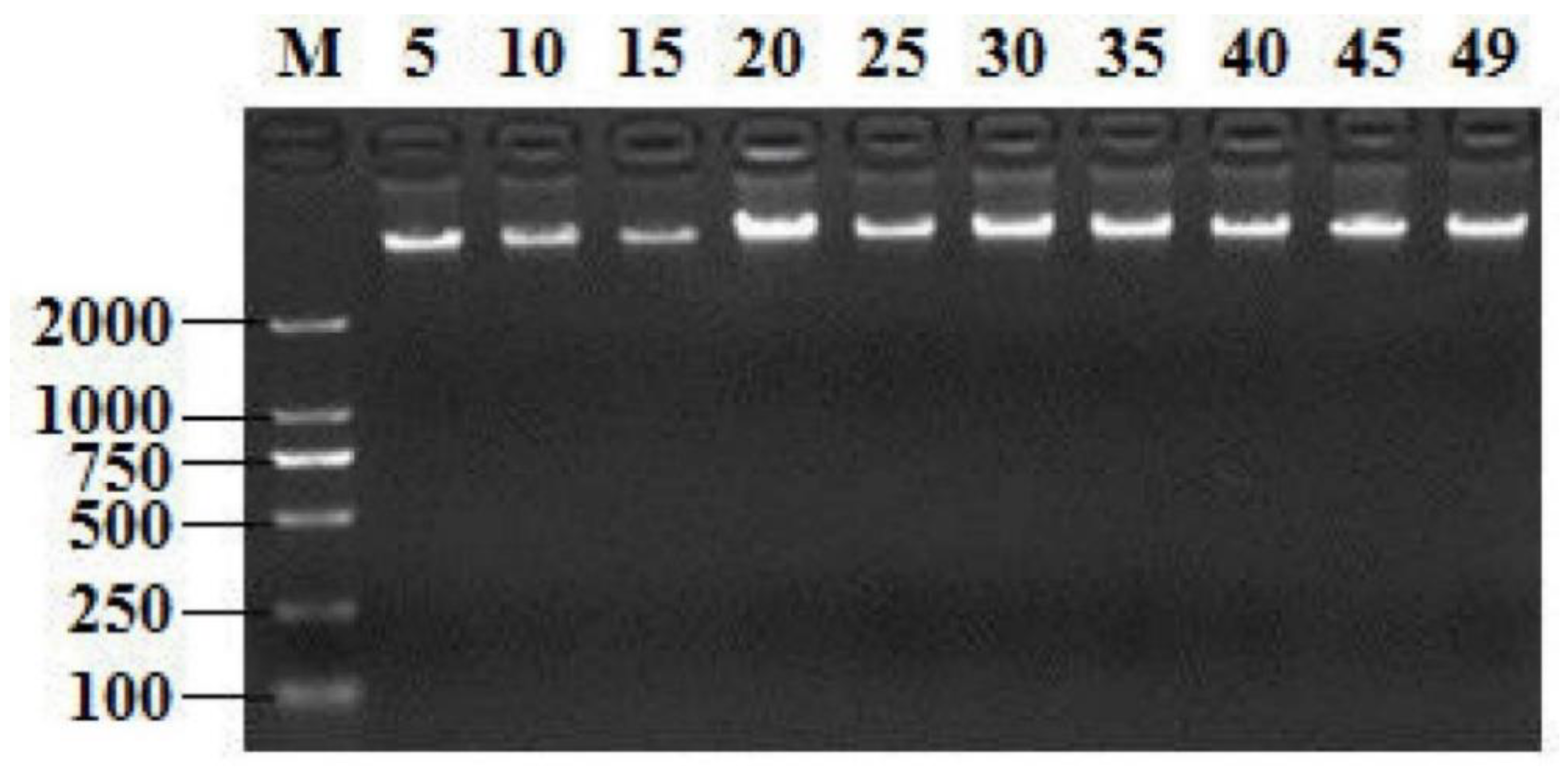
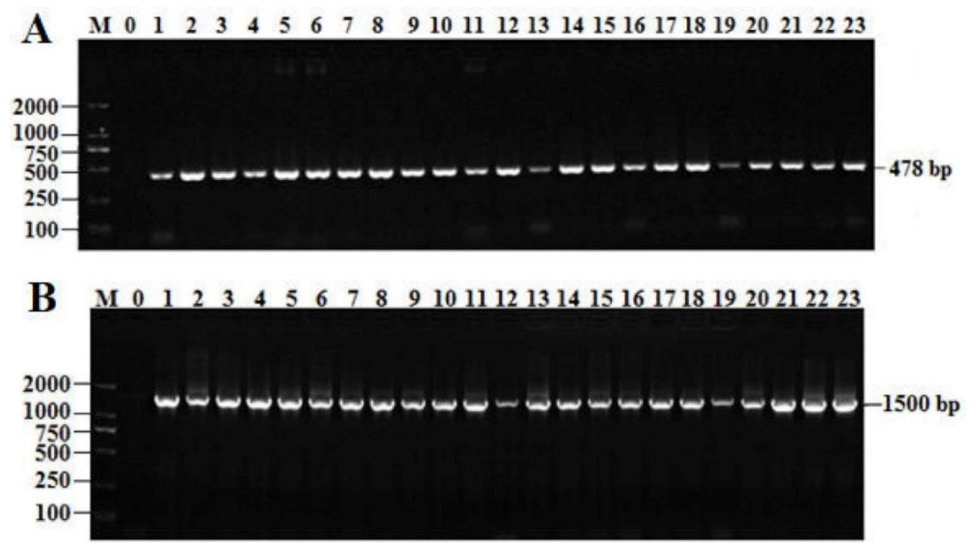
2.1. Screening T. Halophilus Isolates by GroEL Gene and 16S rRNA Gene

Extracting high-quality DNA from isolates is crucial for downstream molecular experiments. All the electropherograms showed the extracted DNAs with good quality; the partial results are shown in Figure 1. The PCR products of the groEL gene and the 16S rRNA gene amplified with primers were analyzed by 2% agarose gel, as shown in Figure 2. The groEL gene and16S rRNA gene amplification primers specifically extended the single target band to the isolated strains, which had no non-specific products and the background was clean. Hence, a total of 49 candidate strains were screened.

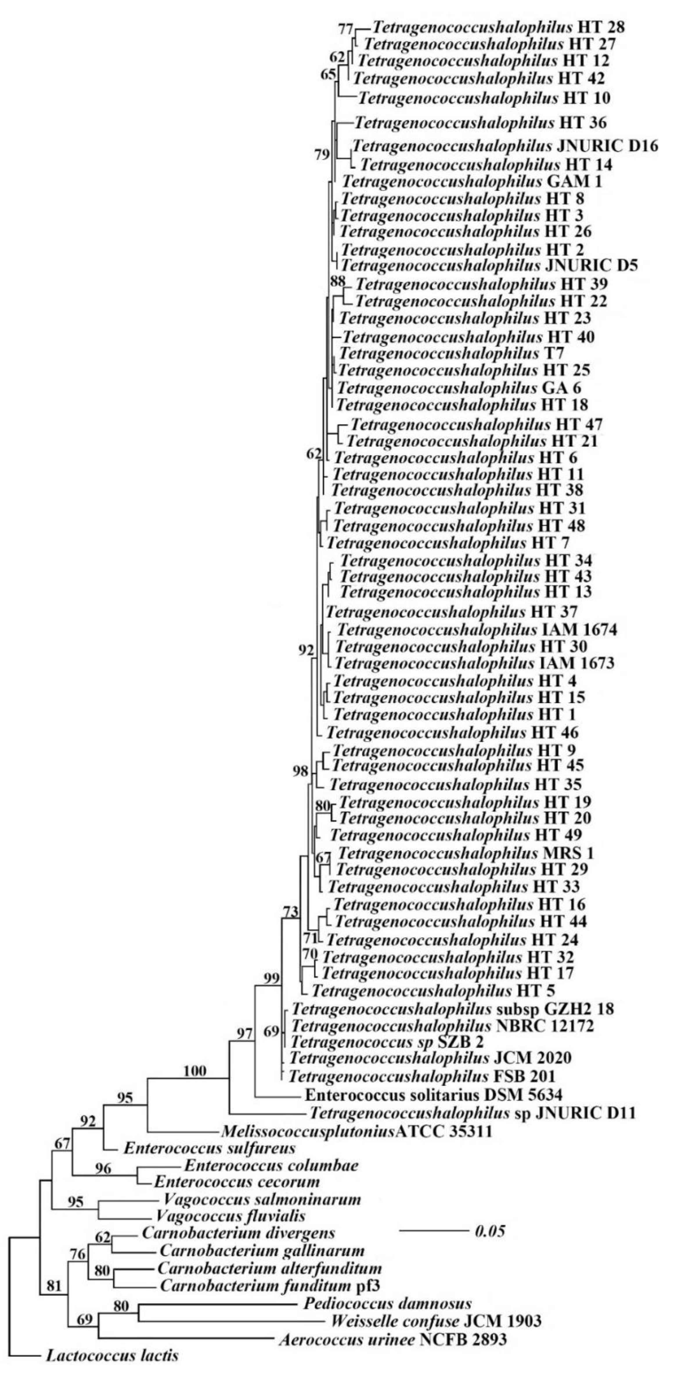
2.2. Identification of the Selected Bacteria

The sequence complements generated in this study were based primarily on BLAST search alignments and recent related literature. Raw sequences were assembled with BioEdit v. 7.2.5. Multiple sequence alignments were made using BioEdit 7.2 and ClustalX v.1. The alignments were examined visually and improved where necessary. Phylogenetic analyses of the combined aligned consisted of maximum likelihood (ML), MrBayes, and maximum parsimony (MP) analyses. Ambiguously aligned regions were removed from all analyses and gaps were treated as ‘‘missing data’’ in the phylogenetic analyses. Related sequences were selected to reveal the closest matching sequence to T. halophilus. A similarity to the 16S rRNA gene sequence of the T. halophilus was greater than 99% and was identified as a T. halophilus.
It can be seen from Figure 3 that the 49 isolates screened have 100% homology with Tetragenococcus sp. JNURIC D11 (GenBank: GQ150505.1) and they are clustered into the same branch with T. halophilus JCM2020 (GenBank: LC269262.1), T. halophilus NBRC12172 (GenBank: NR_076296.1), T. halophilus subsp. GZH2-18 (GenBank: MG654641.1), Tetragenococcus sp. JNURIC D16 (GenBank: GQ150510.1), T. halophilus MRS1 (GenBank: MK063722.1), and T. halophilus IAM1674 (GenBank: EU689055.1), and also have the closest relationship. From this, it can be seen that the 49 strains isolated from the Xuanwei ham are T. halophilus in the genus Tetragenococcus.

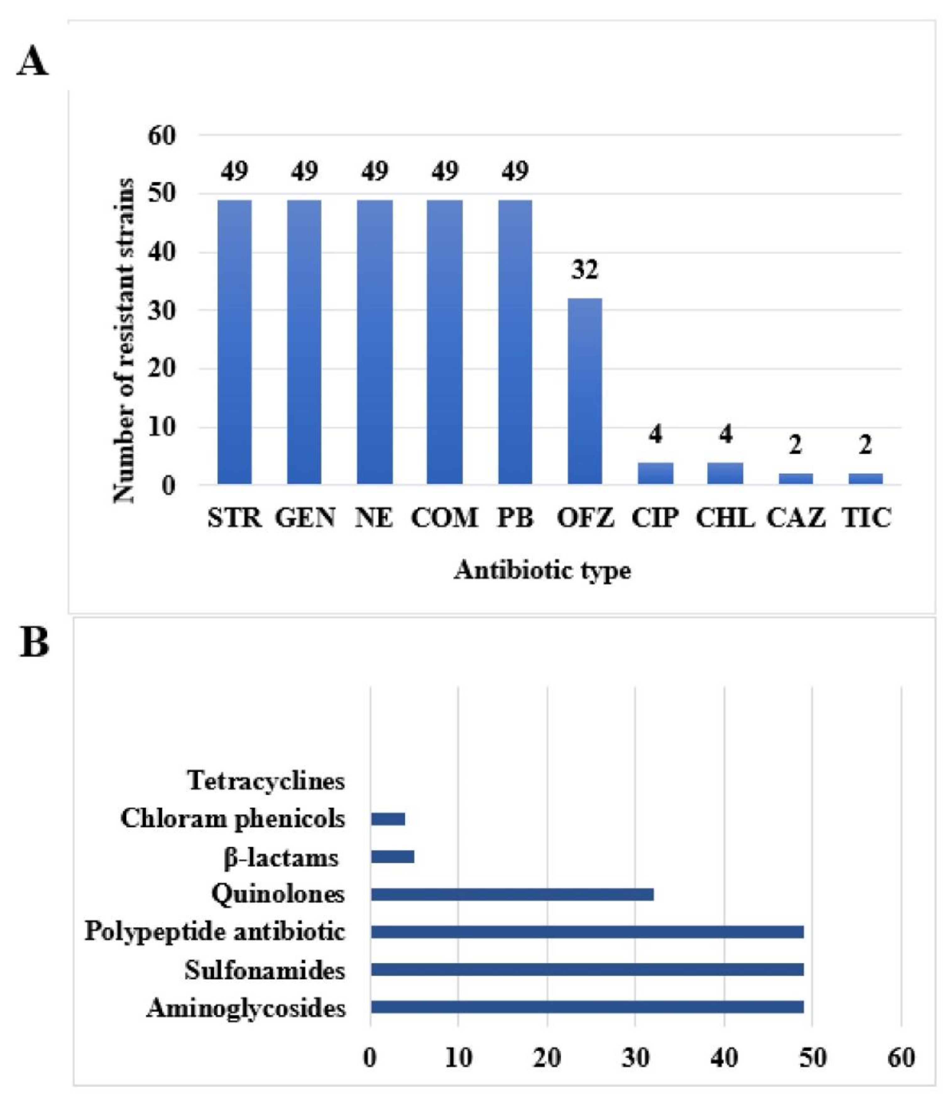
2.3. Experimental Results of Drug resistance Phenotype of 49 Isolates of T. Halophilus

A total of 19 antibiotic susceptibility tests were performed on 49 strains of T. halophilus using the K–B method. The size of the inhibition zone was explained by reference to the CLSI report method. It can be seen from Table 1 that the resistance rate of T. halophilic isolated from Xuanwei ham to gentamicin, streptomycin, neomycin, polymyxin B, and cotrimoxazole reached 100%, the resistance rate to ofloxacin reached 65.31%, the intermediate rate reached 30.61%, the resistance rate to ciprofloxacin and chloramphenicol was 8.16%, and the intermediate rate of ciprofloxacin was 24.49%. The intermediate rate of the chloramphenicol was 10.2%. The resistance rate to ticarcillin and ceftazidime was 4.08%, while the intermediate rate for ceftazidime was 10.2%, and there was susceptibility for ticarcillin, penicillin, ampicillin, amoxicillin, cefradine, cefotaxime, tetracyclines. The sensitivity rate of minocycline, doxycycline, and vancomycin reached 100% and the intermediate rate to amoxicillin was 4.08%.
As can be seen from Figure 4A, 49 strains of T. halophilus are resistant to gentamicin, streptomycin, neomycin, polymyxin B, and sulfamethoxazole, 32 strains were resistant to ofloxacin, 4 strains are resistant to ciprofloxacin and chloramphenicol, and 2 strains are resistant to ticarcillin and ceftazidime. It can be seen from Figure 4B that 49 strains are mainly resistant to aminoglycosides, sulfonamides, polypeptide, and quinolone antibiotics, followed by β-lactams and chloramphenicol, and there was no resistance to tetracyclines, temporarily.

2.4. Drug Resistance Spectrum of 49 Strains of T. Halophilus

As shown in Table 2 and Table 3, the isolates of 49 strains of T. halophilus showed high concentration of drug resistance, with a total of 8 drug resistance spectra and no 0 or 9 resistant strains. The least number of resistant strains were 8 (only 6 strains), the most of which were 1, 2, 3, 4, and 5 (49 strains each, accounting for 100%), followed by 6 resistant strains (32 strains, accounting for 65.31%), most of which were concentrated in this range.

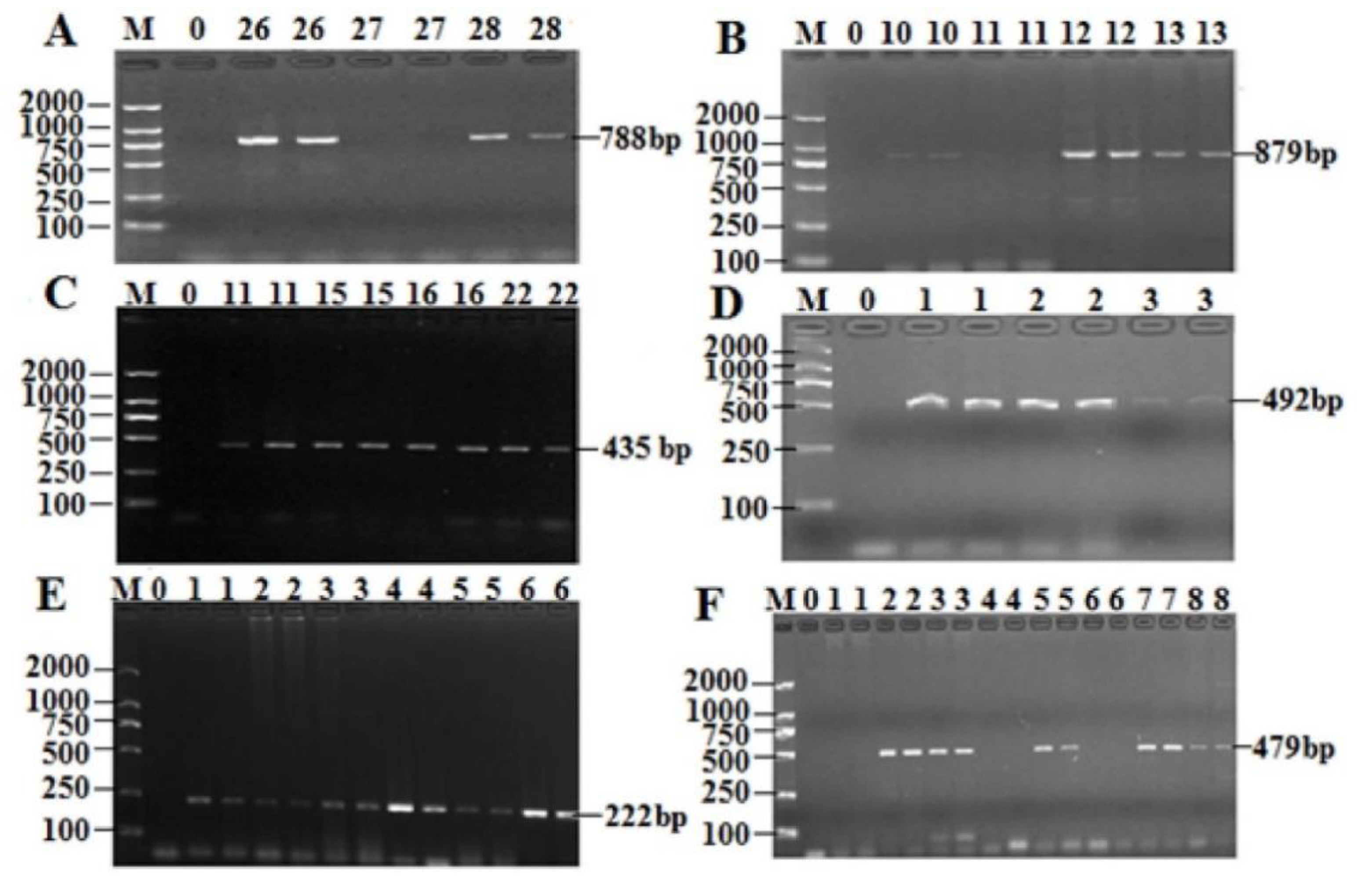
2.5. Prevalence of the AR Genes Among 49 Isolates of T. Halophilus

All of the 49 strains of T. halophilus isolates isolated from Xuanwei ham were subjected to PCR detection of their AR gene patterns. The results are shown in Figure 5 and Table 4. Among the drug resistance genes, two drug resistance genes of aminoglycoside drugs were detected, the highest of which was acrB, with a positive rate of 89.79%. The positive detection rate of aadA1 was 28.57%. Only the TEM gene was detected in the β-lactams and the positive rate was 47.7%. As long as Sul2 was detected in the three genes of sulfa drugs, the positive rate was as high as 77.55%. Only the GyrB gene was detected in quinolones and the positive rate was 75.51%. The positive rate of EmgrB gene in polypeptide antibiotic was 61.22%. Among the 6 genes of tetracyclines, no drug resistance gene was detected. Chloramphenicol was also not detected in resistant genes.

2.6. Analysis of Drug Resistance Phenotype and Genotype Matching Rate of Isolates of T. Halophilus
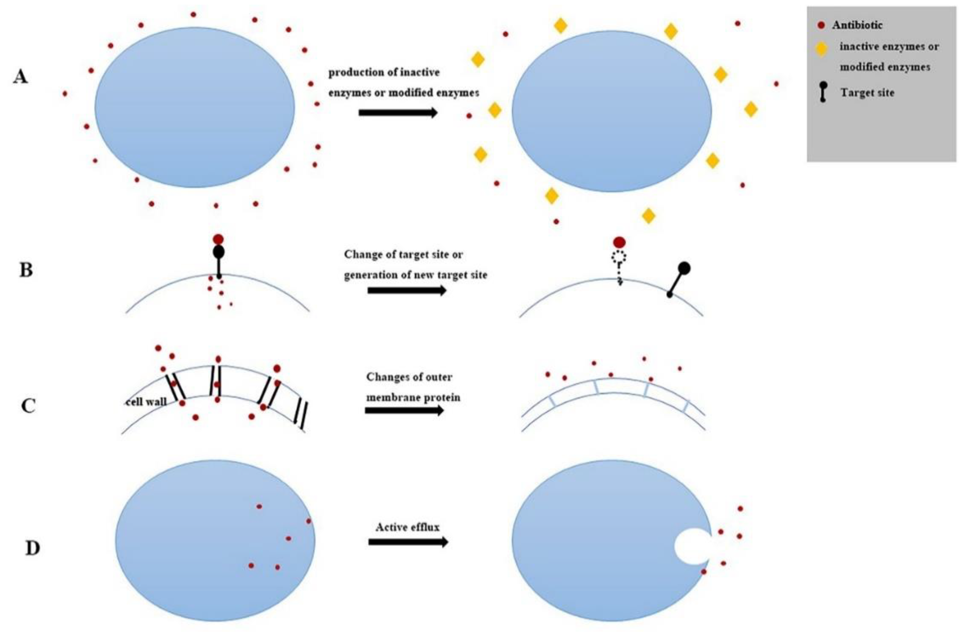
Bacterial resistance to antibiotics is an antibiotic phenomenon in nature, but with the widespread use of antibiotics, bacterial resistance has gradually increased. The mechanism of bacterial drug resistance, which is commonly manifested in two aspects, genetic mechanism and biochemical (protein) mechanism, tends to be more complex. Figure 6 and Figure 7 show two resistance mechanisms.
The drug resistance of 49 isolates of T. halophilus was studied and the strains with the resistant phenotype and the resistant genes were detected for conformity analysis. As shown in Table 5, 49 isolates of T. halophilus were found to have resistance phenotypes and genotypes to tetracyclines, aminoglycosides, quinolones, sulfonamides, polypeptide antibiotic, and β-lactams, with the coincidence rates of 100%, 91.84%, 86.49%, 79.17%, 61.22%, and 62.5%, respectively. However, the resistant phenotype was detected for chloramphenicol and no drug resistance gene was detected.
Among the antimicrobial activities of bacteria, β-lactams is the most widely used drug in clinical applications. Its resistance mechanism is that bacteria produces β- lactamase or changes in PBPs, as shown in Figure 7A,B. Bacterial resistance to aminoglycosides and chloramphenics is mainly to produce inactivating enzymes, aminoglycoside inactivating enzymes including aminoglycoside acyltransferase, aminoglycoside adenosine transferase, and aminoglycoside phosphotransferase. A chloramphenicol inactivating enzyme is chloramphenicol acyltransferase (CAT), as shown in Figure 7A. For polypeptide vancomycin, tetracycline, quinolones, sulfonamides and aminoglycosides, drug resistance can also be induced by changing the target site, as shown in Figure 7B. On the other hand, the cell envelope and active efflux system are widespread in Gram-negative and Gram-positive bacteria (Figure 7C,D). In this study, for sulfonamides, aminoglycosides, chloramphenicol, and polypeptides, there were more resistant phenotype strains than resistant genotype strains. This may be influenced by resistance mechanisms at the biochemical (protein) level.
At the same time, genetic and biochemical mechanisms are mutually reinforcing. For example, most Gram-negative bacteria are resistant to polypeptide vancomycin. The reason for this is that it contains a resistance gene and an inactivating enzyme gene by itself or by gene mutation, shown in Figure 6A,B. As a consequence of the wide spread of resistance genes among different species of bacteria and the transformation of strains (inducing drug resistance) in the production of antibiotics, as shown in Figure 6B,C, a large number of resistant bacteria have increased.
After the sequenced results were compared with the reference sequences on GenBank, the homology of the above six antibiotic resistance gene sequencing results was more than 99%.

3. Discussion

Xuanwei ham is a high-salty and delicious dried ham cured by pickling, drying, and fermenting, while T. halophilus has been found in a variety of fermented foods and syrups. However, at present, there is almost no literature that reports on the screening of T. halophilus bacteria from Xuanwei ham. The T. halophilus has the potential effects of improving food flavor and increasing flavor substance content as a starter. So, in this study, 49 strains of T. halophilus was isolated from Xuanwei ham, which further enriched the microbial resources in the Xuanwei ham, and we assessed the antibiotic resistance safety of the 49 T. halophilus strains. The drug sensitivity results showed that the sensitivity rate of 49 isolates to penicillin, ampicillin, amoxicillin, cefradine, cefotaxime, tetracyclines, minocycline, doxycycline, and vancomycin reached 100%, which was roughly the same as the reported by Jeong et al. [22].
A total of 8 kinds of antibiotics were selected for drug sensitivity tests, which showed that almost all strains were sensitive to 5 kinds of antibiotics, including ampicillin, penicillin, tetracyclines, and vancomycin. The drug resistance rate of gentamicin and erythromycin were 11.54% and 3.85% respectively. All strains were resistant to ciprofloxacin and the drug resistance rate was up to 100%. All the strains in this study were resistant to gentamicin and the drug resistance rate was as high as 100%. The resistance rate to ciprofloxacin was 8.16%. In addition, the resistance rate of streptomycin, neomycin, polymyxin B, and compound xinomin reached 100%, ofloxacin reached 65.31%, chloramphenicol was 8.16%, and ticarcillin and ceftazidime was 4.08%.
The difference may be caused by the difference in the host of the T. halophilus. It has been reported that sulfonamides, aminoglycans, and quinolones have been widely used in pig and poultry production. In 2014, the World Health Organization (WHO) global surveillance report on antibiotic resistance showed evidence of a link between the use of antimicrobial agents in food animals and the emergence of resistance in common pathogens [23]. Teuber et al. stated, “The problem of drug resistance in human medicine will not be solved because resistant genes are constantly pouring into the human microflora through the food chain [24].” Berends et al. concluded that “most of the problems of drug-resistant bacteria in humans are related to the medical use of antimicrobials, particularly the limited impact of veterinary use [25].” However, these authors acknowledge that the impact of antibiotics as feed additives is extremely worrying. Therefore, it can be speculated that the antibiotic resistance of the T. halophilic isolate in Xuanwei ham may be mainly originated from the host. Pigs may only partially metabolize when they consume feed containing antibiotic additives or are injected with drugs containing antibiotics, and part of the antibiotics remain in the body.
On the other hand, the high concentration of the drug resistance spectrum indicates that there may be a problem of drug resistance gene transfer between these strains. At the same time, 26 common drug resistance genes of T. halophilic were detected. The drug-resistant phenotype and genotype of T. halophilic have a high coincidence rate, indicating that the drug-resistant phenotype of 49 isolates of T. halophilus isolates has a certain correlation with the drug resistance genes from themselves. Among them, some strains of aminoglycosides, sulfonamides, and chloramphenicol antibiotics detected the corresponding drug resistance phenotype, but no drug resistance genes related to the strain were detected. According to reports in the literature, when a bacterial strain has a drug-resistant phenotype to a certain antibiotic, but does not contain the corresponding drug-resistant gene, it can be inferred that the bacterial strain’s resistance to this antibiotic may be inherent resistance and the presence of a drug-resistant phenotype does not necessarily carry the corresponding drug-resistant gene [26]. Casado and Muñoz et al. tested the resistance of Lactobacillus pentosus and Leuconostoc to some antibiotics and found that no drug resistance genes were detected in strains with resistant phenotypes [27]. Zhang Hongmei et al. reported that 7 strains of LAB isolated from yoghurt were resistant to ampicillin and tetracyclines, but only 5 strains were detected that carried the ampicillin resistant gene Amp and no tetracyclines resistant gene was detected [28]. Hummel et al. found that some lactic acid bacteria strains showed low resistance to ampicillin, penicillin, chloramphenicol, and tetracyclines, but no known resistance genes were detected, although some strains have cat genes, none of these strains was phenotypically resistant to chloramphenicol, and these cat genes were silenced under both induction and non-induction conditions by reverse transcription PCR [29].
In this study, the resistance gene related to the isolated strain was detected in β-lactams and quinolone antibiotics, but the number of resistant phenotype strains was less than that of resistant genotype strains. It is possible that the strain carries drug resistance genes associated with it, but the drug resistance genes may exist but remain silent. Qin Yuxuan et al. tested the drug resistance of lactic acid bacteria isolated from commercially available yoghurt and found that all strains were sensitive to erythromycin and tetracyclines, but detected the corresponding drug resistance genes [30]. This once again proved that there is no drug resistance phenotype that may also carry a drug resistance gene, possibly because the drug resistance gene it carries is not expressed or expressed insufficiently.
At present, whether or not they carry antibiotic resistance genes has become an important indicator to evaluate the safety of bacterial strains and to determine whether they can be used as a safe strain for food. In this paper, the drug resistance gene of T. halophilic has a high coincidence rate with the drug resistance phenotype, but it has not reached full compliance. It is necessary to continue to do more work in the detection of drug resistance genes of T. halophilic, such as the way in which T. halophilic acquire resistance, whether there is resistance gene transfer between strains, and factors affecting drug resistance gene transfer.

4. Materials and Methods

4.1. Material

For the current study, a total of 49 T. halophilus strains isolated from two Xuanwei hams were used. The Xuanwei hams in this study were gathered from local markets in Kunming, Yunnan province.
The high-speed desktop centrifuge (H318K) was purchased from Hunan Kecheng equipment Co., Ltd. (Hunan, China), the Ultra-micro UV spectrophotometer (NanoDrop 2000) was purchased from Thermo Fisher Scientific (Waltham, MA, USA), and the Gel imaging system (Gel DOC XR) was purchased from Bio-Rad Laboratories (Hercules, CA, USA).

4.2. Xuanwei Ham Samples and Bacterial Strain Isolation

The 25 g sample was aseptically chopped and immersed in 225 mL of 0.85% sterile normal saline for 15 minutes. The same was sealed on a shaker and was shaken at 280 r/min, 4 °C for 30 min. The sample solution was diluted continuously from 10−1 to 10−7 for use.
A total of 0.1 mL of each dilution (10−1–10−7) was pour-plated with DeMan Rogosa Sharpe (MRS) medium (Oxoid, Basingstoke, UK) containing 1% (w/v) Agar, 3% (w/v) NaCl [31], and 0.2% (w/v) CaCO3 at 30 °C for 3–4 days under semi-anaerobic conditions. Separation and purification of bacterial colonies used the same type of agar medium incubated at 30 °C for 2 days. After purifications, bacterial isolate strains were enriched and cultured in MRS medium containing 3% (w/v) NaCl at 30 °C for 48 h. The isolated strains were preserved in a ratio of 30% glycerol to 1:1 and stored at −80 °C.

4.3. Identification of Isolates by groEL Gene and 16S rRNA Gene

Genomic DNA of isolates was extracted using a kit (TaKaRa MiniBEST Bacteria Genomic DNA Extraction Kit Ver.3.0). Amplification primers of the 16S rRNA gene and groEL gene were performed and are shown in Table 6. PCR was performed using an ABI SimpliAmp thermal cycler (Applied Biosystems) in a 25 µL reaction system containing 2.5 µL of 10 × PCR Buffer(Mg2+ plus), 2 µL of dNTP Mixture(2.5 mM), 1 μL of each primer (10 µM), 0.2 μL of rTaq DNA Polymerase (5 U/µL) (TaKaRa Biotechnology, Beijing, China), 2 µL of DNA template, and 16.3 µL of ultrapure water. The 16S rRNA amplification was carried out the with the following program: Initial denaturation at 94 °C for 2 min; 30 cycles of denaturation at 94 °C for 30 s, primer annealing at 54 °C for 30 s and primer extension 1 min at 72 °C; and a final extension of at 72 °C for 10 min. During groEL gene amplification, the procedures applied for 16S rRNA amplification were followed, but annealing was performed at 54 °C instead of 58 °C.
The 16S rRNA and groEL PCR products were further analyzed on 2% agarose gel with ethidium bromide (0.1 g/mL) and run with 1 × TAE buffer, using a 2-kb ladder Maker (TsingKe, Beijing, China) for molecular weight standards. The PCR products were purified and sequenced using a custom service provided by Sangon Co. Ltd. (Shanghai, China). The 16S rDNA sequence analysis was carried out using and Illumina Miseq Sequencing Instrument and the Miseq Reagent Kit V3. The 16S rRNA gene sequence similarities were searched and identification of these isolates was determined through a search of the GenBank DNA database using the BLAST algorithm. The phylogenetic positions of the isolates were inferred by 16SrRNA gene sequence analysis.

4.4. Antibiotic Sensitivity Tests of Isolates

T. halophilus strains were cultured twice in MRS broth containing 3% NaCl and matched to a McFarland 0.5 turbidity standard (BIO-KONT, Shenzhen, China). Antibiotic diffusion tests were determined by the Bauer−Kirby method according to the guidelines of the Clinical and Laboratory Standards Institute (CLSI). The phenotypic antimicrobial sensitivity response of each T. halophilus isolate was evaluated using panel of 20 antimicrobial discs. The antimicrobial agents used included 10 µg penicillin (PEN), 10 μg amoxicillin (AMO), 10 μg ampicillin (AMP), 10 μg streptomycin (STR), 10 μg gentamicin (GEN), 30 μg tetracyclines (TET), 300 μg polymyxin B (PB), 1.25 μg compound sulfamethoxazole (COM), 5 μg ciprofloxacin (CIP), 5 μg ofloxacin (OFZ), 30μg ceftazidime pentahydrate (CAZ), 30 μg ticarcillin (TIC), 30 μg chloramphenicol (CHL), 30 μg cefradine (CE), 30 μg cefotaxime sodium (CTX), 30 μg neomycin (NE), 30 μg minocycline (MH), 30 μg doxycycline (DO), and 30 μg vancomycin (VA). All the antibiotics discs were purchased from the company Oxoid (UK). Measurement of the diameter of the zone of inhibition was to the nearest millimeter. Since CLSI has not yet defined the inhibition zone diameter value of the Tetragenococcus genus antibiotic, according to the taxonomic position, Enterococcus is the closest to Tetragenococcus in phylogeny. Therefore, the T. halophilus isolates were classified as susceptible, intermediate, and resistant according to the definition of antimicrobial circle diameter for the genus Enterococcus interpretative standards of CLSI.

4.5. Antibiotic Resistance Gene Detection of Isolates

All primers were designed using Vector NTI Advance 10 software according the antibiotic resistance gene information provided by the Antibiotic Resistance Genes Database (ARGD) and some primers were introduced in the references and synthesized by Sangon Co. Ltd. (Shanghai, China). The detailed sequences of these primers, optimum annealing temperatures, and product size are listed in Table 7. The PCR amplification in a 25 µL reaction system contained 2.5 µL 10× Ex Taq Buffer, 2 µL dNTP Mixture (2.5 mM), 1 μL of each primer, 0.2 μL ExTaq DNA Polymerase (TaKaRa Biotechnology, Beijing, China), 2 µL DNA template, and 16.3 µL ddH2O.

5. Conclusions

We assessed the prevalence of antibiotic resistance phenotypes (K-B method) and genotypes (PCR method) of 49 T. halophilus strains isolated from Xuawei ham in China. The results showed that 49 isolates can be considered as completely susceptible to penicillin, ampicillin, amoxicillin, cefradine, cefotaxime, tetracyclines, minocycline, doxycycline and vancomycin, but resistant to gentamicin, streptomycin, neomycin, polymyxinB and cotrimoxazole, which were consistent with the previous reports. These resistances were sufficiently high to consider the potential for acquisition of transmissible determinants. Among the 26 antibiotic resistance genes, 5 varieties of antibiotic resistance genes, including acrB, blaTEM, AAda1, SulII and GyrB, were detected, and the detection rates were 89.79%, 47.7%, 16.33%, 77.55%, and 75.51%, respectively. The potential acquisition of transmissible determinants for antibiotic resistance and antibiotic resistance genes identified in this study necessitate the need for a thorough antibiotic resistance safety assessment of T. halophilus before it can be considered for use in food fermentation processes.

Author Contributions

Methodology, Y.L.; formal analysis, Y.L. and L.S.; validation, C.Z. and Z.L.; writing—original draft preparation, Y.L.; writing—review and editing, L.S. and Y.S.; supervision, Y.S.; funding acquisition Y.S.

Funding

This research was funded by the Key R & D Project of Yunnan Province: Social Development Field, grant number 2018BC006 and the National Natural Science Foundation of China, grant number 31801635.

Conflicts of Interest

The authors declare no conflict of interest.

References

  1. Collins, M.D.; Williams, A.M.; Wallbanks, S. The phylogeny of Aerococcus and Pediococcus as determined by 16S rRNA sequence analysis: Description of Tetragenococcus gen. nov. FEMS Microbiol. Lett. 1990, 58, 255–262. [Google Scholar] [CrossRef] [PubMed]
  2. Gurtler, M.; Ganzle, M.G.; Wolf, G.; Hammes, W.P. Physiological diversity among strains of Tetragenococcus halophilus. Syst. Appl. Microbiol. 1998, 21, 107–112. [Google Scholar] [CrossRef]
  3. Liao, D.; Fang, F.; Chen, J.; Du, G.; Liu, L. Isolation and Arginine Metabolism Detection of a Tetragenococcus halophilus Strain. J. Food Sci. Biotechnol. 2016, 35, 48–53. [Google Scholar]
  4. Devi, K.R.; Deka, M.; Jeyaram, K. Bacterial dynamics during yearlong spontaneous fermentation for production of ngari, a dry fermented fish product of Northeast India. Int. J. Food Microbiol. 2015, 199, 62–71. [Google Scholar] [CrossRef] [PubMed]
  5. Hanagata, H.; Shida, O.; Takagi, H. Taxonomic homogeneity of a salt-tolerant lactic acid bacteria isolated from shoyu mash. J. Gen. Appl. Microbiol. 2003, 49, 95–100. [Google Scholar] [CrossRef] [PubMed] [Green Version]
  6. Juste, A.; Lievens, B.; Frans, I.; Marsh, T.L.; Klingeberg, M.; Michiels, C.W.; Willems, K.A. Genetic and physiological diversity of Tetragenococcus halophilus strains isolated from sugar- and salt-rich environments. Microbiology 2008, 154, 2600–2610. [Google Scholar] [CrossRef] [PubMed] [Green Version]
  7. Kim, T.W.; Lee, J.H.; Park, M.H.; Ki, H.Y. Analysis of Bacterial and Fungal Communities in Japanese- and Chinese-Fermented Soybean Pastes Using Nested PCR-DGGE. Curr. Microbiol. 2010, 60, 315–320. [Google Scholar] [CrossRef] [PubMed]
  8. Onda, T.; Yanagida, F.; Uchimura, T.; Tsuji, M.; Ogino, S.; Shinohara, T.; Yokotsuka, K. Widespread distribution of the bacteriocin-producing lactic acid cocci in Miso-paste products. J. Appl. Microbiol. 2002, 92, 695–705. [Google Scholar] [CrossRef] [PubMed]
  9. Roling, W.; van Verseveld, H.W. Characterization of Tetragenococcus halophila Populations in Indonesian Soy Mash (Kecap) Fermentation. Appl. Environ. Microbiol. 1996, 62, 1203–1207. [Google Scholar] [Green Version]
  10. Udomsil, N.; Rodtong, S.; Tanasupawat, S.; Yongsawatdigul, J. Proteinase-producing halophilic lactic acid bacteria isolated from fish sauce fermentation and their ability to produce volatile compounds. Int. J. Food Microbiol. 2010, 141, 186–194. [Google Scholar] [CrossRef]
  11. Udomsil, N.; Rodtong, S.; Choi, Y.J.; Hua, Y.; Yongsawatdigul, J. Use of Tetragenococcus halophilus as a Starter Culture for Flavor Improvement in Fish Sauce Fermentation. J. Agric. Food Chem. 2011, 59, 8401–8408. [Google Scholar] [CrossRef] [PubMed]
  12. Cui, R.Y.; Zheng, J.; Wu, C.D.; Zhou, R.Q. Effect of different halophilic microbial fermentation patterns on the volatile compound profiles and sensory properties of soy sauce moromi (vol 239, pg 321, 2014). Eur. Food Res. Technol. 2015, 240, 669–670. [Google Scholar] [CrossRef]
  13. He, G.; Huang, J.; Liang, R.; Wu, C.; Zhou, R. Comparing the differences of characteristic flavour between natural maturation and starter culture for Mucor-type Douchi. Int. J. Food Sci. Technol. 2016, 51, 1252–1259. [Google Scholar] [CrossRef]
  14. Kuda, T.; Izawa, Y.; Ishii, S.; Takahashi, H.; Torido, Y.; Kimura, B. Suppressive effect of Tetragenococcus halophilus, isolated from fish-nukazuke, on histamine accumulation in salted and fermented fish. Food Chem. 2012, 130, 569–574. [Google Scholar] [CrossRef]
  15. Ikuko, N.; Toshinori, I.; Tadao, E.; Yoshihiro, D.; Yoshiaki, O.; Akio, O. Clinical efficacy of halophilic lactic acid bacterium Tetragenococcus halophilus Th221 from soy sauce moromi for perennial allergic rhinitis. Allergol. Int. 2009, 58, 179–185. [Google Scholar]
  16. Adams, M.R. Safety of industrial lactic acid bacteria. J. Biotechnol. 1999, 68, 171–178. [Google Scholar] [CrossRef]
  17. Jonkers, D.M.A.E. Microbial perturbations and modulation in conditions associated with malnutrition and malabsorption. Best Pract. Res. Clin. Gastroenterol. 2016, 30, 161–172. [Google Scholar] [CrossRef]
  18. Wang, J.; Li, M.; Wang, J.; Liu, M.; Yang, K.; Zhang, J.; Fan, M.; Wei, X. Antibiotic Resistance of Coagulase-Negative Staphylococci and Lactic Acid Bacteria Isolated from Naturally Fermented Chinese Cured Beef. J. Food Prot. 2018, 81, 2054–2063. [Google Scholar] [CrossRef]
  19. Pesavento, G.; Ducci, B.; Comodo, N.; Lo Nostro, A. Antimicrobial resistance profile of Staphylococcus aureus isolated from raw meat: A research for methicillin resistant Staphylococcus aureus (MRSA). Food Control 2007, 18, 196–200. [Google Scholar] [CrossRef]
  20. Sorum, H.; L’Abee-Lund, T.M. Antibiotic resistance in food-related bacteriaA result of interfering with the global web of bacterial genetics. Int. J. Food Microbiol. 2002, 78, 43–56. [Google Scholar] [CrossRef]
  21. Graveland, H.; Duim, B.; van Duijkeren, E.; Heederik, D.; Wagenaar, J.A. Livestock-associated methicillin-resistant Staphylococcus aureus in animals and humans. Int. J. Med. Microbiol. 2011, 301, 630–634. [Google Scholar] [CrossRef] [PubMed]
  22. Jeong, D.W.; Heo, S.; Lee, J.H. Safety assessment of Tetragenococcus halophilus isolates from doenjang, a Korean high-salt-fermented soybean paste. Food Microbiol. 2017, 62, 92–98. [Google Scholar] [CrossRef] [PubMed]
  23. Sundsfjord, A.; Simonsen, G.S.; Courvalin, P. Human infections caused by glycopeptide-resistant Enterococcus spp.: Are they a zoonosis? Clin. Microbiol. Infect. 2001, 7, 16–33. [Google Scholar] [CrossRef] [PubMed]
  24. Teuber, M. Spread of antibiotic resistance with food-borne pathogens. Cell. Mol. Life Sci. 1999, 56, 755–763. [Google Scholar] [CrossRef] [PubMed]
  25. Berends, B.R.; van den Bogaard, A.E.; van Knapen, F.L.; Snijders, J.M. Human health hazards associated with the administration of antimicrobials to slaughter animals. Part II. An assessment of the risks of resistant bacteria in pigs and pork. Vet. Q. 2001, 23, 10–21. [Google Scholar] [CrossRef]
  26. Song, X.; Li, S.; Ma, C.; Li, Z.; Li, S. Antibiotic resistance in fermented food lactic acid bacteria-a review. Microbiol. China 2015, 42, 207–213. [Google Scholar]
  27. Casado Muñoz Mdel, C.; Benomar, N.; Lerma, L.L.; Gálvez, A.; Abriouel, H. Antibiotic resistance of Lactobacillus pentosus and Leuconostoc pseudomesenteroides isolated from naturally-fermented Aloreña table olives throughout fermentation process. Int. J. Food Microbiol. 2014, 172, 110–118. [Google Scholar] [CrossRef]
  28. Zhang, H.; Huang, S.; Zhou, H. Two kinds of antibiotics resistance of lactic acid bacteria isolated from yogurt. China Public Health 2010, 26, 511–512. [Google Scholar]
  29. Hummel, A.S.; Hertel, C.; Holzapfel, W.H.; Franz, C.M. Antibiotic resistances of starter and probiotic strains of lactic acid bacteria. Appl. Environ. Microbiol. 2007, 73, 730–739. [Google Scholar] [CrossRef]
  30. Qin, Y.; Li, J.; Wang, Q.; Gao, K.; Zhu, B.; Lv, N. Identification of lactic acid bacteria in commercial yogurt and their antibiotic resistance. Wei Sheng Wu Xue Bao 2013, 53, 889–897. [Google Scholar]
  31. Lu, L.K. The Role of Tetragenococcus halophilus’ Gene clpB, dnaK, hrcA in Saline Adaption. Master’s Thesis, South China University of Technology, Guangzhou, China, 2013. [Google Scholar]
  32. Li, X.; Wu, J.; Tian, T.; Yue, X. Isolation, Identification and Preliminary Screening of Acid-Tolerant Lactic Acid Bacteria from Naturally Fermented Pickle Juices from Daqing. Food Sci. 2014, 35, 150–154. [Google Scholar]
  33. Guo, M.L. Analysis of Prosciutto Microflora and the Effect of Superior Strains on the Composition of Microflora in Fermented Sausage. Master’s Thesis, Yangzhou University, Yangzhou, China, 2015. [Google Scholar]
  34. Ma, C. Detection and Sequence Analysis of Correlative Virulence Gene and Drug Resistance Genes of Superiority Escherichia Coli Serotypes Isolated from Cow Mastitis. Master’s Thesis, Ningxia University, Ningxia, China, 2014. [Google Scholar]
  35. Yue, S. Detection and Analysis of Drug Resistance Genes and Virulence Genes of E. coli in Beef Cattle in Parts of Heilongjiang Province. Master’s Thesis, Heilongjiang Bayi Agricultural University, Daqing, China, 2017. [Google Scholar]
  36. Ma, M.; Wang, H.; Yu, Y.; Li, C.; Zhang, D.; Yang, Y.; Liu, S. Detection of Antimicrobial Resistance Genes of Pathogenic Salmonella from Swine. Acta Vet. Et Zootech. Sin. 2006, 37, 65–70. [Google Scholar]
  37. Li, L.; Shen, J.; Wu, J.; Wang, Y.; Wu, C. Expression of marA, soxS and ramA genes and efflux pump genes acrA and acrB in ciprofloxacin-resistant Salmonella. Vet. Sci. China 2009, 39, 928–932. [Google Scholar]
  38. Li, C.Y.; Yang, Q. Surveillance of antimicrobial resistance and vancomycin resistance genes in Enterococcus species. Chin. J. Health Lab. Technol. 2007, 17, 2244–2246. [Google Scholar]
  39. Liu, Y. Emergence of Plasmid-Mediated Colistin Resistance Gene mcr-1 in Enterobacteriacea; South China University of Technology: Guangzhou, China, 2016. [Google Scholar]
Figure 1. Partial electropherogram result of genomic DNA of isolated strains. M: DNA Maker DL2000; Lanes 5–49: DNA bands of 10 isolated strains.
Antibiotics 08 00151 g001
Figure 2. The PCR (polymerase chain reaction) results of the screened T. halophilus strains. (A) The screened T. halophilus strains by amplification of groEL gene; Lane M: DNA Marker DL2000; Lane 0: negative; Lane 1–23: Candidate strain of T. halophilus; (B) Electrophoresis of amplification product; Lane M: DNA Maker; Lane 0: Negative control; Lanes 1–23: The band of PCR amplification products of DNA primer PCR.
Antibiotics 08 00151 g002
Figure 3. Phylogenetic analysis of 49 strains of T halophilus based on 16S rRNA sequence.
Antibiotics 08 00151 g003
Figure 4. Distribution of drug resistance of 49 strains of T. halophilus. (A) Number of bacterial strains —by type of antibiotic; (B) Number of bacterial strains —by categories of antibiotic.
Antibiotics 08 00151 g004
Figure 5. PCR detection results of some T. halophilus resistance genes. (A) blaTEM gene; (B) GyrB gene; (C) SulII gene; (D) EmgrB gene; (E) acrB gene; (F) aadA1 gene; M: DL2000Marker, 0: Negative control.
Antibiotics 08 00151 g005
Figure 6. Gene mechanism. (A) Inherent drug resistance. Resistance genes exist in bacteria and are passed on from generation to generation. (B) Gene mutation or acquisition of new genes. Pressure to increase antibiotic production causes bacteria to mutate genes into drug resistance and, at the same time, bacteria can easily acquire resistance by ingesting resistance genes released after the death of another drug-resistant bacteria. (C) Integrons mediate drug resistance. Under the catalysis of integrase, integrons can capture and express exogenous genes, especially drug-resistant genes, so that drug-resistant genes can be transmitted between different species.
Antibiotics 08 00151 g006
Figure 7. Biochemical (protein) mechanism. (A) Production of inactivated or modified enzymes. (B) Change of target site or generation of new target site. This makes is difficult for antibiotics to bind to cells, thus reducing the inhibitory effect of antibiotics. (C) Changes of outer membrane protein. Long-term drug effects can stimulate adventitia proteins to alter the cell wall structure and reduce permeability, thus hindering the penetration of antibiotics. (D) Active efflux system. When energy is available, membrane proteins selectively or non-selectively expel antibiotics from the cell, reducing drug concentration and leading to drug resistance.
Antibiotics 08 00151 g007
Table 1. Pharmacokinetic phenotypic test results of 49 strains of T. halophilus.
AntibioticIsolates of ResistantIsolates of SusceptibleResistance Rate (R)Intermediate Rate (I)Susceptible Rate (S)
PEN0490%(0/49)0%(0/49)100%(49/49)
AMO0470%(0/49)4.08%(2/49)95.90%(47/49)
AMP0490%(0/49)0%(0/49)100%(49/49)
TIC2474.08%(2/49)0%(0/49)95.9%(47/49)
CE0490%(0/49)0%(0/49)100%(49/49)
CAZ2424.08%(2/49)10.20%(5/49)85.70%(42/49)
CTX0490%(0/49)0%(0/49)100%(49/49)
GEN490100%(49/49)0%(0/49)0%(0/49)
STR490100%(49/49)0%(0/49)0%(0/49)
NE490100%(49/49)0%(0/49)0%(0/49)
TET0490%(0/49)0%(0/49)100%(0/49)
MH0490%(0/49)0%(0/49)100%(0/49)
DO0490%(0/49)0%(0/49)100%(0/49)
PB490100%(49/49)0%(0/49)0%(0/49)
VA0490%(0/49)0%(0/49)100%(0/49)
COM490100%(0/49)0%(0/49)0%(0/49)
CHL4408.16%(0/49)10.20%(5/49)81.63%(40/49)
OFZ32265.31%(32/49)30.61%(15/49)4.08%(2/49)
CIP4338.16%(4/49)24.49%(12/49)67.35%(33/49)
Table 2. Drug resistance spectrum of 49 strains of T. halophilus.
Type of ResistanceResistant SpectrumIsolates
5GEN-STR-NEO-PB-COM49
6GEN-STR-NEO-PB-COM-OFZ32
7GEN-STR-NEO-PB-COM-OFZ-CIP4
GEN-STR-NEO-PB-COM-OFZ-CAZ2
GEN-STR-NEO-PB-COM-OFZ-TLC2
GEN-STR-NEO-PB-COM-OFZ-CHL2
8GEN-STR-NEO-PB-COM-OFZ-CIP-CHL4
GEN-STR-NEO-PB-COM-OFZ-CIP-CAZ1
Table 3. Multi-drug resistance of 49 strains of T. halophilus.
Type of ResistanceNumber of IsolatesProportion (%)
000
149100%
249100%
349100%
449100%
549100%
63265.31%
71224.50%
8510.20%
Table 4. Detection rate of 26 drug resistance genes in 49 strains of T. halophilus.
AntibioticAR GenePositive IsolatesTotally IsolatesPositive Rates
Tetracyclinestet(K)0490%
tet(L)0490%
tet(M)0490%
tet(O)0490%
tet(S)0490%
tet(W)0490%
β-lactamsblaTEM84916.33%
bl3-vim0490%
blaOXA0490%
blaSHV0490%
SulfonamidesSul10490%
Sul2384977.55%
Sul30490%
Aminoglycosidesaac(3′)-IIa0490%
acrB444989.79%
aadB0490%
aadA1144928.57%
Chloram PhenicolsfloR0490%
Cat10490%
QuinolonesGyrA0490%
GyrB374975.51%
ParC0490%
Polypeptide AntibioticVanC10490%
EmgrB304961.22%
Table 5. Drug-resistant phenotype and genotype coincidence rate of T. halophilus.
AntibioticResistant PhenotypeResistant GeneCompliance Rate (%)
Tetracyclines00(0/0)100%
β-lactams58(5/8)62.5%
Sulfonamides4938(38/49)79.17%
Aminoglycosides4945(45/49)91.84%
Chloram phenicols4000
Quinolones3237(32/37)86.49%
Polypeptide Antibiotic4930(30/49)61.22%
Table 6. Primers sequences of PCR amplification.
PrimersGene Sequences (5’–3’)References
27-FAGATTTGATCCTGGCTCAG[32]
1492-RCTACGGCTACCTTGTTACGA
GroEL-FCGTCGTCAATGCTYAATGG[33]
GroEL-RTGCTGCCAGAAGAAACTTCA
Table 7. Primers sequences of PCR amplification.
AntibioticAR GeneGene Sequences (5‘–3‘)Annealing Temp. (°C)Product Size (bp)Ref.
Tetracyclinestet(K)F: TTAGGTGAAGGGTTAGGTCC
R: GCAAACTCATTCCAGAAGCA
59697[30]
tet(O)F: AACTTAGGCATTCTGGCTCAC
R: TCCCACTGTTCCATATCGTCA
55515[30]
tet(S)F: CATTTGGTCTTATTGGATCG
R: ATTACACTTCCGATTTCGG
55456[30]
tet(W)F: GAGAGCCTGCTATATGCCAGC
R: GGGCGTATCCACAATGTTAAC
58168[30]
tet(L)F: TCATCATCTCCTGATTTTAC
R: AGTAAAAACAAGCAGAGCAT
601464This study
tet(M)F: GTTAAATAGTGTTCTTGGAG
R: CTAAGATATGGCTCTAACAA
53501This study
β-lactamsblaTEMF: CAGAAACGCTGGTGAAAG
R: TTACCAATGGTTAATCAGTGAG
54788[34]
bl3-vimFF: TTGGTCTACATGACCGCGTCTGTA
R: AGATCGGCATCGGCCACGTT
59623This study
blaOXAF: TTTTCTGTTGTTTGGGTTTC
R: TTTCTTGGCTTTTATGCTTG
53447[35]
blaSHVF: TGTATTATCTCCCTGTTAGC
R: TTAGCGTTGCCAGTGCTC
55843[35]
SulfonamidesSul1F: TCGGACAGGGCGTCTAAG
R: GGGTATCGGAGCGTTTGC
63925[35]
Sul2F: CCTGTTTCGTCCGACACAGA
R: GAAGCGCAGCCGCAATTCAT
55435This study
Sul3F: ATGAGCAAGATTTTTGGAATCGTA
R: CTAACCTAGGGCTTTGGATATTT
59792[36]
Aminoglycosidesaac (3′)-IIaF: GGCGACTTCACCGTTTCT
R: GGACCGATCACCCTACGAG
54412[35]
acrBF: CGTGAGCGTTGAGAAGTCCT
R: GGCGTCAGTTGGTATTTGGT
58222[37]
aadBF: GAGGAGTTGGACTATGGATT
R: CTTCATCGGCATAGTAAAA
53208This study
aadA1F: TTTGCTGGTTACGGTGAC
R: GCTCCATTGCCCAGTCG
58497[36]
ChloramphenicolsfloRF: GAACACGACGCCCGCTAT
R: TTCCGCTTGGCCTATGAG
54601[35]
Cat1F: AGTGGAATAACGAACGAGC
R: TCAGCAAGCGATATACGCAG
57470This study
QuinolonesGyrAF: GGTGACGTAATCGGTAAATA
R: ACCATGGTGCAATGCCACCA
53810[35]
GyrBF: GGACAAAGAAGGCTACAGCA
R: CGTCGCGTTGTACTCAGATA
53879[35]
ParCF: CTGGGTAAATACCATCCGCAC
R: CGGTTCATCTTCATTACGAA
53987[35]
PolypeptidesVanC1F: GGTATCAAGGAAACCTC
R: CTTCCGCCATCATAGCT
55822[38]
EmgrBF: CCGCTGAGTAATAATCCTAT
R: TACAACCAAAGACGCAAT
48492[39]

