
Structural Modifications of 3-Triazeneindoles and Their Increased Activity Against Mycobacterium tuberculosis

Abstract
:1. Introduction
2. Results and Discussion
3. Materials and Methods
3.1. Compounds Synthesis
3.2. Mycobacteria
3.3. Animals
3.4. MIC Evaluation
3.5. Serum and Lung Supernatant Inhibition Assay
Author Contributions
Funding
Conflicts of Interest
References
- Hoagland, D.; Liu, J.; Lee, R.B.; Lee, R.E. New agents for the treatment of drug-resistant Mycobacterium tuberculosis. Adv. Drug Deliv. Rev. 2016, 102, 55–72. [Google Scholar] [CrossRef] [PubMed] [Green Version]
- Quan, D.; Nagalingam, G.; Payne, R.; Triccas, J.A. New tuberculosis drug leads from naturally occurring compounds. Int. J. Infect. Dis. 2017, 56, 212–220. [Google Scholar] [CrossRef] [PubMed] [Green Version]
- Wellington, S.; Hung, D.T. The expanding diversity of Mycobacterium tuberculosis drug targets. ACS Infect. Dis. 2018, 4, 696–714. [Google Scholar] [CrossRef] [PubMed]
- Nunes, J.E.S.; Duque, M.A.; de Freitas, T.F.; Galina, L.; Timmers, L.F.S.M.; Bizarro, C.V.; Machado, P.; Basso, L.A.; Ducati, R.G. Mycobacterium tuberculosis shikimate pathway enzymes as targets for the rational design of anti-tuberculosis drugs. Molecules 2020, 25, 1259. [Google Scholar] [CrossRef] [Green Version]
- Miggiano, R.; Morrone, C.; Rossi, F.; Rizzi, M. Targeting genome integrity in Mycobacterium tuberculosis: From nucleotide synthesis to DNA replication and repair. Molecules 2020, 25, 1205. [Google Scholar] [CrossRef] [Green Version]
- Kadura, S.; King, N.; Nakhoul, M.; Zhu, H.; Theron, G.; Köser, C.U.; Farhat, M. Systematic review of mutations associated with resistance to the new and repurposed Mycobacterium tuberculosis drugs bedaquiline, clofazimine, linezolid, delamanid and pretomanid. J. Antimicrob. Chemother. 2020, dkaa136. [Google Scholar] [CrossRef]
- Nikonenko, B.V.; Kornienko, A.; Majorov, K.; Ivanov, P.; Kondratieva, T.; Korotetskaya, M.; Apt, A.S.; Salina, E.; Velezheva, V. In Vitro Activity of 3-Triazeneindoles against Mycobacterium tuberculosis and Mycobacterium avium. Antimicrob. Agents Chemother. 2016, 60, 6422–6424. [Google Scholar] [CrossRef] [Green Version]
- Velezheva, V.; Brennan, P.; Ivanov, P.; Kornienko, A.; Lyubimov, S.; Kazarian, K.; Nikonenko, B.; Majorov, K.; Apt, A. Synthesis and antituberculosis activity of indole-pyridine derived hydrazides, hydrazide-hydrazones, and thiosemicarbazones. Bioorg. Med. Chem. Lett. 2016, 26, 978–985. [Google Scholar] [CrossRef]
- Orme, I. Tuberculosis drug screening program. Search for new drugs for treatment of tuberculosis. Antimicrob. Agents Chemother. 2001, 45, 1943–1946. [Google Scholar] [CrossRef] [Green Version]
- Rautio, J.; Kumpulainen, H.; Heimbach, T.; Oliyai, R.; Oh, D.; Järvinen, T.; Savolainen, J. Prodrugs: Design and clinical applications. Nat. Rev. Drug Discov. 2008, 7, 255–270. [Google Scholar] [CrossRef] [PubMed]
- Onajole, O.K.; Pieroni, M.; Tipparaju, S.K.; Lun, S.; Stec, J.; Chen, G.; Gunosewoyo, H.; Guo, H.; Ammerman, N.C.; Bishai, W.R.; et al. Preliminary structure-activity relationships and biological evaluation of novel antitubercular indolecarboxamide derivatives against drug-susceptible and drug-resistant Mycobacterium tuberculosis strains. J. Med. Chem. 2013, 56, 4093–4103. [Google Scholar] [CrossRef] [PubMed]
- Simakov, S.V.; Velezheva, V.S.; Kozik, T.A.; Ershova, Y.A.; Chernov, V.A.; Suvorov, N.N. Synthesis and antitumor activity of some 4-oxo-1,2,3-triazino[5,6-b]indoles and 1,1-dialkyl-3-[indol-3-yl]triazenes. Pharm. Chem. J. 1983, 17, 707–712. [Google Scholar] [CrossRef]
- Nesterova, I.N.; Velezheva, V.S.; Alekseeva, L.M.; Padeiskaya, E.N.; Radkevich, T.P.; Baklanova, O.V.; Golovanova, E.A. Synthesis and antibacterial ctivity of 3-substituted-5H-4-oxo-1,2,3-triazino[5,4-b]indoles and 1,1-dialkyl(1-aryl)-3-(2-ethoxycarbonylindol-3-yl)triazenes. Pharm. Chem. J. 1990, 24, 813–817. [Google Scholar] [CrossRef]
- Jayadevappa, A.; Mahadevan, H.; Hulikal, K.M.V. Efficient Synthesis of 2-Ethoxycarbonyl Indoles. Synth. Commun. 2009, 39, 2505–2515. [Google Scholar] [CrossRef]
- Lyadova, I.V.; Eruslanov, E.B.; Khaidukov, S.V.; Yeremeev, V.V.; Majorov, K.B.; Pichugin, A.V.; Nikonenko, B.V.; Kondratieva, T.K.; Apt, A.S. Comparative analysis of T lymphocytes recovered from the lungs of mice genetically susceptible, resistant and hyper-resistant to Mycobacterium tuberculosis-triggered disease. J. Immunol. 2000, 165, 5921–5931. [Google Scholar] [CrossRef] [Green Version]
- Yajko, D.M.; Nassos, P.S.; Hadley, W.K. Broth microdilution testing on susceptibilities to 30 antimicrobial agents of Mycobacterium avium strains from patients with acquired immune deficiency syndrome. Antimicrob. Agents Chemother. 1987, 31, 1579–1584. [Google Scholar] [CrossRef] [PubMed] [Green Version]
- Dubuisson, T.; Bogatcheva, E.; Krishnan, M.Y.; Collins, M.T.; Einck, L.; Nacy, C.A.; Reddy, V.M. In vitro antimicrobial activities of capuramycin analogues against non-tuberculous mycobacteria. J. Antimicrob. Chemother. 2010, 65, 2590–2597. [Google Scholar] [CrossRef]
- Bogatcheva, E.; Hanrahan, C.; Nikonenko, B.; de los Santos, G.; Reddy, V.; Chen, P.; Barbosa, F.; Einck, L.; Nacy, C.; Protopopova, M. Identification of SQ609 as a lead compound from a library of dipiperidines. Bioorg. Med. Chem. Lett. 2011, 21, 5353–5357. [Google Scholar] [CrossRef] [Green Version]
- Majorov, K.B.; Lyadova, I.V.; Kondratieva, T.K.; Eruslanov, E.B.; Rubakova, E.I.; Orlova, M.O.; Mischenko, V.V.; Apt, A.S. Different innate ability of I/St and A/Sn mice to combat virulent Mycobacterium tuberculosis: Phenotypes expressed in lung and extra-pulmonary macrophages. Infect. Immun. 2003, 71, 697–707. [Google Scholar] [CrossRef] [Green Version]
- Nikonenko, B.V.; Samala, R.; Einck, L.; Nacy, C.A. Rapid, simple in vivo screen for new drugs active against Mycobacterium tuberculosis. Antimicrob. Agents Chemother. 2004, 48, 4550–4555. [Google Scholar] [CrossRef] [Green Version]
- Nikonenko, B.V.; Reddy, V.M.; Protopopova, M.; Bogatcheva, E.; Einck, L.; Nacy, C.A. Activity of SQ641, a capuramycin analog, in a murine model of tuberculosis. Antimicrob. Agents Chemother. 2009, 53, 3138–3139. [Google Scholar] [CrossRef] [PubMed] [Green Version]


| Molecular Formula | Against H37Rv | Against CN-40 | |
|---|---|---|---|
| 112 |  | 0.19–0.25 | 0.34–0.45 |
| 214 |  | 0.095–0.13 | 0.17–0.23 |
| 236 |  | 0.021–0.028 | 0.28–0.38 |
| 250 |  | 0.071–0.095 | 0.071–0.095 |
| 251 |  | 0.071–0.095 | 0.071–0.095 |
| 276 |  | 0.19–0.25 | 0.34–0.45 |
| 281 |  | 1.90–2.53 | 1.00–2.53 |
| 282 |  | 1.90–2.53 | 2.53–3.38 |
| 289 |  | 0.53–0.71 | 0.3–0.4 |
| 290 |  | 1.9–2.53 | >4.5 |
| 300 |  | 0.3–0.4 | 0.3–0.4 |
| 311 |  | 1.69–2.25 | 2.25–3.0 |
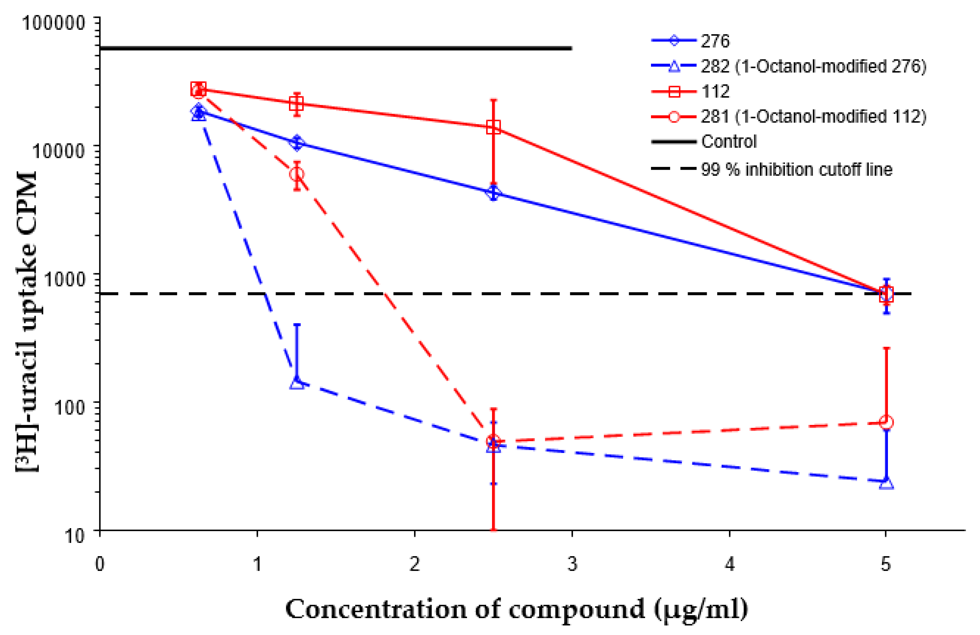
| Compound | TU276 | TU282 * | TU112 | TU281 ** |
|---|---|---|---|---|
| MIC for intra-macrophage mycobacteria (µg/mL) | 5.0 | 1.25 | 5.0 | 2.5 |
| MIC for free mycobacteria (µg/mL) | 0.19–0.25 | 1.92–2.53 | 0.19–0.25 | 1.90–2.53 |
| Sera from Intact Mice | Sera from TU112-Treated Mice | Serum from INH-Treated Mouse | Lung Tissue from Intact Mice | Lung Tissue from TU112-Treated Mice | ||
|---|---|---|---|---|---|---|
| 3.6 ± 3.0 | 15 min | 1 h | 4 h | 243 ± 172 | 2.0 ± 1.4 | 17.6 ± 8.7 |
| 64 ± 39 | 70 ± 35 | 64 ± 39 | ||||
| ANOVA, p = 0.017 | ANOVA, p = 0.0044 | |||||
© 2020 by the authors. Licensee MDPI, Basel, Switzerland. This article is an open access article distributed under the terms and conditions of the Creative Commons Attribution (CC BY) license (http://creativecommons.org/licenses/by/4.0/).
Share and Cite
Majorov, K.B.; Nikonenko, B.V.; Ivanov, P.Y.; Telegina, L.N.; Apt, A.S.; Velezheva, V.S. Structural Modifications of 3-Triazeneindoles and Their Increased Activity Against Mycobacterium tuberculosis. Antibiotics 2020, 9, 356. https://0-doi-org.brum.beds.ac.uk/10.3390/antibiotics9060356
Majorov KB, Nikonenko BV, Ivanov PY, Telegina LN, Apt AS, Velezheva VS. Structural Modifications of 3-Triazeneindoles and Their Increased Activity Against Mycobacterium tuberculosis. Antibiotics. 2020; 9(6):356. https://0-doi-org.brum.beds.ac.uk/10.3390/antibiotics9060356
Chicago/Turabian StyleMajorov, Konstantin B., Boris V. Nikonenko, Pavel Yu. Ivanov, Lyudmila N. Telegina, Alexander S. Apt, and Valeria S. Velezheva. 2020. "Structural Modifications of 3-Triazeneindoles and Their Increased Activity Against Mycobacterium tuberculosis" Antibiotics 9, no. 6: 356. https://0-doi-org.brum.beds.ac.uk/10.3390/antibiotics9060356