Article

Phenolic Content and Antimicrobial and Anti-Inflammatory Effects of Solidago virga-aurea, Phyllanthus niruri, Epilobium angustifolium, Peumus boldus, and Ononis spinosa Extracts

1
Department of Pharmacy, Università degli Studi “Gabriele d’Annunzio”, via dei Vestini 31, 66100 Chieti, Italy
2
Department of Chemistry, Biology and Biotechnology, University of Perugia, 06100 Perugia, Italy
3
Omega Pharma Srl, Via Milano 129, 22063 Cantù (CO), Italy
4
Department of Biology, Science Faculty, Selcuk Universtiy, Campus, Konya, 42130 Konya, Turkey
5
Veridia Italia Srl, via Raiale 285, 65100 Pescara, Italy
*
Authors to whom correspondence should be addressed.
Submission received: 7 October 2020 / Revised: 30 October 2020 / Accepted: 4 November 2020 / Published: 6 November 2020

Abstract

:
Prostatitis is an inflammatory condition that is related to multiple infectious agents, including bacteria and fungi. Traditional herbal extracts proved efficacious in controlling clinical symptoms associated with prostatitis. In this context, the aim of the present study was to explore the efficacy of extracts from Solidago virga-aurea, Ononis spinosa, Peumus boldus, Epilobium angustifolium, and Phyllanthus niruri against bacterial (Escherichia coli, Pseudomonas aeruginosa, Staphylococcus aureus, Bacillus cereus) and fungi strains (Candida albicans; C. tropicalis) involved in prostatitis. Additionally, anti-mycotic effects were tested against multiple species of dermatophytes (Trichophyton rubrum, T. tonsurans, T. erinacei, Arthroderma crocatum, A. quadrifidum, A. gypseum, A. currey, and A. insingulare). Antioxidant effects were also evaluated in isolated rat prostates challenged with lipopolysaccharide (LPS), and phytochemical analyses were conducted to identify and quantify selected phenolic compounds, in the extracts. Finally, a bioinformatics analysis was conducted to predict putative human and microbial enzymes targeted by extracts’ phytocompounds and underlying the observed bio-pharmacological effects. The phytochemical analysis highlighted that rutin levels could be crucial for explaining the highest antibacterial activity of P. boldus extract, especially against E. coli and B. cereus. On the other hand, in the E. angustifolium extract, catechin concentration could partially explain the highest efficacy of this extract in reducing lipid peroxidation, in isolated rat prostates stimulated with LPS. Concluding, the results of the present study showed moderate antimicrobial and anti-inflammatory effects induced by water extracts of S. virga-aurea, P. boldus, E. angustifolium, P. niruri, and O. spinosa that could be related, at least partially, to the phenolic composition of the phytocomplex.

1. Introduction

Prostatitis is an inflammatory condition that is related to multiple infectious agents, including bacteria and fungi [1,2,3]. The inflammatory pathways underlying prostatitis have been increasingly studied in recent years, and herbal extracts, especially in combination, have been revealed as a promising tool for the management of clinical symptoms related to the burden of inflammation/oxidative stress occurring in prostatitis [4]. Strictly related to this disorder, inflammation of the lower urinary tract (LUTS) is reported as the second most common human infectious disorder [5]. Currently, α-blockers represent the first-choice treatment for LUTS in patients diagnosed with both prostatitis and benign prostate hyperplasia (BPH) [6]. On the other hand, traditional herbal extracts were efficacious in controlling the inflammatory component associated with these disorders [7,8,9,10], with the efficacy that could be improved through their pharmacological associations [11]. This is consistent, at least in part, with the scientific literature suggesting the benefits of the combination of herbal extracts, usually employed as food supplements, for treating chronic inflammatory diseases [12,13,14,15,16]. Increasing attention to alternative medications, including nutraceuticals and herbal preparations that could display a lower grade of side effects, has led to the exploration of extracts prepared from plants traditionally used by folk populations to manage inflammatory conditions [14]. These extracts, especially those prepared via traditional infusions or decoctions, could not only be effective and safe due to their long-term use, but also represent novel strategies for implementing local botanical chain productions [15,16].
Phyllanthus niruri is a traditional Ayurvedic remedy that has long been employed in a wide range of inflammatory disorders. It is also able to induce anti-proliferative effects on human prostate cancer PC3 cell lines [17]. Boldine, the main active principle of Peumus boldus, revealed to be effective in inducing apoptosis in human bladder cancer cells, as well [18]. Ononis spinosa is a folk remedy used in the treatment of urinary tract infections and stones [19]. Analogous traditional uses have been described for Solidago virga-aurea, although the scientific literature reported anti-proliferative effects, possibly related to the presence of terpene and phenolic compounds, in the phytocomplex [20,21]. Similarly, Epilobium angustifolium has been shown to be effective in preventing BPH [22] due to its richness in phenolic compounds [23].
In this context, the aim of the present study was to explore the antimicrobial and anti-inflammatory effects of water extracts from S. virga-aurea, O. spinosa, P. boldus, E. angustifolium, and P. niruri. Although the water solubility of plant secondary metabolites could be limited, previous studies of ours suggested the use of microwave- and ultrasound-assisted methods for improving phenol extraction that was paralleled by increased anti-inflammatory, antioxidant, and antimicrobial effects [16,24,25]. In the present study, the extracts’ antimicrobial activity was tested against bacterial (Escherichia coli, Pseudomonas aeruginosa, Staphylococcus aureus, and Bacillus cereus) and fungi strains (Candida albicans, C. tropicalis) involved in prostatitis and LUTS [2,3,26,27,28]. Additionally, anti-mycotic activities were also tested against multiple species of dermatophytes (Trichophyton rubrum, T. tonsurans, T. erinacei, Arthroderma crocatum, A. quadrifidum, A. gypseum, A. Currey, and A. insingulare). This is consistent with the incidence of dermatophyte infections in patients suffering from urinary tract disorders, including renal failure and prostate adenocarcinoma [29,30,31]. Anti-inflammatory effects, in terms of reduction of prostaglandin production, were also evaluated in a preclinical model of prostatitis consisting of isolated rat prostate specimens challenged with lipopolysaccharide (LPS). The study also compared the efficacy of single extracts and their pharmacological association (Fluxonorm®), and the obtained results supported the rationale for their use to treat clinical symptoms related to prostatitis and LUTS. In order to improve our knowledge about the mechanism of action of the studied herbal extracts, a fingerprint analysis via high-performance liquid chromatography coupled to ultraviolet and mass spectrometry (HPLC-UV-MS) was conducted to identify and quantify selected phenolic compounds, in the phytocomplex. Specifically, gallic acid, rutin, catechin, and epicatechin were selected considering their intrinsic antimicrobial and anti-inflammatory properties [32,33]. Finally, a bioinformatics analysis was also carried out to predict putative human and microbial enzymes targeted by the extracts’ phytocompounds and underlying the observed bio-pharmacological effects.

2. Materials and Methods

2.1. Plant Material

Dry extracts of Phyllanthus niruri L. (family Phyllanthaceae), Ononis spinosa L. (family Fabaceae), Solidago virga-aurea L. (family Asteraceae), Peumus boldus Molina (family Monimiaceae), Epilobium angustifolium L. (family Onagraceae), and the registered trademark formula Fluxonorm® (O. spinosa/S. virga-aurea/P. niruri/P. boldus/E. angustifolium 12.5:12.5:18.7:25.0:31.2), were provided as dried materials by OMEGA PHARMA Srl (Cantù, Italy). Before testing, extracts were rehydrated via ultrasound-assisted extraction at 60 °C for one hour.

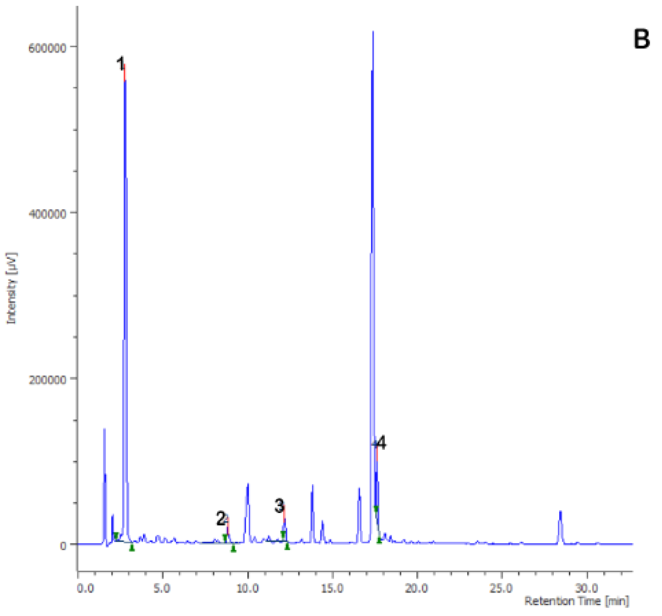
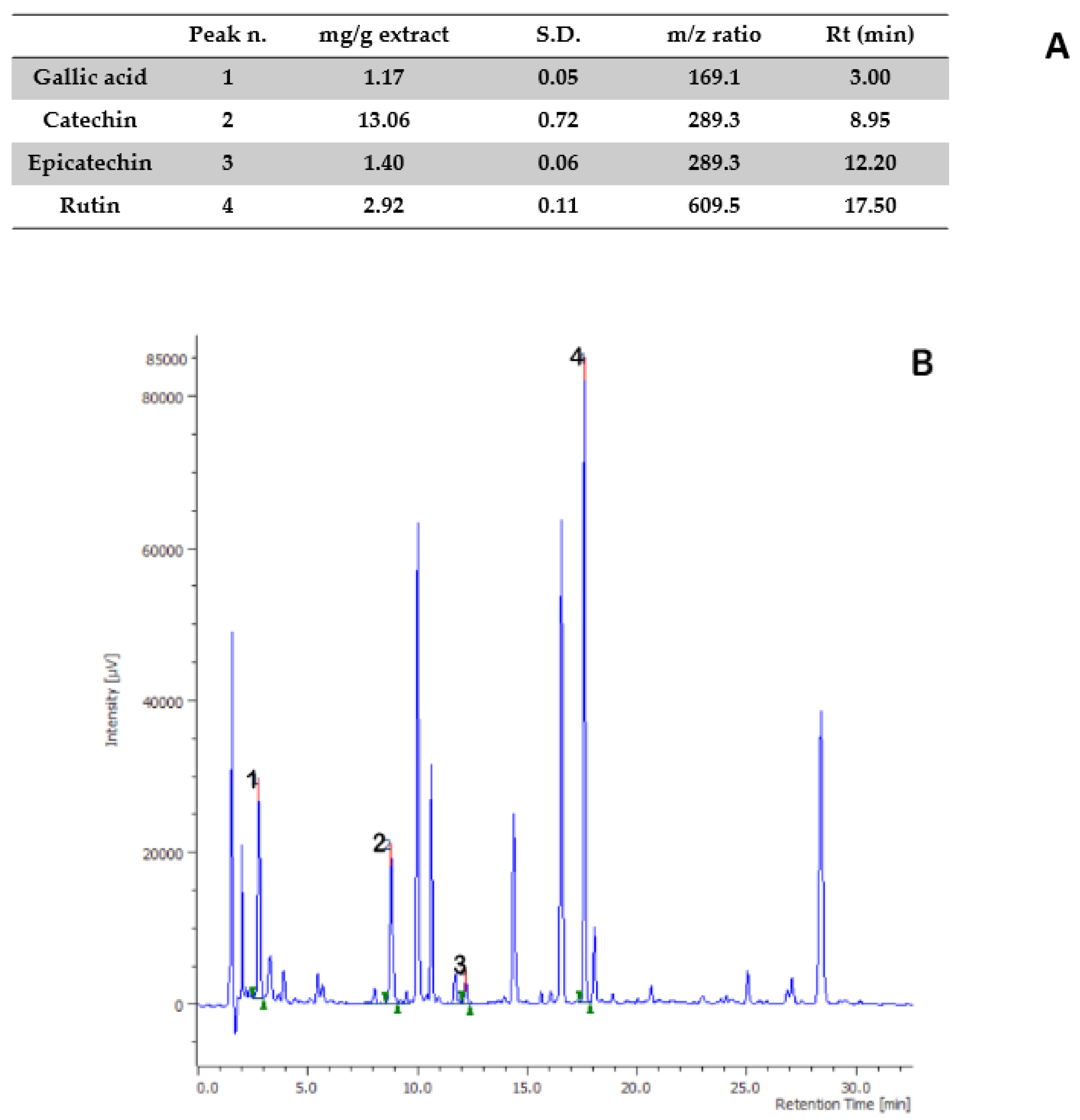
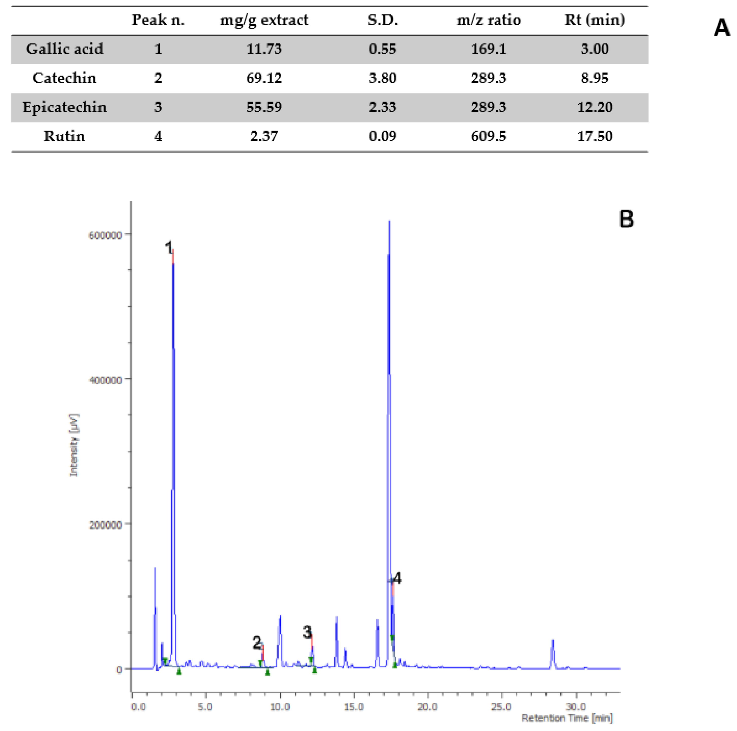
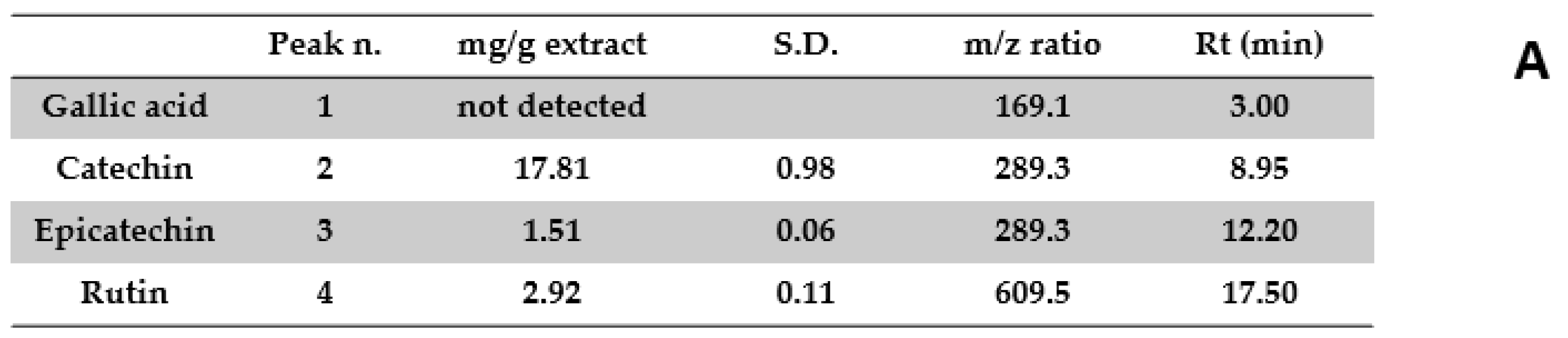
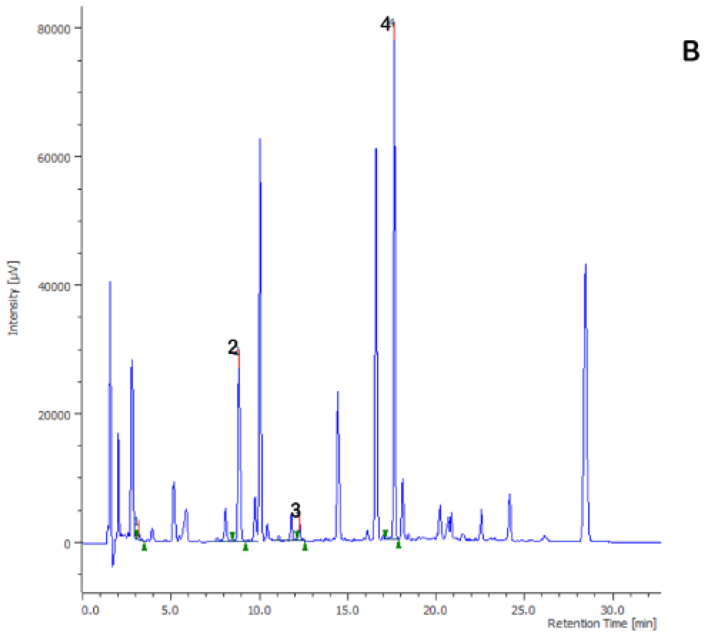
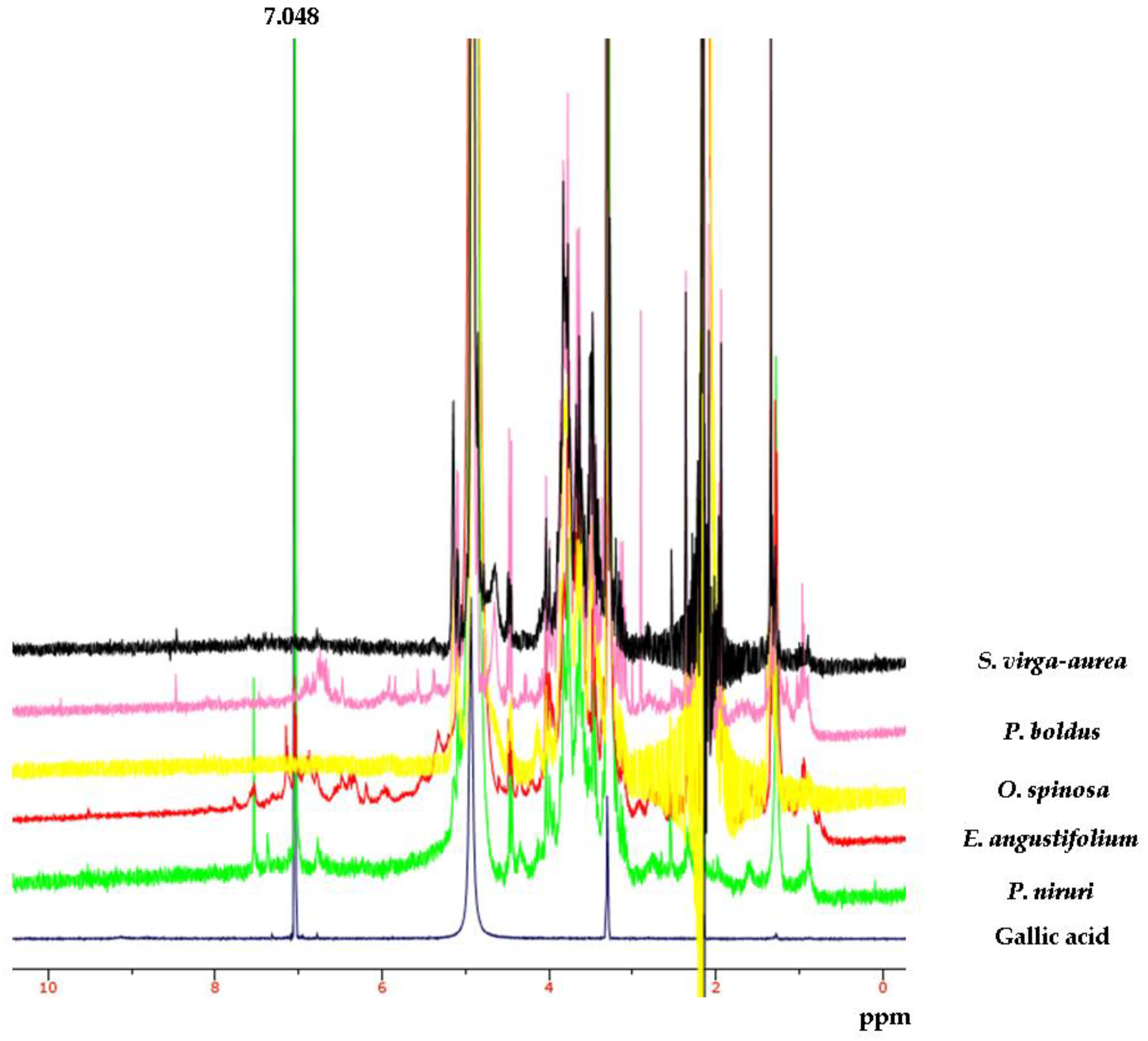
2.2. Phytochemical Analysis

P. niruri, E. angustifolium, P. boldus, S. virga-aurea, and O. spinosa extracts (10 mg/mL) were analyzed for phenol quantitative determination using a reversed-phase HPLC-UV-MS in gradient elution mode. The separation was conducted within 60 min, starting from the conditions: water/acetonitrile: 93:7, (v/v). The details about the gradient are listed in Table 1.
The HPLC apparatus consisted of two PU-2080 PLUS chromatographic pumps, a DG-2080-54 line degasser, a mix-2080-32 mixer, a UV-2075 PLUS UV detector, an AS-2057 PLUS autosampler, and CO-2060 PLUS column thermostat, all made by JASCO (Tokyo, Japan). The used mass spectrometer was an Expression-L compact mass spectrometer (Advion, Ithaca, NY, USA). Integration was performed by JASCO ChromNAV2 chromatography software, and the separation was performed by gradient elution on an Agilent (Santa Clara, CA, USA) InfinityLab Poroshell 120 reverse phase column (C18, 150 mm × 4.6 mm ID, 2.7 µm). The analytical conditions for the separation of gallic acid, catechin, epicatechin and rutin were in agreement with the study by Rodríguez-Delgado and colleagues [34]. Extracts were qualitatively analyzed with an Advion Expression-L compact mass spectrometer (MS) in negative ion mode. MS signal identification was realized through comparison with standard solutions and MS spectra present in the recognized Mass Bank Europe database (https://massbank.eu/MassBank/). Quantitative determination of phenolic compounds identified by MS analysis was performed via a UV detector at 240 nm wavelength. Quantification was done through 7 point calibration curves, with linearity coefficients (R2) > 0.999, in the concentration range of 2–140 µg/mL. The area under the curve obtained from the HPLC chromatograms was used to quantify analyte concentration in the extracts.
Protonic nuclear magnetic resonance (1H-NMR) analysis was conducted with a Varian 300 MHz spectrometer using a standard proton pulse sequence (s2pul). Samples were prepared as follows: 10 mg/mL gallic acid and 50 mg/mL extract were sonicated in CD3OD for 30 min at room temperature. Next, 600 µL of each sample was transferred into the NMR tube and analyzed with the following parameters: 1.706 s acquisition time, 4803.1 Hz width, and 64 scans.

2.3. Artemia salina Lethality Test

The cytotoxicity limit of the extracts in the range 0.1–20 mg/mL was evaluated through a lethality bioassay of the brine shrimp Artemia salina, as previously reported [16]. The experiments were carried out in triplicate.

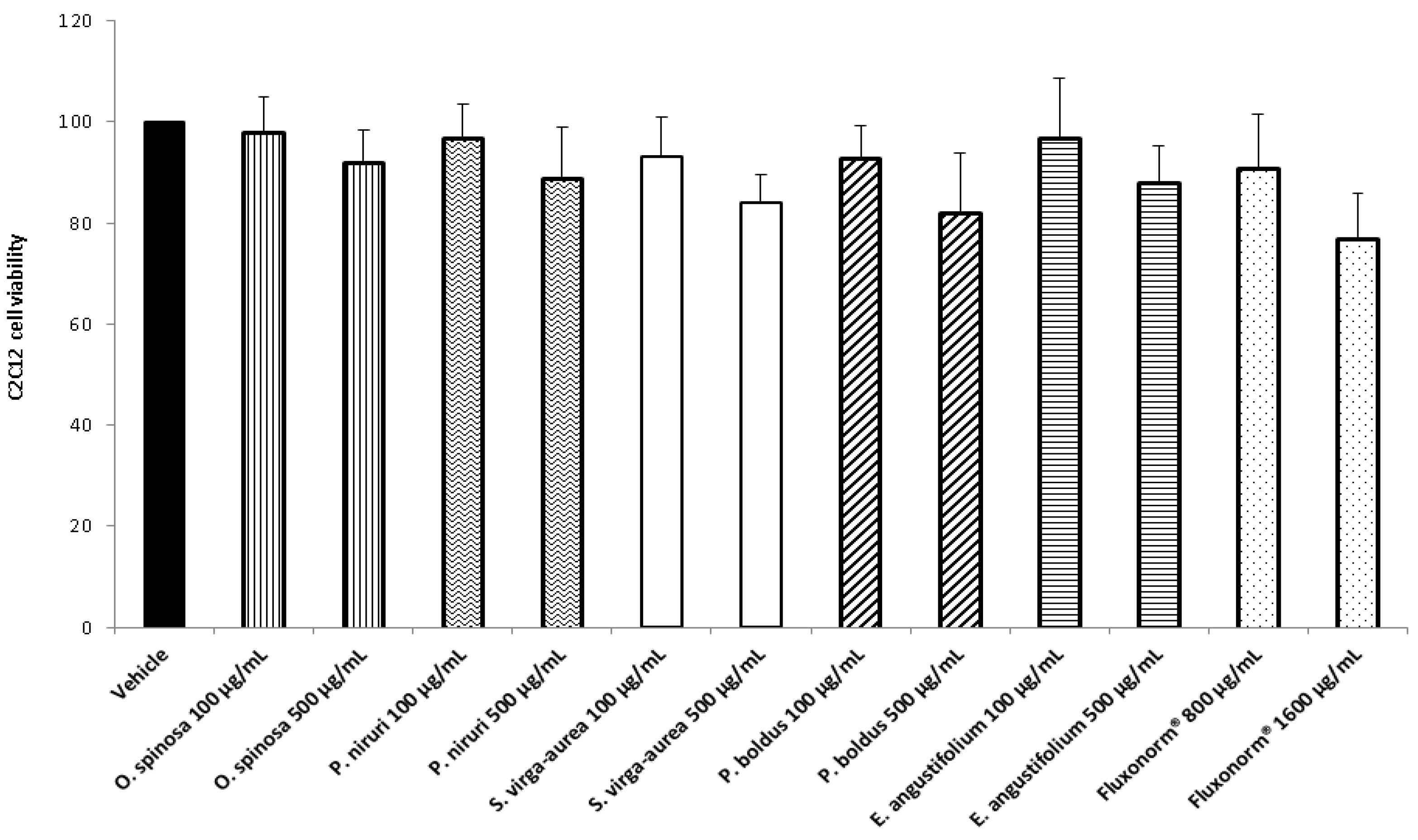
2.4. Cell Cultures and Viability Test

The effects of the extracts (100–500 µg/mL) on myocyte C2C12 and prostate cancer PC3 cell viability were determined through the 3-(4,5-dimethylthiazol-2-yl)-2,5-diphenyltetrazolium bromide (MTT) test. The experimental conditions were fully described in our previous paper [35].

2.5. Ex Vivo Pharmacological Study
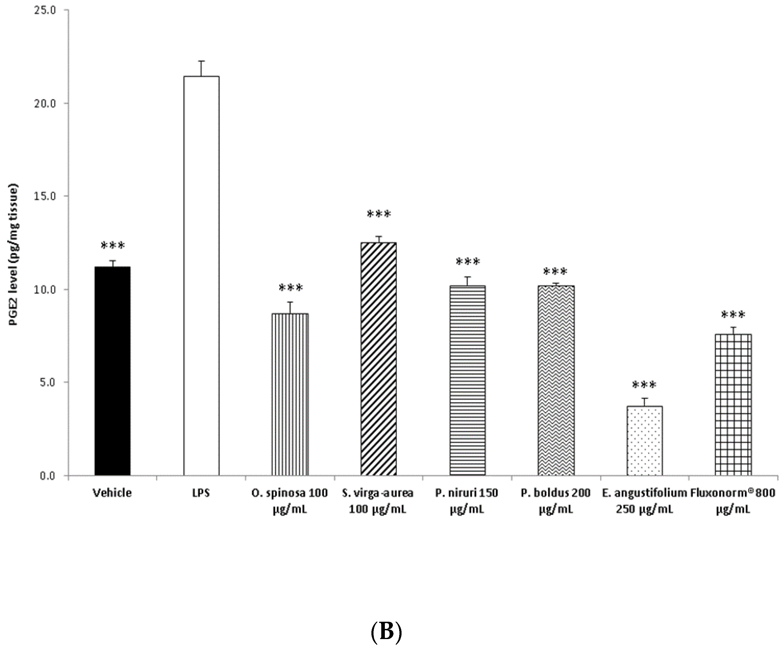
The protective effects of the extracts were investigated through an ex vivo model consisting of isolated prostate challenged with E. coli lipopolysaccharide (LPS) at 10 µg/mL, as previously reported [9]. Prostates were collected from 24 Sprague Dawley rats (200–250 g). The study was approved by the Local Ethical Committee and the Italian Ministry of Health. The approval number of the study was the following: authorization N. F4738.N.XTQ, delivered for the period 11 November 2018–11 November 2019. During the study, the prostate specimens were separately treated with the extracts of O. spinosa, S. virga-aurea, P. niruri, P. boldus, and E. angustifolium (100, 100, 150, 200, and 250 µg/mL, respectively) or Fluxonorm® (800 µg/mL). Afterward, the prostate tissue supernatants were assayed, as described below.

2.6. 8-Iso-PGF and PGE2 Radioimmunoassay

The levels of 8-iso-PGF and PGE2 in the supernatants were determined by radioimmunoassay (RIA). The detailed protocol was described in our previous study [36].

2.7. Antibacterial and Antimycotic Activities
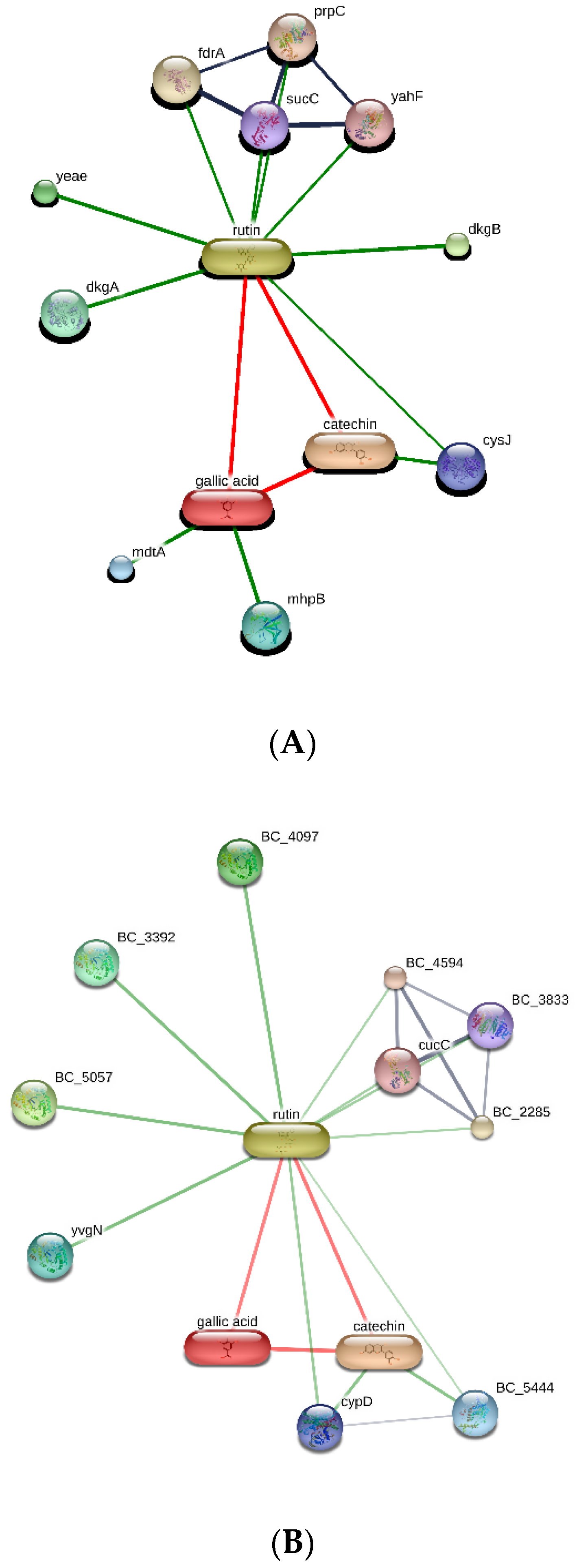
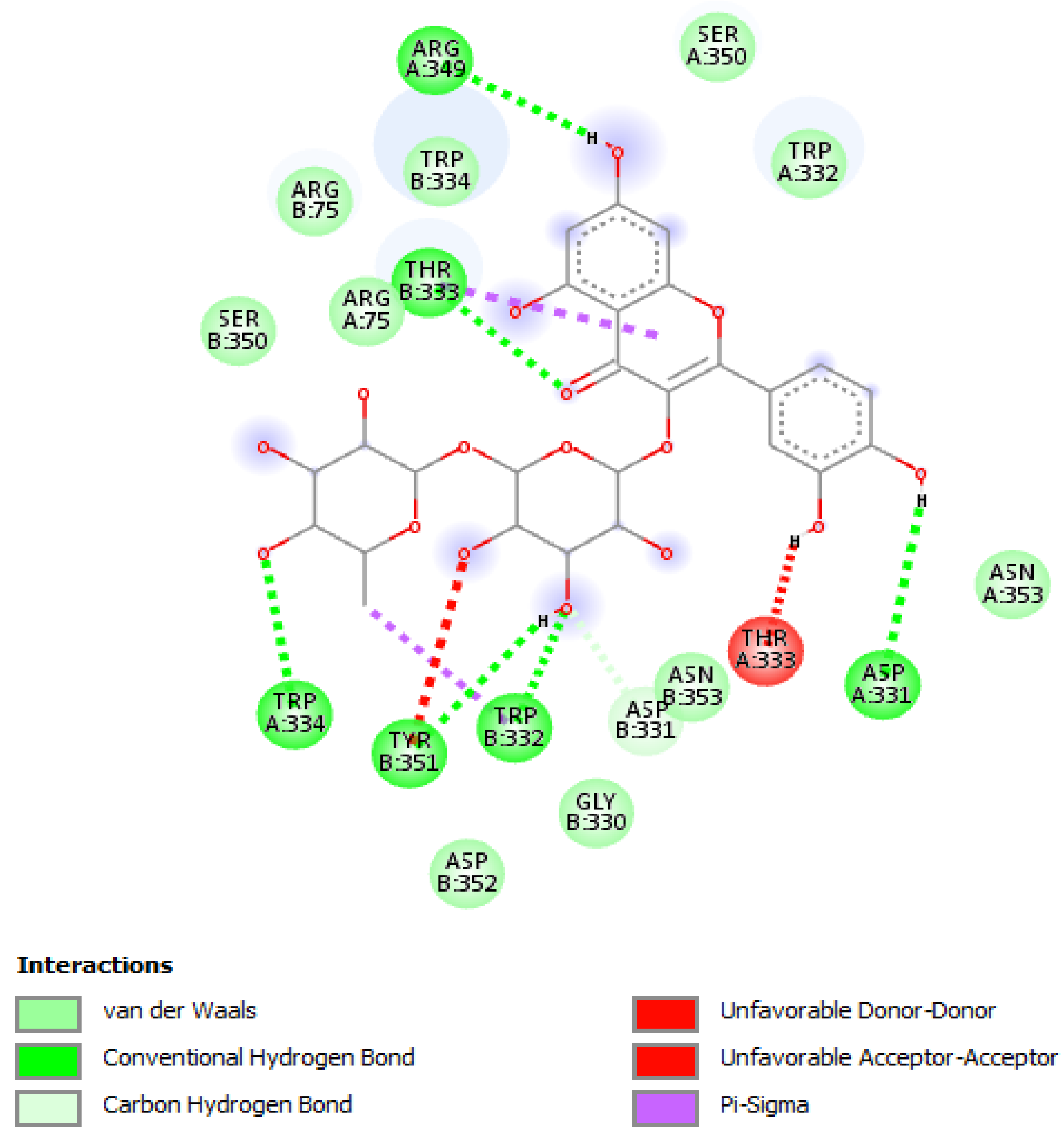
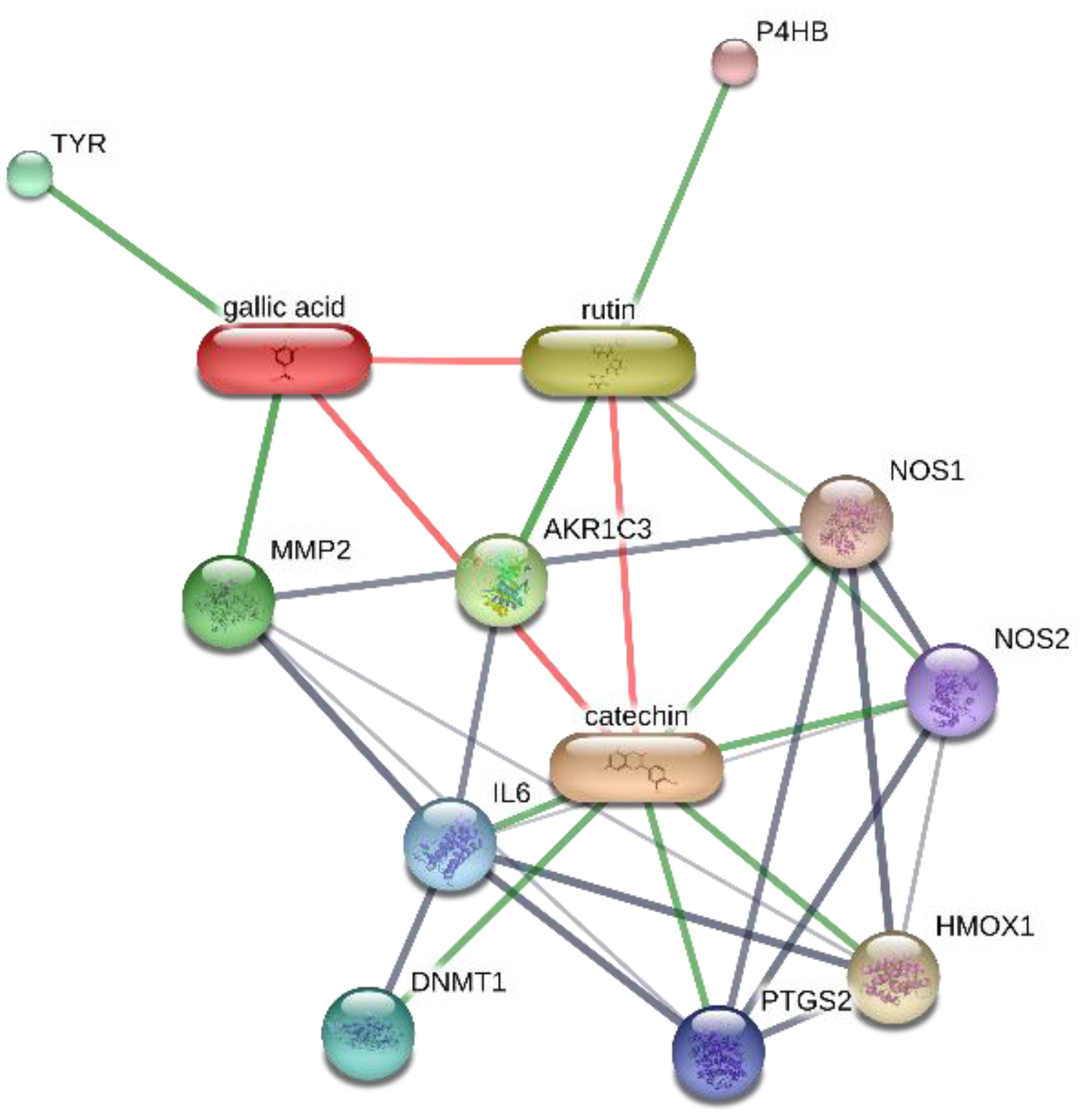
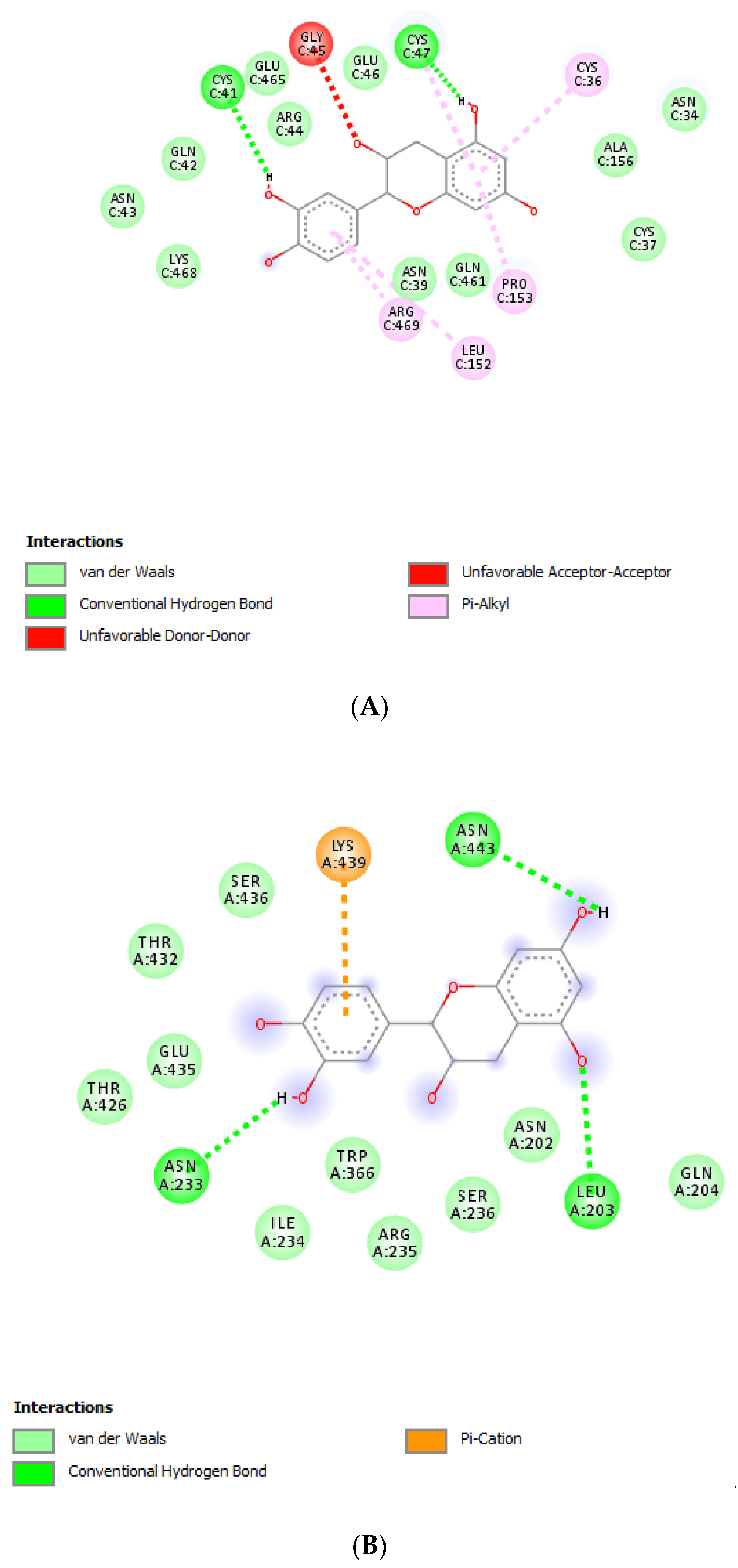
The water extracts of P. niruri, O. spinosa, S. virga-aurea, P. boldus, and E. angustifolium were assayed for antibacterial activity against the following Gram-negative and Gram-positive strains: E. coli (ATCC 10536); E. coli (PeruMycA 2); E. coli (PeruMycA 3); P. aeruginosa (PeruMycA 5); S. typhy (PeruMycA 7); B. cereus (PeruMycA 4); B. subtilis (PeruMycA 6); S. aureus (ATCC 6538). Additionally, the same extracts were assayed for antimycotic effects against different Candida and dermatophyte species: C. tropicalis (DBVPG 6184); C. albicans (DBVPG 6379); C. parapsilosis (DBVPG 6551); C. albicans (DBVPG 6183); T. mentagrophytes (CCF 4823); T. tonsurans (CCF 4834); T. rubrum (CCF 4879); T. rubrum (CCF 4933); A. crocatum (CCF 5300); A. quadrifidum (CCF 5792); T. erinacei (CCF 5930); A. gypseum (CCF 6261); A. currey (CCF 5207); A. insingulare (CCF 5417). The antimicrobial activities of the aforementioned extracts were compared to reference drugs. To investigate the extracts effects on bacterial, Candida, and dermatophyte growth, the inhibitory activity was compared to ciprofloxacin, fluconazole and griseofulvin, respectively. The detailed protocols of the present antimicrobial assays have been fully described in recently published papers of ours [37,38].

2.8. Bioinformatics

Putative targets were identified according to the bioinformatic method recently described by Gu and colleagues [39]. Briefly, microbial and human proteins targeted by P. niruri, O. spinosa, S. virga-aurea, P. boldus, and E. angustifolium extracts were predicted using the bioinformatics platform STITCH (http://stitch.embl.de/cgi/network.pl). Docking calculations were conducted using AutoDock Vina PyRx 0.8 software. Crystal structures of target proteins were derived from the Protein Data Bank (PDB) with PDB IDs as follows: 1MJT (nitric oxide synthase (NOS) from S. aureus), 2FLQ (nitric oxide synthase (NOS) from B. cereus), and 1CX2 [cyclooxygenase-2 (COX-2)]. Discovery studio 2020 visualizer was employed to investigate the protein–ligand non-bonding interactions.

2.9. Statistical Analysis

The experimental data related to in vitro and ex vivo studies were analyzed through the analysis of variance (ANOVA) followed by the Newman–Keuls post hoc test. GraphPad Prism software was employed for the statistical analysis, while the software G*Power (v3.1.9.4, University of Kiel, Kiel, Germany) was used for calculating the animal number for the experiments, where p < 0.05 was considered statistically significant.

3. Results and Discussion

Aiming to investigate the putative mechanism of action of the tested extracts, HPLC-UV-MS fingerprint analysis was carried out in order to measure the levels of selected phenolic compounds, namely gallic acid, catechin, epicatechin, and rutin, that play a master role in the antioxidant/anti-inflammatory response following herbal extract administration [40]. Specifically, the results of HPLC-UV-MS analysis depicted in Figure 1, Figure 2, Figure 3, Figure 4 and Figure 5 show that the level of gallic acid is higher in P. niruri and E. angustifolium extracts, whereas P. boldus, O. spinosa and S. virga-aurea do not show a relevant amount of this compound. These data were also consistent with the 1H-NMR analyses (Figure 6), where the gallic acid protons signal (H-2, H-6) was detected at 7.048 ppm, mainly in P. niruri and E. angustifolium extracts, respectively. In the same extracts, the catechin fraction was also present at higher concentrations, compared to S. virga-aurea, P. boldus, and O. spinosa extracts, whereas P. boldus extract showed higher rutin concentration. In each figure, the concentration data were substantiated by related UV and MS chromatograms that highlighted a complete separation of the four measured phenolics within a 20 min HPLC run. Specifically, each analyte peak was qualitatively analyzed with MS detector in negative ion mode and the presence of gallic acid (m/z = 169.1; retention time = 3.00 min), catechin (m/z = 289.3; retention time = 8.95 min), epicatechin (m/z = 289.3; retention time = 12.20 min), and rutin (m/z = 609.5; retention time = 17.50 min) was identified through comparison with related standards and with MS data collected by MassBank Europe (https://massbank.eu/MassBank/). According to the quantitative analysis conducted through the UV detector (set at 240 nm wavelength), E. angustifolium displayed the richest phenolic profile from both qualitative and quantitative points of view. This was consistent, albeit partially, with literature data suggesting the richness in phenolic compounds of E. angustifolium as a key factor influencing its efficacy in BHP [22,23].
The extracts were also assayed for the brine shrimp (A. salina) lethality test to evaluate the biocompatibility limit in the standard range (0.01–10 mg/mL) [41]. The brine shrimp test yielded LC50 values >10 mg/mL that were indicatory for choosing a concentration limit at least ten-fold lower than LC50 for the following bio-pharmacological tests [16]. In this regard, the extracts were assayed for antimicrobial activity against multiple bacterial and fungi strains (Table 2, Table 3 and Table 4), including Candida and dermatophyte species, that could be involved in infectious disorders of the urinary tract [28,29,30,31]. It is of noteworthy interest that most of the tested extracts showed bacteriostatic and mycostatic effects at concentrations much lower than LC50 values calculated via the brine shrimp test. In the present multidirectional study, the inhibitory effects of tested extracts on bacterial and fungi strains were expressed as µg/mL in order to better compare the concentrations of the extracts active as antimicrobials with those active as anti-inflammatory/antioxidant agents [16,37,38]. On the other hand, multiple studies suggest the use of arbitrary units to express the antimicrobial activity of herbal extracts as well [42,43,44]. The spectrum of antimicrobial activity seems to be paralleled, at least partially, by the extract qualitative composition. In this regard, E. angustifolium was effective against a larger number of microbial species compared to the other tested extracts that displayed lower levels of catechins. The quantitative HPLC analysis also suggested that E. angustifolium could have a higher content of total phenols, thus substantiating the wider spectrum of antimycotic activity [16]. Nevertheless, P. boldus was the most active as antibacterial agent against both E. coli and B. cereus. It is sensitive to note that P. boldus displayed the highest rutin concentration that could be crucial for the highest antibacterial activity against these strains. This hypothesis seemed to be validated by the bioinformatics analysis conducted through the STITCH platform that highlighted a prominent position for rutin in the scenario of putative interactions between identified phytocompounds and bacterial proteins involved in oxidative metabolism (Figure 7A,B). Regarding the B. cereus, a further docking analysis was conducted to calculate the affinity of rutin towards B. cereus nitric oxide synthase (BC_44). Intriguingly, the docking run yielded an affinity constant (Ki) in the sub-micromolar range (Figure 8). This result adds to our recent observation about the putative interactions between BC_44 and resveratrol [37]. Nevertheless, the inhibitory effect induced by rutin on B. cereus occurred at concentrations higher than 250 µg/mL, thus excluding the interaction of this flavonoid with BC_44 as a key event underlying the antimicrobial effect induced by P. boldus. In this regard, we hypothesize that total phenolic compound content could be involved in the antibacterial activity [16], as also suggested by the P. boldus HPLC-UV-MS chromatogram and 1H-NMR spectrum showing numerous signals related to unidentified phenolic compounds present in this extract.
The different pattern of antimicrobial activity also supports the rationale for the pharmacological association of tested extracts in order to improve their efficacy. In this regard, in vitro tests were conducted on the non-tumoral cell line C2C12 to expand our comprehension about the intrinsic tolerability and protective effects of the extracts employed as both single treatment and pharmacological associations (Fluxonorm®). Specifically, MTT assay was carried out, and extracts were tested in the concentration range 100–500 µg/mL, whereas the Fluxonorm® formula was assayed in the range from 800–1600 µg/mL. The null effect on the cell viability after challenging the cells with the extracts (Figure 9) further confirmed the biocompatibility of the selected water extracts. The pharmacological association Fluxonorm®, which was the resulting algebraic sum of the aforementioned extracts used as single ingredients in the ratio reported in Section 2.1, was completely tolerated at the lowest tested concentration (800 µg/mL). By contrast, the cell viability tended to decrease under the biocompatibility limit (80%) at the highest formula concentration (1600 µg/mL). It is rational to hypothesize that the reduced cell viability at the highest tested concentration could be related to the paradoxical phenolic compound-induced oxidative stress that often occurs at elevated concentrations [45,46]. Conversely, under the limit of biocompatibility, the single extracts and Fluxonorm® were able to contrast the burden of oxidative stress and inflammation in isolated rat prostate specimens challenged with LPS, an ex vivo model of prostatitis [11,47]. All extracts were able to completely blunt the LPS-induced level of both PGE2 and 8-iso-PGF (Figure 10A,B). PGE2 is the main prostanoid synthesized by cyclo-oxygenase (COX)-2 in inflammatory conditions [48], and up-regulated levels of PGE2 are found in prostate inflammation and cancer [49,50]. While, 8-iso-PGF is an isomer of classical prostaglandins that is principally produced by oxidative stress-mediated pathways, including reactive oxygen/nitrogen species (ROS/RNS), that could cause disruptive peroxidation reactions on cellular substrates, such as proteins, lipids and nucleic acids [51]. In this regard, 8-iso-PGF deriving from ROS/RNS peroxidation of membrane arachidonic acid represents a diagnostic biomarker for evaluating lipid peroxidation in vivo [52]. On the other hand, a paradoxically reduced level of 8-iso-PGF has been observed in superficial bladder cancer, possibly related to the putative vasoconstrictor role of this prostanoid [53]. Currently, the reduced levels of both PGE2 and 8-iso-PGF following extract treatment are consistent with the qualitative and quantitative profile of the analyzed phytochemicals. However, the best pharmacological profile was showed by E. angustifolium, which influenced the antioxidant/anti-inflammatory role of Fluxonorm® the most. This is probably related to its content in catechin and epicatechin, which are known to behave as COX-2 inhibitors [54]. It is sensitive to highlight the clinical study carried out by Micali and colleagues [55], which evidenced a significant reduction in prostate-specific antigen following green tea catechin administration, thus suggesting its potential efficacy in treating/preventing inflammatory prostate diseases. Furthermore, the antiproliferative effects of green tea deriving catechins were demonstrated in multiple cancer cell lines, including prostate cancer LNCaP cells [56]. The pivotal role of catechin in the anti-inflammatory response related to extract treatment was also indicated by bioinformatics analysis (Figure 11). In this regard, besides COX-2, different pro-inflammatory proteins were predicted to be targeted by catechin, including inducible nitric oxide synthase (iNOS). Additionally, the sub-micromolar/micromolar affinity of catechin towards COX-2 and iNOS (Figure 12A,B) agreed with catechin levels in all tested extracts. The highest efficacy of E. angustifolium in contrasting the burden of oxidative stress/inflammation in isolate prostate specimens was consistent with the recent findings by Deng and colleagues (2019), as well as the capability of E. angustifolium extract to counteract the oxidative stress in male rats subcutaneously injected with testosterone in order to induce BHP [22]. The same authors also suggested that phytocomplex phenolic compounds could be crucial for substantiating the antioxidant/anti-inflammatory effects of E. angustifolium in the prostate. Finally, prostate cancer PC3 cells were treated with single extracts and Fluxonorm®. Specifically, E. angustifolium, P. niruri, P. boldus, and Fluxonorm® showed similar antiproliferative effects (Figure 13) that could be related, at least partially, to multiple anti-inflammatory and antioxidant mechanisms [57,58,59]. In this context, it is sensitive to hypothesize that phenolic compounds present in the tested extracts could be responsible for the observed antiproliferative effects. However, these effects agree only partially with the levels of selected phenolic compounds measured via HPLC-UV-MS analyses. The chromatograms of the same analyses, but also the NMR spectra, suggest the presence of other unidentified phenolic compounds that could be crucial for the observed bio-pharmacological effects.

4. Conclusions

The results of the present study showed moderate antimicrobial and anti-inflammatory effects induced by water extracts of S. virga-aurea, P. boldus, E. angustifolium, P. niruri, and O. spinosa, that could be related, at least partially, to the phenolic composition of the phytocomplex. Future phytochemical investigations may further unravel the relationships between extract composition and bio-pharmacological effects.

Author Contributions

Conceptualization, M.P. (Massimiliano Petrucci), P.A., G.O., and C.F.; methodology, P.A., L.M., G.O. and G.Z.; software, C.F.; validation, P.A., G.O., L.M., G.Z. and C.F.; formal analysis, C.F.; investigation, G.A.F., A.C., L.R., S.L., R.V., M.D.M., F.B., M.P. (Matteo Politi) and G.A.; resources, G.O. and C.F.; data curation, P.A., G.Z. and L.M.; writing—original draft preparation, C.F. and P.A.; writing—review and editing, C.F., L.M. and G.O.; visualization, R.V. and L.B.; supervision, L.B.; project administration, P.A., C.F. and G.O.; funding acquisition, M.P. (Massimiliano Petrucci). All authors have read and agreed to the published version of the manuscript.

Funding

This work was supported by OMEGA PHARMA Srl (Cantù, Italy).

Acknowledgments

The authors would like to thank Lucio Boschi (Veridia Italia Srl) for technical assistance during the study that was conducted within a joint project between the Department of Pharmacy of “G. d’Annunzio” University and Veridia Srl (Research Program 2020–2023). Study Coordinators were Giustino Orlando, Luigi Menghini, and Claudio Ferrante.

Conflicts of Interest

The study was supported by OMEGA PHARMA Srl.

References

  1. Steenkamp, V.; Gouws, M.C.; Gulumian, M.; Elgorashi, E.E.; van Staden, J. Studies on antibacterial, anti-inflammatory and antioxidant activity of herbal remedies used in the treatment of benign prostatic hyperplasia and prostatitis. J. Ethnopharmacol. 2006, 103, 71–75. [Google Scholar] [CrossRef]
  2. Delcaru, C.; Podgoreanu, P.; Alexandru, I.; Popescu, N.; Măruţescu, L.; Bleotu, C.; Mogoşanu, G.D.; Chifiriuc, M.C.; Gluck, M.; Lazăr, V. Antibiotic Resistance and Virulence Phenotypes of Recent Bacterial Strains Isolated from Urinary Tract Infections in Elderly Patients with Prostatic Disease. Pathogens 2017, 6, 22. [Google Scholar] [CrossRef]
  3. Delcaru, C.; Alexandru, I.; Podgoreanu, P.; Grosu, M.; Stavropoulos, E.; Chifiriuc, M.C.; Lazar, V. Microbial Biofilms in Urinary Tract Infections and Prostatitis: Etiology, Pathogenicity, and Combating strategies. Pathogens 2016, 5, 65. [Google Scholar] [CrossRef] [PubMed] [Green Version]
  4. Marzano, R.; Dinelli, N.; Ales, V.; Bertozzi, M.A. Effectiveness on urinary symptoms and erectile function of Prostamev Plus® vs only extract Serenoa repens. Arch. Ital. Urol. Androl. 2015, 87, 25–27. [Google Scholar] [CrossRef] [Green Version]
  5. Bjorling, D.E.; Wang, Z.Y.; Bushman, W. Models of inflammation of the lower urinary tract. Neurourol. Urodyn. 2011, 30, 673–682. [Google Scholar] [CrossRef] [Green Version]
  6. Nickel, J.C. Alpha-blockers for the treatment of prostatitis-like syndromes. Rev. Urol. 2006, 8 (Suppl. S4), S26–S34. [Google Scholar] [PubMed]
  7. Stamatiou, K.; Pierris, N. Serenoa repens extract additionally to quinolones in the treatment of chronic bacterial prostatitis. The preliminary results of a long term observational study. Arch. Ital. Urol. Androl. 2013, 85, 190–196. [Google Scholar] [CrossRef] [PubMed] [Green Version]
  8. Cai, T.; Morgia, G.; Carrieri, G.; Terrone, C.; Imbimbo, C.; Verze, P.; Mirone, V. IDIProst® Gold Study Group. An improvement in sexual function is related to better quality of life, regardless of urinary function improvement: Results from the IDIProst® Gold Study. Arch. Ital. Urol. Androl. 2013, 85, 184–189. [Google Scholar] [CrossRef] [PubMed]
  9. Wang, M.; Ma, H.L.; Liu, B.; Wang, H.B.; Xie, H.; Li, R.D.; Wang, J.F. Pinus massoniana bark extract protects against oxidative damage in L-02 hepatic cells and mice. Am. J. Chin. Med. 2010, 38, 909–919. [Google Scholar] [CrossRef] [PubMed]
  10. Iglesias-Gato, D.; Carsten, T.; Vesterlund, M.; Pousette, A.; Schoop, R.; Norstedt, G. Androgen-independent effects of Serenoa repens extract (Prostasan®) on prostatic epithelial cell proliferation and inflammation. Phytother. Res. 2012, 26, 259–264. [Google Scholar] [CrossRef]
  11. Chiavaroli, A.; Recinella, L.; Ferrante, C.; Locatelli, M.; Carradori, S.; Macchione, N.; Zengin, G.; Leporini, L.; Leone, S.; Martinotti, S.; et al. Crocus sativus, Serenoa repens and Pinus massoniana extracts modulate inflammatory response in isolated rat prostate challenged with LPS. J. Biol. Regul. Homeost. Agents 2017, 31, 531–541. [Google Scholar] [PubMed]
  12. Menghini, L.; Ferrante, C.; Leporini, L.; Recinella, L.; Chiavaroli, A.; Leone, S.; Pintore, G.; Vacca, M.; Orlando, G.; Brunetti, L. A natural formula containing lactoferrin, Equisetum arvensis, soy isoflavones and vitamin D3 modulates bone remodeling and inflammatory markers in young and aged rats. J. Biol. Regul. Homeost. Agents 2016, 30, 985–996. [Google Scholar]
  13. Tabatabaei-Malazy, O.; Larijani, B.; Abdollahi, M. Targeting metabolic disorders by natural products. J. Diabetes Metab. Disord. 2015, 14, 57. [Google Scholar] [CrossRef] [Green Version]
  14. Jones, W.P.; Chin, Y.W.; Kinghorn, A.D. The role of pharmacognosy in modern medicine and pharmacy. Curr. Drug Targets. 2006, 7, 247–264. [Google Scholar] [CrossRef]
  15. Chichiriccò, G.; Ferrante, C.; Menghini, L.; Recinella, L.; Leone, S.; Chiavaroli, A.; Brunetti, L.; Di Simone, S.; Ronci, M.; Piccone, P.; et al. Crocus sativus by-products as sources of bioactive extracts: Pharmacological and toxicological focus on anthers. Food Chem. Toxicol. 2019, 126, 7–14. [Google Scholar] [CrossRef]
  16. Ferrante, C.; Recinella, L.; Ronci, M.; Menghini, L.; Brunetti, L.; Chiavaroli, A.; Leone, S.; Di Iorio, L.; Carradori, S.; Tirillini, B.; et al. Multiple pharmacognostic characterization on hemp commercial cultivars: Focus on inflorescence water extract activity. Food Chem. Toxicol. 2019, 125, 452–461. [Google Scholar] [CrossRef] [PubMed]
  17. Tang, Y.Q.; Jaganath, I.; Manikam, R.; Sekaran, S.D. Phyllanthus Suppresses Prostate Cancer Cell, PC-3, Proliferation and Induces Apoptosis through Multiple Signalling Pathways (MAPKs, PI3K/Akt, NFκB, and Hypoxia). Evid. Based. Complement. Alternat. Med. 2013, 2013, 609581. [Google Scholar] [CrossRef] [Green Version]
  18. Gerhardt, D.; Bertola, G.; Dietrich, F.; Figueiró, F.; Zanotto-Filho, A.; Moreira Fonseca, J.C.; Morrone, F.B.; Barrios, C.H.; Battastini, A.M.; Salbego, C.G. Boldine induces cell cycle arrest and apoptosis in T24 human bladder cancer cell line via regulation of ERK, AKT, and GSK-3β. Urol. Oncol. 2014, 32, 36.e1–36.e9. [Google Scholar] [CrossRef]
  19. Gampe, N.; Darcsi, A.; Kursinszki, L.; Béni, S. Separation and characterization of homopipecolic acid isoflavonoid ester derivatives isolated from Ononis spinosa L. root. J. Chromatogr. B Analyt. Technol. Biomed. Life Sci. 2018, 1091, 21–28. [Google Scholar] [CrossRef]
  20. Choi, S.Z.; Choi, S.U.; Lee, K.R. Phytochemical constituents of the aerial parts from Solidago virga-aurea var. gigantea. Arch. Pharm. Res. 2004, 27, 164–168. [Google Scholar] [CrossRef]
  21. Choi, S.Z.; Choi, S.U.; Bae, S.Y.; Pyo, S.; Lee, K.R. Immunobiological [correction of Immunobioloical] activity of a new benzyl benzoate from the aerial parts of Solidago virga-aurea var. gigantea. Arch. Pharm. Res. 2005, 28, 49–54. [Google Scholar] [CrossRef] [PubMed]
  22. Deng, L.; Zong, W.; Tao, X.; Liu, S.; Feng, Z.; Lin, Y.; Liao, Z.; Chen, M. Evaluation of the therapeutic effect against benign prostatic hyperplasia and the active constituents from Epilobium angustifolium L. J. Ethnopharmacol. 2019, 232, 1–10. [Google Scholar] [CrossRef] [PubMed]
  23. Schepetkin, I.A.; Ramstead, A.G.; Kirpotina, L.N.; Voyich, J.M.; Jutila, M.A.; Quinn, M.T. Therapeutic Potential of Polyphenols from Epilobium Angustifolium (Fireweed). Phytother. Res. 2016, 30, 1287–1297. [Google Scholar] [CrossRef] [Green Version]
  24. Locatelli, M.; Ferrante, C.; Carradori, S.; Secci, D.; Leporini, L.; Chiavaroli, A.; Leone, S.; Recinella, L.; Orlando, G.; Martinotti, S.; et al. Optimization of Aqueous Extraction and Biological Activity of Harpagophytum procumbens Root on Ex Vivo Rat Colon Inflammatory Model. Phytother. Res. 2017, 31, 937–944. [Google Scholar] [CrossRef]
  25. Recinella, L.; Chiavaroli, A.; Ronci, M.; Menghini, L.; Brunetti, L.; Leone, S.; Tirillini, B.; Angelini, P.; Covino, S.; Venanzoni, R.; et al. Multidirectional Pharma-Toxicological Study on Harpagophytum procumbens DC. ex Meisn.: An IBD-Focused Investigation. Antioxidants (Basel) 2020, 9, 168. [Google Scholar] [CrossRef] [Green Version]
  26. Mishra, P.P.; Prakash, V.; Singh, K.; Mog, H.; Agarwal, S. Bacteriological Profile of Isolates From Urine Samples in Patients of Benign Prostatic Hyperplasia and or Prostatitis Showing Lower Urinary Tract Symptoms. J. Clin. Diagn. Res. 2016, 10, 16–18. [Google Scholar] [CrossRef]
  27. Jain, S.; Samal, A.G.; Das, B.; Pradhan, B.; Sahu, N.; Mohapatra, D.; Behera, P.K.; Satpathi, P.S.; Mohanty, A.K.; Satpathi, S.; et al. Escherichia coli, a common constituent of benign prostate hyperplasia-associated microbiota induces inflammation and DNA damage in prostate epithelial cells. Prostate 2020. [Google Scholar] [CrossRef]
  28. Odabasi, Z.; Mert, A. Candida urinary tract infections in adults. World J. Urol. 2019. [Google Scholar] [CrossRef] [PubMed]
  29. Irimie, M.; Tătaru, A.; Oantă, A.; Moga, M. In vitro susceptibility of dermatophytes isolated from patients with end-stage renal disease: A case-control study. Mycoses 2014, 57, 129–134. [Google Scholar] [CrossRef]
  30. Magagnin, C.M.; Stopiglia, C.D.; Vieira, F.J.; Heidrich, D.; Machado, M.; Vetoratto, G.; Lamb, F.M.; Scroferneker, M.L. Antifungal susceptibility of dermatophytes isolated from patients with chronic renal failure. An. Bras. Dermatol. 2011, 86, 694–701. [Google Scholar] [CrossRef] [PubMed]
  31. Casamiquela, K.M.; Cohen, P.R. Radiation port dermatophytosis: Tinea corporis occurring at the site of irradiated skin. Dermatol. Online J. 2012, 18, 5. [Google Scholar]
  32. Sousa, V.; Luís, Â.; Oleastro, M.; Domingues, F.; Ferreira, S. Polyphenols as resistance modulators in Arcobacter butzleri. Folia Microbiol. (Praha) 2019, 64, 547–554. [Google Scholar] [CrossRef]
  33. Koudoufio, M.; Desjardins, Y.; Feldman, F.; Spahis, S.; Delvin, E.; Levy, E. Insight into Polyphenol and Gut Microbiota Crosstalk: Are Their Metabolites the Key to Understand Protective Effects against Metabolic Disorders? Antioxidants (Basel) 2020, 9, 982. [Google Scholar] [CrossRef] [PubMed]
  34. Rodríguez-Delgado, M.A.; Malovaná, S.; Pérez, J.P.; Borges, T.; García Montelongo, F.J. Separation of phenolic compounds by high-performance liquid chromatography with absorbance and fluorimetric detection. J. Chromatogr. A 2001, 912, 249–257. [Google Scholar] [CrossRef]
  35. Orlando, G.; Leone, S.; Ferrante, C.; Chiavaroli, A.; Mollica, A.; Stefanucci, A.; Macedonio, G.; Dimmito, M.P.; Leporini, L.; Menghini, L.; et al. Effects of Kisspeptin-10 on Hypothalamic Neuropeptides and Neurotransmitters Involved in Appetite Control. Molecules 2018, 23, 3071. [Google Scholar] [CrossRef] [Green Version]
  36. Chiavaroli, A.; Brunetti, L.; Orlando, G.; Recinella, L.; Ferrante, C.; Leone, S.; Di Michele, P.; Di Nisio, C.; Vacca, M. Resveratrol inhibits isoprostane production in young and aged rat brain. J. Biol. Regul. Homeost. Agents 2010, 24, 441–446. [Google Scholar]
  37. Ferrante, C.; Angelini, P.; Venanzoni, R.; Angeles Flores, G.; Tirillini, B.; Recinella, L.; Chiavaroli., A.; Brunetti, L.; Leone, S.; Di Simone, S.C.; et al. Antimicrobial, Antioxidant, and Antiproliferative Effects of Coronilla minima: An Unexplored Botanical Species. Antibiotics (Basel) 2020, 9, 611. [Google Scholar] [CrossRef]
  38. Angelini, P.; Venanzoni, R.; Angeles Flores, G.; Tirillini, B.; Orlando, G.; Recinella, L.; Chiavaroli, A.; Brunetti, L.; Leone, S.; Di Simone, S.C.; et al. Evaluation of Antioxidant, Antimicrobial and Tyrosinase Inhibitory Activities of Extracts from Trichophyton goniospermum, an Edible Wild Mushroom. Antibiotics (Basel) 2020, 9, 513. [Google Scholar] [CrossRef]
  39. Gu, L.; Lu, J.; Li, Q.; Wu, N.; Zhang, L.; Li, H.; Xing, W.; Zhang, X. A network-based analysis of key pharmacological pathways of Andrographis paniculata acting on Alzheimer’s disease and experimental validation. J. Ethnopharmacol. 2019, 251, 112488. [Google Scholar] [CrossRef]
  40. Daglia, M.; Di Lorenzo, A.; Nabavi, S.F.; Talas, Z.S.; Nabavi, S.M. Polyphenols: Well beyond the antioxidant capacity: Gallic acid and related compounds as neuroprotective agents: You are what you eat! Curr. Pharm. Biotechnol. 2014, 15, 362–372. [Google Scholar] [CrossRef] [PubMed]
  41. Özdemir, Z.; Utku, S.; Mathew, B.; Carradori, S.; Orlando, G.; Di Simone, S.; Alagöz, M.A.; Özçelik, A.B.; Uysal, M.; Ferrante, C. Synthesis and biological evaluation of new 3(2H)-pyridazinone derivatives as non-toxic anti-proliferative compounds against human colon carcinoma HCT116 cells. J. Enzym. Inhib. Med. Chem. 2020, 35, 1100–1109. [Google Scholar] [CrossRef] [Green Version]
  42. Huang, E.; Yang, X.; Zhang, L.; Moon, S.H.; Yousef, A.E. New Paenibacillus strain produces a family of linear and cyclic antimicrobial lipopeptides: Cyclization is not essential for their antimicrobial activity. FEMS Microbiol. Lett. 2017, 364. [Google Scholar] [CrossRef] [Green Version]
  43. Guo, Y.; Huang, E.; Yang, X.; Zhang, L.; Yousef, A.E.; Zhong, J. Isolation and characterization of a Bacillus atrophaeus strain and its potential use in food preservation. Food Control. 2016, 60, 511–518. [Google Scholar] [CrossRef]
  44. Yang, X.; Huang, E.; Yuan, C.; Zhang, L.; Yousef, A.E. Isolation and Structural Elucidation of Brevibacillin, an Antimicrobial Lipopeptide from Brevibacillus laterosporus That Combats Drug-Resistant Gram-Positive Bacteria. Appl. Environ. Microbiol. 2016, 82, 2763–2772. [Google Scholar] [CrossRef] [Green Version]
  45. Orlando, G.; Ferrante, C.; Zengin, G.; Sinan, K.I.; Bene, K.; Diuzheva, A.; Jekő, J.; Cziáky, Z.; Simone, S.D.; Recinella, L.; et al. Qualitative Chemical Characterization and Multidirectional Biological Investigation of Leaves and Bark Extracts of Anogeissus leiocarpus (DC.) Guill. & Perr. (Combretaceae). Antioxidants (Basel) 2019, 8, 343. [Google Scholar] [CrossRef] [Green Version]
  46. di Giacomo, V.; Ferrante, C.; Ronci, M.; Cataldi, A.; Di Valerio, V.; Rapino, M.; Recinella, L.; Chiavaroli, A.; Leone, S.; Vladimir-Knežević, S.; et al. Multiple pharmacological and toxicological investigations on Tanacetum parthenium and Salix alba extracts: Focus on potential application as anti-migraine agents. Food Chem. Toxicol. 2019, 133, 110783. [Google Scholar] [CrossRef]
  47. Abnosi, M.H.; Yari, S. The toxic effect of gallic acid on biochemical factors, viability and proliferation of rat bone marrow mesenchymal stem cells was compensated by boric acid. J. Trace Elem. Med. Biol. 2018, 48, 246–253. [Google Scholar] [CrossRef]
  48. Locatelli, M.; Macchione, N.; Ferrante, C.; Chiavaroli, A.; Recinella, L.; Carradori, S.; Zengin, G.; Cesa, S.; Leporini, L.; Leone, S.; et al. Graminex Pollen: Phenolic Pattern, Colorimetric Analysis and Protective Effects in Immortalized Prostate Cells (PC3) and Rat Prostate Challenged with LPS. Molecules 2018, 23, 1145. [Google Scholar] [CrossRef] [Green Version]
  49. Koeberle, A.; Werz, O. Inhibitors of the microsomal prostaglandin E(2) synthase-1 as alternative to non steroidal anti-inflammatory drugs (NSAIDs)-a critical review. Curr. Med. Chem. 2009, 16, 4274–4296. [Google Scholar] [CrossRef] [PubMed]
  50. Brunetti, L.; Leone, S.; Chiavaroli, A.; Orlando, G.; Recinella, L.; Ferrante, C.; Di Nisio, C.; Verratti, V.; Vacca, M. Cafeteria diet increases prostaglandin E2 levels in rat prostate, kidney and testis. Int. J. Immunopathol. Pharmacol. 2010, 23, 1073–1078. [Google Scholar] [CrossRef] [Green Version]
  51. Verratti, V.; Brunetti, L.; Ferrante, C.; Orlando, G.; Recinella, L.; Chiavaroli, A.; Leone, S.; Wang, R.; Berardinelli, F. Physiological and pathological levels of prostaglandin E2 in renal parenchyma and neoplastic renal tissue. Prostaglandins Other Lipid Mediat. 2019, 141, 11–13. [Google Scholar] [CrossRef] [PubMed]
  52. Uttara, B.; Singh, A.V.; Zamboni, P.; Mahajan, R.T. Oxidative stress and neurodegenerative diseases: A review of upstream and downstream antioxidant therapeutic options. Curr. Neuropharmacol. 2009, 7, 65–74. [Google Scholar] [CrossRef] [PubMed] [Green Version]
  53. Praticò, D. Alzheimer’s disease and oxygen radicals: New insights. Biochem. Pharmacol. 2002, 63, 563–567. [Google Scholar] [CrossRef]
  54. Verratti, V.; Brunetti, L.; Tenaglia, R.; Chiavaroli, A.; Ferrante, C.; Leone, S.; Orlando, G.; Berardinelli, F.; Di Giulio, C.; Vacca, M. Physiological analysis of 8-ISO-PGF2 alpha: A homeostatic agent in superficial bladder cancer. J. Biol. Regul. Homeost. Agents 2011, 25, 71–76. [Google Scholar]
  55. Al-Sayed, E.; Abdel-Daim, M.M. Analgesic and anti-inflammatory activities of epicatechin gallate from Bauhinia hookeri. Drug Dev. Res. 2018, 79, 157–164. [Google Scholar] [CrossRef]
  56. Micali, S.; Territo, A.; Pirola, G.M.; Ferrari, N.; Sighinolfi, M.C.; Martorana, E.; Navarra, M.; Bianchi, G. Effect of green tea catechins in patients with high-grade prostatic intraepithelial neoplasia: Results of a short-term double-blind placebo controlled phase II clinical trial. Arch. Ital. Urol. Androl. 2017, 89, 197–202. [Google Scholar] [CrossRef] [Green Version]
  57. Hayakawa, S.; Ohishi, T.; Miyoshi, N.; Oishi, Y.; Nakamura, Y.; Isemura, M. Anti-Cancer Effects of Green Tea Epigallocatchin-3-Gallate and Coffee Chlorogenic Acid. Molecules 2020, 25, 4553. [Google Scholar] [CrossRef]
  58. Reddivari, L.; Vanamala, J.; Safe, S.H.; Miller, J.C., Jr. The bioactive compounds alpha-chaconine and gallic acid in potato extracts decrease survival and induce apoptosis in LNCaP and PC3 prostate cancer cells. Nutr. Cancer 2010, 62, 601–610. [Google Scholar] [CrossRef]
  59. Afsar, T.; Trembley, J.H.; Salomon, C.E.; Razak, S.; Khan, M.R.; Ahmed, K. Growth inhibition and apoptosis in cancer cells induced by polyphenolic compounds of Acacia hydaspica: Involvement of multiple signal transduction pathways. Sci. Rep. 2016, 6, 23077. [Google Scholar] [CrossRef]
Figure 1. Quantitative analysis of Phyllanthus niruri water extract. (A): Levels (mg/g extract) of gallic acid (1), catechin (2), epicatechin (3), and rutin (4). (B): Chromatogram of selected phenolic compounds.
Antibiotics 09 00783 g001aAntibiotics 09 00783 g001b
Figure 2. Quantitative analysis of Ononis spinosa water extract. (A): Levels (mg/g extract) of gallic acid (1), catechin (2), epicatechin (3), and rutin (4). (B): Chromatogram of selected phenolic compounds.
Antibiotics 09 00783 g002
Figure 3. Quantitative analysis of Epilobium angustifolium water extract. (A): Levels (mg/g extract) of gallic acid (1), catechin (2), epicatechin (3), and rutin (4). (B): Chromatogram of selected phenolic compounds.
Antibiotics 09 00783 g003
Figure 4. Quantitative analysis of Solidago virga-aurea water extract. (A): Levels (mg/g extract) of gallic acid (1), catechin (2), epicatechin (3), and rutin (4). (B): Chromatogram of selected phenolic compounds.
Antibiotics 09 00783 g004aAntibiotics 09 00783 g004b
Figure 5. Quantitative analysis of Peumus boldus water extract. (A): Levels (mg/g extract) of gallic acid (1), catechin (2), epicatechin (3), and rutin (4). (B): Chromatogram of selected phenolic compounds.
Antibiotics 09 00783 g005
Figure 6. From top to bottom, 1H-NMR spectra of Solidago virga-aurea (black), Peumus boldus (pink), Ononis spinosa (yellow), Epilobium angustifolium (red), and Phyllanthus niruri (green) extracts and gallic acid standard solution (blue). The gallic acid protons signal (H-2, H-6) is visible at 7.048 ppm and is detected in P. niruri and E. angustifolium extracts.
Antibiotics 09 00783 g006
Figure 7. Targets–components analysis conducted on the STITCH bioinformatics platform for unraveling putative interactions between extracts’ phytochemicals and putative proteins involved in the oxidative metabolism of E. coli (subfigure (A)) and B. cereus (subfigure (B)). In both subfigures, the prominent position of rutin is highlighted with regards to its putative interactions with bacterial enzymes, among which is B. cereus nitric oxide synthase (BC_44).
Antibiotics 09 00783 g007
Figure 8. Putative interactions between rutin and B. cereus nitric oxide synthase (BC_5444; PDB: 2FLQ). Free energy of binding (ΔG) and affinity (Ki) are −8.9 kcal/mol and 0.3 µM, respectively.
Antibiotics 09 00783 g008
Figure 9. Effect of Phyllanthus niruri, Ononis spinosa, Solidago virga-aurea, Peumus boldus, and Epilobium angustifolium extracts (100–500 µg/mL) and Fluxonorm® (800–1600 µg/mL) on myocyte C2C12 cell viability (MTT test).
Antibiotics 09 00783 g009
Figure 10. (A) Effect of extracts of Phyllanthus niruri 100 µg/mL, Ononis spinosa 100 µg/mL, Solidago virga-aurea 150 µg/mL, Peumus boldus 200 µg/mL, and Epilobium angustifolium 250 µg/mL water extracts and Fluxonorm® 800µg/mL on 8-iso-prostaglandin (PG)F production in isolated rat prostates challenged with E. coli lipopolysaccharide (LPS: 10 µg/mL). ANOVA, p < 0.0001, post-hoc ***p < 0.001 vs. LPS group. (B) Effect of water extracts of P. niruri 100 µg/mL, O. spinosa 100 µg/mL, S. virga-aurea 150 µg/mL, P. boldus 200 µg/mL, and E. angustifolium 250 µg/mL water extracts and Fluxonorm® 800 µg/mL on prostaglandin (PG)E2 production in isolated rat prostate challenged with E. coli lipopolysaccharide (LPS: 10 µg/mL). ANOVA, p < 0.0001, post-hoc ***p < 0.001 vs. LPS group.
Antibiotics 09 00783 g010aAntibiotics 09 00783 g010b
Figure 11. Targets–components analysis related to the putative human proteins that are principally targeted by catechin.
Antibiotics 09 00783 g011
Figure 12. (A) Putative interactions between catechin and human cyclooxygenase-2 (COX-2; PDB: 1CX2). Free energy of binding (ΔG) and affinity (Ki) are −9.5 kcal/mol and 0.1 µM, respectively; (B) Putative interactions between catechin and human inducible nitric oxide synthase (iNOS; PDB: 1DD7). Free energy of binding (ΔG) and affinity (Ki) are −7.5 kcal/mol and 3.2 µM, respectively.
Antibiotics 09 00783 g012
Figure 13. Effect of extracts of Phyllanthus niruri 100 µg/mL, Ononis spinosa 100 µg/mL, Solidago virga-aurea 150 µg/mL, Peumus boldus 200 µg/mL, and Epilobium angustifolium 250 µg/mL water extracts and Fluxonorm® 800 µg/mL on prostate cancer PC3 cell line viability. ANOVA, p < 0.01, post-hoc * p < 0.05 vs. CTR group.
Antibiotics 09 00783 g013
Table 1. Gradient elution.
Time (min)Flow%A%B
0–0.501 mL/min937
257228
307228
387525
45298
48937
58937
Table 2. Minimal inhibitory concentrations (MICs) of plant extracts against dermatophytes strains.
Dermatophyte Strains (ID)Minimum Inhibitory Concentration (MIC) *
P. boldus
(µg/mL)
E. angustifolium
(µg/mL)
O. spinosa
(µg/mL)
P. niruri
(µg/mL)
S. virga-aurea
(µg/mL)
Griseofulvin
(µg/mL)
T. mentagrophytes (CCF 4823)78.74 (62.5–125)99.21 (62.25–125>250198.42 (125–250)>2502.52 (2–4)
T. tonsurans (CCF 4834)9.84 (7.81–15.62)12.4 (7.81–15.62)78.74 (62.5–125)49.60 (31.25–62.5)49.6 (31.25–62.5)0.198 (0.125–0.25)
T. rubrum (CCF 4879)>25078.74 (62.5–125)99.21 (62.5–125)78.74 (62.5–125)99.21 (62.5–125)3.175 (2–4)
T. rubrum (CCF 4933)24.80 (15.62–31.25)24.80 (15.62–31.25)49.6 (31.25–62.5)49.6 (31.25–62.5)99.21 (62.5–125)1.26 (1–2)
A. crocatum (CCF 5300)19.68 (15.62–31.25)19.68 (15.62–31.25)24.80 (15.62–31.25)39.37 (31.25–62.5)78.74 (62.5–125)>8
A. quadrifidum (CCF 5792)39.37 (31.25–62.25)78.74 (62.5–125)198.42 (125–250)>250198.42 (125–250)>8
T. erinacei (CCF 5930)>250198.42 (125–250)>250>250>2503.174 (2–4)
A. gypseum (CCF 6261)157.49 (125–250)157.49 (125–250)>250>250>2501.587 (1–2)
A. currey (CCF 5207)24.80 (15.62–31.25)39.37 (31.25–62.5)78.74 (62.5–125)49.6 (31.25–62.5)78.74 (62.5–125)>8
A. insingulare (CCF 5417)39.37 (31.25–62.25)49.61 (31.25–62.5)99.21 (62.5–125)78.74 (62.5–125)99.21 (62.5–125)>8
* MIC values are reported as geometric means of three independent replicates (n = 3). MIC range concentrations are reported within brackets.
Table 3. Minimal inhibitory concentrations (MICs) of plant extracts against yeasts strains.
Yeast Strains (ID)Minimum Inhibitory Concentration (MIC) *
P. boldus
(µg/mL)
E. angustifolium
(µg/mL)
O. spinosa
(µg/mL)
P. niruri
(µg/mL)
S. virga-aurea
(µg/mL)
Fluconazole
(µg/mL)
C. tropicalis (DBVPG 6184)157.49 (125–250)49.60 (31.25–62.5)78.74 (62.5–125)99.21 (62.5–125)49.60 (31.25–62.5)2
C. albicans (DBVPG 6379)>250198.42 (125–250)198.42 (125–250)(≥250)>2501
C. parapsilosis (DBVPG 6551)198.42 (125–250)(≥250)>250>250198.42 (125–250)4
C. albicans (DBVPG 6183)99.21 (62.5–125)157.49 (125–250)>250>250>2502
* MIC values are reported as geometric means of three independent replicates (n = 3). MIC range concentrations are reported within brackets.
Table 4. Minimal inhibitory concentrations (MICs) of plant extracts against bacterial strains.
Bacterial Strains (ID)Minimum Inhibitory Concentration (MIC) *
P. boldus
(µg/mL)
E. angustifolium
(µg/mL)
O. spinosa
(µg/mL)
P. niruri
(µg/mL)
S. virga-aurea
(µg/mL)
Ciprofloxacin
(µg/mL)
Gram−
E. coli (ATCC 10536) 24.80 (7.81–15.625)78.74 (62.5–125)49.60 (31.25–62.5)39.37 (31.25–62.5)78.74 (62.5–125)<0.12
E. coli (PeruMycA 2) 39.37 (31.25–62.5)157.49 (125–250)99.21 (62.5–125)49.60 (31.25–62.5)49.60 (31.25–62.5)1.23 (1.95–0.98)
E. coli (PeruMycA 3) 99.21 (62.5–125)198.42 (125–250)157.49 (125–250)(≥250)>2500.62 (0.98–0.49)
P. aeruginosa (PeruMycA 5) 78.74 (62.5–125)99.21 (62.5–125)49.60 (31.25–62.5)24.8 (15.62–31.25)39.37 (31.25–62.5)1.23 (1.95–0.98)
S. typhy (PeruMycA 7) 157.49 (125–250)198.42 (125–250)(≥250)198.42 (125–250)(≥250)0.38 (0.49–0.24)
Gram+
B. cereus (PeruMycA 4) 78.74 (62.5–125)99.21 (62.5–125)198.42 (125–250)49.60 (31.25–62.5)78.74 (62.5–125)<0.12
B. subtilis (PeruMycA 6) 157.49 (125–250)198.42 (125–250)(≥250)99.21 (62.5–125)157.49 (125–250)<0.12
S. aureus (ATCC 6538) 198.42 (125–250)198.42 (125–250)99.21 (62.5–125)78.74 (62.5–125)99.21 (62.5–125)0.62 (0.98–0.49)
* MIC values are reported as geometric means of three independent replicates (n = 3). MIC range concentrations are reported within brackets.
Publisher’s Note: MDPI stays neutral with regard to jurisdictional claims in published maps and institutional affiliations.

