Intolerable Burden of Impetigo in Endemic Settings: A Review of the Current State of Play and Future Directions for Alternative Treatments


Abstract
:1. Introduction
1.1. Background
1.2. Microbiology of Impetigo
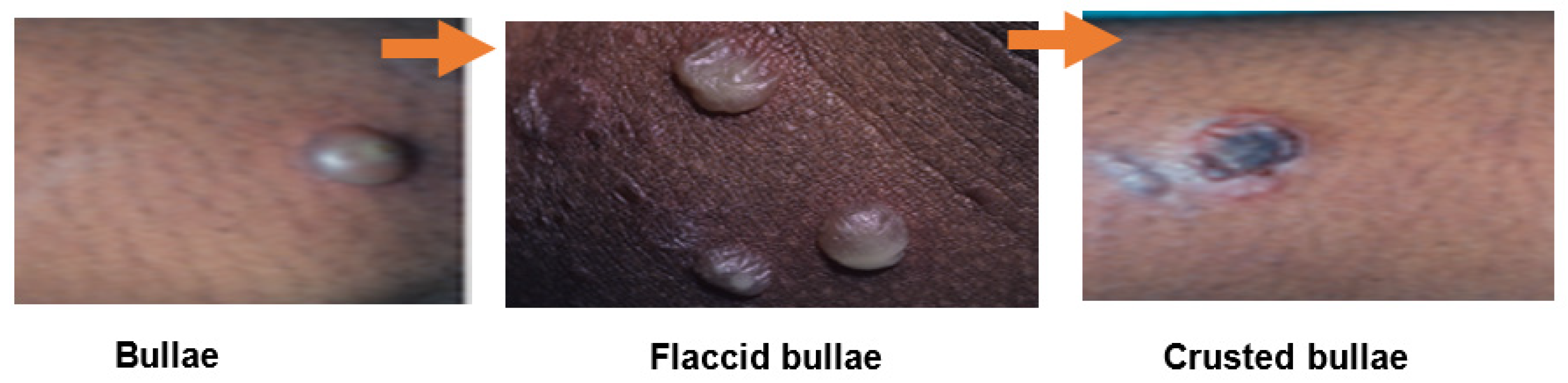
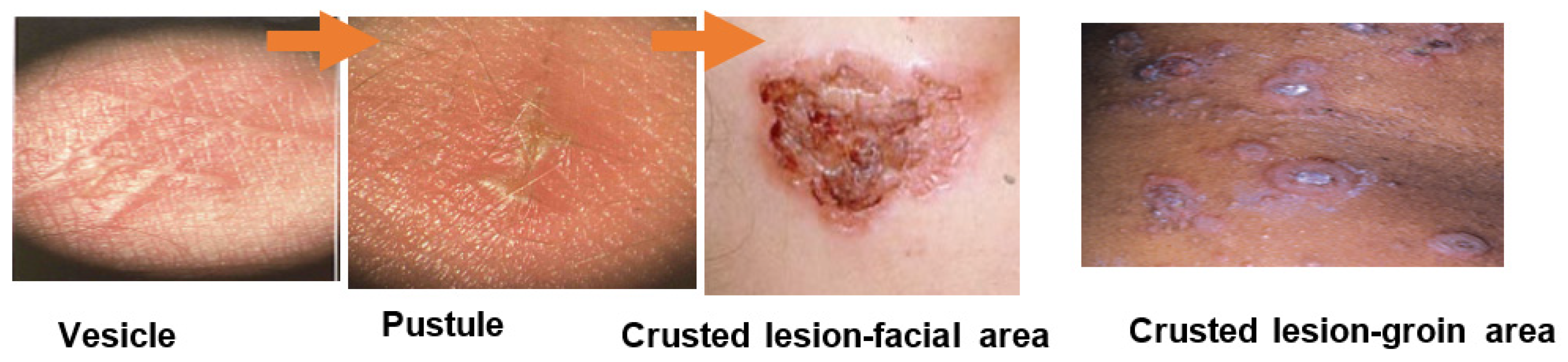
1.3. Clinical Presentation
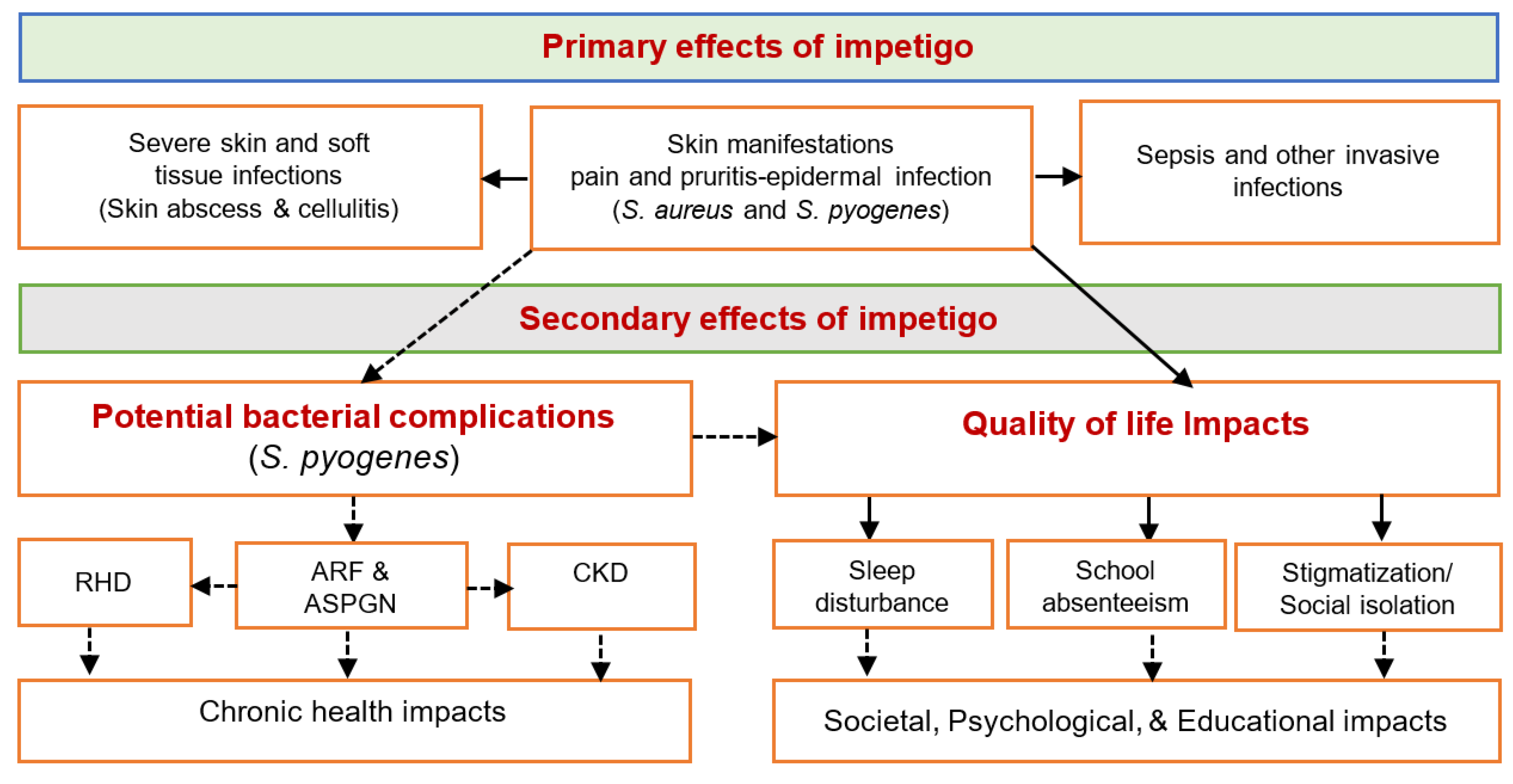
1.4. Morbidity from Impetigo
2. Current Impetigo Treatments and Challenges
3. Potential Antimicrobial Candidates for Impetigo
3.1. Tea Tree Oil
3.2. Manuka Oil
3.3. Hydrogen Peroxide
4. Summary
Author Contributions
Funding
Conflicts of Interest
References
- World health organization. Epidemiology and Management of Common Skin Diseases in Children in Developing Countries; World Health Organization: Geneva, Switzerland, 2005. [Google Scholar]
- Hay, R.J.; Augustin, M.; Griffiths, C.E.M.; Sterry, W. The global challenge for skin health. Br. J. Dermatol. 2015, 172, 1469–1472. [Google Scholar] [CrossRef] [PubMed] [Green Version]
- Kyu, H.H.; Abate, D.; Abate, K.H.; Abay, S.M.; Abbafati, C.; Abbasi, N.; Abbastabar, H.; Abd-Allah, F.; Abdela, J.; Abdelalim, A.; et al. Global, regional, and national disability-adjusted life-years (DALYs) for 359 diseases and injuries and healthy life expectancy (HALE) for 195 countries and territories, 1990–2017: A systematic analysis for the Global Burden of Disease Study 2017. Lancet 2018, 392, 1859–1922. [Google Scholar] [CrossRef] [Green Version]
- Cole, C.; Gazewood, J. Diagnosis and treatment of impetigo. Am. Fam Physician 2007, 75, 859–864. [Google Scholar] [PubMed]
- Bowen, A.C.; Mahe, A.; Hay, R.J.; Andrews, R.M.; Steer, A.C.; Tong, S.Y.; Carapetis, J.R. The global epidemiology of impetigo: A systematic review of the population prevalence of impetigo and pyoderma. PLoS ONE 2015, 10, e0136789. [Google Scholar] [CrossRef] [Green Version]
- Romani, L.; Steer, A.C.; Whitfeld, M.J.; Kaldor, J.M. Prevalence of scabies and impetigo worldwide: A systematic review. Lancet Infect. Dis. 2015, 15, 960–967. [Google Scholar] [CrossRef]
- Hay, R.J.; Johns, N.E.; Williams, H.C.; Bolliger, I.W.; Dellavalle, R.P.; Margolis, D.J.; Marks, R.; Naldi, L.; Weinstock, M.A.; Wulf, S.K.; et al. The global burden of skin disease in 2010: An analysis of the prevalence and impact of skin conditions. J. Investig. Dermatol. 2014, 134, 1527–1534. [Google Scholar] [CrossRef] [Green Version]
- Bowen, A.C.; Carapetis, J.R.; Currie, B.J.; Fowler, V., Jr.; Chambers, H.F.; Tong, S.Y.C. Sulfamethoxazole-trimethoprim (cotrimoxazole) for skin and soft tissue infections including impetigo, cellulitis, and abscess. Open Forum Infect. Dis. 2017, 4, ofx232. [Google Scholar] [CrossRef] [Green Version]
- Romani, L.; Whitfeld, M.J.; Koroivueta, J.; Kama, M.; Wand, H.; Tikoduadua, L.; Tuicakau, M.; Koroi, A.; Ritova, R.; Andrews, R.; et al. The epidemiology of scabies and impetigo in relation to demographic and residential characteristics: Baseline findings from the skin health intervention Fiji trial. Am. J. Trop. Med. Hyg. 2017, 97, 845–850. [Google Scholar] [CrossRef]
- Hartman-Adams, H.; Banvard, C.; Juckett, G. Impetigo: Diagnosis and treatment. Am. Fam. Physician 2014, 90, 229–235. [Google Scholar]
- Pereira, L.B. Impetigo-review. An. Bras. Dermatol. 2014, 89, 293–299. [Google Scholar] [CrossRef]
- Sladden, M.J.; Johnston, G.A. Common skin infections in children. BMJ Clin. Res. Ed. 2004, 329, 95–99. [Google Scholar] [CrossRef] [PubMed] [Green Version]
- Koning, S.; van der Sande, R.; Verhagen, A.P.; van Suijlekom-Smit, L.W.; Morris, A.D.; Butler, C.C.; Berger, M.; van der Wouden, J.C. Interventions for impetigo. Cochrane Database Syst. Rev. 2012, 1, Cd003261. [Google Scholar] [CrossRef] [PubMed] [Green Version]
- Aung, P.T.Z.; Cuningham, W.; Hwang, K.; Andrews, R.M.; Carapetis, J.R.; Kearns, T.; Clucas, D.; McVernon, J.; Simpson, J.A.; Tong, S.Y.C.; et al. Scabies and risk of skin sores in remote Australian Aboriginal communities: A self-controlled case series study. PLoS Negl. Trop. Dis. 2018, 12, e0006668. [Google Scholar] [CrossRef] [PubMed]
- Yeoh, D.K.; Bowen, A.C.; Carapetis, J.R. Impetigo and scabies–disease burden and modern treatment strategies. J. Infect. 2016, 72, S61–S67. [Google Scholar] [CrossRef]
- Yeoh, D.K.; Anderson, A.; Cleland, G.; Bowen, A.C. Are scabies and impetigo “normalised”? A cross-sectional comparative study of hospitalised children in northern Australia assessing clinical recognition and treatment of skin infections. PLoS Negl. Trop. Dis. 2017, 11, e0005726. [Google Scholar] [CrossRef] [Green Version]
- Edge, R.; Argáez, C. Topical Antibiotics for Impetigo: A Review of the Clinical Effectiveness and Guidelines [Internet]. Available online: https://0-www-ncbi-nlm-nih-gov.brum.beds.ac.uk/books/NBK447580 (accessed on 4 July 2020).
- Bowen, A.C.; Tong, S.Y.; Chatfield, M.D.; Carapetis, J.R. The microbiology of impetigo in indigenous children: Associations between Streptococcus pyogenes, Staphylococcus aureus, scabies, and nasal carriage. BMC Infect. Dis. 2014, 14, 727. [Google Scholar] [CrossRef] [Green Version]
- Bowen, A.C.; Tong, S.Y.; Andrews, R.M.; O’Meara, I.M.; McDonald, M.I.; Chatfield, M.D.; Currie, B.J.; Carapetis, J.R. Short-course oral co-trimoxazole versus intramuscular benzathine benzylpenicillin for impetigo in a highly endemic region: An open-label, randomised, controlled, non-inferiority trial. Lancet 2014, 384, 2132–2140. [Google Scholar] [CrossRef] [Green Version]
- Bangert, S.; Levy, M.; Hebert, A.A. Bacterial resistance and impetigo treatment trends: A review. Pediatr. Dermatol. 2012, 29, 243–248. [Google Scholar] [CrossRef]
- Vila, J.; Hebert, A.A.; Torrelo, A.; Lopez, Y.; Tato, M.; Garcia-Castillo, M.; Canton, R. Ozenoxacin: A review of preclinical and clinical efficacy. Expert Rev. Anti Infect. Ther. 2019, 17, 159–168. [Google Scholar] [CrossRef]
- Steer, A.C.; Danchin, M.H.; Carapetis, J.R. Group A streptococcal infections in children. J. Paediatr. Child. Health 2007, 43, 203–213. [Google Scholar] [CrossRef]
- Leyden, J.J.; Stewart, R.; Kligman, A.M. Experimental infections with group A streptococci in humans. J. Investig. Dermatol. 1980, 75, 196–201. [Google Scholar] [CrossRef] [PubMed] [Green Version]
- Dajani, A.S.; Wannamaker, L.W. Experimental infection of the skin in the hamster simulating human impetigo. I. natural history of the infection. J. Infect. Dis. 1970, 122, 196–204. [Google Scholar] [CrossRef] [PubMed]
- Australian Commission on safety and quality in health care (ACSQHC). AURA 2019: Third Australian Report on Antimicrobial Use and Resistance in Human Health; ACSQHC: Sydney, Australia, 2019.
- Engelman, D.; Hofer, A.; Davis, J.S.; Carapetis, J.R.; Baird, R.W.; Giffard, P.M.; Holt, D.C.; Tong, S.Y. Invasive Staphylococcus aureus infections in children in tropical Northern Australia. J. Pediatr. Infect. Dis. Soc. 2014, 3, 304–311. [Google Scholar] [CrossRef] [PubMed] [Green Version]
- The Australian Healthy Skin Consortium. National healthy Skin Guideline for the Prevention, Treatment and public Health Control of Impetigo, Scabies, Crusted Scabies and Tinea for Indigenous Populations and Communities in Australia. Available online: https://infectiousdiseases.telethonkids.org.au/siteassets/media-docs---wesfarmers-centre/national-healthy-skin-guideline---1st-ed.-2018.pdf (accessed on 21 March 2019).
- Boyd, R.; Patel, M.; Currie, B.J.; Holt, D.C.; Harris, T.; Krause, V. High burden of invasive group A streptococcal disease in the Northern Territory of Australia. Epidemiol Infect. 2016, 144, 1018–1027. [Google Scholar] [CrossRef] [Green Version]
- Carapetis, J.R.; Steer, A.C.; Mulholland, E.K.; Weber, M. The global burden of group A streptococcal diseases. Lancet Infect. Dis. 2005, 5, 685–694. [Google Scholar] [CrossRef]
- Davidson, L.; Knight, J.; Bowen, A.C. Skin infections in Australian Aboriginal children: A narrative review. Med. J. Aust. 2020, 212, 231–237. [Google Scholar] [CrossRef] [Green Version]
- Blyth, C.C.; Robertson, P.W.; Rosenberg, A.R. Post-streptococcal glomerulonephritis in Sydney: A 16-year retrospective review. J. Paediatr. Child. Health 2007, 43, 446–450. [Google Scholar] [CrossRef]
- Chaturvedi, S.; Boyd, R.; Krause, V. Acute post-streptococcal glomerulonephritis in the Northern Territory of Australia: A review of data from 2009 to 2016 and comparison with the literature. Am. J. Trop. Med. Hyg. 2018, 99, 1643–1648. [Google Scholar] [CrossRef]
- Bennett, J.; Moreland, N.J.; Oliver, J.; Crane, J.; Williamson, D.A.; Sika-Paotonu, D.; Harwood, M.; Upton, A.; Smith, S.; Carapetis, J.; et al. Understanding group A streptococcal pharyngitis and skin infections as causes of rheumatic fever: Protocol for a prospective disease incidence study. BMC Infect. Dis. 2019, 19, 633. [Google Scholar] [CrossRef] [Green Version]
- Parks, T.; Smeesters, P.R.; Steer, A.C. Streptococcal skin infection and rheumatic heart disease. Curr. Opin. Infect. Dis. 2012, 25, 145–153. [Google Scholar] [CrossRef]
- O’Sullivan, L.; Moreland, N.J.; Webb, R.H.; Upton, A.; Wilson, N.J. Acute rheumatic fever after group A streptococcus pyoderma and group G streptococcus pharyngitis. Pediatr. Infect. Dis. J. 2017, 36, 692–694. [Google Scholar] [CrossRef] [PubMed]
- McDonald, M.I.; Towers, R.J.; Andrews, R.M.; Benger, N.; Currie, B.J.; Carapetis, J.R. Low rates of streptococcal pharyngitis and high rates of pyoderma in Australian Aboriginal communities where acute rheumatic fever is hyperendemic. Clin. Infect. Dis. 2006, 43, 683–689. [Google Scholar] [CrossRef] [PubMed] [Green Version]
- RHDAustralia (ARF/RHD Writing Group) National Heart Foundation of Australia and the Cardiac Society of Australia and New Zealand. Australian Guideline for Prevention, Diagnosis and Management of Acute Rheumatic Fever and Rheumatic Heart Disease. Available online: https://www.rhdaustralia.org.au/arf-rhd-guideline (accessed on 8 July 2020).
- Australian Institute of Health and Welfare. Rheumatic heart Disease and Acute Rheumatic Fever in Australia: 1996–2012; Cat. no. CVD 60 ed.; Australian Institute of Health and Welfare: Canberra, Australia, 2013; p. 60.
- Mitchell, A.G.; Belton, S.; Johnston, V.; Read, C.; Scrine, C.; Ralph, A.P. Aboriginal children and penicillin injections for rheumatic fever: How much of a problem is injection pain? Aust. N. Z. J. Public Health 2018, 42, 46–51. [Google Scholar] [CrossRef] [PubMed] [Green Version]
- D’Cunha, N.M.; Peterson, G.M.; Baby, K.E.; Thomas, J. Impetigo: A need for new therapies in a world of increasing antimicrobial resistance. J. Clin. Pharm. Ther. 2018, 43, 150–153. [Google Scholar] [CrossRef] [Green Version]
- Vogel, A.; Lennon, D.; Best, E.; Leversha, A. Where to from here? The treatment of impetigo in children as resistance to fusidic acid emerges. N. Z. Med. J. 2016, 129, 77–83. [Google Scholar]
- eTG Complete-Therapeutic Guidelines. Impetigo. Available online: https://tgldcdp.tg.org.au/viewTopic?topicfile=impetigo#toc_d1e207 (accessed on 8 July 2020).
- George, A.; Rubin, G. A systematic review and meta-analysis of treatments for impetigo. Br. J. Gen. Pract. 2003, 53, 480–487. [Google Scholar]
- Loadsman, M.E.N.; Verheij, T.J.M.; van der Velden, A.W. Impetigo incidence and treatment: A retrospective study of Dutch routine primary care data. Fam. Pract. 2019, 36, 410–416. [Google Scholar] [CrossRef]
- Stevens, D.L.; Bisno, A.L.; Chambers, H.F.; Everett, E.D.; Dellinger, P.; Goldstein, E.J.C.; Gorbach, S.L.; Hirschmann, J.V.; Kaplan, E.L.; Montoya, J.G.; et al. Practice guidelines for the diagnosis and management of skin and soft-tissue infections. Clin. Infect. Dis. 2005, 41, 1373–1406. [Google Scholar] [CrossRef]
- Pangilinan, R.; Tice, A.; Tillotson, G. Topical antibiotic treatment for uncomplicated skin and skin structure infections: Review of the literature. Expert Rev. Anti Infect. Ther. 2009, 7, 957–965. [Google Scholar] [CrossRef]
- Mertz, P.M.; Marshall, D.A.; Eaglstein, W.H.; Piovanetti, Y.; Montalvo, J. Topical mupirocin treatment of impetigo is equal to oral erythromycin therapy. Arch. Dermatol. 1989, 125, 1069–1073. [Google Scholar] [CrossRef]
- Barton, L.L.; Friedman, A.D.; Sharkey, A.M.; Schneller, D.J.; Swierkosz, E.M. Impetigo contagiosa III. Comparative efficacy of oral erythromycin and topical mupirocin. Pediatr. Dermatol. 1989, 6, 134–138. [Google Scholar] [CrossRef] [PubMed]
- Werner, A.H.; Russell, A.D. Mupirocin, fusidic acid and bacitracin: Activity, action and clinical uses of three topical antibiotics. Vet. Dermatol. 1999, 10, 225–240. [Google Scholar] [CrossRef]
- World Health Organization. Antimicrobial Resistance: Global Report on Surveillance. Available online: https://www.who.int/drugresistance/documents/surveillancereport/en/ (accessed on 5 July 2020).
- Bohaty, B.R.; Choi, S.; Cai, C.; Hebert, A.A. Clinical and bacteriological efficacy of twice daily topical retapamulin ointment 1% in the management of impetigo and other uncomplicated superficial skin infections. Int. J. Womens Dermatol. 2015, 1, 13–20. [Google Scholar] [CrossRef] [PubMed] [Green Version]
- World Health Organization. WHO Publishes List of Bacteria for which New Antibiotics Are Urgently Needed. Available online: https://www.who.int/en/news-room/detail/27-02-2017-who-publishes-list-of-bacteria-for-which-new-antibiotics-are-urgently-needed (accessed on 1 October 2020).
- Rijnders, M.I.; Wolffs, P.F.; Hopstaken, R.M.; den Heyer, M.; Bruggeman, C.A.; Stobberingh, E.E. Spread of the epidemic European fusidic acid-resistant impetigo clone (EEFIC) in general practice patients in the south of The Netherlands. J. Antimicrob Chemother 2012, 67, 1176–1180. [Google Scholar] [CrossRef] [PubMed]
- Van Bijnen, E.M.; Paget, W.J.; den Heijer, C.D.; Stobberingh, E.E.; Bruggeman, C.A.; Schellevis, F.G. Primary care treatment guidelines for skin infections in Europe: Congruence with antimicrobial resistance found in commensal Staphylococcus aureus in the community. BMC Fam. Pract. 2014, 15, 175. [Google Scholar] [CrossRef] [PubMed] [Green Version]
- Dobie, D.; Gray, J. Fusidic acid resistance in Staphylococcus aureus. Arch. Dis. Child. 2004, 89, 74–77. [Google Scholar] [CrossRef] [Green Version]
- Katopodis, G.D.; Grivea, I.N.; Tsantsaridou, A.J.; Pournaras, S.; Petinaki, E.; Syrogiannopoulos, G.A. Fusidic acid and clindamycin resistance in community-associated, methicillin-resistant Staphylococcus aureus infections in children of Central Greece. BMC Infect. Dis. 2010, 10, 351. [Google Scholar] [CrossRef] [Green Version]
- Khoshnood, S.; Heidary, M.; Asadi, A.; Soleimani, S.; Motahar, M.; Savari, M.; Saki, M.; Abdi, M. A review on mechanism of action, resistance, synergism, and clinical implications of mupirocin against Staphylococcus aureus. Biomed. Pharmacother. 2019, 109, 1809–1818. [Google Scholar] [CrossRef]
- Iovino, S.M.; Krantz, K.D.; Blanco, D.M.; Fernández, J.A.; Ocampo, N.; Najafi, A.; Memarzadeh, B.; Celeri, C.; Debabov, D.; Khosrovi, B.; et al. NVC-422 topical gel for the treatment of impetigo. Int. J. Clin. Exp. Pathol. 2011, 4, 587–595. [Google Scholar]
- Mulvey, M.R.; MacDougall, L.; Cholin, B.; Horsman, G.; Fidyk, M.; Woods, S. Community-associated methicillin-resistant Staphylococcus aureus, Canada. Emerg. Infect. Dis. 2005, 11, 844–850. [Google Scholar] [CrossRef]
- Nicholson, A.M.; Thorns, C.; Wint, H.; Didier, M.; Willis, R.; McMorris, N.; Castle, D.; Maharaj, N.; Orrett, F.A. The detection of mupirocin resistance and the distribution of methicillin-resistant Staphylococcus aureus at the university hospital of the West Indies, Jamaica. West. Indian Med. J. 2010, 59, 509–513. [Google Scholar] [PubMed]
- Upton, A.; Lang, S.; Heffernan, H. Mupirocin and Staphylococcus aureus: A recent paradigm of emerging antibiotic resistance. J. Antimicrob. Chemother. 2003, 51, 613–617. [Google Scholar] [CrossRef] [PubMed] [Green Version]
- Antonov, N.K.; Garzon, M.C.; Morel, K.D.; Whittier, S.; Planet, P.J.; Lauren, C.T. High prevalence of mupirocin resistance in Staphylococcus aureus isolates from a pediatric population. Antimicrob. Agents Chemother. 2015, 59, 3350–3356. [Google Scholar] [CrossRef] [PubMed] [Green Version]
- Orrett, F.A. The emergence of mupirocin resistance among clinical isolates of methicillin-resistant Staphylococcus aureus in Trinidad: A first report. Jpn. J. Infect. Dis. 2008, 61, 107–110. [Google Scholar] [PubMed]
- Farrell, D.J.; Robbins, M.; Rhys-Williams, W.; Love, W.G. Investigation of the potential for mutational resistance to XF-73, retapamulin, mupirocin, fusidic acid, daptomycin, and vancomycin in methicillin-resistant Staphylococcus aureus isolates during a 55-passage study. Antimicrob. Agents Chemother. 2011, 55, 1177–1181. [Google Scholar] [CrossRef] [PubMed] [Green Version]
- McNeil, J.C.; Hulten, K.G.; Kaplan, S.L.; Mason, E.O. Decreased susceptibilities to retapamulin, mupirocin, and chlorhexidine among Staphylococcus aureus isolates causing skin and soft tissue infections in otherwise healthy children. Antimicrob. Agents Chemother. 2014, 58, 2878–2883. [Google Scholar] [CrossRef] [Green Version]
- Eliopoulos, G.M.; Huovinen, P. Resistance to trimethoprim-sulfamethoxazole. Clin. Infect. Dis. 2001, 32, 1608–1614. [Google Scholar] [CrossRef] [Green Version]
- Agostino, J.W.; Ferguson, J.K.; Eastwood, K.; Kirk, M.D. The increasing importance of community-acquired methicillin-resistant Staphylococcus aureus infections. Med. J. Aust. 2017, 207, 388–393. [Google Scholar] [CrossRef]
- Britton, P.N.; Andresen, D.N. Paediatric community-associated Staphylococcus aureus: A retrospective cohort study. J. Paediatr. Child. Health 2013, 49, 754–759. [Google Scholar] [CrossRef]
- Oliver, S.J.; Cush, J.; Ward, J.E. Community-based prescribing for impetigo in remote Australia: An opportunity for antimicrobial stewardship. Front. Public Health 2017, 5, 158. [Google Scholar] [CrossRef] [Green Version]
- Thomas, S.; Crooks, K.; Islam, F.; Massey, P.D. Community-associated methicillin-resistant Staphylococcus aureus infections in Aboriginal children attending hospital emergency departments in a regional area of New South Wales, Australia: A seven-year descriptive study. West. Pac. Surveill Response J. 2017, 8, 6–12. [Google Scholar] [CrossRef] [PubMed] [Green Version]
- Tängdén, T.; Pulcini, C.; Aagaard, H.; Balasegaram, M.; Hara, G.L.; Nathwani, D.; Sharland, M.; Theuretzbacher, U.; Cars, O. Unavailability of old antibiotics threatens effective treatment for common bacterial infections. Lancet Infect. Dis. 2018, 18, 242–244. [Google Scholar] [CrossRef]
- Therapeutic Goods Administration. Sulfamethoxazole-Medicine Shortage Information. Available online: https://apps.tga.gov.au/Prod/msi/Search/Details/sulfamethoxazole (accessed on 10 July 2020).
- Bowen, A. Antibiotic Shortages are Putting Aboriginal Kids at Risk. Available online: https://theconversation.com/antibiotic-shortages-are-putting-aboriginal-kids-at-risk-114355 (accessed on 29 April 2020).
- Russell, K.; Nicholson, R.; Naidu, R. Reducing the pain of intramuscular benzathine penicillin injections in the rheumatic fever population of Counties Manukau district health board. J. Paediatr. Child. Health 2014, 50, 112–117. [Google Scholar] [CrossRef] [PubMed]
- Hancock, R.E.W. Mechanisms of action of newer antibiotics for Gram-positive pathogens. Lancet Infect. Dis. 2005, 5, 209–218. [Google Scholar] [CrossRef]
- Doron, S.; Davidson, L.E. Antimicrobial stewardship. Mayo Clin. Proc. 2011, 86, 1113–1123. [Google Scholar] [CrossRef] [Green Version]
- Den Heijer, C.D.J.; Van Bijnen, E.M.E.; Paget, W.J.; Pringle, M.; Goossens, H.; Bruggeman, C.A.; Schellevis, F.G.; Stobberingh, E.E. Prevalence and resistance of commensal Staphylococcus aureus, including meticillin-resistant S aureus, in nine European countries: A cross-sectional study. Lancet Infect. Dis. 2013, 13, 409–415. [Google Scholar] [CrossRef] [Green Version]
- Appiah, T.; Boakye, Y.D.; Agyare, C. Antimicrobial Activities and time-kill kinetics of extracts of selected Ghanaian mushrooms. Evid. Based Complement. Altern. Med. 2017, 2017, 4534350. [Google Scholar] [CrossRef] [Green Version]
- Donsì, F.; Ferrari, G. Essential oil nanoemulsions as antimicrobial agents in food. J. Biotechnol. 2016, 233, 106–120. [Google Scholar] [CrossRef]
- Deyno, S.; Mtewa, A.G.; Abebe, A.; Hymete, A.; Makonnen, E.; Bazira, J.; Alele, P.E. Essential oils as topical anti-infective agents: A systematic review and meta-analysis. Complement. Med. Ther. 2019, 47, 102224. [Google Scholar] [CrossRef]
- Solórzano-Santos, F.; Miranda-Novales, M.G. Essential oils from aromatic herbs as antimicrobial agents. Curr. Opin. Biotechnol. 2012, 23, 136–141. [Google Scholar] [CrossRef]
- Orchard, A.; van Vuuren, S. Commercial essential oils as potential antimicrobials to treat skin diseases. Evid. Based Complement. Altern. Med. 2017, 2017, 4517971. [Google Scholar] [CrossRef] [PubMed] [Green Version]
- Muroi, H.; Kubo, I. Antibacterial activity of anacardic acid and totarol, alone and in combination with methicillin, against methicillin-resistant Staphylococcus aureus. J. Appl. Bacteriol. 1996, 80, 387–394. [Google Scholar] [CrossRef] [PubMed]
- Kubo, I.; Muroi, H.; Himejima, M. Antibacterial activity of totarol and its potentiation. J. Nat. Prod. 1992, 55, 1436–1440. [Google Scholar] [CrossRef] [PubMed]
- Muroi, H.; Kubo, I. Bactericidal effects of anacardic acid and totarol on methicillin-resistant Staphylococcus aureus (MRSA). Biosci. Biotechnol. Biochem. 1994, 58, 1925–1926. [Google Scholar] [CrossRef]
- Carson, C.F.; Hammer, K.A.; Riley, T.V. Melaleuca alternifolia (tea tree) oil: A review of antimicrobial and other medicinal properties. Clin. Microbiol. Rev. 2006, 19, 50–62. [Google Scholar] [CrossRef] [Green Version]
- Thomas, J.; Carson, C.F.; Peterson, G.M.; Walton, S.F.; Hammer, K.A.; Naunton, M.; Davey, R.C.; Spelman, T.; Dettwiller, P.; Kyle, G.; et al. Therapeutic potential of tea tree oil for scabies. Am. J. Trop. Med. Hyg. 2016, 94, 258–266. [Google Scholar] [CrossRef] [Green Version]
- Hammer, K.A.; Carson, C.F.; Riley, T.V.; Nielsen, J.B. A review of the toxicity of Melaleuca alternifolia (tea tree) oil. Food Chem. Toxicol. 2006, 44, 616–625. [Google Scholar] [CrossRef]
- Caelli, M.; Porteous, J.; Carson, C.F.; Heller, R.; Riley, T.V. Tea tree oil as an alternative topical decolonization agent for methicillin-resistant Staphylococcus aureus. J. Hosp. Infect. 2000, 46, 236–237. [Google Scholar] [CrossRef]
- Dryden, M.S.; Dailly, S.; Crouch, M. A randomized, controlled trial of tea tree topical preparations versus a standard topical regimen for the clearance of MRSA colonization. J. Hosp. Infect. 2004, 56, 283–286. [Google Scholar] [CrossRef]
- Enshaieh, S.; Jooya, A.; Siadat, A.; Iraji, F. The efficacy of 5% topical tea tree oil gel in mild to moderate acne vulgaris: A randomized, double-blind placebo-controlled study. Indian J. Dermatol. Venereol. Leprol. 2007, 73, 22–25. [Google Scholar] [CrossRef]
- Satchell, A.C.; Saurajen, A.; Bell, C.; Barnetson, R.S. Treatment of dandruff with 5% tea tree oil shampoo. J. Am. Acad. Dermatol. 2002, 47, 852–855. [Google Scholar] [CrossRef] [PubMed] [Green Version]
- Syed, T.A.; Qureshi, Z.A.; Ali, S.M.; Ahmad, S.; Ahmad, S.A. Treatment of toenail onychomycosis with 2% butenafine and 5% Melaleuca alternifolia (tea tree) oil in cream. Trop. Med. Int. Health 1999, 4, 284–287. [Google Scholar] [CrossRef] [PubMed] [Green Version]
- Tong, M.M.; Altman, P.M.; Barnetson, R.S. Tea tree oil in the treatment of Tinea pedis. Australas. J. Dermatol. 1992, 33, 145–149. [Google Scholar] [CrossRef] [PubMed]
- Lee, R.L.P.; Leung, P.H.M.; Wong, T.K.S. A randomized controlled trial of topical tea tree preparation for MRSA colonized wounds. Int. J. Nurs. Sci. 2014, 1, 7–14. [Google Scholar] [CrossRef] [Green Version]
- Lauten, J.D.; Boyd, L.; Hanson, M.B.; Lillie, D.; Gullion, C.; Madden, T.E. A clinical study: Melaleuca, manuka, calendula and green tea mouth rinse. Phytother. Res. 2005, 19, 951–957. [Google Scholar] [CrossRef]
- Brady, A.; Loughlin, R.; Gilpin, D.; Kearney, P.; Tunney, M. In vitro activity of tea-tree oil against clinical skin isolates of meticillin-resistant and -sensitive Staphylococcus aureus and coagulase-negative staphylococci growing planktonically and as biofilms. J. Med. Microbiol. 2006, 55, 1375–1380. [Google Scholar] [CrossRef] [Green Version]
- Kwieciński, J.; Eick, S.; Wójcik, K. Effects of tea tree (Melaleuca alternifolia) oil on Staphylococcus aureus in biofilms and stationary growth phase. Int. J. Antimicrob. Agents 2009, 33, 343–347. [Google Scholar] [CrossRef]
- Brady, A.J.; Farnan, T.B.; Toner, J.G.; Gilpin, D.F.; Tunney, M.M. Treatment of a cochlear implant biofilm infection: A potential role for alternative antimicrobial agents. J. Laryngol. Otol. 2010, 124, 729–738. [Google Scholar] [CrossRef]
- Park, H.; Jang, C.H.; Cho, Y.B.; Choi, C.H. Antibacterial effect of tea-tree oil on methicillin-resistant Staphylococcus aureus biofilm formation of the tympanostomy tube: An in vitro study. In Vivo 2007, 21, 1027–1030. [Google Scholar]
- Gilbert, P.; Allison, D.G.; McBain, A.J. Biofilms in vitro and in vivo: Do singular mechanisms imply cross-resistance? J. Appl. Microbiol. 2002, 92, 98s–110s. [Google Scholar] [CrossRef]
- Carson, C.F.; Mee, B.J.; Riley, T.V. Mechanism of action of Melaleuca alternifolia (tea tree) oil on Staphylococcus aureus determined by time-kill, lysis, leakage, and salt tolerance assays and electron microscopy. Antimicrob. Agents Chemother. 2002, 46, 1914–1920. [Google Scholar] [CrossRef] [PubMed] [Green Version]
- Hammer, K.A.; Carson, C.F.; Riley, T.V. Frequencies of resistance to Melaleuca alternifolia (tea tree) oil and rifampicin in Staphylococcus aureus, Staphylococcus epidermidis and Enterococcus faecalis. Int. J. Antimicrob. Agents 2008, 32, 170–173. [Google Scholar] [CrossRef] [PubMed]
- Hammer, K.A.; Carson, C.F.; Riley, T.V. Effects of Melaleuca alternifolia (tea tree) essential oil and the major monoterpene component terpinen-4-ol on the development of single- and multistep antibiotic resistance and antimicrobial susceptibility. Antimicrob. Agents Chemother. 2012, 56, 909–915. [Google Scholar] [CrossRef] [Green Version]
- Martin, K.W. Herbal medicines for treatment of bacterial infections: A review of controlled clinical trials. J. Antimicrob. Chemother. 2003, 51, 241–246. [Google Scholar] [CrossRef] [PubMed] [Green Version]
- Stephens, J.M.C.; Molan, P.C.; Clarkson, B.D. A review of Leptospermum scoparium (Myrtaceae) in New Zealand. N. Z. J. Bot. 2005, 43, 431–449. [Google Scholar] [CrossRef]
- Maddocks-Jennings, W.; Wilkinson, J.; Shillington, D.P.; Cavanagh, H. A fresh look at manuka and kanuka essential oils from New Zealand. Int. J. Aromather. 2005, 15, 141–146. [Google Scholar] [CrossRef]
- Mathew, C.; Tesfaye, W.; Rasmussen, P.; Peterson, G.M.; Bartholomaeus, A.; Sharma, M.; Thomas, J. Mānuka Oil—A Review of Antimicrobial and Other Medicinal Properties. Pharmaceuticals 2020, 13, 343. [Google Scholar] [CrossRef]
- Porter, N.G.; Wilkins, A.L. Chemical, physical and antimicrobial properties of essential oils of Leptospermum scoparium and Kunzea ericoides. Phytochemistry 1999, 50, 407–415. [Google Scholar] [CrossRef]
- Perry, N.B.; Van Klink, J.W.; Brennan, N.J.; Harris, W.; Anderson, R.E.; Douglas, M.H.; Smallfield, B.M. Essential oils from New Zealand manuka and kanuka: Chemotaxonomy of kunzea. Phytochemistry 1997, 45, 1605–1612. [Google Scholar] [CrossRef]
- Christoph, F.; Kubeczka, K.H.; Stahl-Biskup, E. The composition of commercial manuka oils from New Zealand. J. Essent. Oil Res. 1999, 11, 705–710. [Google Scholar] [CrossRef]
- Douglas, M.H.; van Klink, J.W.; Smallfield, B.M.; Perry, N.B.; Anderson, R.E.; Johnstone, P.; Weavers, R.T. Essential oils from New Zealand manuka: Triketone and other chemotypes of Leptospermum scoparium. Phytochemistry 2004, 65, 1255–1264. [Google Scholar] [CrossRef] [PubMed]
- Van Klink, J.W.; Larsen, L.; Perry, N.B.; Weavers, R.T.; Cook, G.M.; Bremer, P.J.; MacKenzie, A.D.; Kirikae, T. Triketones active against antibiotic-resistant bacteria: Synthesis, structure-activity relationships, and mode of action. Bioorg. Med. Chem. 2005, 13, 6651–6662. [Google Scholar] [CrossRef] [PubMed]
- Christoph, F.; Kaulfers, P.M.; Stahl-Biskup, E. A comparative study of the in vitro antimicrobial activity of tea tree oils s.l. with special reference to the activity of beta-triketones. Planta Med. 2000, 66, 556–560. [Google Scholar] [CrossRef] [PubMed]
- Costa, R.; Pizzimenti, F.; Marotta, F.; Dugo, P.; Santi, L.; Mondello, L. Volatiles from steam-distilled leaves of some plant species from Madagascar and New Zealand and evaluation of their biological activity. Nat. Prod. Commun. 2010, 5, 1803–1808. [Google Scholar] [CrossRef] [Green Version]
- Fratini, F.; Mancini, S.; Turchi, B.; Friscia, E.; Pistelli, L.; Giusti, G.; Cerri, D. A novel interpretation of the fractional inhibitory concentration index: The case Origanum vulgare L. and Leptospermum scoparium J. R. et G. forst essential oils against Staphylococcus aureus strains. Microbiol. Res. 2017, 195, 11–17. [Google Scholar] [CrossRef]
- Harkenthal, M.; Reichling, J.; Geiss, H.K.; Saller, R. Comparative study on the in vitro antibacterial activity of Australian tea tree oil, cajuput oil, niaouli oil, manuka oil, kanuka oil, and eucalyptus oil. Die Pharm. 1999, 54, 460–463. [Google Scholar]
- Jeong, E.-Y.; Lee, M.-J.; Lee, H.-S. Antimicrobial activities of leptospermone isolated from Leptospermum scoparium seeds and structure-activity relationships of its derivatives against foodborne bacteria. Food Sci. Biotechnol. 2018, 27, 1541–1547. [Google Scholar] [CrossRef]
- Piotr, S.; Magdalena, Z.; Joanna, P.; Barbara, K.; Slawomir, M. Essential oils as potential anti-staphylococcal agents. J. Acta Vet. 2018, 68, 107–195. [Google Scholar] [CrossRef] [Green Version]
- Van Vuuren, S.F.; Docrat, Y.; Kamatou, G.P.P.; Viljoen, A.M. Essential oil composition and antimicrobial interactions of understudied tea tree species. S. Afr. J. Bot. 2014, 92, 7–14. [Google Scholar] [CrossRef] [Green Version]
- Patterson, J.E.; McElmeel, L.; Wiederhold, N.P. In Vitro activity of essential oils against gram-positive and Gram-negative clinical isolates, including carbapenem-resistant enterobacteriaceae. Open Forum Infect. Dis. 2019, 6, 1–4. [Google Scholar] [CrossRef]
- Alnaimat, M.S.; Wainwright, M.; Jaber, S.; Amasha, R. Mechanism of the antibacterial action of (Leptospermum scoparium) oil on methicillin-resistant Staphylococcus aureus (MRSA) and E. coli 2015. In Proceedings of the 2nd Mediterranean Symposium on Medicinal and Aromatic Plants (MESMAP-2), Antalya, Turkey, 22–25 April 2015. [Google Scholar]
- Lis-Balchin, M.; Hart, S.L.; Deans, S.G. Pharmacological and antimicrobial studies on different tea-tree oils (Melaleuca alternifolia, Leptospermum scoparium or Manuka and Kunzea ericoides or Kanuka), originating in Australia and New Zealand. Phytother. Res. 2000, 14, 623–629. [Google Scholar] [CrossRef]
- Chen, C.C.; Yan, S.H.; Yen, M.Y.; Wu, P.F.; Liao, W.T.; Huang, T.S.; Wen, Z.H.; David Wang, H.M. Investigations of kanuka and manuka essential oils for in vitro treatment of disease and cellular inflammation caused by infectious microorganisms. J. Microbiol. Immunol. Infect. 2016, 49, 104–111. [Google Scholar] [CrossRef] [PubMed] [Green Version]
- Williamson, D.A.; Carter, G.P.; Howden, B.P. Current and emerging topical antibacterials and antiseptics: Agents, action, and resistance patterns. Clin. Microbiol. Rev. 2017, 30, 827–860. [Google Scholar] [CrossRef] [PubMed] [Green Version]
- National Institute for Health and Care Excellence (NICE) Guideline 153. Impetigo: Antimicrobial Prescribing Guideline: Evidence Review; National Institute for Health and Care Excellence, Ed.; National Institute for Health and Care Excellence: London, UK, 2020. [Google Scholar]
- Rutala, W.A.; Weber, D.J. Guideline for Disinfection and Sterilization in Healthcare Facilities, 2nd ed.; Centers for Disease Control (CDC), Ed.; Centers for Disease Control (CDC): Atlanta, USA, 2008.
- Christensen, O.B.; Anehus, S. Hydrogen peroxide cream: An alternative to topical antibiotics in the treatment of impetigo contagiosa. Acta Derm. Venereol. 1994, 74, 460–462. [Google Scholar] [CrossRef]
- National Institute for Health and Care Excellence (NICE). Impetigo: Antimicrobial Prescribing. Available online: https://www.nice.org.uk/guidance/ng153/chapter/Recommendations (accessed on 21 July 2020).
- Leversha, A. Randomised Controlled Trial Assessing the Efficacy of Topical Fusidic Acid and Topical Hydrogen Peroxide Cream for Mild Impetigo in School Children. Available online: https://www.anzctr.org.au/Trial/Registration/TrialReview.aspx?id=370307 (accessed on 21 June 2020).
- Finnegan, M.; Linley, E.; Denyer, S.P.; McDonnell, G.; Simons, C.; Maillard, J.-Y. Mode of action of hydrogen peroxide and other oxidizing agents: Differences between liquid and gas forms. J. Antimicrob. Chemother. 2010, 65, 2108–2115. [Google Scholar] [CrossRef] [Green Version]


Publisher’s Note: MDPI stays neutral with regard to jurisdictional claims in published maps and institutional affiliations. |
© 2020 by the authors. Licensee MDPI, Basel, Switzerland. This article is an open access article distributed under the terms and conditions of the Creative Commons Attribution (CC BY) license (http://creativecommons.org/licenses/by/4.0/).
Share and Cite
Abrha, S.; Tesfaye, W.; Thomas, J. Intolerable Burden of Impetigo in Endemic Settings: A Review of the Current State of Play and Future Directions for Alternative Treatments. Antibiotics 2020, 9, 909. https://0-doi-org.brum.beds.ac.uk/10.3390/antibiotics9120909
Abrha S, Tesfaye W, Thomas J. Intolerable Burden of Impetigo in Endemic Settings: A Review of the Current State of Play and Future Directions for Alternative Treatments. Antibiotics. 2020; 9(12):909. https://0-doi-org.brum.beds.ac.uk/10.3390/antibiotics9120909
Chicago/Turabian StyleAbrha, Solomon, Wubshet Tesfaye, and Jackson Thomas. 2020. "Intolerable Burden of Impetigo in Endemic Settings: A Review of the Current State of Play and Future Directions for Alternative Treatments" Antibiotics 9, no. 12: 909. https://0-doi-org.brum.beds.ac.uk/10.3390/antibiotics9120909