Article

Effects of tDCS on Muscle Stiffness in Children with Cerebral Palsy Measured by Myotonometry: A Preliminary Study

1
Diagnostic and Rehabilitation Center ‘Promyk Słońca’, 50-088 Wrocław, Poland
2
College of Physical Education, Yangzhou University, Yangzhou 225000, China
3
Department of Anatomy, Medical University of Warsaw, ul. Chałubińskiego 5, 02-004 Warszawa, Poland
4
Department of Biostructure, University School of Physical Education in Wrocław, 51-612 Wrocław, Poland
*
Author to whom correspondence should be addressed.
Submission received: 15 March 2020 / Revised: 5 April 2020 / Accepted: 8 April 2020 / Published: 10 April 2020

Abstract

:

Featured Application

The transcranial direct current stimulation has a decreasing effect on spasticity of the flexor carpi radialis in children with cerebral palsy and should be implemented in the rehabilitation process.

Abstract

Background: The aim of this study was to investigate the influence of transcranial direct current stimulation (tDCS) on the biceps brachii and flexor carpi radialis stiffness in children with cerebral palsy (CP). The authors also aimed to verify the relationship between spasticity and muscle stiffness. Methods: Twelve children with CP (mean ± SD; age, 8 ± 1.3 years; height, 118.7 ± 4.1 cm; weight, 23.0 ± 2.2 kg) were involved in the study. Muscle stiffness was estimated using a MyotonPRO device in a MultiScan pattern of five measurements. Simultaneously, the tDCS stimulation was performed. Spasticity was assessed by a neurologist using the Ashworth Scale. Results: Stiffness of the flexor carpi radialis muscle decreased significantly after tDCS therapy (p = 0.04). There was no significant change in stiffness of the biceps brachii. For all participants, the Spearman rank correlation showed statistically significant and positive relationships between muscle stiffness and the Ashworth Scale (p = 0.04). Conclusions: Transcranial direct current stimulation has a decreasing effect on stiffness and spasticity of the flexor carpi radialis in children with CP. The MyotonPRO device provides objective data and correlates with spasticity measurements.

1. Introduction

Cerebral palsy (CP) refers to permanent, mutable, motor developmental disorders stemming from a primary brain lesion. It causes secondary musculoskeletal dysfunction and limitations in the activities of daily living [1]. Following the decreased activation of the central nervous system, it leads to abnormalities in the performance of movements [2]. Transcranial direct current stimulation (tDCS) is one of the simplest ways of achieving noninvasive brain stimulation [3].
Although usage of similar technology dates back to the late 1880s, soon after electricity was “rediscovered”, a reappraisal of tDCS took place at the turn of this century [4]. Doctor Walter Paulus and his group in Gottingen, Germany, led a recent resurrection of this technology, and there is now active investigation of tDCS, with over 100 articles written in the past 10 years in peer-reviewed journals [5].
A systematic review of interventions for children with CP showed several methods of managing spasticity, i.e., selective dorsal rhizotomy and administration of diazepam or botulinum toxin. However, tDCS was not mentioned in that review [6]. As a low-cost, easy-to-administer, and well-tolerated technique, tDCS in combination with conventional physical rehabilitation may potentiate neuroplasticity [7]. Therefore, it seems reasonable to investigate tDCS as an optional (additional) method to manage spasticity in CP children.
The main contributors to increased joint resistance, which is a typical symptom in CP, are increased stiffness of muscle and hyperactivity of the stretch reflex (clinically labeled as “spasticity”) [8]. It is generally assumed that increased resistance, explained by an acceleration in involuntary muscle activation during passive movements, is related to impaired voluntary motor function [9]. As stated by Gracies (2005), improvements in the upper limb and hand function can be achieved by spasticity reduction. Precise evaluation of spasticity is crucial to establish the efficacy of medical and physical therapeutic interventions. The Ashworth Scale grades the manual sensation of mechanical resistance experienced by the examiner during a 1-s joint rotation over the full range of motion [10]. Rating depends on examiner experience and testing technique, which may have a significant effect on subjectivity and may not provide a valid measure of spasticity [11].
To support our study with more objective data, we obtained muscle stiffness measurements using a MyotonPRO (Myoton Muscle Diagnostics, Tallinn, Estonia) device for clinical purposes. Myotonometry is becoming increasingly popular in assessing the viscoelastic properties of muscles, both before and after applying physiotherapy treatment [12]. It is non-invasive, portable, and easy to administer [13].
To the best of our knowledge, no study addressed the effect of tDCS on muscle stiffness, assessed by myotonometry, in CP children. Our study aimed to investigate the influence of tDCS on the biceps brachii and flexor carpi radialis stiffness in children with CP and the relationship between spasticity and muscle stiffness.

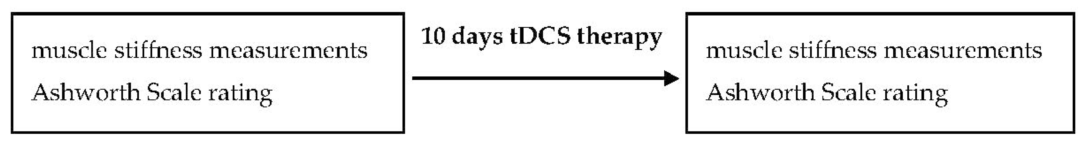
2. Materials and Methods

2.1. Participants

Twelve children diagnosed with CP (mean ± SD; age, 8 ± 1.3 years; height, 118.7 ± 4.1 cm; weight, 23.0 ± 2.2 kg) participated in the study. The exclusion criteria were (1) previous history of any surgeries, (2) orthopedically deformities, (3) epilepsy, (4) metal implants in the skull, and (5) resignation of the legal guardian from further therapy. Thus, six children underwent tDCS therapy and the other six became the control group which had no tDCS therapy. Children from the control group maintained their everyday individual therapy during the course of the study.
Informed consent was obtained from all parents prior to the measurements. The study was approved by the ethics committee of University School of Physical Education in Wrocław (Poland) and conducted according to the Helsinki Declaration.

2.2. Measures

A handheld MyotonPRO device was applied to measure muscle stiffness. The probe of MyotonPRO, placed perpendicular to the skin surface overlying tested muscle, produces a short mechanical impulse. Natural damped oscillations due to the response of soft tissue are then processed by the device. It calculates three basic parameters, including muscle stiffness. It is the biomechanical property of a muscle that resists either contraction or an external force, tending to deform its initial shape [14,15].

2.3. Procedures

Arm and forearm muscles (biceps brachii caput longum and flexor carpi radialis) were tested in the middle point of the muscle belly. For this study, a MultiScan pattern of five measurements was taken, of which the mean value was used. Spasticity was assessed by a neurologist using the Ashworth Scale. During MyotonPRO measurements, participants supervised by an investigator (therapist) were seated on a chair with their backs supported in an upright position. They were instructed to relax their muscles.
The tDCS treatment was applied to the group of six children daily for the period of 10 days (Figure 1). A direct sinusoidal current of 1 mA and 250 Hz was applied over the primary motor cortex for 15 min. Muscle stiffness in all participants was measured on the first (before tDCS) and 10th (after tDCS) days of therapy. The specific electrode position and the strength of current for tDCS therapy were controlled [16]. The anode was placed at the parietal region, and the cathode was placed on the mastoid process. The second electrode pair was used to stimulate the spinal cord; the anode was placed on the cervical vertebrae 3–4 and the cathode was placed on cervical vertebrae 6–7. A direct sinusoidal current of 1 mA and 250 Hz was applied over the primary motor cortex for 15 min.

2.4. Data Analysis

Statistical analysis was performed using statistical software SPSS 18 (SPSS Inc). The interclass correlation coefficient (ICC) was used to determine the intra-rater reliability of the stiffness. Intra-rater reliability was evaluated by ICC (3,1) using a two-way mixed model, consistency type. ICC values can be considered excellent (>0.89), good (0.70–0.89), moderate (0.50–0.69), and poor (<0.49) [14,17]. Measurements (first and 10th days of therapy) were introduced as within-subject factors in a full-factorial, repeated-measures analysis of variance for the dependent variables of muscle stiffness and the Ashworth Scale score. Bonferroni adjustment for multiple comparisons was used as a post hoc test. In all tests, p < 0.05 was considered significant, and partial eta squared (ƞ2) was calculated to assess effect sizes. The data are presented as means ± SD in the text.

3. Results

The relative reliability of myotonometry over therapy was found to be high (ICC = 0.842). Muscle stiffness for flexor carpi radialis decreased significantly from before therapy (295.4 ± 49.8 N/m) to the 10th day of therapy (246 ± 29.7 N/m) (p = 0.04; ƞ2 = 0.47). For the biceps brachii, there was no significant difference between muscle stiffness before therapy (191.4 ± 35.9 N/m) and the 10th day of therapy (210 ± 27.6 N/m) (p = 0.4; ƞ2 = 0.39) (Table 1).
In the control group, muscle stiffness of both investigated muscles did not change significantly throughout the 10 days of the experiment (Table 2).
The Ashworth Scale score for the wrist joint (functionally related to the flexor carpi radialis) decreased from before therapy (3.7 ± 0.5) to the 10th day of therapy (2.3 ± 1.2) (p = 0.02).
In the control group, the Ashworth Scale score for the wrist joint did not change significantly throughout 10 days of the experiment.
For all participants, the Spearman rank correlation showed statistically significant and positive relationships between muscle stiffness and the Ashworth Scale (p = 0.04).

4. Discussion

This preliminary study, even if conducted in a very low number of subjects, provides novel information regarding a possible effect of tDCS on decreasing muscle stiffness in children with CP.
The results of clinical studies demonstrate the potential of tDCS in treating neurological disorders and investigating modulation of cerebral cortex excitability [18,19]. Grecco et al. [20] reported that even a single session of this type of treatment can significantly improve static balance and gait velocity in children with CP. Lazzari et al. [21] conducted a similar research. As they stated, tDCS can prolong and enhance positive effects achieved in physical therapy. Blesneag et al. [22] studied effects of tDCS combined with other physiotherapy modalities for adults with neurological disorders, such as stroke. By changing the membrane potential, a weak electrical current used in tDCS causes an increase in local synaptic efficiency. This can open a pathway to modulate cortex activity and preserve effects of rehabilitation [23]. The presented findings are in agreement with our research, providing evidence on the considerable effectiveness of this treatment.
Although the precise mechanism that underlies tDCS is not yet completely understood, its effects on cortical activity are consistent and reliable [19]. A study conducted by Fecteau et al. [24] showed that tDCS could induce specific changes in motor activity, as a function of targeted brain areas. The primary mechanism of tDCS stimulation of the cerebral cortex is a subthreshold modulation of neuronal resting membrane potential [25]. It is worth emphasizing that tDCS has the unique ability to induce antagonistic effects in cortical excitability according to the parameters of stimulation. Therefore, cathodal stimulation decreases cortical excitability, leading to hypo-polarization of axonal membrane potentials [26], whereas anodal stimulation increases it and leads to neuronal hyper-polarization [27]. Furthermore, stimulation duration over 7 min causes synaptic strengthening under the anode and synaptic weakening under the cathode [19]. We took these variables into consideration when designing our research.
It is generally accepted that the possible cause of spasticity in CP patients lies within a decrease of cortical input in the corticospinal tract. In that case, the effects of anodal tDCS on increasing cortical activity might be an explanation for motor improvements in our study. Those were assessed as a decrease in muscle stiffness and Ashworth Scale score. The presented hypothesis is also supported by studies done by Auvichayapat and colleagues. After evaluating pre- and post-anodal tDCS, they demonstrated a significant decrease in spasticity for the whole upper limb [28].
Chen and Schlaug [29] used tDCS to treat patients with stroke, showing promising results, i.e., a significant reduction of motor impairment in all individuals, and an increased resting state connectivity between ipsilesional motor cortex with contralesional premotor cortex and bilateral precuneus. This gives fresh insight into the treatment and possibilities for individuals with chronic neurological impairments where the time for spontaneous brain recovery is complete. Our research did not focus on spasticity itself, but on general motor function improvements. However, it can be a valuable source of knowledge, as spasticity limits the ability to perform everyday activities.
Our study showed a decrease in muscle stiffness, which is in line with a general improvement in motor function. Moreover, Hesse et al. [30] used spinal cord stimulation at the C3–C4 level combined with tDCS, showing an improvement in arm function in patients with paresis after stroke. Gupta et al. [31] made the assumption that, by modifying the descending corticospinal influences and increasing spinal cord excitability, inhibition to the spinal motor neurons could be restored, thereby reducing clinical spasticity. Muscle stiffness is reported to increase with elevated muscle activation [8]. Therefore, its objective evaluation allows accessing the specific mechanism underlying pathology in neurological disorders [32]. We found high reliability of myotonometry over therapy. Our results are consistent with those of other researchers investigating this matter [33].
For our research, we selected arm and forearm muscles (biceps brachii caput longum and flexor carpi radialis), because of their functional importance in daily living. Brændvik et al. [34] demonstrated that spasticity, measured as involuntary biceps brachii activation during passive elbow extension, contributes to limitations in upper limb activity in children with CP. Bruin et al. [35] also confirmed a connection between increased biceps brachii involuntary activation and the decrease of upper limb activity. Based on their conclusion, the relationship between the ability to voluntarily activate a muscle and activity performance explains how spasticity can contribute to the limitations of daily living activities.
The normal development of postural tone in ontogenesis indicates a dependence of changes in the proximal muscle tone (in the trunk and head) on the distal muscle tension (upper and lower limbs). The passive tone increases during pregnancy in a caudo-cephalic direction. Therefore, infants are born with maximum physiological hypertonia. Subsequently, in the first year period, a caudo-cephalic decrease of passive muscle tone is observed. On the contrary, active tone progresses in a cephalo-caudal direction, starting with the assurance of a head position at three months of age [36].
In cerebral palsy, postural tension is incorrect, and there is a misbalanced distribution of proximal muscle tone with respect to the distal [37]. The area that needs further investigation is why there was a better result for distal muscle (flexor carpi radialis) than for proximal (biceps brachii). Analyzing the general disturbances in the distribution of muscle tension in various parts of the body might bring some explanation. This indicates the need for continuing research within this field using a larger study group.
There are some limitations to the present study. Firstly, the recruited group in this study was too small. The recruitment process for this study was very long, according to the exclusion criteria. However, children with CP are a small selected group inducted by tDCS. Thus, this research assumes the character of a preliminary study. Secondly, the therapy period could include a longer period (than 10 days of intervention). In a previous study, only the effect of a single session was investigated [21].

5. Conclusions

Our study using tDCS therapy and the myotonometry measuring technique provides important information about the influence of tDCS on muscle stiffness reduction in children with CP. A decrease in muscle stiffness has implications for design of the rehabilitation process. MyotonPRO appears to be a reliable assessment tool. Moreover, it provides objective data and correlates with spasticity measurements. Therefore, stiffness might be considered a new parameter in the supervision of clinical changes in cerebral palsy. Additional research on a larger group of CP patients and on other body regions is clearly needed. To support our hypothesis and to increase our experimental results, pediatric professionals could consider incorporating tDCS therapy, with due attention and control, in rehabilitation programs for decreasing spasticity.

Author Contributions

Conceptualization, M.S. and Z.I.; methodology, M.S. and B.J.-G.; formal analysis, M.S., B.C., and Z.I.; investigation, M.S. and B.J.-G.; writing—original draft preparation, M.S. and Z.I.; writing—review and editing, M.S., A.C., and Z.I.; project administration, M.S. and Z.I. All authors read and agreed to the published version of the manuscript.

Funding

This research received no external funding.

Acknowledgments

We are grateful to the Diagnostic and Rehabilitation Center “Promyk Słońca” for the opportunity to perform the presented study. We are grateful for cooperation with the Chinese–Polish Laboratory on Sport and Brain Science.

Conflicts of Interest

The authors declare no conflicts of interest

References

  1. Rosenbaum, P.; Paneth, N.; Leviton, A.; Goldstein, M.; Bax, M.; Damiano, D.; Dan, B.; Jacobsson, B. A report: The definition and classification of cerebral palsy April 2006. Dev. Med. Child Neurol. Suppl. 2007, 109, 8–14. [Google Scholar] [PubMed]
  2. Abbruzzese, G.; Berardelli, A. Sensorimotor integration in movement disorders. Mov. Disord. 2003, 18, 231–240. [Google Scholar] [CrossRef] [PubMed]
  3. Knechtel, L.; Schall, U.; Cooper, G.; Ramadan, S.; Stanwell, P.; Jolly, T.; Thienel, R. Transcranial direct current stimulation of prefrontal cortex: An auditory event-related potential and proton magnetic resonance spectroscopy study. Neurol. Psychiatry Brain Res. 2014, 20, 96–101. [Google Scholar] [CrossRef]
  4. Brunoni, A.R.; Nitsche, M.A.; Bolognini, N.; Bikson, M.; Wagner, T.; Merabet, L.; Edwards, D.J.; Valero-Cabre, A.; Rotenberg, A.; Pascual-Leone, A. Clinical research with transcranial direct current stimulation (tDCS): Challenges and future directions. Brain Stimul. 2012, 5, 175–195. [Google Scholar] [CrossRef] [Green Version]
  5. Paulus, W. Transcranial direct current stimulation (tDCS). In Supplements to Clinical Neurophysiology; Elsevier: Amsterdam, The Netherland, 2003; Volume 56, pp. 249–254. [Google Scholar]
  6. Novak, I.; Mcintyre, S.; Morgan, C.; Campbell, L.; Dark, L.; Morton, N.; Stumbles, E.; Wilson, S.A.; Goldsmith, S. A systematic review of interventions for children with cerebral palsy: State of the evidence. Dev. Med. Child Neurol. 2013, 55, 885–910. [Google Scholar] [CrossRef]
  7. Stagg, C.J.; Best, J.G.; Stephenson, M.C.; O’Shea, J.; Wylezinska, M.; Kincses, Z.T.; Morris, P.G.; Matthews, P.M.; Johansen-Berg, H. Polarity-sensitive modulation of cortical neurotransmitters by transcranial stimulation. J. Neurosci. 2009, 29, 5202–5206. [Google Scholar] [CrossRef]
  8. Barber, L.; Barrett, R.; Lichtwark, G. Passive muscle mechanical properties of the medial gastrocnemius in young adults with spastic cerebral palsy. J. Biomech. 2011, 44, 2496–2500. [Google Scholar] [CrossRef]
  9. Sanger, T.D.; Chen, D.; Delgado, M.R.; Gaebler-Spira, D.; Hallett, M.; Mink, J.W. Definition and classification of negative motor signs in childhood. Pediatrics 2006, 118, 2159–2167. [Google Scholar] [CrossRef] [Green Version]
  10. De Vlugt, E.; de Groot, J.H.; Schenkeveld, K.E.; Arendzen, J.; van Der Helm, F.C.; Meskers, C.G. The relation between neuromechanical parameters and Ashworth score in stroke patients. J. Neuroeng. Rehabil. 2010, 7, 35. [Google Scholar] [CrossRef] [Green Version]
  11. Pandyan, A.D.; Price, C.I.; Barnes, M.P.; Johnson, G.R. A biomechanical investigation into the validity of the modified Ashworth Scale as a measure of elbow spasticity. Clin. Rehabil. 2003, 17, 290–294. [Google Scholar] [CrossRef]
  12. Chuang, L.-L.; Wu, C.-Y.; Lin, K.-C. Reliability, validity, and responsiveness of myotonometric measurement of muscle tone, elasticity, and stiffness in patients with stroke. Arch. Phys. Med. Rehabil. 2012, 93, 532–540. [Google Scholar] [CrossRef] [PubMed]
  13. Ditroilo, M.; Hunter, A.M.; Haslam, S.; De Vito, G. The effectiveness of two novel techniques in establishing the mechanical and contractile responses of biceps femoris. Physiol. Meas. 2011, 32, 1315. [Google Scholar] [CrossRef] [PubMed] [Green Version]
  14. Kawczynski, A.; Mroczek, D.; Andersen, R.E.; Stefaniak, T.; Arendt-Nielsen, L.; Madeleine, P. Trapezius viscoelastic properties are heterogeneously affected by eccentric exercise. J. Sci. Med. Sport 2018, 21, 864–869. [Google Scholar] [CrossRef] [PubMed]
  15. Pozarowszczyk, B.; Pawlaczyk, W.; Smoter, M.; Zarzycki, A.; Mroczek, D.; Kumorek, M.; Witkowski, K.; Kawczyński, A. Effects of Karate Fights on Achilles Tendon Stiffness Measured by Myotonometry. J. Hum. Kinet. 2017, 56, 93–97. [Google Scholar] [CrossRef]
  16. Fregni, F.; Boggio, P.; Nitsche, M.; Pascual-Leone, A. Transcranial direct current stimulation. Br. J. Psychiatry 2005, 186, 446–447. [Google Scholar] [CrossRef] [Green Version]
  17. Zarzycki, A.; Stawarz, M.; Maillette, J.; Lovecchio, N.; Zago, M.; Kawczynski, A.; Klich, S. Acute changes of Achilles tendon thickness investigated by ultrasonography after shotokan and kyokushin karate training. Arch. Budo 2018, 14, 213–218. [Google Scholar]
  18. Ho, K.-A.; Taylor, J.L.; Chew, T.; Gálvez, V.; Alonzo, A.; Bai, S.; Dokos, S.; Loo, C.K. The effect of transcranial direct current stimulation (tDCS) electrode size and current intensity on motor cortical excitability: Evidence from single and repeated sessions. Brain Stimul. 2016, 9, 1–7. [Google Scholar] [CrossRef]
  19. Nitsche, M.A.; Paulus, W. Sustained excitability elevations induced by transcranial DC motor cortex stimulation in humans. Neurology 2001, 57, 1899–1901. [Google Scholar] [CrossRef]
  20. Grecco, L.A.C.; Duarte, N.d.A.C.; Mendonça, M.E.; Cimolin, V.; Galli, M.; Fregni, F.; Oliveira, C.S. Transcranial direct current stimulation during treadmill training in children with cerebral palsy: A randomized controlled double-blind clinical trial. Res. Dev. Disabil. 2014, 35, 2840–2848. [Google Scholar] [CrossRef]
  21. Lazzari, R.D.; Politti, F.; Santos, C.A.; Dumont, A.J.L.; Rezende, F.L.; Grecco, L.A.C.; Ferreira, L.A.B.; Oliveira, C.S. Effect of a single session of transcranial direct-current stimulation combined with virtual reality training on the balance of children with cerebral palsy: A randomized, controlled, double-blind trial. J. Phys. Ther. Sci. 2015, 27, 763–768. [Google Scholar] [CrossRef] [Green Version]
  22. Blesneag, A.; Popa, L.; Stan, A. Non-invasive brain stimulation in early rehabilitation after stroke. J. Med. Life 2015, 8, 52. [Google Scholar] [PubMed]
  23. Moreno-Duarte, I.; Gebodh, N.; Schestatsky, P.; Guleyupoglu, B.; Reato, D.; Bikson, M.; Fregni, F. Transcranial electrical stimulation: Transcranial direct current stimulation (tDCS), transcranial alternating current stimulation (tACS), transcranial pulsed current stimulation (tPCS), and transcranial random noise stimulation (tRNS). In The Stimulated Brain; Elsevier: Amsterdam, The Netherland, 2014; pp. 35–59. [Google Scholar]
  24. Fecteau, S.; Agosta, S.; Hone-Blanchet, A.; Fregni, F.; Boggio, P.; Ciraulo, D.; Pascual-Leone, A. Modulation of smoking and decision-making behaviors with transcranial direct current stimulation in tobacco smokers: A preliminary study. Drug Alcohol Depend. 2014, 140, 78–84. [Google Scholar] [CrossRef] [PubMed] [Green Version]
  25. Antal, A.; Kincses, T.Z.; Nitsche, M.A.; Bartfai, O.; Paulus, W. Excitability changes induced in the human primary visual cortex by transcranial direct current stimulation: Direct electrophysiological evidence. Investig. Ophthalmol. Vis. Sci. 2004, 45, 702–707. [Google Scholar] [CrossRef] [PubMed] [Green Version]
  26. Monte-Silva, K.; Kuo, M.-F.; Liebetanz, D.; Paulus, W.; Nitsche, M.A. Shaping the optimal repetition interval for cathodal transcranial direct current stimulation (tDCS). J. Neurophysiol. 2010, 103, 1735–1740. [Google Scholar] [CrossRef] [PubMed] [Green Version]
  27. Batsikadze, G.; Moliadze, V.; Paulus, W.; Kuo, M.F.; Nitsche, M. Partially non-linear stimulation intensity-dependent effects of direct current stimulation on motor cortex excitability in humans. J. Physiol. 2013, 591, 1987–2000. [Google Scholar] [CrossRef]
  28. Auvichayapat, N.; Amatachaya, A.; Auvichayapat, P. Reduction of spasticity in cerebral palsy by anodal transcranial direct current stimulation. J. Med. Assoc. Thail. 2014, 97, 954–962. [Google Scholar]
  29. Chen, J.L.; Schlaug, G. Increased resting state connectivity between ipsilesional motor cortex and contralesional premotor cortex after transcranial direct current stimulation with physical therapy. Sci. Rep. 2016, 6, 23271. [Google Scholar] [CrossRef]
  30. Hesse, S.; Werner, C.; Schonhardt, E.; Bardeleben, A.; Jenrich, W.; Kirker, S. Combined transcranial direct current stimulation and robot-assisted arm training in subacute stroke patients: A pilot study. Restor. Neurol. Neurosci. 2007, 25, 9–15. [Google Scholar]
  31. Gupta, A.; Kumar, S.N.; Taly, A.B. Neurological and functional recovery in acute transverse myelitis patients with inpatient rehabilitation and magnetic resonance imaging correlates. Spinal Cord 2016, 54, 804–808. [Google Scholar] [CrossRef]
  32. Agyapong-Badu, S.; Warner, M.; Samuel, D.; Stokes, M. Measurement of ageing effects on muscle tone and mechanical properties of rectus femoris and biceps brachii in healthy males and females using a novel hand-held myometric device. Arch. Gerontol. Geriatr. 2016, 62, 59–67. [Google Scholar] [CrossRef] [Green Version]
  33. Bailey, L.; Samuel, D.; Warner, M.; Stokes, M. Parameters representing muscle tone, elasticity and stiffness of biceps brachii in healthy older males: Symmetry and within-session reliability using the MyotonPRO. J. Neurol. Disord. 2013, 1, 1–7. [Google Scholar] [CrossRef] [Green Version]
  34. Braendvik, S.M.; Roeleveld, K. The role of co-activation in strength and force modulation in the elbow of children with unilateral cerebral palsy. J. Electromyogr. Kinesiol. 2012, 22, 137–144. [Google Scholar] [CrossRef] [PubMed]
  35. De Bruin, M.; Veeger, H.E.; Kreulen, M.; Smeulders, M.J.; Bus, S.A. Biceps brachii can add to performance of tasks requiring supination in cerebral palsy patients. J. Electromyogr. Kinesiol. 2013, 23, 516–522. [Google Scholar] [CrossRef] [PubMed]
  36. Chaudhari, S.; Deo, B. Neurodevelopmental assessment in the first year with emphasis on evolution of tone. Indian Pediatrics 2006, 43, 527. [Google Scholar] [PubMed]
  37. Trompetto, C.; Marinelli, L.; Mori, L.; Pelosin, E.; Currà, A.; Molfetta, L.; Abbruzzese, G. Pathophysiology of spasticity: Implications for neurorehabilitation. BioMed Res. Int. 2014, 2014, 354906. [Google Scholar] [CrossRef] [PubMed]
Figure 1. Experimental protocol.
Applsci 10 02616 g001
Table 1. Mean ± SD muscle stiffness (N/m) before and after transcranial direct current (tDCS) therapy.
Sessionsm. Flexor Carpi Radialism. Biceps Brachii
Before therapy295.4 ± 49.8191.4 ± 35.9
After therapy246 ± 29.7 *210 ± 27.6
* Statistically significant changes.
Table 2. Mean ± SD muscle stiffness (N/m) on the first and 10th days of the experiment.
Sessionsm. Flexor Carpi Radialism. Biceps Brachii
1st day of the experiment289.2 ± 50.8187.4 ± 33.8
10th day of the experiment293.4 ± 47.3183.2 ± 31.3

