Applications of Pulsed Light Decontamination Technology in Food Processing: An Overview


Abstract
:Featured Application
Abstract
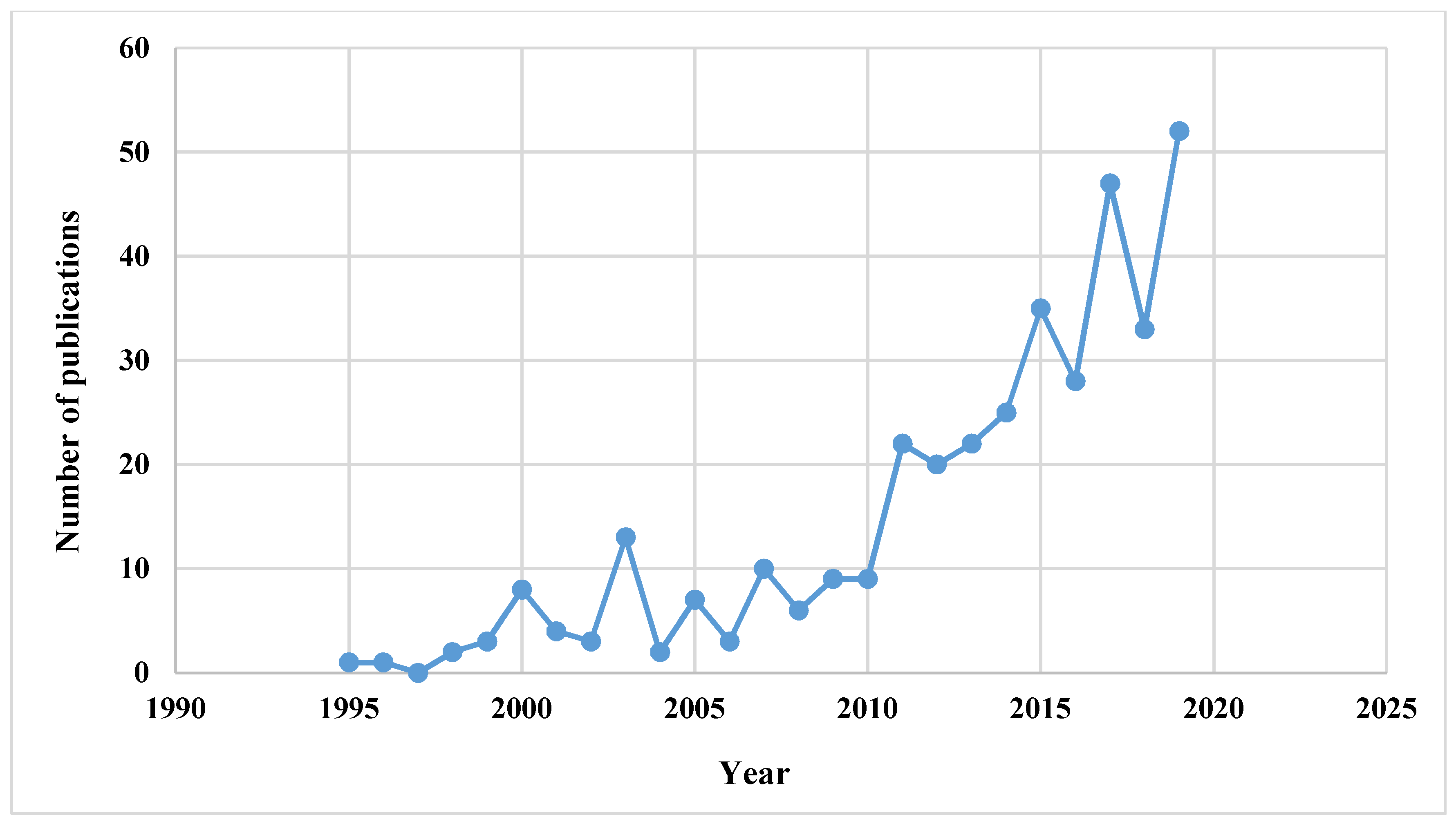
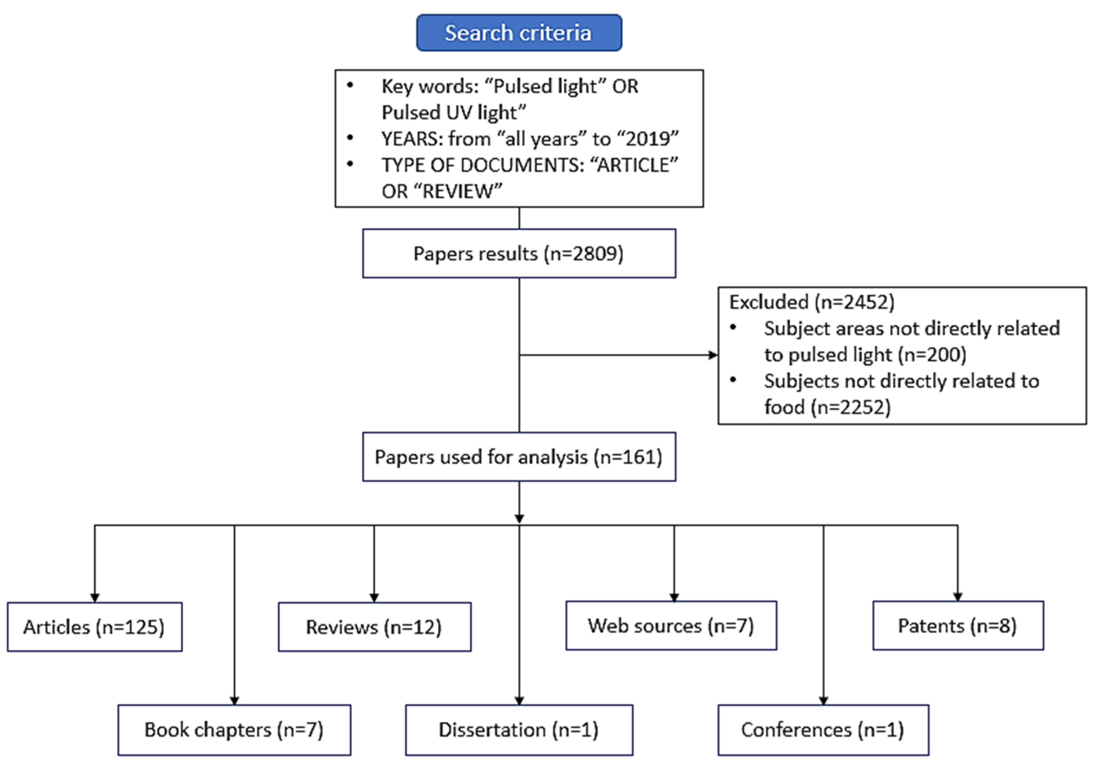
1. Introduction
2. Overview of PL Technology
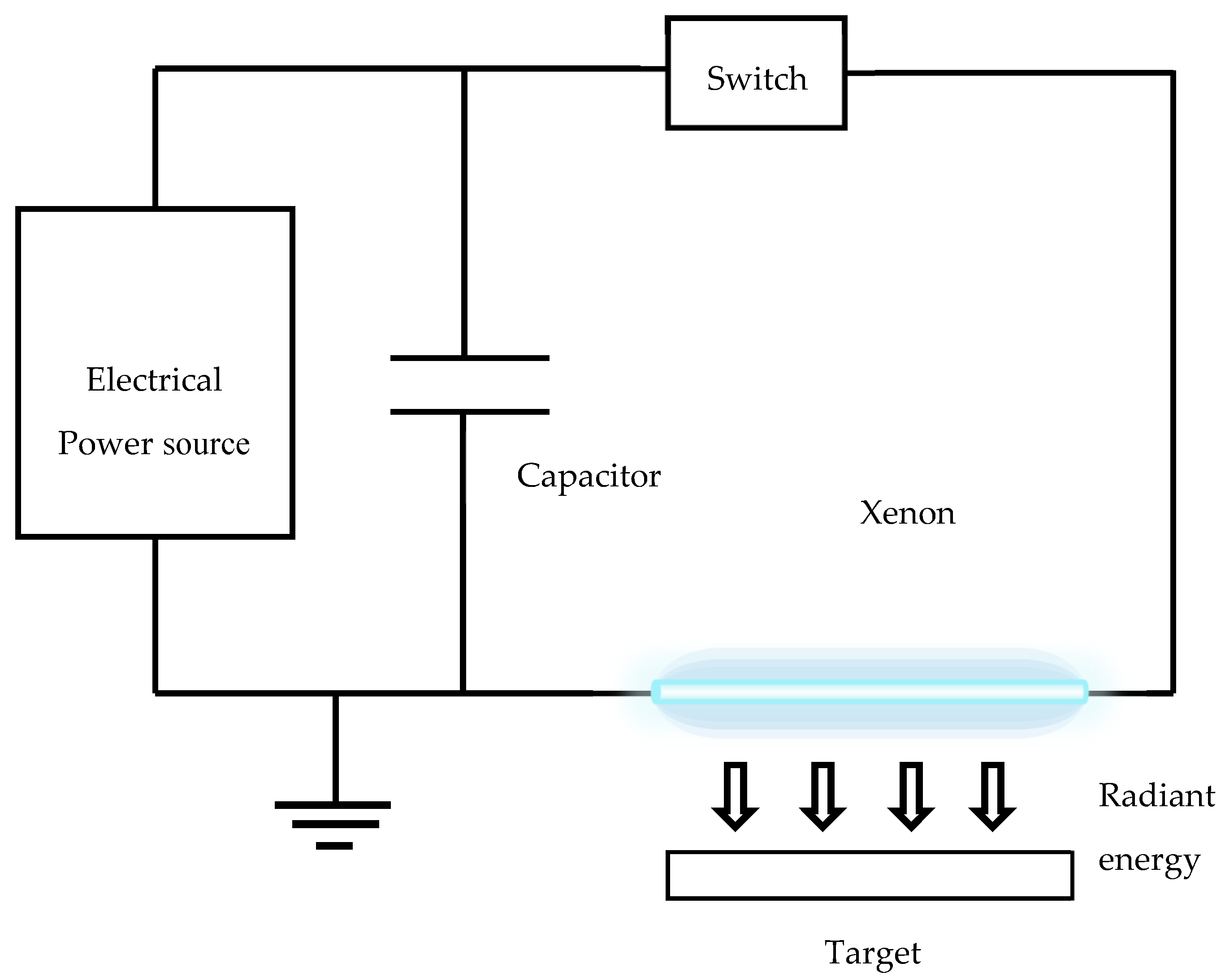
3. Pulsed Light Equipment
- The power unit: Comprising electrical power supplier where high-voltage direct current (DC) power is obtained from low-voltage AC power.
- Pulse configuration device: Comprising of high-voltage capacitors joined in parallel that concentrate energy from the power supplying unit in the charge cycle and release that during the discharge cycle, generating high electrical current. It is also connected to special high-power handling switches that perform on/off cycles of very short time converting the continuous low-electrical power into a pulsed high-electrical power.
- The lamp unit: Comprising specially designed batteries of flashlamps containing gases that are excited due to pulsed high-electrical power from the pulsed configuration device. While transitioning to lower energy or ground states, they give off energy in the form of high intensity pulses. The high-intensity PL is delivered to the products by various lamp footprints and configurations.
4. Mechanism of Microbial Decontamination
4.1. Photo-Chemical Effect
4.2. Photo-Thermal Effect
4.3. Photo-Physical Effect
5. Role of Pulsed Light Technology in Food Safety
5.1. Microbial Decontamination in Food Products
5.2. Food Contact Surface Decontamination
5.3. Food Package Decontamination
6. Effect on Food Quality Characteristics
6.1. Effects on Organoleptic Properties (Color, Texture and Flavor) of Food Products
6.2. Effects on Physico-Chemical Properties of Foods
6.3. Effects on Nutrients and Bio-Active Components
6.4. Effects on Allergens, Toxins and Anti-nutritional Factors
6.5. Effects on Enzymes
6.6. Effects on Post-Harvest Physiology of Fresh Produces
7. Pulsed Light Technology for Enhancement of Bioactive Components in Foods
8. Future Challenges, Trends and Scope
- The total cumulative treatment, in terms of total fluence shall not exceed 12 J/cm2
- The duration of pulses is < 2 ms
- Typical pulse frequencies used in range of 1 to 20 pulses per second
9. Conclusions
Author Contributions
Funding
Acknowledgments
Conflicts of Interest
References
- Pollock, A.M.; Pratap Singh, A.; Ramaswamy, H.S.; Ngadi, M.O. Pulsed light destruction kinetics of L. monocytogenes. LWT Food Sci. Technol. 2017, 84, 114–121. [Google Scholar] [CrossRef]
- Rahman, M.S. Food Preservation: Overview. In Handbook of Food Preservation, 2nd ed.; Rahman, M.S., Ed.; CRC Press: Boca Raton, FL, USA, 2007; pp. 3–17. [Google Scholar]
- Pratap Singh, A.; Singh, A.; Ramaswamy, H.S. Heat transfer phenomena during thermal processing of liquid particulate mixtures—A review. Crit. Rev. Food Sci. Nutr. 2015, 57, 1350–1364. [Google Scholar] [CrossRef] [PubMed]
- Palmieri, L.; Cacace, D. High Intensity Pulsed Light Technology. In Emerging Technologies for Food Processing; Sun, D.-W., Ed.; Academic Press: Cambridge, MA, USA; Elsevier Ltd.: Amsterdam, The Netherlands, 2005; pp. 279–306. [Google Scholar]
- Krishnamurthy, K.; Tewari, J.C.; Irudayaraj, J.; Demirci, A. Microscopic and spectroscopic evaluation of inactivation of Staphylococcus aureus by pulsed UV light and infrared heating. Food Bioprocess Technol. 2010, 3, 93–104. [Google Scholar] [CrossRef]
- Dunn, J.; Ott, T.; Clark, W. Pulsed-light treatment of food and packaging. Food Technol. 1995, 49, 95–98. [Google Scholar]
- Dunn, J. Pulsed light and pulsed electric field for foods and eggs. Poultry Sci. 1996, 75, 1133–1136. [Google Scholar] [CrossRef]
- Buchovec, I.; Paskeviciute, E.; Luksiene, Z.B. Photosensitization-based inactivation of food pathogen Listeria monocytogenes in vitro and on the surface of packaging material. J. Photoch. Photobio. B Biol. 2010, 99, 9–14. [Google Scholar] [CrossRef]
- Woodling, S.E.; Moraru, C.I. Effect of spectral range in surface inactivation of Listeria innocua using broad-spectrum pulsed light. J. Food Prot. 2007, 70, 909–916. [Google Scholar] [CrossRef]
- Rajkovic, A.; Tomasevic, I.; Smigic, N.; Uyttendaele, M.; Radovanovic, R.; Devlieghere, F. Pulsed ultraviolet light as an intervention strategy against Listeria monocytogenes and Escherichia coli O157:H7 on the surface of a meat slicing knife. J. Food Eng. 2010, 100, 446–451. [Google Scholar] [CrossRef]
- Food and Drug Administration. Code of Federal Regulations; 21CFR179.41; FDA: Silver Spring, MD, USA, 1996. [Google Scholar]
- Barbosa-Canovas, G.V.; Schaffner, D.W.; Pierson, M.; Zhang, Q.H. Pulsed light technology. J. Food Sci. 2000, 65 (Suppl. S8), 82–85. [Google Scholar] [CrossRef]
- Elmnasser, N.; Guillou, S.; Leroi, F.; Orange, N.; Bakhrouf, A.; Federighi, M. Pulsed-light system as a novel food decontamination technology: A review. Can. J. Microbiol. 2007, 53, 813–821. [Google Scholar] [CrossRef] [Green Version]
- Koutchma, T.; Forney, L.J.; Moraru, C.I. Ultraviolet Light in Food Technology: Principles and Applications; CRC Press: Boca Raton, FL, USA; Taylor & Francis group: Abingdon, UK, 2009. [Google Scholar]
- Abida, J.; Rayees, B.; Masoodi, F.A. Pulsed light technology: A novel method for food preservation. Int. Food Res. J. 2014, 21, 839–848. [Google Scholar]
- Ortega-Rivas, E. Pulsed light Technology. In Non-Thermal Food Engineering Operations; Ortega-Rivas, E., Ed.; Springer: New York, NY, USA, 2012; pp. 263–272. [Google Scholar]
- Mandal, R.; Shi, Y.; Singh, A.; Yada, R.Y.; Pratap Singh, A. Food Safety and Preservation. In Encyclopedia of Gastroenterology, 2nd ed.; Academic Press, Elsevier Ltd.: Amsterdam, The Netherlands, 2020; pp. 467–479. [Google Scholar]
- Bintsis, T.; Litopoulou-Tzanetaki, E.; Robinson, R.K. Existing and potential applications of ultraviolet light in the food industry—A critical review. J. Sci. Food Agric. 2000, 80, 637–645. [Google Scholar] [CrossRef]
- Gómez-López, V.M.; Bolton, J.R. An approach to standardize methods for fluence determination in bench-scale pulsed light experiments. Food Bioprocess Technol. 2016, 9, 1040–1048. [Google Scholar] [CrossRef]
- Gómez-López, V.M. Pulsed Light Technology. In Handbook of Food Safety Engineering; Sun, D.-W., Ed.; Blackwell Publishing Ltd.: Singapore, 2012; pp. 643–665. [Google Scholar]
- MacGregor, S.J.; Rowan, N.J.; McIlvaney, L.; Anderson, J.G.; Fouracre, R.A.; Farish, O. Light inactivation of food related pathogenic bacteria using a pulsed power source. Lett. Appl. Microbiol. 1998, 27, 67–70. [Google Scholar] [CrossRef] [PubMed]
- Hillegas, S.L.; Demirci, A. Inactivation of Clostridium sporogenes in Clover Honey by Pulsed UV-Light Treatment; V. Manuscript FP 03 009; American Society of Agricultural and Biological Engineers: St. Joseph, MI, USA, 2003. [Google Scholar]
- Bhavya, M.L.; Hebbar, H.U. Pulsed light processing of foods for microbial safety. Food Qual. Saf. 2017, 1, 187–202. [Google Scholar]
- Zou, X.Y.; Lin, Y.L.; Xu, B.; Cao, T.C.; Tang, Y.L.; Pan, Y.; Gao, Z.-C.; Gao, N.Y. Enhanced inactivation of E. coli by pulsed UV-LED irradiation during water disinfection. Sci. Total Environ. 2019, 650, 210–215. [Google Scholar] [CrossRef] [PubMed]
- Krishnamurthy, K.; Demirci, A.; Irudayaraj, J. Inactivation of Staphylococcus aureus by pulsed UV-light sterilization. J. Food Prot. 2004, 67, 1027–1030. [Google Scholar] [CrossRef]
- Holck, A.L.; Liland, K.H.; Drømtorp, S.M.; Carlehög, M.; McLeod, A. Comparison of UV-C and Pulsed UV Light Treatments for Reduction of Salmonella, Listeria monocytogenes, and Enterohemorrhagic Escherichia coli on Eggs. J. Food Prot. 2018, 81, 6–16. [Google Scholar] [CrossRef]
- Huang, Y.; Chen, H. A novel water-assisted pulsed light processing for decontamination of blueberries. Food Microbiol. 2014, 40, 1–8. [Google Scholar] [CrossRef]
- Bialka, K.L.; Demirci, A. Efficacy of Pulsed UV-Light for the Decontamination of Escherichia coli O157:H7 and Salmonella spp. on Raspberries and Strawberries. J. Food Sci. 2008, 73, 201–207. [Google Scholar] [CrossRef]
- Hiramoto, T. Method of Sterilization. U.S. Patent 4,464,336, 7 August 1984. [Google Scholar]
- Dunn, J.E.; Clark, R.W.; Asmus, J.F.; Pearlman, J.S.; Boyer, K.; Painchaud, F.; Hofmann, G.A. Methods for Preservation of Foodstuffs. U.S. Patent 4,871,559, 3 October 1989. [Google Scholar]
- Wekhof, A.; Trompeter, F.J.; Franken, O. Pulsed UV disintegration (PUVD): A new sterilisation mechanism for packaging and broad medical-hospital applications. In Proceedings of the First International Conference on Ultraviolet Technologies, Washington, DC, USA, 14–16 June 2001. [Google Scholar]
- Wekhof, A. Sterilising Packaging and Preserving Foodstuffs with Pulsed Light; Newsletter of International UV Association; International UV Association: Washington, DC, USA, 2002; Volume 4. [Google Scholar]
- Jun, S.; Irudayaraj, J.; Demirci, A.; Geiser, D. Pulsed UV-light treatment of corn meal for inactivation of Aspergillus niger spores. Int. J. Food Sci. Technol. 2003, 38, 883–888. [Google Scholar] [CrossRef]
- Fine, F.; Gervais, P. Efficiency of pulsed UV light for microbial decontamination of food powders. J. Food Prot. 2004, 67, 787–792. [Google Scholar] [CrossRef] [PubMed]
- Choi, M.S.; Cheigh, C.I.; Jeong, E.A.; Shin, J.K.; Chung, M.S. Nonthermal sterilization of Listeria monocytogenes in infant foods by intense pulsed-light treatment. J. Food Eng. 2010, 97, 504–509. [Google Scholar] [CrossRef]
- Cheigh, C.I.; Hwang, H.J.; Chung, M.S. Intense pulsed light (IPL) and UV-C treatments for inactivating Listeria monocytogenes on solid medium and seafoods. Food Res. Int. 2013, 54, 745–752. [Google Scholar] [CrossRef]
- Ferrario, M.; Guerrero, S. Effect of a continuous flow-through pulsed light system combined with ultrasound on microbial survivability, colour and sensory shelf life of apple juice. Innov. Food Sci. Emerg. Technol. 2016, 34, 214–224. [Google Scholar] [CrossRef]
- Sonenshein, A.L. Killing of Bacillus Spores by high-Intensity Ultraviolet Light; Xenon Corporation: Wilmington, MA, USA, 2003. [Google Scholar]
- Rajkovic, A.; Tomasevic, I.; De Meulenaer, B.; Devlieghere, F. The effect of pulsed UV light on Escherichia coli O157: H7, Listeria monocytogenes, Salmonella typhimurium, Staphylococcus aureus and staphylococcal enterotoxin A on sliced fermented salami and its chemical quality. Food Control 2017, 73, 829–837. [Google Scholar] [CrossRef]
- Lytbot. Meet the Lytbot—A highly Innovative First Line of Defence and Attack. Available online: https://solarislyt.com/the-lytbot/ (accessed on 18 March 2020).
- Gómez-López, V.M.; Ragaerta, P.; Debeverea, J.; Devliegherea, F. Pulsed light for food decontamination: A review. Trends Food Sci. Technol. 2007, 18, 464–473. [Google Scholar] [CrossRef]
- Wang, T.; MacGregor, S.J.; Anderson, J.G.; Woolsey, G.A. Pulsed ultra-violet inactivation spectrum of Escherichia coli. Water Res. 2005, 39, 2921–2925. [Google Scholar] [CrossRef]
- Ramos-Villarroel, A.Y.; Martín-Belloso, O.; Soliva-Fortuny, R. Bacterial inactivation and quality changes in fresh-cut avocado treated with intense light pulses. Eur. Food Res. Technol. 2011, 233, 395–402. [Google Scholar] [CrossRef]
- Rowan, N.J.; MacGregor, S.J.; Anderson, J.G.; Fouracre, R.A.; McIlvaney, L.; Farish, O. Pulsed-light inactivation of food-related microorganisms. Appl. Environ. Microbiol. 1999, 65, 1312–1315. [Google Scholar] [CrossRef] [Green Version]
- Anderson, J.G.; Rowan, N.J.; MacGregor, S.J.; Fouracre, R.A.; Farish, O. Inactivation of food-borne enteropathogenic bacteria and spoilage fungi using pulsed-light. IEEE Trans. Plasma Sci. 2000, 28, 83–88. [Google Scholar] [CrossRef]
- Gómez-López, V.M.; Devlieghere, F.; Bonduelle, V.; Debevere, J. Factors affecting the inactivation of microorganisms by intense light pulses. J. Appl. Microinghbiol. 2005, 99, 460–470. [Google Scholar] [CrossRef] [PubMed]
- Gómez-López, V.M.; Koutchma, T.; Linden, K. Ultraviolet and pulsed light processing of fluid foods. In Novel Thermal and Non-Thermal Technologies for Fluid Foods; Cullen, P.J., Tiwari, B.K., Valdramidis, V.P., Eds.; Academic Press: London, UK, 2012; pp. 185–223. [Google Scholar]
- Lukšiene, Ž. New approach to inactivate harmful and pathogenic microorganisms: Photosensitization. Food Technol. Biotech. 2005, 43, 411–418. [Google Scholar]
- Lukšiene, Ž.; Buchovec, I.; Paškevičiūtė, E. Inactivation of food pathogen Bacillus cereus by photosensitization in vitro and on the surface of packaging material. J. Appl. Microbiol. 2009, 107, 2037–2046. [Google Scholar] [CrossRef] [PubMed]
- Nicorescu, I.; Nguyen, B.; Moreau-Ferret, M.; Agoulon, A.; Chevalier, S.; Orange, N. Pulsed light inactivation of Bacillus subtilis vegetative cells in suspensions and spices. Food Control 2013, 31, 151–157. [Google Scholar] [CrossRef]
- Xu, W.; Wu, C. The impact of pulsed light on decontamination, quality, and bacterial attachment of fresh raspberries. Food Microbiol. 2016, 57, 135–143. [Google Scholar] [CrossRef] [PubMed] [Green Version]
- Krishnamurthy, K.; Demirci, A.; Irudayaraj, J.M. Inactivation of Staphylococcus aureus in milk using flow-through pulsed UV-light treatment system. J. Food Sci. 2007, 72, 233–239. [Google Scholar] [CrossRef] [PubMed]
- Takeshita, K.; Shibato, J.; Sameshima, T.; Fukunaga, S.; Isobe, S.; Arihara, K.; Itoh, M. Damage of yeasts induced by pulsed light irradiation. Int. J. Food Microbiol. 2003, 85, 151–158. [Google Scholar] [CrossRef]
- Ramos-Villarroel, A.Y.; Aron-Maftei, N.; Martín-Belloso, O.; Soliva-Fortuny, R. The role of pulsed light spectral distribution in the inactivation of Escherichia coli and Listeria innocua on fresh-cut mushrooms. Food Control 2012, 24, 206–213. [Google Scholar] [CrossRef]
- Macias-Rodriguez, B.; Yang, W.; Schneider, K.; Rock, C. Pulsed UV light as a postprocessing intervention for decontamination of hard-cooked peeled eggs. Int. J. Food Sci. Technol. 2014, 49, 2472–2480. [Google Scholar] [CrossRef]
- Oms-Oliu, G.; Martín-Belloso, O.; Soliva-Fortuny, R. Pulsed Light Treatments for Food Preservation. A Review. Food Bioprocess Technol. 2010, 3, 13–23. [Google Scholar] [CrossRef]
- Roberts, P.; Hope, A. Virus inactivation by high intensity broad spectrum pulsed light. J. Virol. Methods 2003, 110, 61–65. [Google Scholar] [CrossRef]
- Dunn, J.E.; Clark, R.W.; Bushnell, A.H.; Salisbury, K.J. Deactivation of Organisms Using High-Intensity Pulsed Polychromatic Light. U.S. Patent 6228332, 8 May 2001. [Google Scholar]
- Marquenie, D.; Geeraerd, A.H.; Lammertyn, J.; Soontjens, C.; Van Impe, J.F.; Michiels, C.W.; Nicolaï, B.N. Combinations of pulsed white light and UV-C or mild heat treatment to inactivate conidia of Botrytis cinerea and Monilia fructigena. Int. J. Food Microbiol. 2003, 85, 185–196. [Google Scholar] [CrossRef]
- Lagunas-Solar, M.C.; Piña, C.; MacDonald, J.D.; Bolkan, L. Development of pulsed UV light processes for surface fungal disinfection of fresh fruits. J. Food Prot. 2006, 69, 376–384. [Google Scholar] [CrossRef]
- Uesugi, A.R.; Woodling, S.E.; Moraru, C.I. Inactivation kinetics and factors of variability in the pulsed light treatment of Listeria innocua cells. J. Food Prot. 2007, 70, 2518–2525. [Google Scholar] [CrossRef]
- Maclean, M.; MacGregor, S.J.; Anderson, J.G.; Woolsey, G. High-intensity narrow-spectrum light inactivation and wavelength sensitivity of Staphylococcus aureus. FEMS Microbiol. Lett. 2008, 285, 227–232. [Google Scholar] [CrossRef] [Green Version]
- Hierro, E.; Manzano, S.; Ordóñez, J.A.; de la Hoz, L.; Fernández, M. Inactivation of Salmonella enterica serovar enteritidis on shell eggs by pulsed light technology. Int. J. Food Microbiol. 2009, 135, 125–130. [Google Scholar] [CrossRef]
- Sauer, A.; Moraru, C.I. Inactivation of Escherichia coli ATCC 25922 and Escherichia coli O157:H7 in apple juice and apple cider, using pulsed light treatment. J. Food Prot. 2009, 72, 937–944. [Google Scholar] [CrossRef]
- Jean, J.; Morales-Rayas, R.; Anoman, M.-N.; Lamhoujeb, S. Inactivation of hepatitis A virus and norovirus surrogate in suspension and on food-contact surfaces using pulsed UV light (pulsed light inactivation of food-borne viruses). Food Microbiol. 2011, 28, 568–572. [Google Scholar] [CrossRef]
- Palgan, I.; Caminiti, I.M.; Muñoz, A.; Noci, F.; Whyte, P.; Morgan, D.J.; Cronin, D.A.; Lyng, J.G. Effectiveness of High Intensity Light Pulses (HILP) treatments for the control of Escherichia coli and Listeria innocua in apple juice, orange juice and milk. Food Microbiol. 2011, 28, 14–20. [Google Scholar] [CrossRef]
- Lukšiene, Ž.; Buchovec, I.; Viskelis, P. Impact of high-power pulsed light on microbial contamination, health promoting components and shelf life of strawberries. Food Technol. Biotechol. 2013, 51, 284–292. [Google Scholar]
- Artíguez, M.L.; de Marañón, I.M. Inactivation of spores and vegetative cells of Bacillus subtilis and Geobacillus stearothermophilus by pulsed light. Innov. Food Sci. Emerg. Technol. 2015, 28, 52–58. [Google Scholar] [CrossRef]
- Yi, J.Y.; Bae, Y.-K.; Cheigh, C.-I.; Chung, M.-S. Microbial inactivation and effects of interrelated factors of intense pulsed light (IPL) treatment for Pseudomonas aeruginosa. LWT Food Sci. Technol. 2017, 77, 52–59. [Google Scholar] [CrossRef]
- Arroyo, C.; Dorozko, A.; Gaston, E.; O’Sullivan, M.; Whyte, P.; Lyng, J.G. Light based technologies for microbial inactivation of liquids, bead surfaces and powdered infant formula. Food Microbiol. 2017, 67, 49–57. [Google Scholar] [CrossRef]
- Huang, Y.; Ye, M.; Cao, X.; Chen, H. Pulsed light inactivation of murine norovirus, Tulane virus, Escherichia coli O157:H7 and Salmonella in suspension and on berry surfaces. Food Microbiol. 2017, 61, 1–4. [Google Scholar] [CrossRef] [Green Version]
- Faghihzadeh, F.; Anaya, N.M.; Hadjeres, H.; Boving, T.B.; Oyanedel-Craver, V. Pulse UV light effect on microbial biomolecules and organic pollutants degradation in aqueous solutions. Chemosphere 2019, 216, 677–683. [Google Scholar] [CrossRef]
- Rice, J. Sterilizing with light and electrical impulses: Technological alternative to hydrogen peroxide, heat and irradiation. Food Process. 1994, 7, 66. [Google Scholar]
- Dunn, J.; Ott, T.M.; Clark, W. Prolongation of Shelf-Life in Perishable Food Products. U.S. Patent 5489442 A, 6 February 1996. [Google Scholar]
- Shuwaish, A.; Figueroa, J.E.; Silva, J.L. Pulsed-light-treated prepackaged catfish fillets. In Proceedings of the 2000 IFT Annual Meeting, Dallas, TX, USA, 10–14 June 2000. [Google Scholar]
- Mimouni, A. Application de la lumiere pulsee en agroalimentaire. Ind. Aliment. Agricoles 2000, 8, 37–39. [Google Scholar]
- Smith, W.L.; Lagunas-Solar, M.C.; Cullor, J.S. Use of pulsed ultraviolet laser light for the cold pasteurization of bovine milk. J. Food Prot. 2002, 65, 1480–1482. [Google Scholar] [CrossRef]
- Marquenie, D.; Michiels, C.W.; Van Impe, J.F.; Schrevens, E.; Nicolaï, B.N. Pulsed white light in combination with UV-C and heat to reduce storage rot of strawberry. Postharvest Biol. Tec. 2003, 28, 455–461. [Google Scholar] [CrossRef]
- Sharma, R.R.; Demirci, A. Inactivation of Escherichia coli O157:H7 on inoculated alfalfa seeds with pulsed ultraviolet light and response surface modeling. J. Food Sci. 2003, 68, 1448–1453. [Google Scholar] [CrossRef]
- Gómez-López, V.M.; Devlieghere, F.; Bonduelle, V.; Debevere, J. Intense light pulses decontamination of minimally processed vegetables and their shelf-life. Int. J. Food Microbiol. 2005, 103, 79–89. [Google Scholar] [CrossRef] [PubMed]
- Ozer, N.P.; Demirci, A. Inactivation of Escherichia coli O157:H7 and Listeria monocytogenes inoculated on raw salmon fillets by pulsed UV-light treatment. Int. J. Food Sci. Technol. 2006, 41, 354–360. [Google Scholar] [CrossRef]
- Bialka, K.L.; Demirci, A. Decontamination of Escherichia coli O157:H7 and Salmonella enterica on blueberries using ozone and pulsed UV-light. J. Food Sci. 2007, 72, 391–396. [Google Scholar] [CrossRef] [PubMed]
- Paškevičiūte, E.; Lukšiene, Ž. High-power pulsed light for decontamination of chicken breast surface. Cheminė Technologija 2009, 4, 53. [Google Scholar]
- Keklik, N.M.; Demirci, A.; Puri, V.M. Decontamination of unpackaged and vacuum-packaged boneless chicken breast with pulsed UV-light. Poultry Sci. 2010, 89, 570–581. [Google Scholar] [CrossRef] [PubMed]
- Keklik, N.M.; Demirci, A.; Puri, V.M. Inactivation of Listeria monocytogenes on unpackaged and vacuum-packaged chicken frankfurters using pulsed UV-light. J. Food Sci. 2009, 74, 431–439. [Google Scholar] [CrossRef]
- Uesugi, A.R.; Moraru, C.I. Reduction of Listeria on Ready-to-Eat Sausages after Exposure to a Combination of Pulsed Light and Nisin. J. Food Prot. 2009, 72, 347–353. [Google Scholar] [CrossRef]
- Keklik, N.M.; Demirci, A.; Patterson, P.H.; Puri, V.M. Pulsed UV light inactivation of Salmonella enteritidis on egg shells and its effects on egg quality. J. Food Prot. 2010, 73, 1408–1415. [Google Scholar] [CrossRef]
- Pataro, G.; Muñoz, A.; Palgan, I.; Noci, F.; Ferrari, G.; Lyng, J.G. Bacterial inactivation in fruit juices using a continuous flow Pulsed Light (PL) system. Food Res. Int. 2011, 44, 1642–1648. [Google Scholar] [CrossRef]
- Lasagabaster, A.; Arboleya, J.C.; Martínez de Marañón, I. Pulsed light technology for surface decontamination of eggs: Impact on Salmonella inactivation and egg quality. Innov. Food Sci. Emerg. Technol. 2011, 12, 124–128. [Google Scholar] [CrossRef]
- Caminiti, I.M.; Palgan, I.; Noci, F.; Muñoz, A.; Whyte, P.; Cronin, D.A.; Morgan, D.J.; Lyng, J.G. The effect of pulsed electric fields (PEF) in combination with high intensity light pulses (HILP) on Escherichia coli inactivation and quality attributes in apple juice. Innov. Food Sci. Emerg. Technol. 2011, 12, 118–123. [Google Scholar] [CrossRef]
- Muñoz, A.; Palgan, I.; Noci, F.; Morgan, D.J.; Cronin, D.A.; Whyte, P.; Lyng, J.G. Combinations of high intensity light pulses and thermosonication for the inactivation of Escherichia coli in orange juice. Food Microbiol. 2011, 28, 1200–1204. [Google Scholar] [CrossRef] [PubMed]
- Muñoz, A.; Caminiti, I.M.; Palgan, I.; Pataro, G.; Noci, F.; Morgan, D.J.; Denis, A.; Cronin, D.A.; Whyte, P.; Ferrari, G.; et al. Effects on Escherichia coli inactivation and quality attributes in apple juice treated by combinations of pulsed light and thermosonication. Food Res. Int. 2012, 45, 299–305. [Google Scholar] [CrossRef]
- Lukšiene, Ž.; Buchovec, I.; Kairyte, K.; Paškevičiūtė, E.; Viskelis, P. High-power pulsed light for microbial decontamination of some fruits and vegetables with different surfaces. J. Food Agric. Environ. 2012, 10, 162–167. [Google Scholar]
- Ramos-Villarroel, A.Y.; Aron-Maftei, N.; Martín-Belloso, O.; Soliva-Fortuny, R. Influence of spectral distribution on bacterial inactivation and quality changes of fresh-cut watermelon treated with intense light pulses. Postharvest Biol. Technol. 2012, 69, 32–39. [Google Scholar] [CrossRef]
- Miller, B.M.; Sauer, A.; Moraru, C.I. Inactivation of Escherichia coli in milk and concentrated milk using pulsed-light treatment. J. Dairy Sci. 2012, 95, 5597–5603. [Google Scholar] [CrossRef] [Green Version]
- Ganan, M.; Hierro, E.; Hospital, X.F.; Barroso, E.; Fernández, M. Use of pulsed light to increase the safety of ready-to-eat cured meat products. Food Control 2013, 32, 512–517. [Google Scholar] [CrossRef]
- Aguiló-Aguayo, I.; Charles, F.; Renard, C.M.; Page, D.; Carlin, F. Pulsed light effects on surface decontamination, physical qualities and nutritional composition of tomato fruit. Postharvest Biol. Technol. 2013, 86, 29–36. [Google Scholar] [CrossRef]
- Ferrario, M.; Alzamora, S.M.; Guerrero, S. Inactivation kinetics of some microorganisms in apple, melon, orange and strawberry juices by high intensity light pulses. J. Food Eng. 2013, 118, 302–311. [Google Scholar] [CrossRef]
- Anugu, A.K. Microbial Inactivation and Allergen Mitigation of Food Matrix by Pulsed Ultraviolet Light. Ph.D. Thesis, University of Florida, Gainesville, FL, USA, 2013. Available online: http://ufdc.ufl.edu/UFE0045406/00001 (accessed on 18 March 2020).
- Ignat, A.; Manzocco, L.; Maifreni, M.; Bartolomeoli, I.; Nicoli, M.C. Surface decontamination of fresh-cut apple by pulsed light: Effects on structure, colour and sensory properties. Postharvest Biol. Technol. 2014, 91, 122–127. [Google Scholar] [CrossRef]
- Innocente, N.; Segat, A.; Manzocco, L.; Marino, M.; Maifreni, M.; Bortolomeoli, I.; Ignat, A.; Nicoli, M.C. Effect of pulsed light on total microbial count and alkaline phosphatase activity of raw milk. Int. Dairy J. 2014, 39, 108–112. [Google Scholar] [CrossRef]
- Can, F.O.; Demirci, A.; Puri, V.M.; Gourama, H. Decontamination of hard cheeses by pulsed UV-light. J. Food Prot. 2014, 77, 1723–1731. [Google Scholar] [CrossRef] [PubMed]
- Manzocco, L.; Maifreni, M.; Anese, M.; Munari, M.; Bartolomeoli, I.; Zanardi, S.; Suman, M.; Nicoli, M.C. Effect of pulsed light on safety and quality of fresh egg pasta. Food Bioprocess Tech. 2014, 7, 1973–1980. [Google Scholar] [CrossRef]
- Artíguez, M.L.; de Marañón, I.M. Improved process for decontamination of whey by a continuous flow-through pulsed light system. Food Control 2015, 47, 599–605. [Google Scholar] [CrossRef]
- Nicorescu, I.; Nguyen, B.; Chevalier, S.; Orange, N. Effects of pulsed light on the organoleptic properties and shelf-life extension of pork and salmon. Food Control 2014, 44, 138–145. [Google Scholar] [CrossRef]
- Proulx, J.; Hsu, L.C.; Miller, B.M.; Sullivan, G.; Paradis, K.; Moraru, C.I. Pulsed-light inactivation of pathogenic and spoilage bacteria on cheese surface. J. Dairy Sci. 2015, 98, 5890–5898. [Google Scholar] [CrossRef]
- Huang, Y.; Chen, H. Inactivation of Escherichia coli O157:H7, Salmonella and human norovirus surrogate on artificially contaminated strawberries and raspberries by water-assisted pulsed light treatment. Food Res. Int. 2015, 72, 1–7. [Google Scholar] [CrossRef]
- Kramer, B.; Wunderlich, J.; Muranyi, P. Pulsed Light Decontamination of Endive Salad and Mung Bean Sprouts and Impact on Colour and Respiration Activity. J. Food Prot. 2015, 78, 340–348. [Google Scholar] [CrossRef]
- Ferrario, M.; Alzamora, S.M.; Guerrero, S. Study of pulsed light inactivation and growth dynamics during storage of Escherichia coli ATCC 35218, Listeria innocua ATCC 33090, Salmonella Enteritidis MA44 and Saccharomyces cerevisiae KE162 and native flora in apple, orange and strawberry juices. Int. J. Food Sci. Technol. 2015, 50, 2498–2507. [Google Scholar] [CrossRef]
- Kasahara, I.; Carrasco, V.; Aguilar, L. Inactivation of Escherichia coli in goat milk using pulsed ultraviolet light. J. Food Eng. 2015, 152, 43–49. [Google Scholar] [CrossRef]
- Agüero, M.V.; Jagus, R.J.; Martín-Belloso, O.; Soliva-Fortuny, R. Surface decontamination of spinach by intense pulsed light treatments: Impact on quality attributes. Postharvest Biol. Technol. 2016, 121, 118–125. [Google Scholar] [CrossRef] [Green Version]
- Koh, P.C.; Noranizan, M.A.; Karim, R.; Hanani, Z.A.N. Microbiological stability and quality of pulsed light treated cantaloupe (Cucumis melo L. reticulatus cv. Glamour) based on cut type and light fluence. J. Food Sci. Technol. 2016, 53, 1798–1810. [Google Scholar] [CrossRef] [PubMed] [Green Version]
- Preetha, P.; Venugopal, A.P.; Varadharaju, N.; Kennedy, Z.J. Inactivation of Escherichia coli in Tender Coconut (Cocos nucifera L.) Water by Pulsed Light Treatment. Int. J. Curr. Microbiol. Appl. Sci. 2017, 6, 1453–1461. [Google Scholar] [CrossRef]
- Valdivia-Nájar, C.G.; Martín-Belloso, O.; Giner-Seguí, J.; Soliva-Fortuny, R. Modeling the Inactivation of Listeria innocua and Escherichia coli in Fresh-Cut Tomato Treated with Pulsed Light. Food Bioprocess Tech. 2017, 10, 266–274. [Google Scholar] [CrossRef] [Green Version]
- Valdivia-Nájar, C.G.; Martín-Belloso, O.; Soliva-Fortuny, R. Impact of pulsed light treatments and storage time on the texture quality of fresh-cut tomatoes. Innov. Food Sci. Emerg. Technol. 2018, 45, 29–35. [Google Scholar] [CrossRef] [Green Version]
- De Sousa, A.E.D.; de Almeida Lopes, M.M.; Moreira, A.D.R.; Macedo, J.J.N.; Moura, C.F.H.; de Aragão, F.A.S.; Zocolo, G.J.; de Miranda, M.R.A.; de Oliveira Silva, E. Induction of postharvest resistance in melon using pulsed light as abiotic stressor. Sci. Hortic. 2019, 246, 921–927. [Google Scholar] [CrossRef]
- Mukhopadhyay, S.; Sokorai, K.; Ukuku, D.O.; Fan, X.; Olanya, M.; Juneja, V. Effects of pulsed light and sanitizer wash combination on inactivation of Escherichia coli O157: H7, microbial loads and apparent quality of spinach leaves. Food Microbiol. 2019, 82, 127–134. [Google Scholar] [CrossRef]
- Cao, X.; Huang, R.; Chen, H. Evaluation of Food Safety and Quality Parameters for Shelf Life Extension of Pulsed Light Treated Strawberries. J. Food Sci. 2019, 84, 1494–1500. [Google Scholar] [CrossRef]
- Leng, J.; Mukhopadhyay, S.; Sokorai, K.; Ukuku, D.O.; Fan, X.; Olanya, M.; Juneja, V. Inactivation of Salmonella in cherry tomato stem scars and quality preservation by pulsed light treatment and antimicrobial wash. Food Control 2020, 110, 107005. [Google Scholar] [CrossRef]
- Heinrich, V.; Zunabovic, M.; Varzakas, T.; Bergmair, J.; Kneifel, W. Pulsed Light Treatment of Different Food Types with a Special Focus on Meat: A Critical Review. Crit. Rev. Food Sci. Nutr. 2015, 56, 591–613. [Google Scholar] [CrossRef] [PubMed]
- Vimont, A.; Fliss, I.; Jean, J. Efficacy and mechanisms of murine norovirus inhibition by pulsed-light technology. Appl. Environ. Microbiol. 2015, 81, 2950–2957. [Google Scholar] [CrossRef] [Green Version]
- Dunn, J.E.; Clark, R.W.; Asmus, J.F.; Pearlman, J.S.; Boyer, K.; Pairchaud, F.; Hofmann, G. Methods for Preservation of Foodstuffs. U.S. Patent 5,034,235, 23 July 1991. [Google Scholar]
- Paškevičiūte, E.; Buchovec, I.; Lukšiene, Ž. High-power pulsed light for decontamination of chicken from food pathogens: A study on antimicrobial efficiency and organoleptic properties. J. Food Safety 2011, 31, 61–68. [Google Scholar] [CrossRef]
- Musgrove, M.T.; Jones, D.R.; Northcutt, J.K.; Cox, N.A.; Harrison, M.A. Shell rinse and shell crush methods for the recovery of aerobic microorganisms and Enterobacteriaceae from shell eggs. J. Food Prot. 2005, 68, 2144–2148. [Google Scholar] [CrossRef] [PubMed]
- Huffman, D.E.; Slifko, T.R.; Salisbury, K.; Rose, J.B. Inactivation of bacteria, virus and Cryptosporidium by a point-of-use device using pulsed broad spectrum white light. Water Res. 2000, 34, 2491–2498. [Google Scholar] [CrossRef]
- Food Safety Magazine. Shedding Light on Food Safety: Applications of Pulsed Light Processing. 2014. Available online: https://www.foodsafetymagazine.com/magazine-archive1/junejuly-2014/shedding-light-on-food-safety-applications-of-pulsed-light-processing/ (accessed on 4 October 2019).
- Woodling, S.E.; Moraru, C.I. Influence of Surface Topography on the Effectiveness of Pulsed Light Treatment for the Inactivation of Listeria innocua on Stainless-steel Surfaces. J. Food Sci. 2005, 70, 345–351. [Google Scholar] [CrossRef]
- Turtoi, M.; Nicolau, A. Intense light pulse treatment as alternative method for mould spores destruction on paper-polyethylene packaging material, J. Food Eng. 2007, 83, 47253. [Google Scholar] [CrossRef]
- Lopes, M.M.; Silva, E.O.; Canuto, K.M.; Silva, L.M.; Gallão, M.I.; Urban, L.; Fernando Ayala-Zavala, J.; Miranda, M.R.A. Low fluence pulsed light enhanced phytochemical content and antioxidant potential of ‘Tommy Atkins’ mango peel and pulp. Innov. Food Sci. Emerg. Technol. 2016, 33, 216–224. [Google Scholar] [CrossRef]
- Proulx, J.; Sullivan, G.; Marostegan, L.F.; VanWees, S.; Hsu, L.C.; Moraru, C.I. Pulsed light and antimicrobial combination treatments for surface decontamination of cheese: Favorable and antagonistic effects. J. Dairy Sci. 2017, 100, 1664–1673. [Google Scholar] [CrossRef]
- Dunn, J.; Clark, W.R.; Asmus, J.F.; Pearlman, J.S.; Boyer, K.; Pairchaud, F.; Hofmann, G. Methods and Apparatus for Preservation of Foodstuffs. U.S. Patent Application No. WO 88/03369, 3 October 1989. [Google Scholar]
- Hierro, E.; Manzano, S.; Ordóñez, J.A.; de la Hoz, L.; Fernández, M. Efficacy of pulsed light for shelf-life extension and inactivation of Listeria monocytogenes on ready-to-eat cooked meat products. Innov. Food Sci. Emerg. Technol. 2011, 12, 275–281. [Google Scholar] [CrossRef]
- Wambura, P.; Verghese, M. Effect of pulsed ultraviolet light on quality of sliced ham. LWT Food Sci. Technol. 2011, 44, 2173–2179. [Google Scholar] [CrossRef]
- Tomasevic, I.; Rajkovic, A. The Sensory Quality of Meat, Game, Poultry, Seafood and Meat Products as Affected by Intense Light Pulses: A Systematic Review. Procedia Food Sci. 2015, 5, 285–288. [Google Scholar] [CrossRef] [Green Version]
- Tonon, F.; Agoulon, A. Lumiere pulse, principe et application au cas des solutions liquides. In Industries Agro-Alimentaires, la Conservation de Demain, 4th ed.; Commission Européenne: Talence, France, 20 November 2003. [Google Scholar]
- Elmnasser, N.; Dalgalarrondo, M.; Orange, N.; Bakhrouf, A.; Haertlé, T.; Federighi, M.; Chobert, J.M. Effect of pulsed-light treatment on milk proteins and lipids. J. Agric. Food Chem. 2008, 56, 1984–1991. [Google Scholar] [CrossRef]
- Fernández, M.; Ganan, M.; Guerra, C.; Hierro, E. Protein oxidation in processed cheese slices treated with pulsed light technology. Food Chem. 2014, 159, 388–390. [Google Scholar] [CrossRef]
- Cantos, E.; Espín, J.C.; Tomás-Barberán, F.A. Postharvest induction modeling method using UV irradiation pulses for obtaining resveratrol-enriched table grapes: A new ‘‘functional’’ fruit? J. Agric. Food Chem. 2001, 49, 5052–5058. [Google Scholar] [CrossRef]
- Pataro, G.; Sinik, M.; Capitoli, M.M.; Donsì, G.; Ferrari, G. The influence of post-harvest UV-C and pulsed light treatments on quality and antioxidant properties of tomato fruits during storage. Innov. Food Sci. Emerg. Technol. 2015, 30, 103–111. [Google Scholar] [CrossRef]
- Braga, T.R.; Silva, E.O.; Rodrigues, S.; Fernandes, F.A. Drying of mangoes (Mangifera indica L.) applying pulsed UV light as pretreatment. Food Bioprod. Process. 2019, 114, 95–102. [Google Scholar] [CrossRef] [Green Version]
- Wiktor, A.; Mandal, R.; Singh, A.; Pratap Singh, A. Pulsed Light treatment below a Critical Fluence (3.82 J/cm2) minimizes photo-degradation and browning of a model Phenolic (Gallic Acid) Solution. Foods 2019, 8, 380. [Google Scholar] [CrossRef] [Green Version]
- Chung, S.Y.; Yang, W.; Krishnamurthy, K. Effects of pulsed UV-light on peanut allergens in extracts and liquid peanut butter. J. Food Sci. 2008, 73, 400–404. [Google Scholar] [CrossRef]
- Meinlschmidt, P.; Ueberham, E.; Lehmann, J.; Reineke, K.; Schlüter, O.; Schweiggert-Weisz, U.; Eisner, P. The effects of pulsed ultraviolet light, cold atmospheric pressure plasma, and gamma-irradiation on the immunoreactivity of soy protein isolate. Innov. Food Sci. Emerg. Technol. 2016, 38, 374–383. [Google Scholar] [CrossRef]
- Yang, W.W.; Chung, S.-Y.; Ajayi, O.; Krishnamurthy, K.; Konan, K.; Goodrich-Schneider, R. Use of Pulsed Ultraviolet Light to Reduce the Allergenic Potency of Soybean Extracts. Int. J. Food Eng. 2010, 6, 1–12. [Google Scholar] [CrossRef]
- Shriver, S.; Yang, W.; Chung, S.Y.; Percival, S. Pulsed ultraviolet light reduces immunoglobulin E binding to Atlantic white shrimp (Litopenaeus setiferus) extract. Int. J. Environ. Res. Public Health 2011, 8, 2569–2583. [Google Scholar] [CrossRef] [PubMed]
- Li, Y.; Yang, W.; Chung, S.Y.; Chen, H.; Ye, M.; Teixeira, A.A.; Gregory, J.F.; Welt, B.A.; Shriver, S. Effect of pulsed ultraviolet light and high hydrostatic pressure on the antigenicity of almond protein extracts. Food Bioprocess Tech. 2013, 6, 431–440. [Google Scholar] [CrossRef]
- Panozzo, A.; Manzocco, L.; Lippe, G.; Nicoli, M.C. Effect of pulsed light on structure and immunoreactivity of gluten. Food Chem. 2016, 194, 366–372. [Google Scholar] [CrossRef]
- Orcajo, J.; Lavilla, M.; Martínez-de-Marañón, I. Effect of Pulsed Light treatment on β-lactoglobulin immunoreactivity. LWT Food Sci. Technol. 2019, 112, 108231. [Google Scholar] [CrossRef]
- Moreau, M.; Lescure, G.; Agoulon, A.; Svinareff, P.; Orange, N.; Feuilloley, M. Application of the pulsed light technology to mycotoxin degradation and inactivation. J. Appl. Toxicol. 2013, 33, 357–363. [Google Scholar] [CrossRef]
- Funes, G.J.; Gómez, P.L.; Resnik, S.L.; Alzamora, S.M. Application of pulsed light to patulin reduction in McIlvaine buffer and apple products. Food Control 2013, 30, 405–410. [Google Scholar] [CrossRef]
- Wang, B.; Mahoney, N.E.; Pan, Z.; Khir, R.; Wu, B.; Ma, H.; Zhao, L. Effectiveness of pulsed light treatment for degradation and detoxification of aflatoxin B 1 and B 2 in rough rice and rice bran. Food Control 2016, 59, 461–467. [Google Scholar] [CrossRef]
- Chen, D.; Chen, P.; Cheng, Y.; Peng, P.; Liu, J.; Ma, Y.; Liu, Y.; Ruan, R. Deoxynivalenol decontamination in raw and germinating barley treated by plasma-activated water and intense pulsed light. Food Bioprocess Tech. 2019, 12, 246–254. [Google Scholar] [CrossRef]
- Pellicer, J.A.; Navarro, P.; Gómez-López, V.M. Pulsed light inactivation of polygalacturonase. Food Chem. 2019, 271, 109–113. [Google Scholar] [CrossRef]
- Jeon, M.S.; Park, K.M.; Yu, H.; Park, J.Y.; Chang, P.S. Effect of intense pulsed light on the deactivation of lipase: Enzyme-deactivation kinetics and tertiary structural changes by fragmentation. Enzyme Microb. Technol. 2019, 124, 63–69. [Google Scholar] [CrossRef] [PubMed] [Green Version]
- Teichmann, A.; Dutta, P.C.; Staffas, A.; Jägerstad, M. Sterol and vitamin D2 concentrations in cultivated and wild grown mushrooms: Effects of UV irradiation. LWT Food Sci. Techol. 2007, 40, 815–822. [Google Scholar] [CrossRef]
- Koyyalamudi, S.R.; Jeong, S.C.; Pang, G.; Teal, A.; Biggs, T. Concentration of vitamin D2 in white button mushrooms (Agaricus bisporus) exposed to pulsed UV light. J. Food Compost. Anal. 2011, 24, 976–979. [Google Scholar] [CrossRef]
- Kalaras, M.D.; Beelman, R.B. Vitamin D2 Enrichment in Fresh Mushrooms Using Pulsed UV Light. Foodscience. 2008. Available online: https://foodscience.psu.edu/directory/rbb6/VitaminDEnrichment.pdf (accessed on 10 October 2019).
- Kalaras, M.D.; Beelman, R.B.; Elias, R.J. Effects of Postharvest Pulsed UV Light Treatment of White Button Mushrooms (Agaricus bisporus) on Vitamin D2 Content and Quality Attributes. J. Agric. Food Chem. 2011, 60, 220–225. [Google Scholar] [CrossRef] [PubMed]
- Chen, S.Y.; Huang, S.J.; Cheng, M.C.; Chen, Y.K.; Yang, S.C.; Mau, J.L. Enhancement of vitamin D2 content in Pleurotus mushrooms using pulsed light. J. Food Process. Pres. 2015, 39, 2027–2034. [Google Scholar] [CrossRef]
- Chalupa, W.F.; Schroeder, G.M. Method and Apparatus for Vitamin D Enhancement in Dried Mushroom Powder. U.S. Patent Application No. 13/469,168, 4 September 2014. [Google Scholar]
- Schroeder, G.M. Method and Apparatus for Vitamin D Enhancement in Mushrooms. U.S. Patent No. 8,545,915, 1 October 2013. [Google Scholar]


| Paper Title, Authors and Year of Publication | Key Contents |
|---|---|
| Pulsed light technology; Barbosa-Canovas et al.; 2000 |
|
| Pulsed light for food decontamination: a review; Gómez-López et a.; 2007 |
|
| Pulsed-light system as a novel food decontamination technology: a review; Elmnasser et al.; 2007 |
|
| Pulsed Light Treatments for Food Preservation. A Review; Oms-Oliu et al.; 2008 |
|
| The Sensory Quality of Meat, Game, Poultry, Seafood and Meat Products as Affected by Intense Light Pulses: A Systematic Review; Tomasevic and Rajkovic; 2015 |
|
| Pulsed Light Treatment of Different Food Types with a Special Focus on Meat: A Critical Review; Heinrich et al.; 2016 |
|
| Pulsed light processing of foods for microbial safety; Bhavya and Hebbar; 2017 |
|
| PL System | Characteristics | References |
|---|---|---|
| Pure Bright™ | System for sterilizing liquid products (fruit juices) | [30] |
| Annular PL processing chamber with PL lamp inside and housed inside a highly reflective material Tube-quartz made; arrangement-spherical, spiral, etc. Electrodes- metallic (tungsten) electrodes Flashlamps-filled with inert gases like xenon, krypton or a noble gases mixture at various pressures | ||
| System for sterilization of a flexible film for aseptic packaging | ||
| Flexible film drawn through rollers into an absorption enhancement solution and then making them into a cylindrical tube (with product filler and lamp inside) like structure, sealing the longitudinal and bottom seals. The lamp disinfects the interior. Then product fills them and film is drawn forward. Then another seal is done transversally to totally seal the first package and create bottom seal of next package. | ||
| System for sterilizing preformed containers | ||
| Packaging containers moved in line under the flashlamps and then pre-sterilized product fills them sequentially. | ||
| Wek-tec® | The system had a stainless steel enclosure, Test area dimensions: 16 cm wide × 12 cm deep × 15 cm high. Flashlamps – linear; xenon filled Fluence- 0.1 J/cm2 to 8 J/cm2 Pulse duration - 200 µs | [31] |
| Sterilization system for inner surfaces of preformed containers | The containers for packaging milk, juices placed on a conveyer belt, which move under the lamps that flash them. Then, the container progresses forward. | [32] |
| Bench top PL system | System comprises a treatment chamber and a control module Treatment chamber-stainless steel Lamps-lamp house is at the top center and filled with Xenon gas. The control module and light source connected by a control cable, to modulate the electric current to get specific pulse repetition rate, pulse width and peak power. | [33] |
| OneShot EN2/2143–1 unit | 3-fluidized bed PUV system mounted with adjustable air nozzles to blow compressed air for fluidization of the food powders; Water circulation in surrounding quartz jacket around the flash lamp to counter overheating and a reflecting cylinder. | [34] |
| RDT350 model, La Calhene, USA | Treatment chamber-250 cm diameter Lamps no.-8 xenon lamps arranged on the periphery of the chamber Distance of samples from the xenon lamps was 13.5 cm Lamps Fluence-1.5 J/cm2 Pulse duration-300 µs Wavelength-between 200–1200 nm | [13] |
| Laboratory-scale PL system for sterilization of infant foods | Pulse discharge by quartz lamp with an oscilloscope to get the exponential decay pulse. Water bath dissipates the heat generated during the operation | [35] |
| Intense PL system for sea foods decontamination | Lab scale system for decontamination of microorganisms in solid foods and sea foods. Input voltage of 220 V alternating current (AC) supply rectified and transformed to a maximum voltage 50KV Lamps- quartz lamp in which the Xenon gas is filled at 450 Torr pressure Length-145 mm Outer diameter-7.14 mm Wavelength- 200–1100 nm ranging from UV-C to IR | [36] |
| Continuous-flow through PL system | For liquid food products like sugar syrup, juices | [37] |
| SteriPulse® XL-3000 system | Fluence: 1.27 J/cm2 per pulse Pulse duration-360 µs;3 pulses/s Broad spectrum UV with high germicidal power Stainless steel sterilization chamber, removable lamp housing | [38] |
| Claranor, France | PL system with multiple xenon lamps | [39] |
| The LytBot, Solaris Disinfection Inc., Canada | PL system for Hospital Disinfection from | [40] |
| Microorganism | Media | Pulse Energy (J/cm2) | Treatment Time (μs) a | Log10 Reductions | Reference |
|---|---|---|---|---|---|
| B. subtilis | Tryptic soy broth | 4 | 2 | 10 | [30] |
| 1 | 4 | 10 | |||
| E. coli | Tryptic soy agar | 4 | 1 | 10 | |
| 1.5 | 2 | 10 | |||
| A. niger | Potato dextrose agar | 4 | 4 | 10 | |
| 12 | 1 | 10 | |||
| S. aureus | Tryptic soy agar | 0.75 | 2 | 10 | |
| 0.2 | 4 | 8 | |||
| Saccharomyces cerevisiae | Tryptic soy agar | 0.4 | 4 | 10 | |
| E. coli O157:H7 | Tryptone Soya-Yeast Extract Agar | 3 | 1–512 | 6.82 | [21] |
| L. monocytogenes 4b | 6.25 | ||||
| Pseudomonas aeruginosa | Tryptone Soya-yeast Extract Agar | 3 | 20 | 5.8 | [44] |
| B. cereus | 4.9 | ||||
| E. coli O157:H7 | 6.2 | ||||
| L. monocytogenes | 4.4 | ||||
| S. enteritidis | 5.6 | ||||
| S. cerevisiae | 4.9 | ||||
| S. aureus | 5.1 | ||||
| B. cereus, E. coli, and S. enteritidis | Tryptones soy-yeast extract broth | 3 | 85 | About 8 | [45] |
| A. niger, Fusarium culmorum | Malt extract agar | 4.5 | |||
| A. niger | Buffer solution | 1 | 1000 | 4.8 | [31] |
| Cryptosporidium parvum oocysts | Solution containing approximately 25 × 106 Cryptosporidium oocysts | 0.11 | - | 3–5 | [58] |
| 0.22 | - | ||||
| B. subtilis | Sterile deionized water | 1–4 | 2–5 | [38] | |
| S. cerevisiae | Sterile potassium phosphate buffer suspensions | 0.7 | 1200 | 6 | [53] |
| Botrytis cinerea | Rose Bengal agar | - | 250 s | 3 | [59] |
| Monilinia fructigena | Malt extract agar | - | 250 s | 4 | |
| S. aureus | Baird-Parker agar plates | 5.6 | 5400 | 7.5 | [25] |
| 5.6 | 5400 | 8.5 | |||
| Alicyclobacillus acidoterrestris | Orange serum agar | 7 | 1500 | >5.2 | [46] |
| B. circulans | Nutrient agar | >4.1 | |||
| Brochotrix thermosphacta | Nutrient agar | 3.1 | |||
| Leuconostoc mesenteroides | de Man, Rogosa and Sharpe agar | 4.0 | |||
| Photobacterium phosphoreum | Nutrient agar | >4.4 | |||
| P. fluorescens | Nutrient agar | 4.2 | |||
| Shewanella putrefaciens | Nutrient agar | 3.9 | |||
| Clostridium perfringens | Nutrient agar | >2.9 | |||
| B. cereus | Nutrient agar | 3.0 | |||
| E. coli O157:H7 | Nutrient agar | 4.7 | |||
| L. monocytogenes | Nutrient agar | 2.8 | |||
| S. typhimurium | Nutrient agar | 3.2 | |||
| Shigella flexnii | Nutrient agar | 3.8 | |||
| S. aureus | Nutrient agar | 5.1 | |||
| Yersina enterocolitica | Nutrient agar | 3.9 | |||
| A. flavus | Potato dextrose agar | 2.2 | |||
| Candida lambica | Yeast glucose choramphenicol | 2.8 | |||
| Rhodotorula mucilaginosa | Yeast glucose choramphenicol | >2.8 | |||
| B. cinerea | Potato dextrose agar | 1.2 | |||
| Alternaria alternate, A. niger, B. cinerea, F. oxysporum, F. roseum, M. fructicola, Penicillium expansum, P. digitatum, Phytophthora citrophthora and Rhizopus stolonifer | Solid culture media | Up to 0.2 | - | Controlled completely | [60] |
| L. innocua | Clear liquid broth | 12 | - | >6 | [61] |
| S. aureus, E. coli NCTC 9001, Methicillin-resistant S. aureus (MRSA) LMG 15,975 and MRSA 16a | Phosphate-buffered saline suspension | 630 | 30 min | 5 for S. aureus and MRSA strains Negligible for E. coli NCTC 9001 | [62] |
| S. erterica serovar Enteritidis | Noble agar | 0.7 | - | 6.7 | [63] |
| E. coli ATCC 25,922 and E. coli O157:H7 | Butterfield’s phosphate buffer | 13.1 | 4 s | >8.5 | [64] |
| Tryptic soy broth | 4 s (12 pulses @ 3 pulses/s) | 3.5 | |||
| Murine norovirus (MNV-1) and hepatitis A virus (HAV) | Viral suspensions | 2 s | 5 | [65] | |
| L. innocua NCTC 11,288 | Maximum recovery diluent | 28 | 8 s | 5.13 | [66] |
| E. coli K12 DSM 160 | 4.67 | ||||
| L. monocytogenes | Tryptone soy agar plate | 0.00175 | 180 s | 6 | [36] |
| B. subtilis strain ATCC 6633 | Cell suspension (OD580 nm - 0.8) | 0.6 | - | 8.7 | [50] |
| B. cereus and | Luria-Bertani Agar | 1.95 | 200 s | About 7 | [67] |
| L. monocytogenes | About 7 | ||||
| B. subtilis | Nutrient agar for vegetative cells Tryptic soy broth for spores (cell density 109 cells/mL in both) | 12 | - | About 4 of sporses >8 for vegetative cells | [68] |
| Geobacillus stearothermophillus | Nutrient agar for both vegetative cells and spores | - | About 0.5 for spores About 1 for vegetative cells | ||
| P. aeruginosa | Plate count agar | 1.13 | 30 s | 3.63 | [69] |
| About 11 | 300 s | About 6.5 | |||
| L. monocytogenes Scott A | Solid tryptic soy agar Petri plates | - | 20 s | 5 | [1] |
| 0.1% peptone water solution in whirl pak bags | - | 93 ± 5 s | 1 | ||
| Bacillus subtilis spores | Clear liquid medium | 0.017 | - | 1 | [70] |
| Murine norovirus (MNV-1) and | Phosphate buffered saline | 4.94 | - | >5.8 | [71] |
| Tulane virus (TV) | - | >6.0 | |||
| E. coli K-12 MG1655 | Lysogeny broth (LB) | 76 J/cm2 wavelength of 190 nm | - | 11 | [72] |
| 95 J/cm2 wavelength of 240 nm | 6 |
| Food Products | Pulse Energy (J/cm2) | Treatment Time (μs) g | Log10 Reductions | Reference |
|---|---|---|---|---|
| Dry, non-creamed cottage cheese curds | 16 | 2 pulses; 0.5 μs; Applied at 3 s interval | PVR a of 96.7% of Pseudomonas | [30] |
| White bread rolls, hard crusted | 16 | 0.5 μs | PVR of 97.2% of mold spores | |
| 16 | 2 pulses; 0.5 μs; Applied at 5 s interval | PVR of 99.8% of mold spores | ||
| Cakes packaged in plastic containers | 16 | 3 pulses; 0.5 μs; Applied at 5 s interval | No mold growth in treated samples during storage at room temperature for 10 days, Untreated samples-moldy in 3 days | |
| Whole fish, treated on the dark side | 10 | 3 flashes; 0.5 μs | 1.93 of coliforms 2 of psychrotrophs | |
| Shrimp | 1–2 | - | Reduction by 10–1000 factors b of Listeria, Salmonella Shelf-life increased by 1 week compared to untreated samples | |
| White bread slices | Broad Spectrum PL | - | Shelf life more than 2 weeks, no surface molds | [73] |
| Meat | Broad Spectrum PL | - | Reduction of Listeria and Salmonella | [73] |
| Chicken wings | Broad Spectrum PL | - | 2 for Salmonella (inoculated) | [6] |
| Frankfurters | 30 | - | 2 of L. innocua (inoculated) | [6] |
| Retail meat | Broad Spectrum PL | - | 1–3 of the lactic, total aerobic, enteric bacteria and Pseudomonas | [6] |
| Commercial eggs | 4 | 8 | Up to 8 of S. Enteriditis (inoculated) | [7] |
| Strawberries | 0.5 | 0.3 ms duration for pulses 4 s | No mold growth after 2 weeks storage | [74] |
| Catfish fillets, pre-packaged | 0.25 | - | Psychrotrophic and coliform bacteria not reduced just after treatment. After storage of 1 week, psychrotrophic reduction to 1 log lesser than untreated ones Coliform reduction from about 50 to less than 10 CFU/g | [75] |
| 0.5 | - | Psychrotrophic reduction to 2 log lower than untreated sample Coliform reduction from about 50 to less than 10 CFU/g | ||
| Eggshells | 1.5 | - | 3 to 6 of Bacillus subtilis spores reduced with no. of pulses ranging from 2 to 6 | [76] |
| Cake | 1.5 | - | 3 to 6 of A. niger spores with 2 to 6 pulses; Shelf-life up to 6 months for treated samples | |
| Bread slices, Packed | 1.5 | - | Shelf life up to 5 months for treated samples | |
| Milk | 25.1 | 114 s | >2.0 of Serratia marcescens | [77] |
| Corn meal | 5.6 c | 100 s | 4.93 of the spore of A. niger | [33] |
| Strawberries | 7 J | 30 | No significant effect on growth of M. fructigena and B. cineria | [78] |
| Alfalfa seeds | 5.6 | 90 s | 4.89 of E. coli O157:H7 | [79] |
| Clover honey | 5.6 | 135 s | 2 mm deep samples: reduction of C. sporogenes spores to 73.9% | [22] |
| 5.6 | 180 s | 8 mm deep samples: reduction of C. sporogenes spores to 89.4% | ||
| Wheat flour | 31.12 | - | 10.1% of S. cerevisiae | [35] |
| Black pepper | 31.12 | - | 44.5% of S. cerevisiae | |
| Shredded spinach | 7 J | 180 d | 0.9 of Mesophilic aerobic microorganisms | [80] |
| Grated celeriac | 0.21 e of Mesophilic aerobic microorganisms | |||
| Chopped green paprika | 0.56 of Mesophilic aerobic microorganisms | |||
| Soybean sprouts | 0.65 of Mesophilic aerobic microorganisms | |||
| Shredded radicchio | 0.79 of Mesophilic aerobic microorganisms | |||
| Grated carrot | 1.64 of Mesophilic aerobic microorganisms | |||
| Shredded iceberg lettuce | 2.04 of Mesophilic aerobic microorganisms 0.46 of aerobic psychrotrophic counts | |||
| Shredded white cabbage | 1.64 of Mesophilic aerobic microorganisms 0.54 of aerobic psychrotrophic counts | |||
| Salmon fillets | 5.6 | 60 s | 1.09 on skin side and 0.3 on muscle side and of E. coli O157:H7 | [81] |
| 5.6 | 60 s | 1.02 on skin side and 0.74 on muscle side of L. monocytogenes Scott A | ||
| Apples, kiwi, orange, lemon, nectarine, peaches, pears, raspberries, table grapes | Up to 0.500 f | < 10 s | >5 for fungi | [60] |
| Milk | 1.27 | - | 0.55–7.26 of S. aureus | [25] |
| Blueberries | 22.6 | 60 s | 4.3 of S. enterica | [82] |
| 22.6 | 60 s | 2.9 of E. coli O157:H7 | ||
| Raspberries | 59.4 | 60 s | 3.4 of Salmonella spp. | [28] |
| 72.0 | 60 s | 3.9 of E. coli O157:H7 | ||
| Strawberries | 64.8 | 60 s | 4.3 of Salmonella spp | |
| 60 s | 3.3 of E. coli O157:H7 | |||
| Chicken breast | 5.4 | 200 s | 2.0 of S. Typhimurium | [83] |
| 16.8 | 15 s | 1.9 of S. Typhimurium | [84] | |
| Chicken frankfurters | 55.9 | 60 s | 1.6 of L. monocytogenes Scott A | [85] |
| Vienna sausages | 9.4 | 4 s | 1.37 of L. innocua | [86] |
| Apple juice | 8.8 | 3 s | 7.29 of E. coli ATCC 2592 | [64] |
| Shell eggs | 12 | - | 2.49 S. Enteriditis | [63] |
| 23.6 | 20 s | >5.3 of S. Enteriditis | [87] | |
| Infant milk powder | - | 9500 | 3 of L. monocytogenes | [35] |
| Fresh-cut avocado | 0.4 | - | 2.97 of L. innocua 3.33 of E.Coli | [43] |
| Apple juice | 4 | - | 4 of E.Coli | [88] |
| 2.98 of L. innocua | ||||
| Orange juice | - | 2.90 of E. Coli | ||
| 0.93 of L. innocua | ||||
| Eggs (unwashed) | 2.1–10.5 | - | 4.9 of Salmonella | [89] |
| Eggs (washed) | - | 5 of Salmonella | ||
| Apple juice | 28 | 8 s | >4.7 of E. coli 1.93 of L. innocua | [66] |
| Orange juice | About 1 for both L. innocua and E. coli | |||
| Milk | 1.06 of E. coli 0.51–0.84 of L. innocua | |||
| Apple juice | 5.1 | 1.52 | 3.9 of E. coli | [90] |
| Orange juice | 5.1 | 2.81 s | 2.42 of E. coli | [91] |
| Apple juice | 5.1 | 300 s | 4.9 of E. coli | [92] |
| Plums, tomatoes, cauliflowers, sweet peppers | 5.4 | 0–200 s | 1.1–1.5 of mesophiles 1.3 -1.8 of B. cereus | [93] |
| Fresh-cut watermelons | 12 | - | >3 of E. coli 2.79 of L. innocua | [94] |
| Fresh-cut mushrooms | 12 | - | 3 of E. coli 2 of L. innocua | [54] |
| Milk (9.8% total solids) | 8.4 | - | 2.5 of E. coli ATCC 25922 | [95] |
| Concentrated milk (45% total solids) | 8.4 | < 1 of E. coli ATCC 25922 | ||
| Skim milk | 14.9 | 3.4 of E. coli ATCC 25922 | ||
| Milk (2% fat) | 14.9 | >2.5 of E. coli ATCC 25922 | ||
| Whole milk | 14.9 | >2.5 of E. coli ATCC 25922 | ||
| Ground spices (caraway, black pepper and red pepper) | 10 | - | 0.8 in ground caraway and black pepper and 1 in ground red pepper for B. subtilis | [50] |
| Shrimp | 12.1 | 1380 s | 2.4 of L. monocytogenes | [36] |
| Salmon | 2.1 of L. monocytogenes | |||
| Flatfish fillets | 1.9 of L. monocytogenes | |||
| Ready-To-Eat dry cured meat products | 11.9 | - | 1.5–1.8 of L. monocytogenes and S. enterica serovar Typhimurium | [96] |
| Tomatoes | 4 | - | 1 of surface microflora | [97] |
| 2.2 | 2.3 of S. cerevisiae | |||
| Strawberries | 3.9 | 200 s | 2.2 of Aerobic mesophilic bacteria | [67] |
| 1 of yeasts and molds | ||||
| 1.5 of B. cereus | ||||
| 1.1 of L. monocytogenes | ||||
| Orange Juice | 71.6 | 60 s | Up to 1 of L. innocua, Up to 2 of E. coli Up to 1.5 of S. Enteritidis Up to 0.5 of S. cerevisiae | [98] |
| Strawberry juice | Up to 0.2 of L. innocua, Up to 0.3 of S. cerevisiae | |||
| Apple juice | Up to 4.5 of L. innocua Up to 2 of E. coli Up to 4 of S. Enteritidis Up to 6 of S. cerevisiae | |||
| Melon juice | Up to 6 of L. innocua and E. coli Up to 5 of S. Enteritidis Up to 7 of S. cerevisiae | |||
| Blueberry wine | - | 60 s | 9.6 ± 0. 9 of S. cerevisiae | [99] |
| White grape wine | - | 40 s | 5.9 ± 0.5 of S. cerevisiae | |
| Blueberries | 5–56.1 | 360 μs | 3.8- > 6.7 of E. coli O157:H7 | [27] |
| 4.8–5.7 of Salmonella | ||||
| Apple juice | 17.5 | - | 3 of L. brevis | [100] |
| Raw milk | 26.25 | - | Up to 3.2 of total microbial count | [101] |
| Packaged cheese | 53.4 | 40 s | 1.25 of P. roqueforti 2.98 of Listeria monocytogenes | [102] |
| Unpackaged cheese | 1.32 of P. roqueforti 3.08 of L. monocytogenes | |||
| Fresh egg pasta | 1.75 | - | 3.3 of S. enterica | [103] |
| Whey | 1.1 | - | 0.5 of L. innocua | [104] |
| Pork and salmon | 30 | - | 0.96 (in pork) and 0.7 (in salmon) of aerobic microorganisms | [105] |
| Cheese | 1.02–12.09 | - | 3.74 ± 0.8 of P. fluorescens 5.41 ± 0.1 of E. coli ATCC 25,922 3.37 ± 0.2 of L. innocua | [106] |
| Strawberries | 63.2 | 60 s | 2.4 of E. coli O157:H7 2.2 of Salmonella 1.8 of Murine norovirus (MNV-1) | [107] |
| Raspberries | 53.9 | 4.5 of E. coli O157:H7 4.4 of Salmonella 3.6 of Murine norovirus (MNV-1) | ||
| Endive salad and | 1 | - | 2.34 of E. coli 2.54 of L. innocua 2.46 of natural microflora | [108] |
| Mung bean sprouts | - | 1.91 of E. coli 1.55 of L. innocua | ||
| Orange Juice | 71.6 | 60 s | 0.3–0.8 of L. innocua, E. coli and S. Enteritidis Upto 0.5 of S. cerevisiae | [109] |
| Strawberry juice | 0.3–0.8 of L. innocua, E. coli and S. Enteritidis | |||
| Apple juice | 1.6 of L. innocua 2.1 of E. coli 2.4 of S. Enteritidis Upto 1 of S. cerevisiae | |||
| Goat milk | 10 | - | 6 of E. coli | [110] |
| Spinach | 4 | - | 0.5–2.2 of initial microbial load | [111] |
| 0.8 | 1.85 of L. innocua 1.72 of E. coli | |||
| 12 | 2.6 of L. innocua 2.3 of E. coli | |||
| Cantaloupe melon | 15.6 | - | 1.39 of total plate count | [112] |
| 1.45 of yeasts and molds | ||||
| Raspberries | 28.2 | 30 s | 4.5 of Salmonella | [51] |
| 3.9 of E. coli O157:H7 | ||||
| Powdered infant formula | 0.199 | - | >99% of L. innocua | [70] |
| 0.398 | - | >99% of B. subtilis spores | ||
| Coconut water | 19.2 | - | 5.2 of E. coli | [113] |
| Strawberry | 22.5 | 24 s | 0.9 ± 0.2 of Murine norovirus 1.9 ± 0.3 of E. coli O157:H7 2.1 ± 0.4 of Salmonella | [71] |
| Blueberry | 3.8 ± 0.6 of Murine norovirus 5.7 ± 1.2 of E. coli O157:H7 4.2 ± 0.2 of Salmonella | |||
| Tomatoes (slices packed in sealed trays | 8 | 30 min | About 1.17 of L. innocua CLIP11262 About 1.34 of E. coli 1.107 | [114] |
| Fresh tomatoes | 8 | - | Up to 2 of psychrophilic bacteria Up to 1.3 of yeasts and molds | [115] |
| Melons | 9 | - | The shelf life was extended 12 days | [116] |
| Spinach | 15.75 | 15 s | 2.7 of E. coli O157:H7 | [117] |
| Strawberries | 3 | 10 s | Up to 0.8 for Salmonella | [118] |
| Tomato stem scar | 31.5 | 30 s | 2.3 for S. enterica | [119] |
© 2020 by the authors. Licensee MDPI, Basel, Switzerland. This article is an open access article distributed under the terms and conditions of the Creative Commons Attribution (CC BY) license (http://creativecommons.org/licenses/by/4.0/).
Share and Cite
Mandal, R.; Mohammadi, X.; Wiktor, A.; Singh, A.; Pratap Singh, A. Applications of Pulsed Light Decontamination Technology in Food Processing: An Overview. Appl. Sci. 2020, 10, 3606. https://0-doi-org.brum.beds.ac.uk/10.3390/app10103606
Mandal R, Mohammadi X, Wiktor A, Singh A, Pratap Singh A. Applications of Pulsed Light Decontamination Technology in Food Processing: An Overview. Applied Sciences. 2020; 10(10):3606. https://0-doi-org.brum.beds.ac.uk/10.3390/app10103606
Chicago/Turabian StyleMandal, Ronit, Xanyar Mohammadi, Artur Wiktor, Anika Singh, and Anubhav Pratap Singh. 2020. "Applications of Pulsed Light Decontamination Technology in Food Processing: An Overview" Applied Sciences 10, no. 10: 3606. https://0-doi-org.brum.beds.ac.uk/10.3390/app10103606