Isolation, Degradation Performance and Field Application of the Metolachlor-Degrading Fungus Penicillium oxalicum MET-F-1

 , , ,
, , ,  and
and 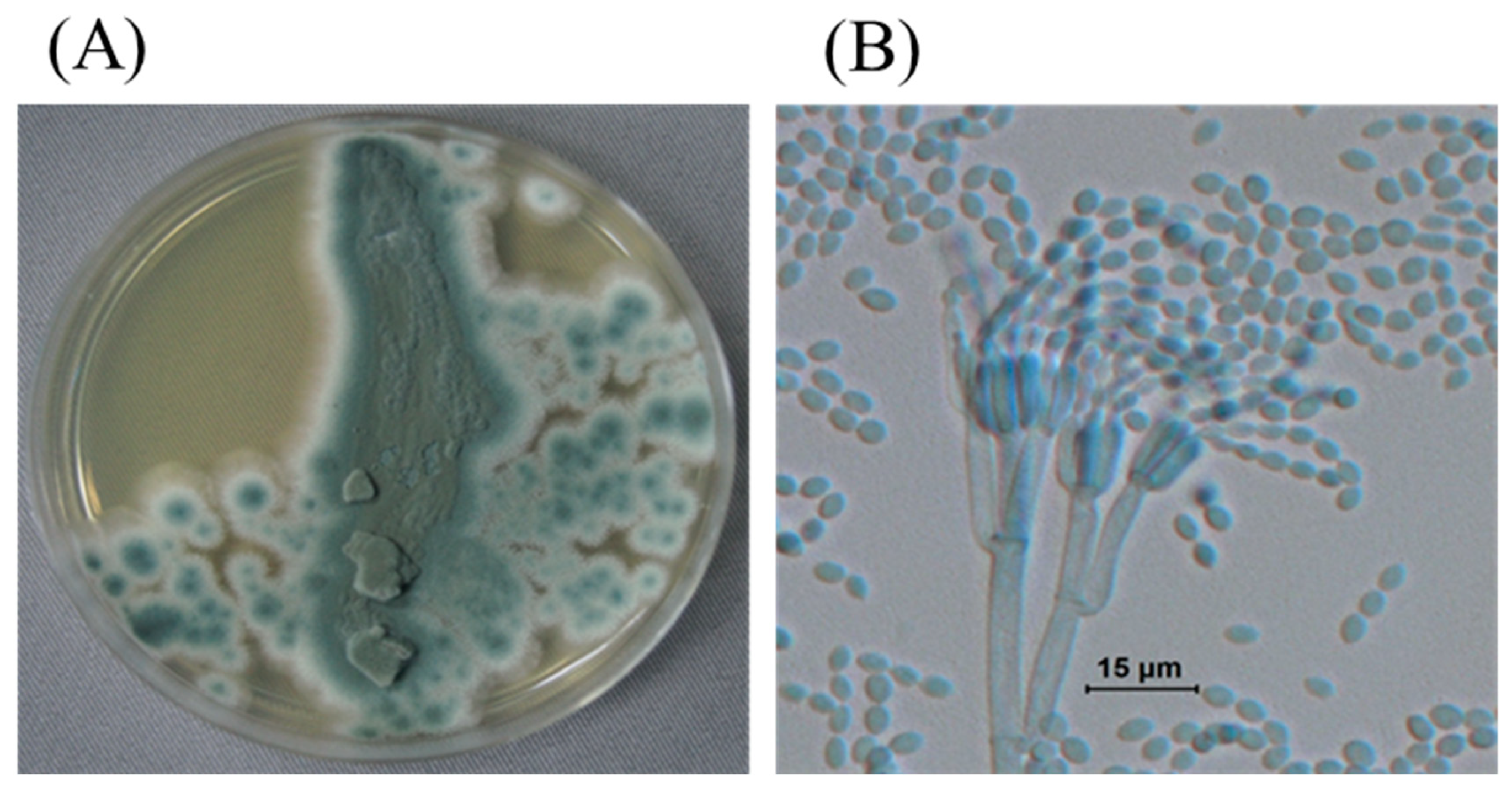
Abstract
:1. Introduction
2. Materials and Methods
2.1. Target Source for Screening Functional Microbes
2.2. Chemicals and Media
2.3. Isolation of Metolachlor-Tolerant Strains
2.4. Screening and Identification of Metolachlor-Degrading Strains
2.5. Optimization of Metolachlor Degradation Performance of Strain MET-F-1
2.6. Metolachlor-Degrading Potential of Strain MET-F-1 in Field Trials
2.7. Determination of Metolachlor and Its Metabolites in the Liquid and Soil Samples
3. Results and Discussion
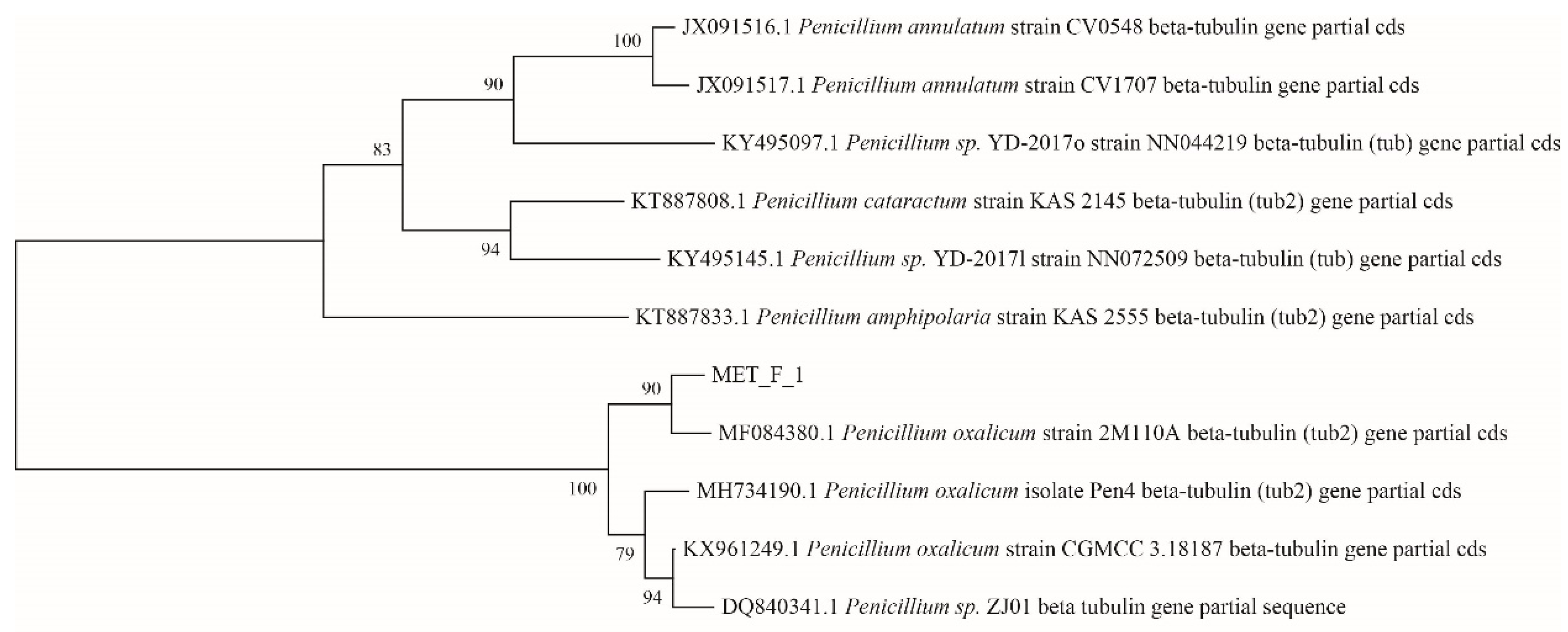
3.1. Isolation and Identification of Metolachlor-Degrading Strain
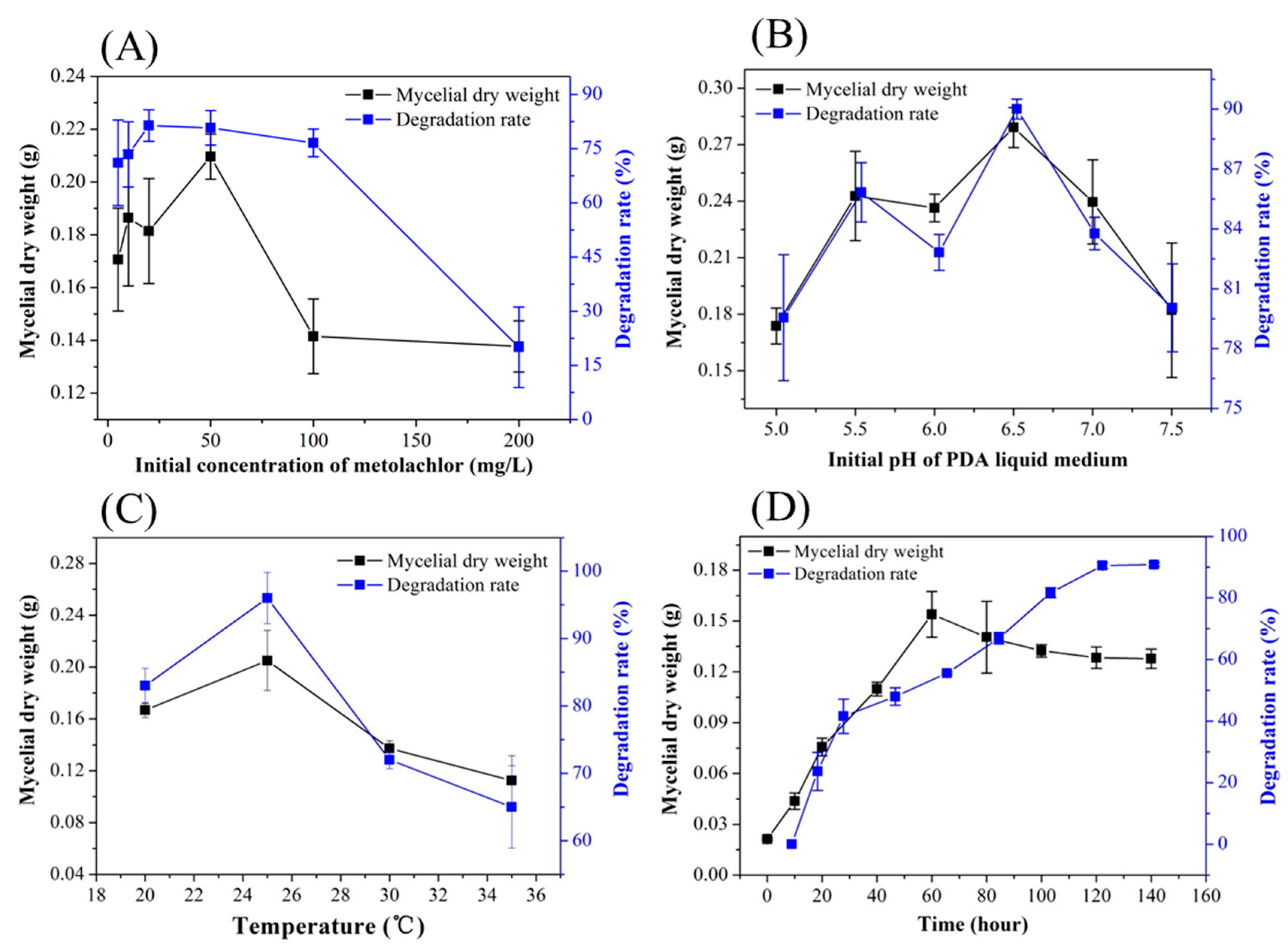
3.2. Optimization of the Metolachlor-Degrading Performance of Strain MET-F-1
3.3. Co-Metabolism of Metolachlor by Strain MET-F-1
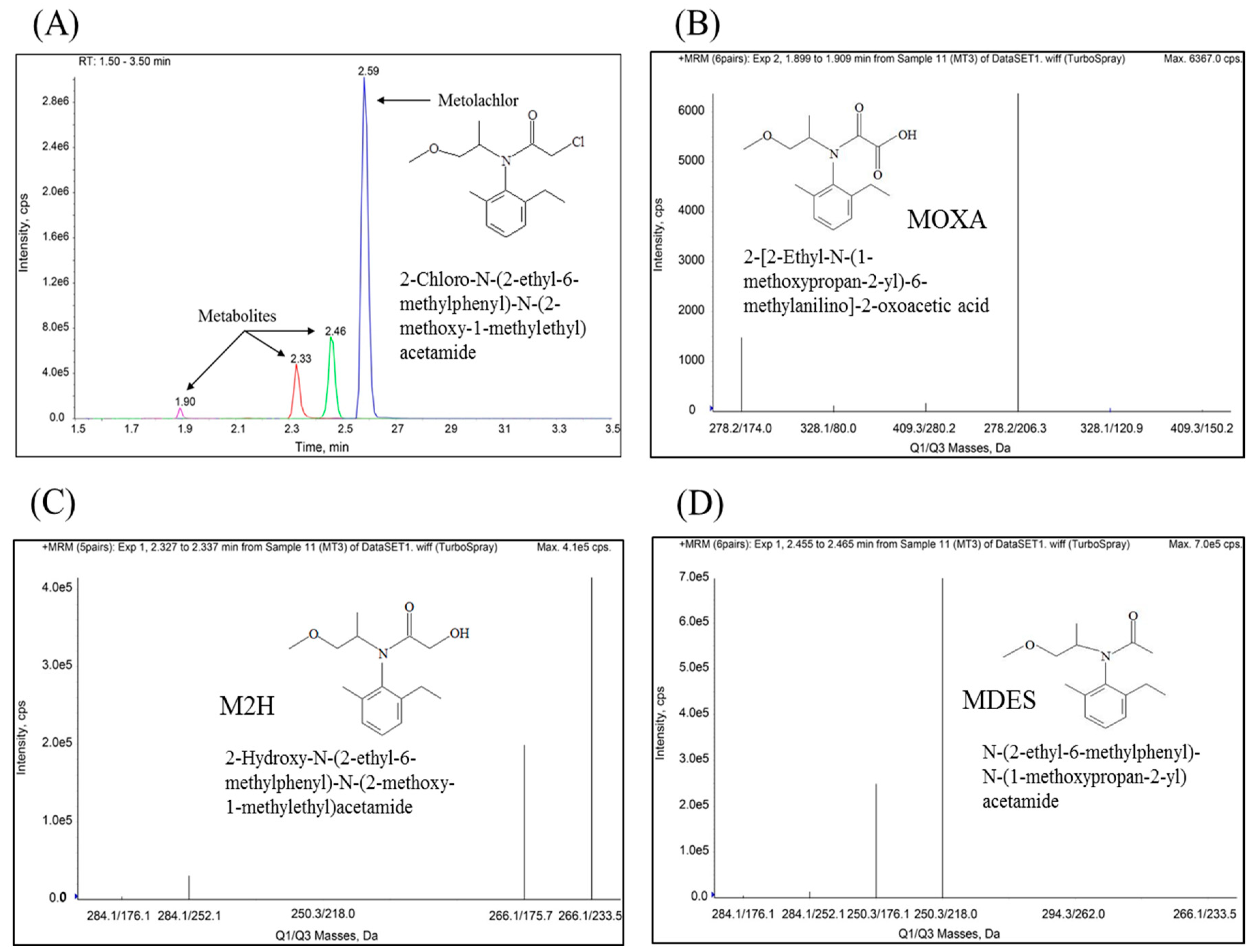
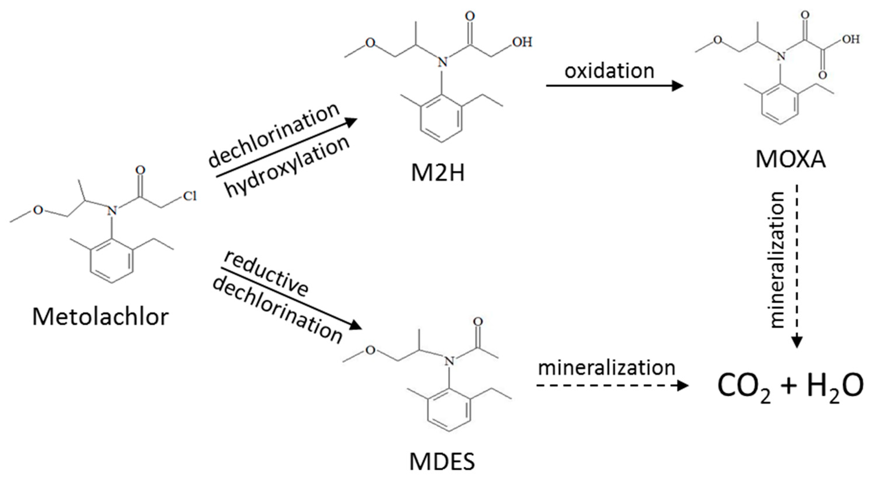
3.4. Metabolites of Metolachlor Degradation by Strain MET-F-1
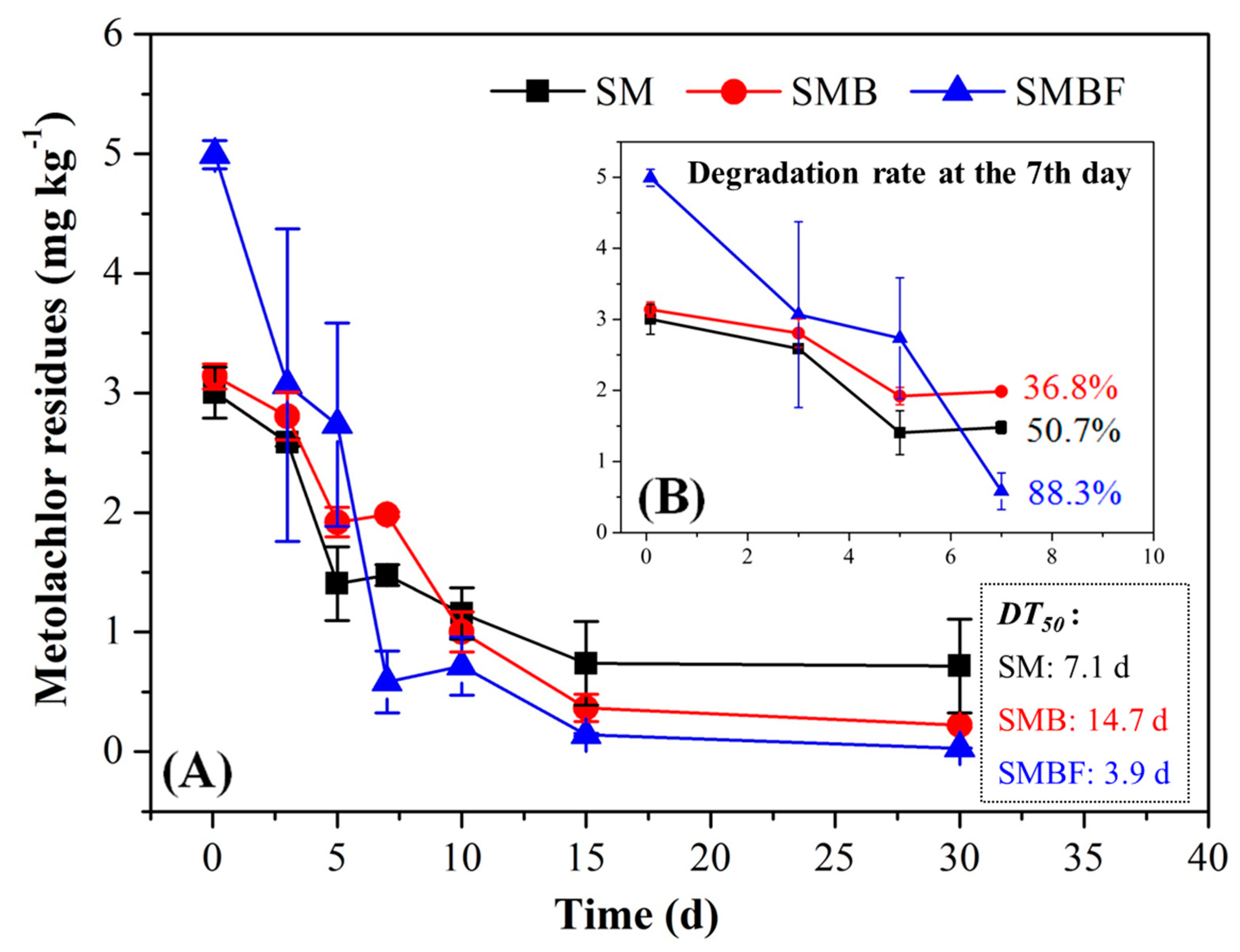
3.5. Degradation Performance of Bran Inoculants of MET-F-1 under Field Conditions
4. Conclusions
Supplementary Materials
Author Contributions
Funding
Conflicts of Interest
References
- Capel, P.D.; McCarthy, K.A.; Barbash, J.E. National, holistic, watershed-scale approach to understand the sources, transport, and fate of agricultural chemicals. J. Environ. Qual. 2008, 37, 983–993. [Google Scholar] [CrossRef]
- Karami-Mohajeri, S.; Abdollahi, M. Toxic influence of organophosphate, carbamate, and organochlorine pesticides on cellular metabolism of lipids, proteins, and carbohydrates. Hum. Exp. Toxicol. 2010, 30, 1119–1140. [Google Scholar] [CrossRef] [PubMed]
- Fenner, K.; Canonica, S.; Wackett, L.P.; Elsner, M. Evaluating Pesticide Degradation in the Environment: Blind Spots and Emerging Opportunities. Science 2013, 341, 752–758. [Google Scholar] [CrossRef] [PubMed] [Green Version]
- O’Connell, P.J.; Harms, C.T.; Allen, J.R. Metolachlor, S-metolachlor and their role within sustainable weed-management. Crop Prot. 1998, 17, 207–212. [Google Scholar] [CrossRef]
- A Sakkas, V.; Arabatzis, I.; Konstantinou, I.K.; Dimou, A.; A Albanis, T.; Falaras, P. Metolachlor photocatalytic degradation using TiO2 photocatalysts. Appl. Catal. B 2004, 49, 195–205. [Google Scholar] [CrossRef]
- Guelfi, D.R.V.; Gozzi, F.; Machulek, A.; Sirés, I.; Brillas, E.; De Oliveira, S.C. Degradation of herbicide S-metolachlor by electrochemical AOPs using a boron-doped diamond anode. Catal. Today 2018, 313, 182–188. [Google Scholar] [CrossRef]
- Liu, C.; Chen, L.; Ding, D.; Cai, T. Sulfate radical induced catalytic degradation of metolachlor: Efficiency and mechanism. Chem. Eng. J. 2019, 368, 606–617. [Google Scholar] [CrossRef]
- Hartnett, S.; Musah, S.; Dhanwada, K.R. Cellular effects of metolachlor exposure on human liver (HepG2) cells. Chemosphere 2013, 90, 1258–1266. [Google Scholar] [CrossRef]
- Caracciolo, A.B.; Giuliano, G.; Grenni, P.; Guzzella, L.; Pozzoni, F.; Bottoni, P.; Fava, L.; Crobe, A.; Orru, M.; Funari, E. Degradation and leaching of the herbicides metolachlor and diuron: A case study in an area of Northern Italy. Environ. Pollut. 2005, 134, 525–534. [Google Scholar] [CrossRef]
- Rose, C.E.; Coupe, R.H.; Capel, P.D.; Webb, R.M. Holistic assessment of occurrence and fate of metolachlor within environmental compartments of agricultural watersheds. Sci. Total Environ. 2018, 612, 708–719. [Google Scholar] [CrossRef]
- Velisek, J.; Stara, A.; Zuskova, E.; Kubec, J.; Buric, M.; Kouba, A. Effects of s-metolachlor on early life stages of marbled crayfish. Pestic. Biochem. Physiol. 2019, 153, 87–94. [Google Scholar] [CrossRef] [PubMed]
- Rice, P.J.; Anderson, T.A.; Coats, J.R. Degradation and persistence of metolachlor in soil: Effects of concentration, soil moisture, soil depth, and sterilization. Environ. Toxicol. Chem. 2002, 21, 2640–2648. [Google Scholar] [CrossRef] [PubMed]
- Wang, Y.; Wang, C.; Li, A.; Gao, J. Biodegradation of pentachloronitrobenzene by Arthrobacter nicotianae DH19. Lett. Appl. Microbiol. 2015, 61, 403–410. [Google Scholar] [CrossRef] [PubMed]
- Castillo, M.A.; Felis, N.; Aragón, P.; Cuesta, G.; Sabater, C. Biodegradation of the herbicide diuron by streptomycetes isolated from soil. Int. Biodeterior. Biodegrad. 2006, 58, 196–202. [Google Scholar] [CrossRef]
- Munoz, A.; Koskinen, W.C.; Cox, L.; Sadowsky, M.J. Biodegradation and mineralization of metolachlor and alachlor by Candida xestobii. J. Agric. Food Chem. 2011, 59, 619–627. [Google Scholar] [CrossRef]
- McGahen, L.L.; Tiedje, J.M. Metabolism of two new acylanilide herbicides, Antor herbicide (H-22234) and Dual (metolachlor) by the soil fungus Chaetomium globosum. J. Agric. Food Chem. 1978, 26, 414–419. [Google Scholar] [CrossRef]
- Wang, Y.-S.; Liu, J.-C.; Chen, W.-C.; Yen, J.-H. Characterization of acetanilide herbicides degrading bacteria isolated from tea garden soil. Microb. Ecol. 2008, 55, 435–443. [Google Scholar] [CrossRef]
- Zhang, J.; Zheng, J.-W.; Liang, B.; Wang, C.-H.; Cai, S.; Ni, Y.-Y.; He, J.; Li, S.-P. Biodegradation of chloroacetamide herbicides by Paracoccus sp. FLY-8 in vitro. J. Agric. Food Chem. 2011, 59, 4614–4621. [Google Scholar] [CrossRef]
- Ma, Y.; Liu, W.; Wen, Y.-Z. Enantioselective Degradation of Rac-Metolachlor and S-Metolachlor in Soil. Pedosphere 2006, 16, 489–494. [Google Scholar] [CrossRef]
- Chen, J.; Cui, B.K.; Dai, Y.C. Global diversity and molecular systematics of Wrightoporia s.l. (Russulales, Basidiomycota). Persoonia 2016, 37, 21–36. [Google Scholar] [CrossRef] [Green Version]
- Sun, Y.; Zhao, L.; Li, X.; Hao, Y.; Xu, H.; Weng, L.; Li, Y. Stimulation of earthworms (Eisenia fetida) on soil microbial communities to promote metolachlor degradation. Environ. Pollut. 2019, 248, 219–228. [Google Scholar] [CrossRef] [PubMed]
- Schroll, R.; Becher, H.H.; Dörfler, U.; Gayler, S.; Grundmann, S.; Hartmann, H.P.; Ruoss, J. Quantifying the effect of soil moisture on the aerobic microbial mineralization of selected pesticides in different soils. Environ. Sci. Technol. 2006, 40, 3305–3312. [Google Scholar] [CrossRef] [PubMed]
- Saxena, A.; Zhang, R.W.; Bollag, J.M. Microorganisms capable of metabolizing the herbicide metolachlor. Appl. Environ. Microbiol. 1987, 53, 390–396. [Google Scholar] [CrossRef] [Green Version]
- Liu, S.Y.; Lu, M.H.; Bollag, J.M. Transformation of metolachlor in soil inoculated with a Streptomyces sp. Biodegradation 1990, 1, 9–17. [Google Scholar] [CrossRef]
- Chen, S.; Deng, Y.; Chang, C.; Lee, J.; Cheng, Y.; Cui, Z.; Zhou, J.; He, F.; Hu, M.; Zhang, L.-H. Pathway and kinetics of cyhalothrin biodegradation by Bacillus thuringiensis strain ZS-19. Sci. Rep. 2015, 5, 8748. [Google Scholar] [CrossRef] [Green Version]
- Cycoń, M.; Żmijowska, A.; Piotrowska-Seget, Z. Enhancement of deltamethrin degradation by soil bioaugmentation with two different strains of Serratia marcescens. Int. J. Environ. Sci. Technol. (Tehran) 2014, 11, 1305–1316. [Google Scholar] [CrossRef] [Green Version]
- Xiao, Y.; Chen, S.; Gao, Y.; Hu, W.; Hu, M.; Zhong, G. Isolation of a novel beta-cypermethrin degrading strain Bacillus subtilis BSF01 and its biodegradation pathway. Appl. Microbiol. Biotechnol. 2015, 99, 2849–2859. [Google Scholar] [CrossRef]
- Cycon, M.; Piotrowska-Seget, Z. Pyrethroid-Degrading Microorganisms and Their Potential for the Bioremediation of Contaminated Soils: A Review. Front. Microb. 2016, 7, 1463. [Google Scholar] [CrossRef] [Green Version]
- Cycon, M.; Mrozik, A.; Piotrowska-Seget, Z. Bioaugmentation as a strategy for the remediation of pesticide-polluted soil: A review. Chemosphere 2017, 172, 52–71. [Google Scholar] [CrossRef]
- Zhao, R.B.; Bao, H.Y.; Liu, Y.X. Isolation and Characterization of Penicillium oxalicum ZHJ6 for Biodegradation of Methamidophos. Agric. Sci. China 2010, 9, 695–703. [Google Scholar] [CrossRef]
- Stamper, D.M.; Tuovinen, O.H. Biodegradation of the acetanilide herbicides alachlor, metolachlor, and propachlor. Crit. Rev. Microbiol. 1998, 24, 1–22. [Google Scholar] [CrossRef] [PubMed]
- Yang, T.; Guo, Y.; Gao, N.; Li, X.; Zhao, J. Modification of a cellulase system by engineering Penicillium oxalicum to produce cellulose nanocrystal. Carbohydr. Polym. 2020, 234, 115862. [Google Scholar] [CrossRef] [PubMed]
- Du, J.; Zhang, X.; Li, X.; Zhao, J.; Liu, G.; Gao, B.; Qu, Y. The cellulose binding region in Trichoderma reesei cellobiohydrolase I has a higher capacity in improving crystalline cellulose degradation than that of Penicillium oxalicum. Bioresour. Technol. 2018, 266, 19–25. [Google Scholar] [CrossRef] [PubMed]
- Olicón-Hernández, D.R.; Camacho-Morales, R.L.; Pozo, C.; González-López, J.; Aranda, E. Evaluation of diclofenac biodegradation by the ascomycete fungus Penicillium oxalicum at flask and bench bioreactor scales. Sci. Total Environ. 2019, 662, 607–614. [Google Scholar] [CrossRef] [PubMed]
- Tian, H.; Ma, Y.J.; Li, W.Y.; Wang, J.W. Efficient degradation of triclosan by an endophytic fungus Penicillium oxalicum B4. Environ. Sci. Pollut. Res. 2018, 25, 8963–8975. [Google Scholar] [CrossRef] [PubMed]
- Aranda, E.; Godoy, P.; Reina, R.; Badia-Fabregat, M.; Rosell, M.; Marco-Urrea, E.; García-Romera, I. Isolation of Ascomycota fungi with capability to transform PAHs: Insights into the biodegradation mechanisms of Penicillium oxalicum. Int. Biodeterior. Biodegrad. 2017, 122, 141–150. [Google Scholar] [CrossRef]
- Vryzas, Z.; Papadakis, E.N.; Oriakli, K.; Moysiadis, T.P.; Papadopoulou-Mourkidou, E. Biotransformation of atrazine and metolachlor within soil profile and changes in microbial communities. Chemosphere 2012, 89, 1330–1338. [Google Scholar] [CrossRef]
- Sun, Y.; Zhao, L.; Li, X.; Xu, H.; Weng, L.; Yang, L.; Li, Y. Response of soil bacterial and fungal community structure succession to earthworm addition for bioremediation of metolachlor. Ecotoxicol. Environ. Saf. 2020, 189, 109926. [Google Scholar] [CrossRef]
- Sanyal, D.; Kulshrestha, G. Metabolism of Metolachlor by Fungal Cultures. J. Agric. Food Chem. 2002, 50, 499–505. [Google Scholar] [CrossRef]
- White, M.P.; Potter, T.L.; Culbreath, A.K. Fungicide dissipation and impact on metolachlor aerobic soil degradation and soil microbial dynamics. Sci. Total Environ. 2010, 408, 1393–1402. [Google Scholar] [CrossRef]
- Liu, S.Y.; Zheng, Z.; Zhang, R.; Bollag, J.M. Sorption and metabolism of metolachlor by a bacterial community. Appl. Environ. Microbiol. 1989, 55, 733–740. [Google Scholar] [CrossRef] [PubMed] [Green Version]
- Shu, Y.L.; Freyer, A.J.; Bollag, J.M. Microbial dechlorination of the herbicide metolachlor. J. Agric. Food Chem. 1991, 39, 631–636. [Google Scholar]
- Karpouzas, D.G.; Morgan, J.A.W.; Walker, A. Isolation and characterisation of ethoprophos-degrading bacteria. FEMS Microbiol. Ecol. 2000, 33, 209–218. [Google Scholar] [CrossRef] [PubMed]
- Fang, H.; Dong, B.; Yan, H.; Tang, F.; Yu, Y. Characterization of a bacterial strain capable of degrading DDT congeners and its use in bioremediation of contaminated soil. J. Hazard. Mater. 2010, 184, 281–289. [Google Scholar] [CrossRef]
- Grundmann, S.; Fuß, R.; Schmid, M.; Laschinger, M.; Ruth, B.; Schulin, R.; Munch, J.C.; Schroll, R. Application of microbial hot spots enhances pesticide degradation in soils. Chemosphere 2007, 68, 511–517. [Google Scholar] [CrossRef]
- Chen, S.; Yang, L.; Hu, M.; Liu, J. Biodegradation of fenvalerate and 3-phenoxybenzoic acid by a novel Stenotrophomonas sp. strain ZS-S-01 and its use in bioremediation of contaminated soils. Appl. Microbiol. Biotechnol. 2011, 90, 755–767. [Google Scholar] [CrossRef]
- Manini, F.; Brasca, M.; Plumed-Ferrer, C.; Morandi, S.; Erba, D.; Casiraghi, M.C. Study of the Chemical Changes and Evolution of Microbiota During Sourdough like Fermentation of Wheat Bran. Cereal Chem. 2014, 91, 342–349. [Google Scholar] [CrossRef]
- Vermeulen, K.; Verspreet, J.; Courtin, C.M.; Haesebrouck, F.; Baeyen, S.; Haegeman, A.; Ducatelle, R.; Van Immerseel, F. Reduced-particle-size wheat bran is efficiently colonized by a lactic acid-producing community and reduces levels of Enterobacteriaceae in the cecal microbiota of broilers. Appl. Environ. Microbiol. 2018, 84, 01343-18. [Google Scholar] [CrossRef] [Green Version]
- Afsar, M.; Radha, S.; Girish, K.; Manonmani, H.K.; Kunhi, A.A.M. Optimization of carbon sources for the preparation of inoculum of hexachlorocy clohexane-degrading microbial consortium. J. Food Sci. Technol. Mysore 2005, 42, 238–241. [Google Scholar]
- Hesseltine, C.W.; Swain, E.W.; Wang, H.L. Production of fungal spores as inocula for oriental fermented foods. Dev. Ind. Microbiol. 1976, 17, 101–115. [Google Scholar]
- Sanghi, A.; Garg, N.; Sharma, J.; Kuhar, K.; Kuhad, R.C.; Gupta, V.K. Optimization of xylanase production using inexpensive agro-residues by alkalophilic Bacillus subtilis ASH in solid-state fermentation. World J. Microbiol. Biotechnol. 2008, 24, 633–640. [Google Scholar] [CrossRef]


Publisher’s Note: MDPI stays neutral with regard to jurisdictional claims in published maps and institutional affiliations. |
© 2020 by the authors. Licensee MDPI, Basel, Switzerland. This article is an open access article distributed under the terms and conditions of the Creative Commons Attribution (CC BY) license (http://creativecommons.org/licenses/by/4.0/).
Share and Cite
Chang, X.; Liang, J.; Sun, Y.; Zhao, L.; Zhou, B.; Li, X.; Li, Y. Isolation, Degradation Performance and Field Application of the Metolachlor-Degrading Fungus Penicillium oxalicum MET-F-1. Appl. Sci. 2020, 10, 8556. https://0-doi-org.brum.beds.ac.uk/10.3390/app10238556
Chang X, Liang J, Sun Y, Zhao L, Zhou B, Li X, Li Y. Isolation, Degradation Performance and Field Application of the Metolachlor-Degrading Fungus Penicillium oxalicum MET-F-1. Applied Sciences. 2020; 10(23):8556. https://0-doi-org.brum.beds.ac.uk/10.3390/app10238556
Chicago/Turabian StyleChang, Xingping, Junfeng Liang, Yang Sun, Lixia Zhao, Bin Zhou, Xiaojing Li, and Yongtao Li. 2020. "Isolation, Degradation Performance and Field Application of the Metolachlor-Degrading Fungus Penicillium oxalicum MET-F-1" Applied Sciences 10, no. 23: 8556. https://0-doi-org.brum.beds.ac.uk/10.3390/app10238556