
Clinical Outcome of a New Surgical Technique for the Treatment of Peri-Implant Dehiscence in the Esthetic Area. A Case Report

 ,
,  ,
, 
Abstract
:1. Introduction
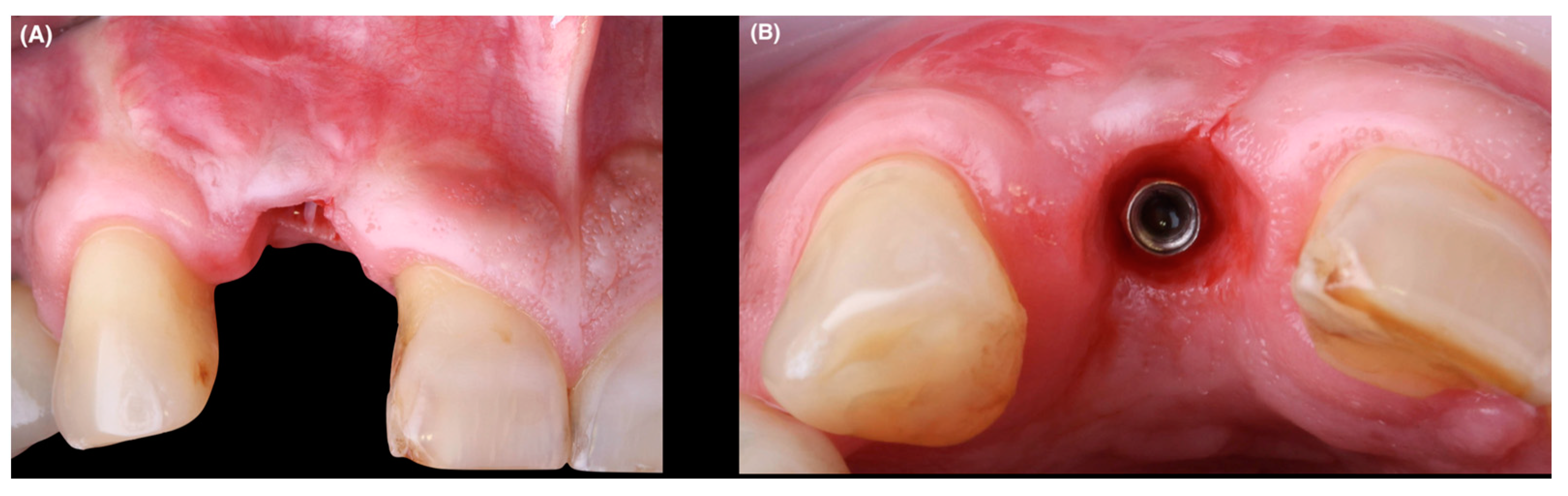
2. Case Report
2.1. Recruitment
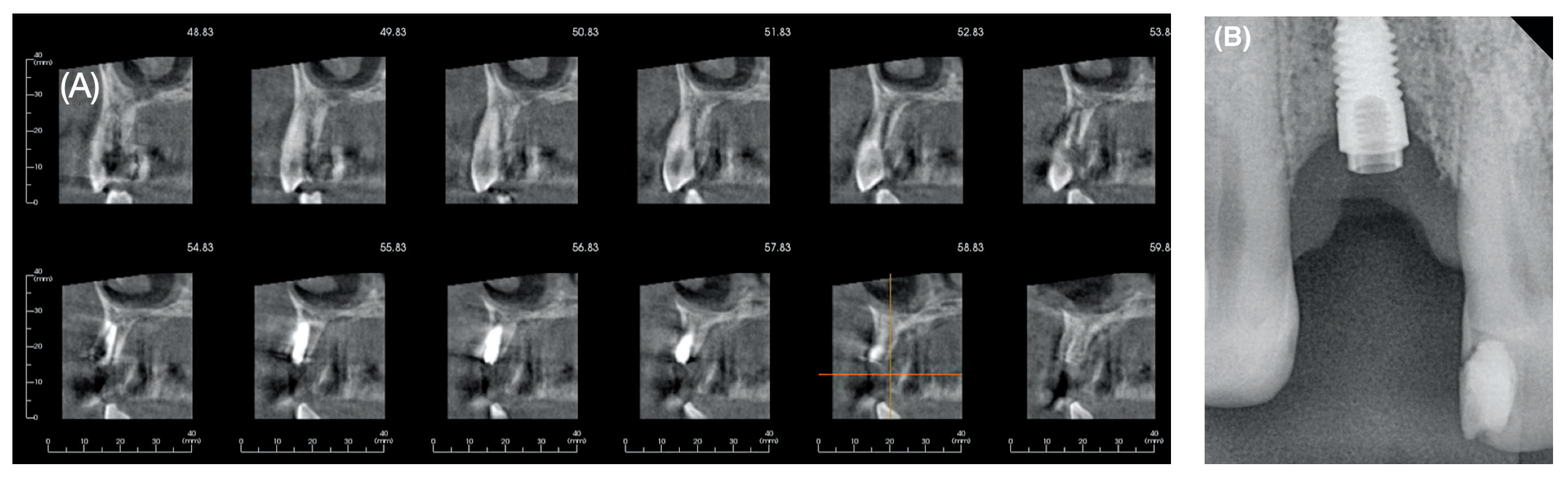
2.2. Baseline Clinical Assessment
- Probing depth (PD) measured in millimeters from the mucosal margin to the bottom of the peri-implant sulcus on the implant and from the gingival margin to the bottom of the gingival sulcus on the adjacent teeth, using a periodontal probe marked millimeter by millimeter adjusting the measurement in multiples of half a millimeter (Colorvue UNC 12, Hu-friedy, Chicago, IL, USA).
- Recession Depth (REC) measured on the implant on the mesio-, mid- and disto- buccal side by means of a digital millimeter ruler. The digital photographs were imported into a presentation software (Keynote®, Apple Inc, Cupertino, California, USA) and perpendicular lines were drawn taking as references the incisal edge and the cemento-enamel junction. The length of the clinical crown of the contralateral homologous tooth 2.2 was measured with a digital caliper from the incisal edge to the cemento-enamel junction. To calculate the initial and final REC on the implant, the length of the clinical crown was subtracted from the length of the clinical crown of the contralateral homologous tooth 2.2. On the teeth, the same probe was used to measure from the cemento-enamel junction to the gingival margin on the vestibular side.
- Gingival index (GI) (Löe and Silness) [29] scored from 0 to 3 according to the extent and severity of bleeding on probing.
- Plaque index (PI) (Silness and Löe) [30] scored from 0 to 3 according to the visibility and severity of plaque accumulation.
- Width of keratinized (WK) mucosa on the adjacent teeth and the 1.2 implant in the mid-vestibular site, recorded using the same periodontal probe.
- Mucosal thickness (MT) on the 1.2 implant in the mid-vestibular site, recorded using a caliper 2mm below the mucosal margin and, on the adjacent teeth using a K#10 endodontic file with rubber stop.
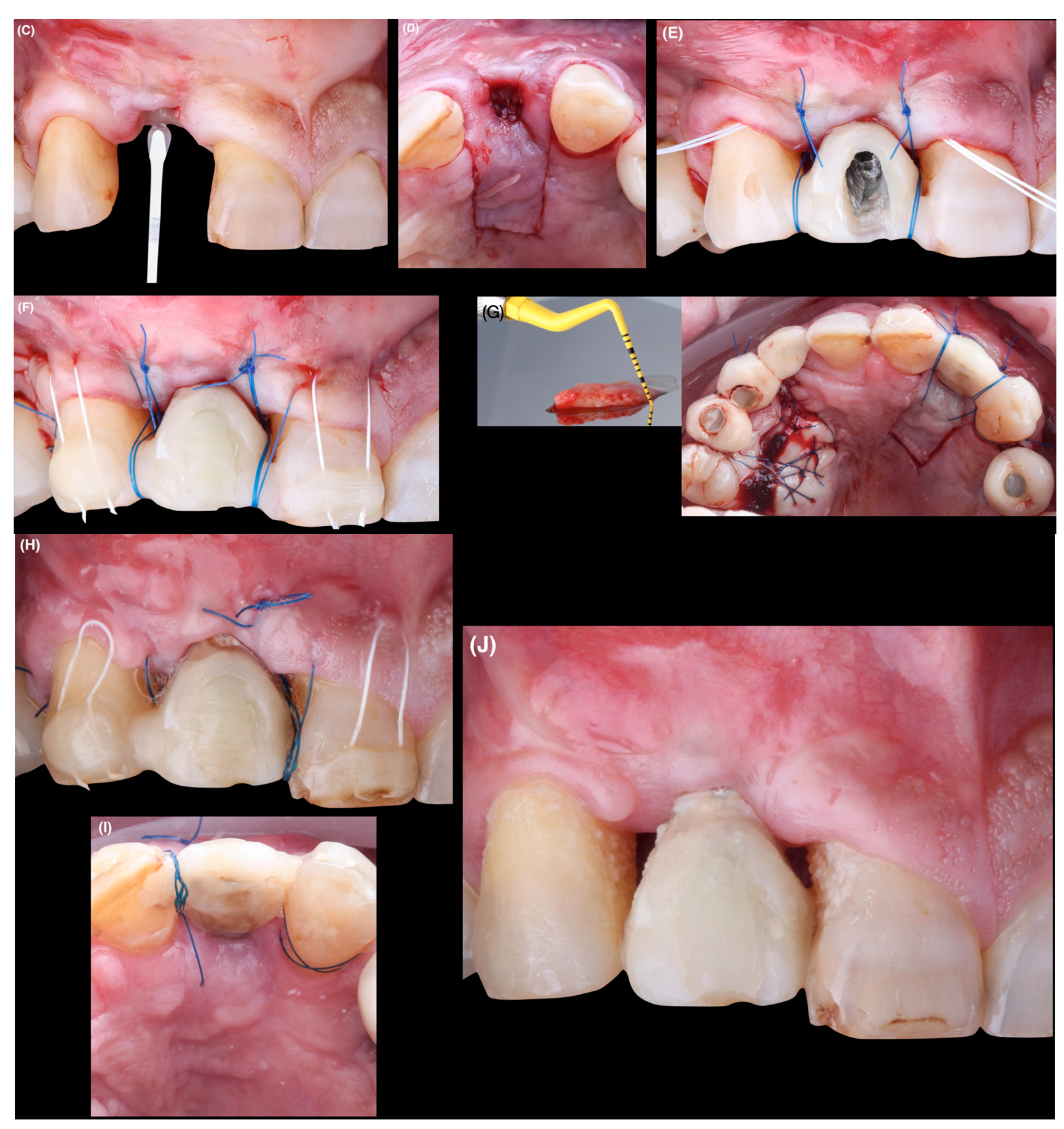
2.3. Surgical Procedure
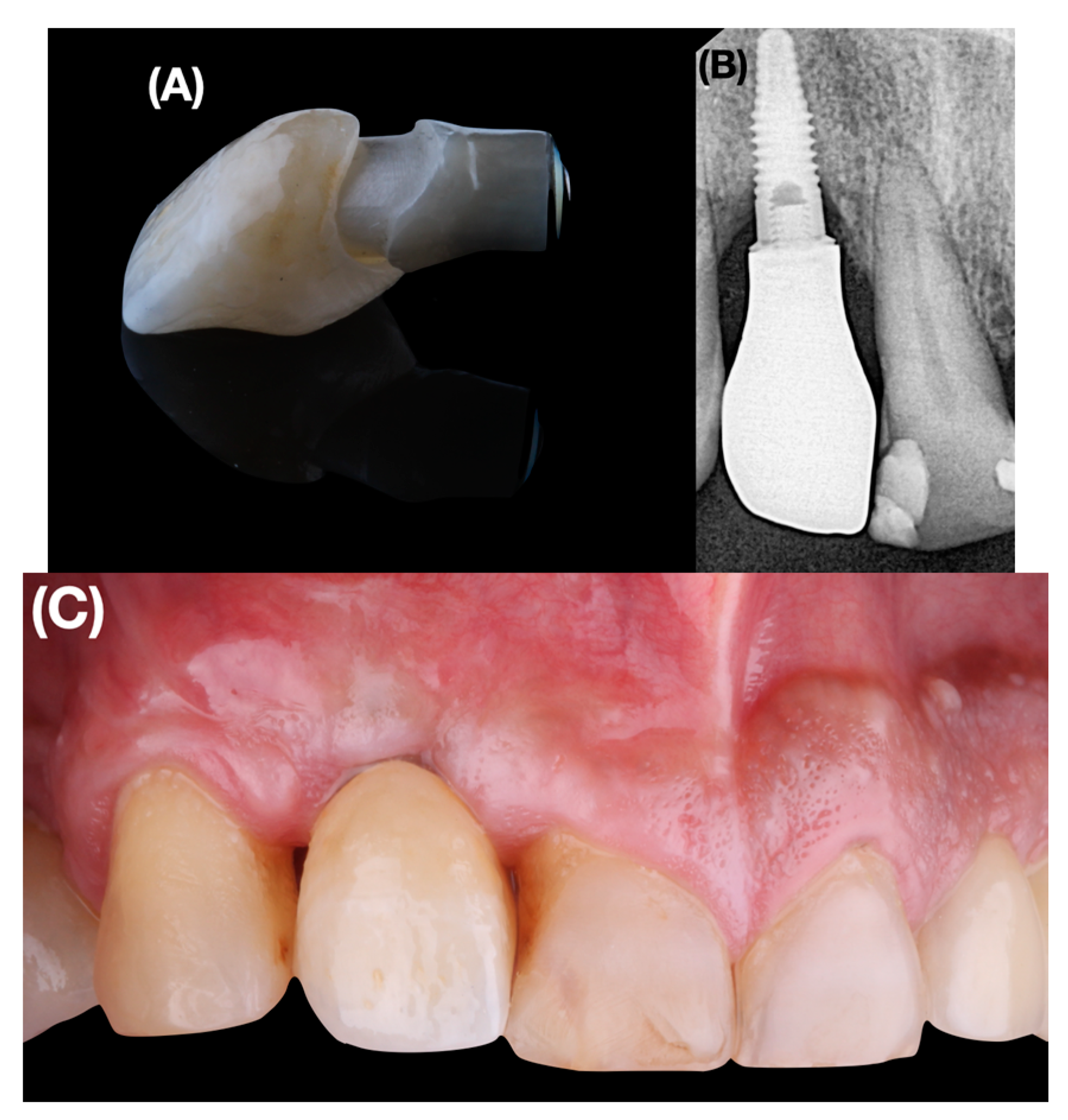
2.4. Restorative Phase
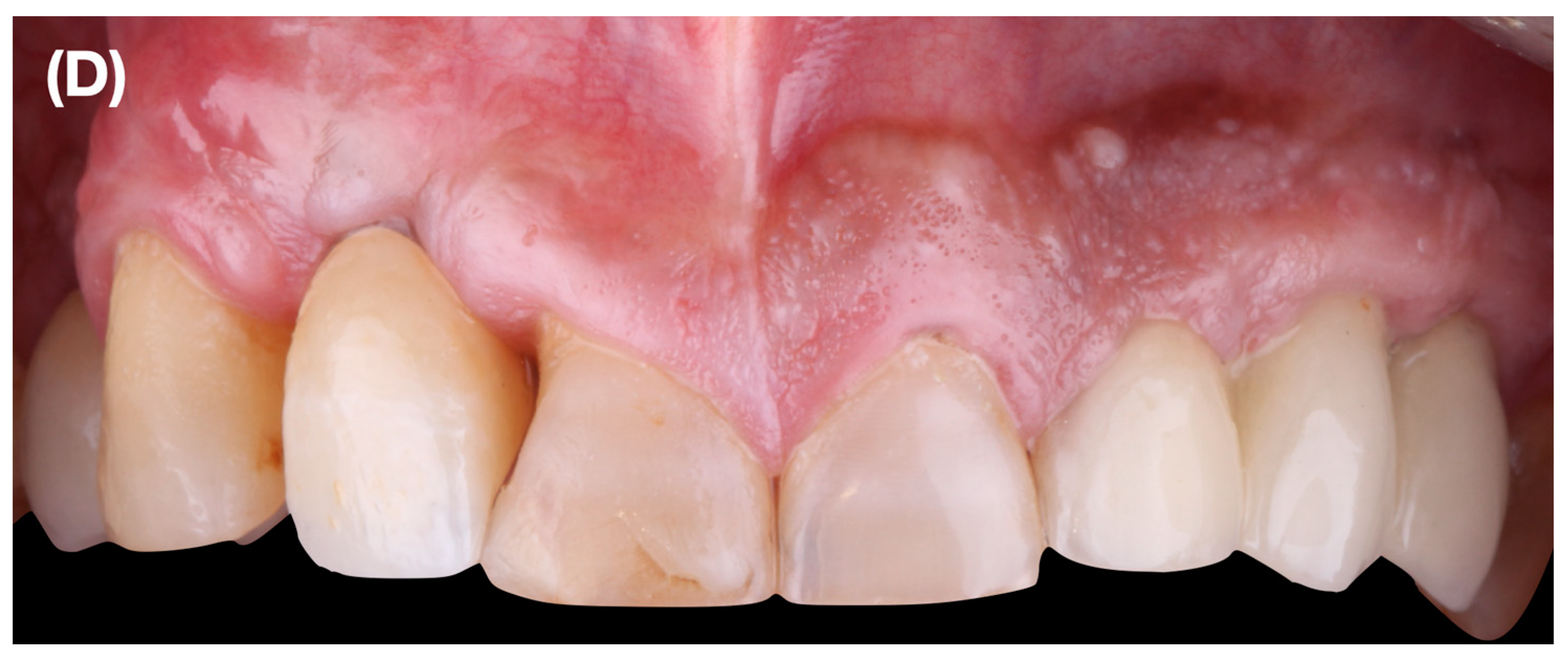
3. Results
3.1. Gingival Parameters
3.2. Esthetic Parameters
4. Discussion
5. Conclusions
Author Contributions
Funding
Institutional Review Board Statement
Informed Consent Statement
Data Availability Statement
Conflicts of Interest
Abbreviations
| PSTD | peri-implant soft tissue dehiscence/deficiency |
| CTG | connective tissue graft |
| CAF | coronally advanced flap |
| KMW | keratinized mucosa width |
| MT | mucosal thickness |
| PMMA | polymethylmethacrylate |
| WES | white esthetic score |
| PES | pink esthetic score |
| PD | probing depth |
| REC | recession depth |
| GI | gingival index |
| PI | plaque index |
References
- Brånemark, R.; Brånemark, P.I.; Rydevik, B.; Myers, R.R. Osseointegration in skeletal reconstruction and rehabilitation: A review. J. Rehabil. Res. Dev. 2001, 38, 175–181. [Google Scholar]
- Diz, P.; Scully, C.; Sanz, M. Dental implants in the medically compromised patient. J. Dent. 2013, 41, 195–206. [Google Scholar] [CrossRef]
- Pellegrini, G.; Francetti, L.; Barbaro, B.; Del Fabbro, M. Novel surfaces and osseointegration in implant dentistry. J. Investig. Clin. Dent. 2018, 9, e12349. [Google Scholar] [CrossRef]
- Goiato, M.C.; dos Santos, D.M.; Santiago, J.F., Jr.; Moreno, A.; Pellizzer, E.P. Longevity of dental implants in type IV bone: A systematic review. Int. J. Oral Maxillofac. Surg. 2014, 43, 1108–1116. [Google Scholar] [CrossRef]
- Pjetursson, B.E.; Thoma, D.; Jung, R.; Zwahlen, M.; Zembic, A. A systematic review of the survival and complication rates of implant-supported fixed dental prostheses (FDPs) after a mean observation period of at least 5 years. Clin. Oral Implants Res. 2012, 23 (Suppl. 6), 22–38. [Google Scholar] [CrossRef] [PubMed]
- Lee, C.T.; Huang, Y.W.; Zhu, L.; Weltman, R. Prevalences of peri-implantitis and peri-implant mucositis: Systematic review and meta-analysis. J. Dent. 2017, 62, 1–12. [Google Scholar] [CrossRef] [PubMed]
- Thoma, D.S.; Buranawat, B.; Hämmerle, C.H.; Held, U.; Jung, R.E. Efficacy of soft tissue augmentation around dental implants and in partially edentulous areas: A systematic review. J. Clin. Periodontol. 2014, 41 (Suppl. 15), S77–S91. [Google Scholar] [CrossRef] [Green Version]
- Changi, K.K.; Finkelstein, J.; Papapanou, P.N. Peri-implantitis prevalence, incidence rate, and risk factors: A study of electronic health records at a U.S. dental school. Clin. Oral Implants Res. 2019, 30, 306–314. [Google Scholar] [CrossRef]
- Krisam, J.; Ott, L.; Schmitz, S.; Klotz, A.L.; Seyidaliyeva, A.; Rammelsberg, P.; Zenthöfer, A. Factors affecting the early failure of implants placed in a dental practice with a specialization in implantology—A retrospective study. BMC Oral Health 2019, 19, 208. [Google Scholar] [CrossRef] [PubMed]
- Renouard, F.; Amalberti, R.; Renouard, E. Are “Human Factors” the Primary Cause of Complications in the Field of Implant Dentistry? Int. J. Oral Maxillofac. Implants 2017, 32, e55–e61. [Google Scholar] [CrossRef] [Green Version]
- Cattoni, F.; Teté, G.; Calloni, A.M.; Manazza, F.; Gastaldi, G.; Capparè, P. Milled versus moulded mock-ups based on the superimposition of 3D meshes from digital oral impressions: A comparative in vitro study in the aesthetic area. BMC Oral Health 2019, 19, 230. [Google Scholar] [CrossRef] [PubMed]
- Crespi, R.; Capparé, P.; Romanos, G.E.; Mariani, E.; Benasciutti, E.; Gherlone, E. Corticocancellous porcine bone in the healing of human extraction sockets: Combining histomorphometry with osteoblast gene expression profiles in vivo. Int. J. Oral Maxillofac. Implants 2011, 26, 866–872. [Google Scholar] [PubMed]
- Crespi, R.; Capparè, P.; Gherlone, E. Comparison of magnesium-enriched hydroxyapatite and porcine bone in human extraction socket healing: A histologic and histomorphometric evaluation. Int. J. Oral Maxillofac. Implants 2011, 26, 1057–1062. [Google Scholar]
- Berglundh, T.; Lindhe, J.; Ericsson, I.; Marinello, C.P.; Liljenberg, B.; Thomsen, P. The soft tissue barrier at implants and teeth. Clin. Oral Implants Res. 1991, 2, 81–90. [Google Scholar] [CrossRef]
- Atsuta, I.; Ayukawa, Y.; Kondo, R.; Oshiro, W.; Matsuura, Y.; Furuhashi, A.; Tsukiyama, Y.; Koyano, K. Soft tissue sealing around dental implants based on histological interpretation. J. Prosthodont. Res. 2016, 60, 3–11. [Google Scholar] [CrossRef]
- Tan, W.L.; Wong, T.L.; Wong, M.C.; Lang, N.P. A systematic review of post-extractional alveolar hard and soft tissue dimensional changes in humans. Clin. Oral Implants Res. 2012, 23 (Suppl. 5), 1–21. [Google Scholar] [CrossRef]
- Mazzotti, C.; Stefanini, M.; Felice, P.; Bentivogli, V.; Mounssif, I.; Zucchelli, G. Soft-tissue dehiscence coverage at peri-implant sites. Periodontology 2000 2018, 77, 256–272. [Google Scholar] [CrossRef] [PubMed]
- Giannobile, W.V.; Jung, R.E.; Schwarz, F.; Groups of the 2nd Osteology Foundation Consensus Meeting. Evidence-based knowledge on the aesthetics and maintenance of peri-implant soft tissues: Osteology Foundation Consensus Report Part 1-Effects of soft tissue augmentation procedures on the maintenance of peri-implant soft tissue health. Clin. Oral Implants Res. 2018, 29 (Suppl. 15), 7–10. [Google Scholar] [CrossRef]
- Monje, A.; Blasi, G. Significance of keratinized mucosa/gingiva on peri-implant and adjacent periodontal conditions in erratic maintenance compliers. J. Periodontol. 2019, 90, 445–453. [Google Scholar] [CrossRef] [PubMed]
- Ioannidis, A.; Cathomen, E.; Jung, R.E.; Fehmer, V.; Hüsler, J.; Thoma, D.S. Discoloration of the mucosa caused by different restorative materials—A spectrophotometric in vitro study. Clin. Oral Implants Res. 2017, 28, 1133–1138. [Google Scholar] [CrossRef] [Green Version]
- Nisapakultorn, K.; Suphanantachat, S.; Silkosessak, O.; Rattanamongkolgul, S. Factors affecting soft tissue level around anterior maxillary single-tooth implants. Clin. Oral Implants Res. 2010, 21, 662–670. [Google Scholar] [CrossRef] [PubMed]
- Berglundh, T.; Armitage, G.; Araujo, M.G.; Avila-Ortiz, G.; Blanco, J.; Camargo, P.M.; Chen, S.; Cochran, D.; Derks, J.; Figuero, E.; et al. Peri-implant diseases and conditions: Consensus report of workgroup 4 of the 2017 World Workshop on the Classification of Periodontal and Peri-Implant Diseases and Conditions. J. Clin. Periodontol. 2018, 45 (Suppl. 20), S286–S291. [Google Scholar] [CrossRef] [Green Version]
- Sanz-Martín, I.; Regidor, E.; Navarro, J.; Sanz-Sánchez, I.; Sanz, M.; Ortiz-Vigón, A. Factors associated with the presence of peri-implant buccal soft tissue dehiscences: A case-control study [published online ahead of print, 2020 Jan 24]. J. Periodontol. 2020. [Google Scholar] [CrossRef]
- Zucchelli, G.; Mazzotti, C.; Mounssif, I.; Mele, M.; Stefanini, M.; Montebugnoli, L. A novel surgical-prosthetic approach for soft tissue dehiscence coverage around single implant. Clin. Oral Implants Res. 2013, 24, 957–962. [Google Scholar] [CrossRef]
- Roccuzzo, M.; Dalmasso, P.; Pittoni, D.; Roccuzzo, A. Treatment of buccal soft tissue dehiscence around single implant: 5-year results from a prospective study. Clin. Oral Investig. 2019, 23, 1977–1983. [Google Scholar] [CrossRef] [PubMed]
- Zucchelli, G.; Tavelli, L.; Stefanini, M.; Barootchi, S.; Mazzotti, C.; Gori, G.; Wang, H.L. Classification of facial peri-implant soft tissue dehiscence/deficiencies at single implant sites in the esthetic zone. J. Periodontol. 2019, 90, 1116–1124. [Google Scholar] [CrossRef] [PubMed]
- Fürhauser, R.; Florescu, D.; Benesch, T.; Haas, R.; Mailath, G.; Watzek, G. Evaluation of soft tissue around single-tooth implant crowns: The pink esthetic score. Clin. Oral Implants Res. 2005, 16, 639–644. [Google Scholar] [CrossRef]
- Belser, U.C.; Grütter, L.; Vailati, F.; Bornstein, M.M.; Weber, H.P.; Buser, D. Outcome evaluation of early placed maxillary anterior single-tooth implants using objective esthetic criteria: A cross-sectional, retrospective study in 45 patients with a 2- to 4-year follow-up using pink and white esthetic scores. J. Periodontol. 2009, 80, 140–151. [Google Scholar] [CrossRef]
- Löe, H.; Silness, J. Periodontal Disease in pregnancy.I. Prevalence and severity. Acta Odontol. Scand. 1963, 21, 533–551. [Google Scholar] [CrossRef] [PubMed]
- Silness, J.; Löe, H. Periodontal disease in pregnancy. II. Correlation between oral hygiene and periodontal condition. Acta Odontol. Scand. 1964, 22, 121–135. [Google Scholar] [CrossRef]
- Bethaz, N.; Romano, F.; Ferrarotti, F.; Mariani, G.M.; Aimetti, M. A mucogingival technique for the treatment of multiple recession defects in the mandibular anterior region: A case series with a 2-year follow-up. Int. J. Periodontics Restor. Dent. 2014, 34, 345–352. [Google Scholar] [CrossRef] [PubMed] [Green Version]
- Tinti, C.; Parma-Benfenati, S. Coronally positioned palatal sliding flap. Int. J. Periodontics Restor. Dent 1995, 15, 298–310. [Google Scholar]
- González-Martín, O.; Lee, E.; Weisgold, A.; Veltri, M.; Su, H. Contour Management of Implant Restorations for Optimal Emergence Profiles: Guidelines for Immediate and Delayed Provisional Restorations. Int. J. Periodontics Restor. Dent. 2020, 40, 61–70. [Google Scholar] [CrossRef] [PubMed]
- Hinds, K.F. Custom impression coping for an exact registration of the healed tissue in the esthetic implant restoration. Int. J. Periodontics Restor. Dent. 1997, 17, 584–591. [Google Scholar]
- Hämmerle, C.H.F.; Tarnow, D. The etiology of hard- and soft-tissue deficiencies at dental implants: A narrative review. J. Periodontol. 2018, 89 (Suppl. 1), S291–S303. [Google Scholar] [CrossRef] [Green Version]
- Wang, Q.; Tang, Z.; Han, J.; Meng, H. The width of keratinized mucosa around dental implants and its influencing factors. Clin. Implant Dent. Relat. Res. 2020, 22, 359–365. [Google Scholar] [CrossRef]
- Sanz, M.; Simion, M.; Working Group 3 of the European Workshop on Periodontology. Surgical techniques on periodontal plastic surgery and soft tissue regeneration: Consensus report of Group 3 of the 10th European Workshop on Periodontology. J. Clin. Periodontol. 2014, 41 (Suppl. 15), S92–S97. [Google Scholar] [CrossRef]
- Tatakis, D.N.; Chambrone, L.; Allen, E.P.; Langer, B.; McGuire, M.K.; Richardson, C.R.; Zabalegui, I.; Zadeh, H.H. Periodontal soft tissue root coverage procedures: A consensus report from the AAP Regeneration Workshop. J. Periodontol. 2015, 86 (Suppl. 2), S52–S55. [Google Scholar] [CrossRef] [PubMed] [Green Version]
- Chambrone, L.; Ortega MA, S.; Sukekava, F.; Rotundo, R.; Kalemaj, Z.; Buti, J.; Prato, G.P.P. Root coverage procedures for treating single and multiple recession-type defects: An updated Cochrane systematic review. J. Periodontol. 2019, 90, 1399–1422. [Google Scholar] [CrossRef] [PubMed]
- Roccuzzo, M.; Gaudioso, L.; Bunino, M.; Dalmasso, P. Surgical treatment of buccal soft tissue recessions around single implants: 1-year results from a prospective pilot study. Clin. Oral Implants Res. 2014, 25, 641–646. [Google Scholar] [CrossRef] [PubMed]
- Schwarz, F.; Mihatovic, I.; Shirakata, Y.; Becker, J.; Bosshardt, D.; Sculean, A. Treatment of soft tissue recessions at titanium implants using a resorbable collagen matrix: A pilot study. Clin. Oral Implants Res. 2014, 25, 110–115. [Google Scholar] [CrossRef] [PubMed]
- Froum, S.J.; Wang, W.C.; Hafez, T.; Suzuki, T.; Yu, Y.C.P.; Cho, S.C. Incision Design and Soft Tissue Management to Maintain or Establish an Interproximal Papilla Around Integrated Implants: A Case Series. Int. J. Periodontics Restor. Dent. 2018, 38, 61–69. [Google Scholar] [CrossRef] [PubMed] [Green Version]
- Sculean, A.; Chappuis, V.; Cosgarea, R. Coverage of mucosal recessions at dental implants. Periodontology 2000 2017, 73, 134–140. [Google Scholar] [CrossRef] [PubMed]
- Zuhr, O.; Rebele, S.F.; Cheung, S.L.; Hürzeler, M.B.; Research Group on Oral Soft Tissue Biology and Wound Healing. Surgery without papilla incision: Tunneling flap procedures in plastic periodontal and implant surgery. Periodontology 2000 2018, 77, 123–149. [Google Scholar] [CrossRef]
- Zucchelli, G.; Felice, P.; Mazzotti, C.; Marzadori, M.; Mounssif, I.; Monaco, C.; Stefanini, M. 5-year outcomes after coverage of soft tissue dehiscence around single implants: A prospective cohort study. Eur. J. Oral Implantol. 2018, 11, 215–224. [Google Scholar] [PubMed]
- Marggraf, E. A direct technique with a double lateral bridging flap for coverage of denuded root surface and gingiva extension. Clinical evaluation after 2 years. J. Clin. Periodontol. 1985, 12, 69–76. [Google Scholar] [CrossRef] [PubMed]
- Raetzke, P.B. Covering localized areas of root exposure employing the “envelope” technique. J. Periodontol. 1985, 56, 397–402. [Google Scholar] [CrossRef] [PubMed]
- Azzi, R.; Etienne, D.; Sauvan, J.L.; Miller, P.D. Root coverage and papilla reconstruction in Class IV recession: A case report. Int. J. Periodontics Restor. Dent. 1999, 19, 449–455. [Google Scholar]
- Sculean, A.; Allen, E.P. The Laterally Closed Tunnel for the Treatment of Deep Isolated Mandibular Recessions: Surgical Technique and a Report of 24 Cases. Int. J. Periodontics Restor. Dent. 2018, 38, 479–487. [Google Scholar] [CrossRef] [PubMed]
- Nemcovsky, C.E. Interproximal papilla augmentation procedure: A novel surgical approach and clinical evaluation of 10 consecutive procedures. Int. J. Periodontics Restor. Dent. 2001, 21, 553–559. [Google Scholar]
- Zadeh, H.H. Minimally invasive treatment of maxillary anterior gingival recession defects by vestibular incision subperiosteal tunnel access and platelet-derived growth factor BB. Int. J. Periodontics Restor. Dent. 2011, 31, 653–660. [Google Scholar]
- Aslan, S.; Buduneli, N.; Cortellini, P. Entire Papilla Preservation Technique: A Novel Surgical Approach for Regenerative Treatment of Deep and Wide Intrabony Defects. Int. J. Periodontics Restor. Dent. 2017, 37, 227–233. [Google Scholar] [CrossRef] [PubMed] [Green Version]
- Moreno Rodríguez, J.A.; Ortiz Ruiz, A.J.; Caffesse, R.G. Periodontal reconstructive surgery of deep intraosseous defects using an apical approach. Non-incised papillae surgical approach (NIPSA): A retrospective cohort study. J. Periodontol. 2019, 90, 454–464. [Google Scholar] [CrossRef] [PubMed]
- Maiorana, C.; Poli, P.P.; Beretta, M. Guided Bone Regeneration and Implant Placement in Association With a Coronally Positioned Palatal Sliding Flap: A 17-Year Follow-Up Case Report. J. Oral Implantol. 2018, 44, 371–376. [Google Scholar] [CrossRef] [PubMed]


| Class | Peri-Implant Soft Tissue Dehiscence Characteristics | Subclass | Recommended Surgical Treatment |
|---|---|---|---|
| I | The soft tissue margin is located at the same level of the ideal position of the gingival margin of the homologous natural tooth, and the color of the abutment/implant is visible only through the mucosa and/or there is a lack of keratinized tissue/soft tissue thickness. | a: The tip of both papillae is ≥3 mm coronal to the ideal position of soft tissue margin of the implant-supported crown. b: The tip of at least one papilla is ≥1 mm but <3 mm coronal to the ideal position of the soft tissue margin of the implant supported crown. c: The height of at least one papilla is <1 mm coronal to the ideal position of the soft tissue margin of the implant-supported crown. | Ia: coronally advanced flap (CAF) or tunnel plus CTG (or other graft substitutes). Ib: Combined prosthetic–surgical approach. |
| II | The soft tissue margin is located more apical to the ideal position of the gingival margin of the homologous natural tooth, and the implant-supported crown profile is located inside (more palatal) the imaginary curve line that connects the profile of the adjacent teeth at the level of the soft tissue margin. | IIa: No crown removal, CAF plus CTG. IIb: Combined prosthetic–surgical approach. IIc: Soft tissue augmentation with submerged healing. | |
| III | The soft tissue margin is located more apical to the ideal position of the gingival margin of the homologous natural tooth. The implant-supported crown profile is located outside (more facially) the imaginary curve line that connects the profile of the adjacent teeth at the level of the soft tissue margin, and the head of the implant (evaluated by removing the crown) is inside (more palatally) the imaginary straight line connecting the profile of the adjacent teeth at the level of the soft tissue margin. | IIIa: Crown removal, CAF plus CTG. IIIb: Combined prosthetic–surgical approach. IIIc: Soft tissue augmentation with submerged healing. | |
| IV | The soft tissue margin is located more apical with respect of the ideal position of the gingival margin of the homologous natural tooth. The implant-supported crown profile is located outside (more facially) the imaginary curve line that connects the profile of the adjacent teeth at the level of the soft tissue margin, and the head of the implant (evaluated by removing the crown) is outside (more facially) the imaginary straight line connecting the profile of the adjacent teeth at the level of the soft tissue margin. | IVa: Combined prosthetic–surgical approach. IVb: Soft tissue augmentation with submerged healing. IVc: Implant removal. |
| PES | Baseline Crown | Final Crown |
|---|---|---|
| Mesial papilla * | 0 | 0 |
| Distal papilla * | 0 | 0 |
| Curvature of facial mucosa ** | 0 | 1 |
| Level of facial mucosa ** | 0 | 1 |
| Soft tissue color and texture ** | 0 | 1 |
| PES score | 0/10 | 3/10 |
| WES | ||
| Form ** | 0 | 1 |
| Volume/outline ** | 0 | 1 |
| Color (hue/value) ** | 1 | 1 |
| Surface texture ** | 0 | 1 |
| Translucency ** | 0 | 1 |
| WES score | 1/10 | 5/10 |
| Baseline | 15 Months | ||||||||||||||||||
|---|---|---|---|---|---|---|---|---|---|---|---|---|---|---|---|---|---|---|---|
| Tooth/Implant | 1.3 | 1.2 | 1.1 | 1.3 | 1.2 | 1.1 | |||||||||||||
| D | C | M | D | C | M | D | C | M | D | C | M | D | C | M | D | C | M | ||
| Parameter | Probing Depth | 4 | 3 | 4 | 3 | 0 | 3 | 2 | 4 | 3 | 4 | 2.5 | 3 | 7 | 7 | 7 | 4 | 4 | 3 |
| Recession Depth | 0 | 0 | 1 | 3 | 5 | 3 | 5 | 0 | 0 | 0 | 1 | 1 | 2 | 3 | 1.5 | 3 | 0 | 0 | |
| Gingival Index | 1 | 3 | 2 | 0 | 0 | 0 | |||||||||||||
| Plaque Index | 1 | 2 | 2 | 1 | 0 | 1 | |||||||||||||
| Width of Keratinized | 2 | 1 | 3 | 2 | 3 | 3 | |||||||||||||
| Mucosal Thickness | 1.3 | 0.3 | 2 | 1.3 | 2.6 | 2 | |||||||||||||
Publisher’s Note: MDPI stays neutral with regard to jurisdictional claims in published maps and institutional affiliations. |
© 2021 by the authors. Licensee MDPI, Basel, Switzerland. This article is an open access article distributed under the terms and conditions of the Creative Commons Attribution (CC BY) license (https://creativecommons.org/licenses/by/4.0/).
Share and Cite
Quispe-López, N.; García-Faria, C.; Mena-Álvarez, J.; Guadilla, Y.; Garrido Martínez, P.; Montero, J. Clinical Outcome of a New Surgical Technique for the Treatment of Peri-Implant Dehiscence in the Esthetic Area. A Case Report. Appl. Sci. 2021, 11, 4781. https://0-doi-org.brum.beds.ac.uk/10.3390/app11114781
Quispe-López N, García-Faria C, Mena-Álvarez J, Guadilla Y, Garrido Martínez P, Montero J. Clinical Outcome of a New Surgical Technique for the Treatment of Peri-Implant Dehiscence in the Esthetic Area. A Case Report. Applied Sciences. 2021; 11(11):4781. https://0-doi-org.brum.beds.ac.uk/10.3390/app11114781
Chicago/Turabian StyleQuispe-López, Norberto, Carmen García-Faria, Jesús Mena-Álvarez, Yasmina Guadilla, Pablo Garrido Martínez, and Javier Montero. 2021. "Clinical Outcome of a New Surgical Technique for the Treatment of Peri-Implant Dehiscence in the Esthetic Area. A Case Report" Applied Sciences 11, no. 11: 4781. https://0-doi-org.brum.beds.ac.uk/10.3390/app11114781