Case Report

Radiographic and Histologic Analysis 1–2 Years after Alveolar Ridge Preservation in Maxillary Premolar and Molar: A Case Report

1
Department of Periodontology, Research Institute for Periodontal Regeneration, Yonsei University College of Dentistry, Seoul 03772, Korea
2
Department of Oral Medicine, Infection and Immunity, Harvard School of Dental Medicine, Boston, MA 02115, USA
*
Author to whom correspondence should be addressed.
Submission received: 17 June 2021 / Revised: 8 July 2021 / Accepted: 15 July 2021 / Published: 17 July 2021
(This article belongs to the Special Issue Clinical Applications for Dentistry and Oral Health)

Abstract

:
After tooth extraction, notable ridge alterations occur due to resorption of bundle bones during the healing process. In areas with thin or damaged socket walls and multiple adjacent tooth extraction, dimensional changes are more prominent in the marginal proportion. In addition to the marginal changes, upper molar teeth are also vulnerable to pneumatization of the maxillary sinus. To reduce dimensional changes in extraction sockets, alveolar ridge preservation (ARP) is favored by many clinicians in areas where a large amount of dimensional change is expected. This case report presents two cases of ARP using collagenated demineralized bovine bone mineral and demineralized porcine bone mineral in the apically involved upper premolar and molar, respectively. Implants were placed one and two years, respectively, after the ARP. Radiographic analyses of residual bone height and volume were measured using cone-beam-computed tomography (CBCT) and histologic analysis of newly formed mineralized bone and residual graft material percentages were measured from the collected tissue samples using a trephine bur. Implants were placed using a simple technique, without any additional bone grafts at the marginal proportion. The ARP technique could maintain the alveolar bone height and volume, as well as minimize the invasiveness of surgical procedures during implant surgery.

1. Introduction

It has been demonstrated that physiologic changes after tooth extraction are inevitable [1,2]. In areas with thin bundle bone or a damaged socket, significant reductions in facial-lingual width and vertical height could be observed [3,4]. To compensate for these post-extraction alterations, alveolar ridge preservation (ARP) has been advocated by many clinicians based upon the rationale that this therapeutic option could reduce the dimensional changes in the ridge after tooth extractions [5,6]. Accompanied by the slow degradation property of the bone substitutes, ARP can maintain both horizontal and vertical dimensions of an extraction socket and ultimately minimize any additional bone grafts when placing an implant [7,8].
Maxillary sinus pneumatization is a physiological process that increases the volume of paranasal sinuses. Pneumatization is especially vulnerable in the posterior maxillary region after a tooth extraction, which could lead to vertical ridge deficiency for implant placement [9,10]. A recent randomized clinical study had revealed that ARP in the posterior maxilla could maintain the vertical bone height and possibly reduce the need for a subsequent sinus augmentation during implant placement in the posterior region [11]. Moreover, a recent retrospective study also reported that the surgical invasiveness of sinus augmentation can be simplified in ARP sites [12].
Previous studies suggest placing implants three to six months after ARP [13,14,15], and based on this evidence, most of the previous studies have evaluated histological data of ARP sites in this short term (3–6 months) [14]. Apart from the appropriate time for implant placement after ARP, it is important for the clinicians to observe and understand the characteristics of bone remodeling of ARP sites, since they will reside intra-orally for much longer periods of time as peri-implant tissues and due to the cumulative long-term survival rates (≥9 years) of implants placed on an ARP site exceeding 95% [12]. Therefore, this clinical report presents two cases of radiographical and histological evaluations of ARP sites one to two years post-operation.

2. Materials and Methods

Both patients provided an informed consent prior to surgical treatment. The clinicians followed treatment procedures corresponding to the Declaration of Helsinki. Ethical approval for this study was obtained from the Institutional Review Board of Yonsei University Dental Hospital (approval no. 2021-0094).

2.1. Case I

A 57-year-old male patient visited the clinic with a chief complaint of tooth mobility and pain on the upper-right second premolar. The patient was systemically healthy. Thorough clinical and radiographical examination revealed full periodontal pocket depth (PPD) on the palatal aspect, with normal PPD on the buccal aspect, easy bleeding on probing (BoP), severe tooth mobility and apically involved intrabony defect with slight palatal marginal bone loss around the tooth (Figure 1e). The tooth had been diagnosed as Stage II, Grade B periodontitis [16] and the post-extraction socket configuration had been classified as subgroup E according to Koo et al. study’s classification of extraction socket defect [17]. During the next appointment, tooth extraction proceeded using a pre-molar extraction forcep without flap elevation (Figure 1a). After a thorough debridement, 0.25 g of collagenated deproteinized bovine bone mineral (collagenated DBBM; Bio-oss collagen®, Geistlich Pharma AG, Wolhusen, Switzerland) was immediately grafted on the extraction socket and a resorbable collagen sponge (Teruplug, Olympus Terumo Biomaterials Corp., Tokyo, Japan) was adapted on top of the grafted site. The socket was sutured with 4-0 absorbable monofilament (Monosyn®, B. Braun, Melsungen, Germany). After the ARP procedure, a peri-apical radiograph was taken (Figure 1g). The patient was prescribed 250 mg of amoxicillin (Kymoxin, Yuhan pharmaceutical, Seoul, Korea), 200 mg of ibuprofen (Carol-F, Ildong pharmaceutical, Seoul, Korea), and 100 mg of rebamipide (Mucosta, Korea Otsuka Pharmaceutical, Seoul, Korea), three times a day, for three days post-operative. Sutures were removed one week after the operation. For miscellaneous reasons, the patient failed to attend regular periodic follow-up schedules and revisited the clinic one-year post extraction for an implant surgery on the operation site. Cone-beam CT (CBCT) was taken for evaluation, and implant planning was proceeded using computer software (OnDemand3D, Cybermed, Seoul, Korea).
During the operation, flaps were elevated after crestal and sulcular incision and the healing status of the surgical site was evaluated. Prior to implantation, a trephine (Ø3.0 Trephine kit, Dentium, Suwon, Korea) with an inner diameter of 2.3 mm was drilled in the centermost area of the implant placement site to obtain the previously grafted bone tissue (6 mm depth). A 4.5 × 10 mm SLA-surfaced implant (TS III, Osstem, Seoul, Korea) was placed with an insertion torque of 50 N/cm (Figure 1c) [18,19]. Additional therapy was not performed, and a healing abutment (5.0 × 4.0 mm, Osstem) was then connected to the fixture. Mesial and distal gingiva was sutured and the same previous medications (amoxicillin, ibuprofen and rebamipide) were prescribed for five days post-operation. Sutures were removed seven days post-operation and the patient attended the clinic monthly for regular follow-up. The implant stability was confirmed by Periotest values (Periotest®, Dentisystem, Budapest, Hungary) [20,21] three months post-operation, and the implant prosthesis was delivered. After a prothesis delivery, the patient underwent a regular periodic follow-up program for any possible post-op complications.

2.2. Case II

A 67-year-old female patient visited the clinic with a chief complaint of toothache and tooth mobility on her upper-right second molar. After a thorough clinical and radiographical examination, the tooth presented with hypermobility, easy BoP and 9 mm of PPD on the mid- and mesio-palatal aspect, with 4–5 mm PPD on the other areas of the tooth. In the peri-apical radiograph, an intrabony defect was extended to the apical side of the tooth. The tooth had been diagnosed as Stage II, Grade B periodontitis [16] and the post-extraction socket configuration was classified as subgroup F (Figure 2e) [17]. The patient was scheduled for an extraction on the next appointment. After the tooth extraction, an exposure to the sinus cavity was observed at the apex of the socket with no sign of sinus membrane perforation. To minimize the pneumatization and to maintain the vertical bone height, ARP proceeded on the extraction site with 0.5 g of deproteinized porcine bone mineral (DPBM; TheGraft®, Purgo, Seoul, Korea). Considering that the sinus cavity connected with the socket apex, the xenogeneic bone minerals were gently grafted. An acellular dermal matrix (Surederm®, Hans Biomed, Seoul, Korea) was then applied over the graft material (Figure 2a). A crisscross suture, with 4-0 absorbable monofilament (Monosyn®, Melsungen, Germany), was applied over the extraction socket to stabilize the grafted materials. The extraction site was covered by a periodontal pack (Coe-pakTM, GC Korea, Seoul, Korea) to prevent any foreign materials from entering the socket. The patient was prescribed 250 mg of amoxicillin (Kymoxin, Yuhan pharmaceutical), 200 mg of ibuprofen (Carol-F, Ildong pharmaceutical), and 100 mg of rebamipide (Mucosta, Korea Otsuka Pharmaceutical) three times a day, for three days post-operation. Sutures were removed 10 days after the operation. The patient was scheduled for a periodic recall appointment and planned for an implantation six -months post-operation. For miscellaneous reasons, the patient failed to attend the recall appointment and came back for an implantation two year post-operation. A CBCT was taken for an evaluation and implant planning (Figure 3c,d).
During the implant planning, from 6 to 7 mm of alveolar bone height was observed on the surgical site and crestal-approached sinus floor elevation was planned during implantation. On the day of the surgery, the same size trephine bur (Ø3.0, Dentium) was drilled in the centermost area of the implant placement site (6 mm depth of the grafted bone tissue) before the flap elevation. Flaps were then raised, and the implantation site was expanded with a 4.0 mm diameter drill. The sinus membrane was elevated using an osteotome and the implantation site was expanded to 4.3 mm diameter. A deproteinized bovine bone mineral (DBBM; Bio-oss®, Geistlich Pharma AG, Wolhusen, Switzerland) was added to the sinus cavity during the crestal-approached sinus floor elevation and 5.0 × 8.0 mm SLA-surfaced implant (Superline III, Dentium) was placed with an insertion torque of 40 N/cm. A healing abutment of 5.5 × 4.0 mm (Dentium) was then connected to the implant. Mesial and distal ends of the gingiva was sutured with 4-0 absorbable monofilament (Monosyn®) and the same previous medications (amoxicillin, ibuprofen and rebamipide) were prescribed for five days post-operation. Sutures were removed 10 days post-operation. The patient attended the clinic monthly for regular follow-up visits, and the implant stability was confirmed by Periotest values (Periotest®) at three months post-operation. An implant prosthesis was then delivered, and the patient followed the regular recall program.

2.3. Histological Preparation and Histomorphometrical Analysis

After collecting the grafted site samples, they were immediately placed into a solution of 10 % formalin and were fixed for two weeks. The collected bone samples were then decalcified using 10% EDTA (Chelator CalTM, National diagnostics, Atlanta, GA, USA). The decalcified samples underwent a dehydration process using ethanol. The samples were embedded in paraffin and the central area was sectioned at 3 µm thickness using an Automated Rotary Microtome (Leica RM 2255, Leica Biosystems, Nussloch, Germany). The specimens were then stained with Hematoxylin-Eosin (H&E) and Masson trichrome (MT) (Figure 4 and Figure 5).
Digital images of the histological slides were captured using a digital camera (Panoramic 250 Flash III, 3D Histech, Budapest, Hungary) and an associated software (Case Viewer 2.0, 3D Histech). The proportion of newly formed bone (NFB), and residual graft material (RGM), were calculated at the apical third (2 mm) of the MT specimen (Figure 5a,b) using a computer software (Photoshop CS6; Adobe). NFB was measured based on morphologic features of mineralized tissues and the presence of osteocytes within the mineralized tissue. RGM was distinguished by mineralized tissues with lamellar structure and lacunae without osteocytes.
In MT staining, the percentages of NFB and RGM in the apical third (2 × 2 mm) of the specimens were evaluated (Figure 5a,b) to eliminate the factors that could influence the results, such as soft tissue ingrowth at the coronal part of the samples and the voids made during the process of bone sample acquisition and histological slide production.

3. Results

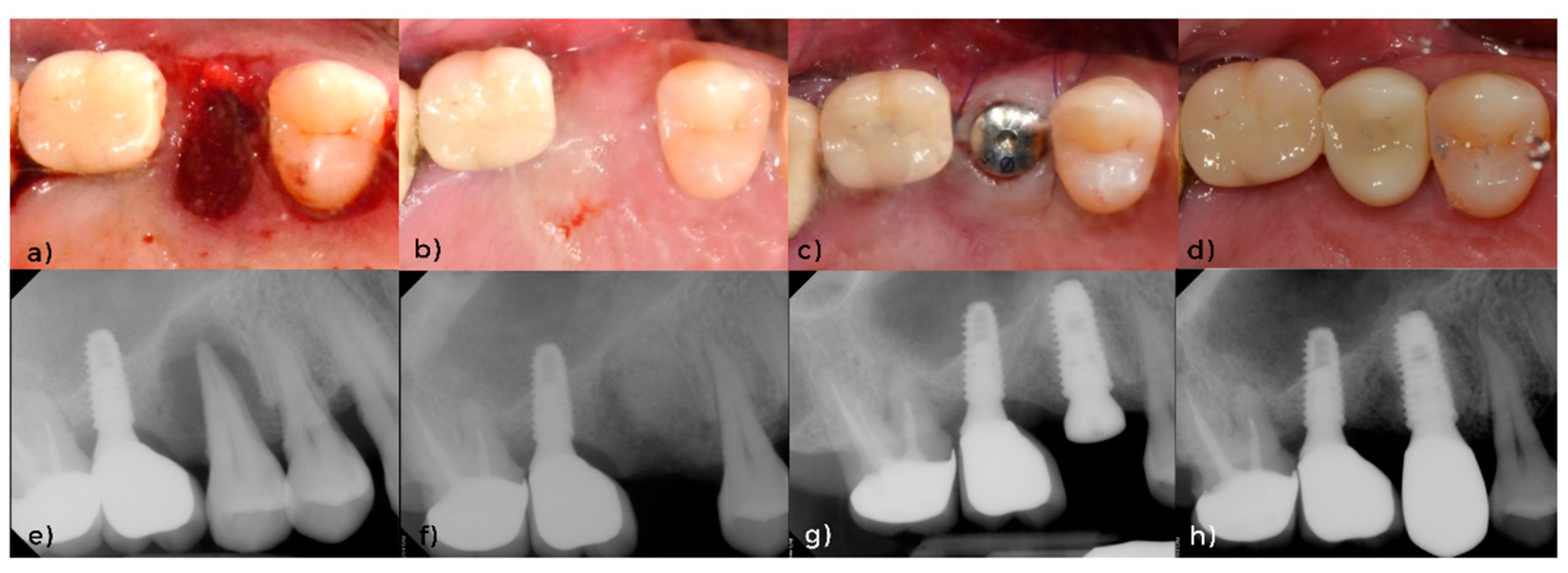
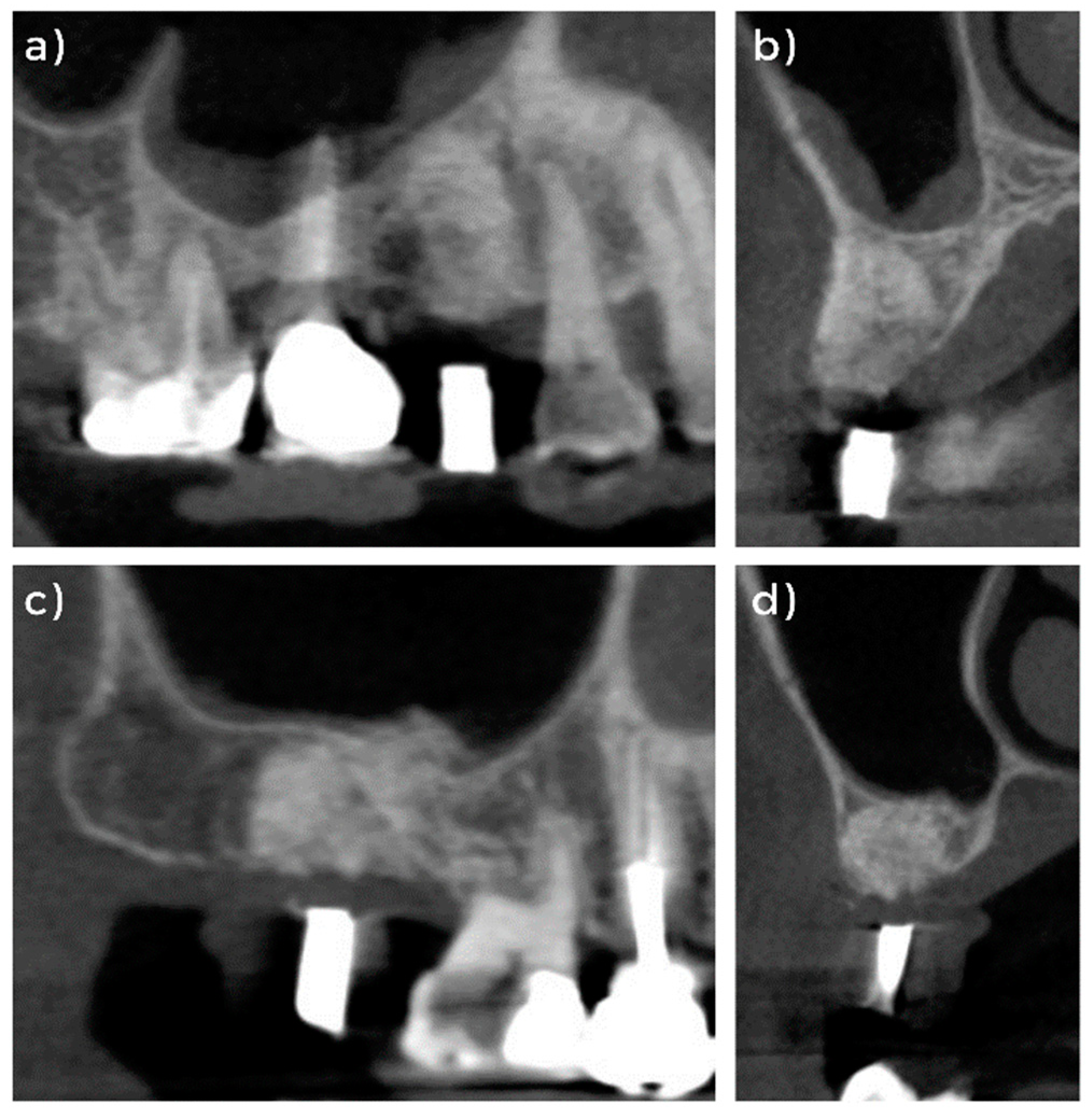
3.1. Clinical and Radiographical Evaluation

There were no signs of negative healing patterns on the surgical sites from the two patients between the ARP and suture removals. During the implant surgery, the clinicians had evaluated the bone quality after flap elevation. Both grated sites had firm tactile sensitivity on the alveolar bone crest, and a high insertion torque of 40 to 50 N/cm was achieved during the fixture installation, indicating that the implants were placed in a normal to hard bone area with reliable primary stability [18,19,22].
From the lateral view of the CBCT radiographs (Figure 3a,c), alveolar bone heights of the two grafted sites are comparative to the adjacent teeth, and the cross-sectional view shows an edentulous bone type A [23]. During an implant planning using CBCT radiographs, from 10 to 12 mm of alveolar bone height was measured in Case I, with a sufficient horizontal alveolar bone width of from 7 to 8 mm. The grated site preserved the shape of the alveolar ridge from the CBCT image (Figure 3a,b) and the implant fixture installation proceeded with no additional procedure.
An alveolar bone height of 6 to 7 mm was observed during an implant planning in Case II with a sufficient horizontal alveolar bone width of from 8 to 9 mm. The grafted site also preserved the shape and height of the alveolar ridge and prevented sinus pneumatization (Figure 3c,d).

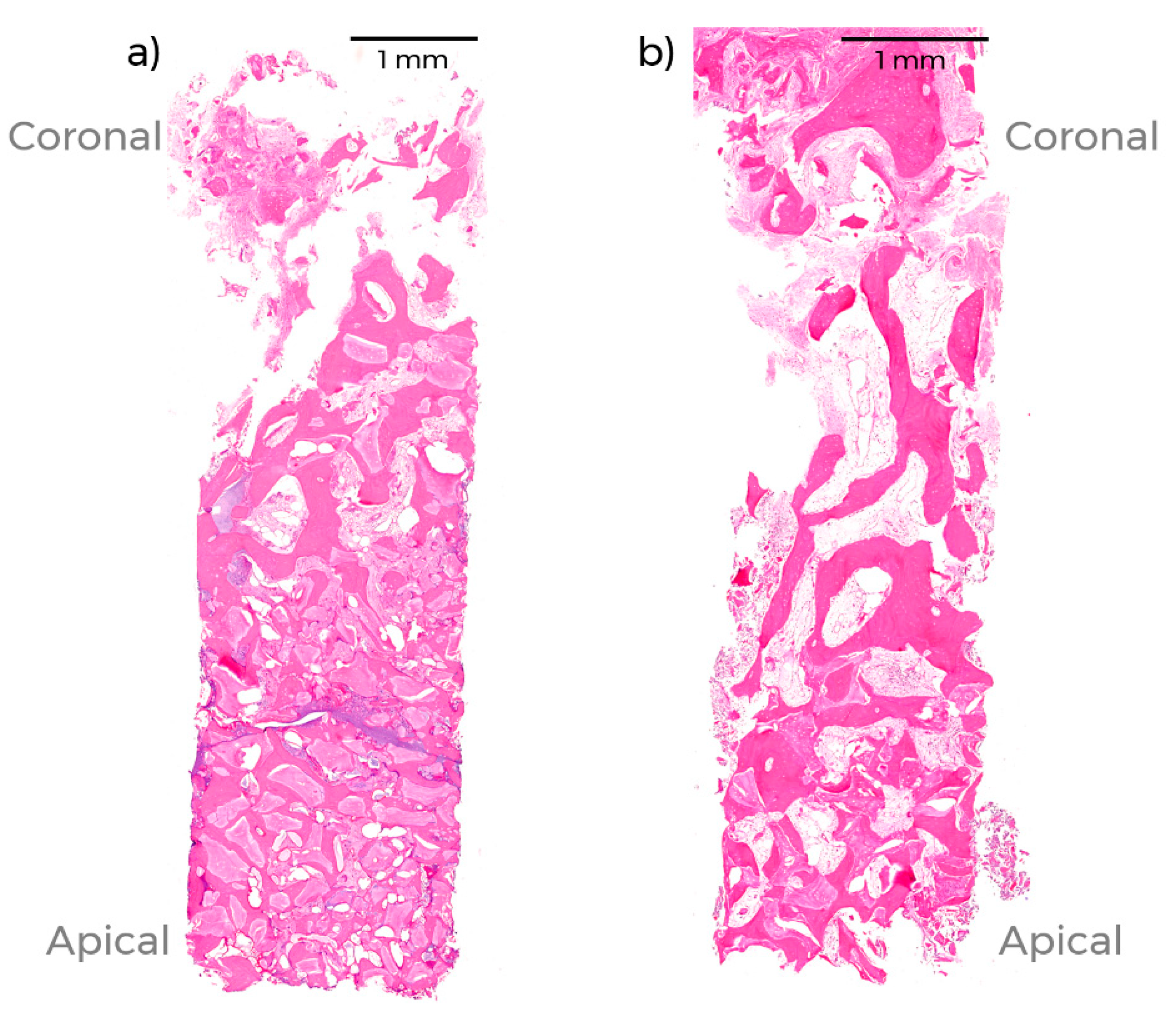
3.2. Histomorphological Evaluation

In H&E staining, the osteocytes were observed in the areas of NFB in both specimens. The NFB was formed around the xenogeneic bone minerals and the fibrovascular tissues, which consisted of connective tissues and vessels, and filled the unmineralized area (Figure 4a,b). There were no conspicuous signs of abnormal healings or inflammation. Both samples show higher compaction of NFB and RGM towards the apical area and that the NFB is observed up to the most coronal area of the histological slides.
In MT staining, DBBM particles in the premolar sample (Figure 5a) present more densely suffused NFB between RGM compared to the larger-sized DPBM particles in the molar sample (Figure 5b). Moreover, NFB tightly fills the spaces between RGM in the premolar sample, whereas higher soft tissue ingrowth between RGM is observed in the molar sample. The percentages of NFB and RGM were 25.3% and 29.0%, respectively, in the first patient; the corresponding percentages in the second patient were 23.3% and 17.5%, respectively.

4. Discussion

This case report analyzed radiographical and histological data of two ARP sites from one to two years post-operation. The first case was diagnosed as Stage II, Grade B periodontitis on the upper-right second premolar while the second case was diagnosed as Stage II, Grade B periodontitis on the upper-right second molar. Both patients had severe alveolar bone resorption extended to the apical aspect and ARP proceeded using xenogeneic graft materials to reduce the dimensional alterations of the alveolar ridge after tooth extraction. The radiographical analysis using CBCT, taking place from one to two years after ARP, presented that the alveolar bone heights remained comparable to the adjacent area and the volume was sufficiently maintained to place an implant without additional horizontal augmentation. Moreover, the high insertion torque achieved in the grafted sites not only provided a reliable primary stability, but also indicated higher initial bone-to-implant contact and ultimately achieved a predictable treatment outcome [18,24].
Previous studies suggested that an intact extraction socket wall may promote hard tissue formation and better outcome for the grafting biomaterials after tooth extraction [8,25]. Despite the large apical bone defect that appeared on the two presented cases, all of the socket walls at the coronal part were intact. However, due to inflammatory bone resorption, only the thin layers of the facial walls remained. It is known that facial walls present a more pronounced reduction after tooth extraction compared to lingual/palatal walls. This is because they are comprised mainly of bundle bones [1,3] and that facial wall thicknesses of less than 2 mm are prone to greater amount of bone loss [26]. Although the thickness of the facial walls was less than 2 mm in both cases, lateral and cross-sectional radiographic images of CBCT exhibited that the alveolar bone heights were comparable to the adjacent area and were maintained one to two years after ARP (Figure 3). With these findings, the authors agree that the biomaterials successfully preserved the socket dimension for a longer period of time.
Histomorphological analysis of the bone samples shows slightly higher NFB (23.5–25.3%) and RGM (17.5–29.0%) percentages compared to a previous clinical study, which evaluated an extraction socket graft that was collected 4 months after the surgery [17]. In this previous study, the mean percentages of NFB and RGM in the DBBM group were 15.1% and 12.7%, respectively. In the DPBM group, the mean percentages of NFB and RGM were 18.5% and 12.2%, respectively. However, a careful interpretation of the data is demanded since the percentages of the previous study analyzed the whole area of the collected sample from a trephine, while this report only analyzed the apical third. With this limited sample, the authors are unable to conclude whether the percentages of NFB increased over time; however, newly formed mineralized bone around the grafted biomaterials appears to be intact for longer period to period. Comparing the histological samples of the two present cases, the residual graft materials of the premolar site (Case I) is more condensed and newly formed bone closely fills the gap between the grafted materials adjunct to the fibrous tissue. Despite the apical lesion that appeared on the premolar, the marginal proportions of the walls were intact and collagenated DBBM enhanced the stability of the grafted material. On the other hand, residual graft materials are more sparsely dispersed in the molar site (Case II) with mineralized bones forming around biomaterials and fibrous tissues filling the gap in between. One possible hypothesis for this that can explain this difference is that, in the molar case, the apex of the socket was exposed to the sinus floor. Therefore, during the condensation process, a minimal amount of pressure was applied to impede the biomaterials from entering the maxillary sinus. Furthermore, the authors hypothesize that the apex aperture, in addition to the larger size of the extraction socket, reduced the bone graft stability. Lastly, fibrous tissue ingrowth from both coronal and apical sides could have promoted a higher rate of fibrous tissue ingrowth.
It is difficult to predict the amount of bone resorption or sinus pneumatization that may have occurred on these sites with periapical lesion. A previous animal study that had compared the healing of an extraction socket in naturally healed sites with grafted sites using collagenated DBBM six months post-operation revealed 35% decrease in the marginal surface area in naturally healed sites, while the marginal surface area in grafted sites only decreased by 12%. Other clinical studies that have evaluated marginal ridge alterations after extraction also showed a similar amount of change [2,27]. Furthermore, sinus pneumatization is more vulnerable in the second molars and in areas with a thin or no layer of bone between the root apex and the sinus floor [11,28]. Considering the above-mentioned findings, ARP was able to minimize the marginal bone resorption and sinus pneumatization in the surgical sites.
Some of the limitations of the current study include the fact that pre- and post-extraction measurements could not be compared because only peri-apical radiographs were taken prior to extraction. Furthermore, the two patients failed to revisit the clinic after ARP. Therefore, any possible complications or other factors that could have influenced the results prior to implantation could not be discussed.

5. Conclusions

Implants should be placed as early as possible, following the other recommended guidelines. Additionally, it is important for the clinicians to understand the long-term healing process and ridge alterations of ARP performed in sites, since the grafted tissue will remain for long periods of time intra-orally as peri-implant tissues. The histological samples presented a densely suffused NFB between RGM, with NFB visible even at the most coronal portion. Moreover, the hard bone density detected during implantation implies that ARP sites could act as stable and reliable peri-implant tissues.

Author Contributions

Conceptualization, S.-W.Y.; methodology, S.-W.Y.; software, S.-W.Y.; validation, Y.W.S., U.-W.J. and J.-K.C.; formal analysis, S.-W.Y.; investigation, S.-W.Y.; resources, U.-W.J.; data curation, S.-W.Y.; writing—original draft preparation, S.-W.Y.; writing—review and editing, Y.W.S., U.-W.J. and J.-K.C.; visualization, S.-W.Y.; supervision, U.-W.J. and J.-K.C.; project administration, J.-K.C.; funding acquisition, U.-W.J. All authors have read and agreed to the published version of the manuscript.

Funding

This work was supported by the Korea Medical Device Development Fund grant funded by the Korea government (the Ministry of Science and ICT, the Ministry of Trade, Industry and Energy, the Ministry of Health & Welfare, the Ministry of Food and Drug Safety) (Project Number: KMDF_PR_20200901_0240).

Institutional Review Board Statement

The study was conducted according to the guidelines of the Declaration of Helsinki. Ethical review and approval were waived for this study by the Institutional Review Board of Yonsei University, since this case report used information on the previously collected data samples.

Informed Consent Statement

Informed consent was obtained from all subjects involved in the study.

Data Availability Statement

The data sets analyzed during the current study are available from the corresponding author by reasonable request.

Conflicts of Interest

The authors declare no conflict of interest.

References

  1. Pietrokovski, J.; Massler, M. Alveolar ridge resorption following tooth extraction. J. Prosthet. Dent. 1967, 17, 21–27. [Google Scholar] [CrossRef]
  2. Schropp, L.; Wenzel, A.; Kostopoulos, L.; Karring, T. Bone healing and soft tissue contour changes following single-tooth extraction: A clinical and radiographic 12-month prospective study. Int. J. Periodontics Restor. Dent. 2003, 23, 313–323. [Google Scholar]
  3. Araújo, M.G.; Lindhe, J. Dimensional ridge alterations following tooth extraction. An experimental study in the dog. J. Clin. Periodontol. 2005, 32, 212–218. [Google Scholar] [CrossRef] [PubMed]
  4. Lim, H.C.; Jung, U.W.; You, H.; Lee, J.S. Randomized clinical trial of ridge preservation using porcine bone/cross-linked collagen vs. bovine bone/non-cross-linked collagen: Cone beam computed tomographic analysis. Clin. Oral Implants Res. 2017, 28, 1492–1500. [Google Scholar] [CrossRef] [PubMed]
  5. Avila-Ortiz, G.; Elangovan, S.; Kramer, K.W.; Blanchette, D.; Dawson, D.V. Effect of alveolar ridge preservation after tooth extraction: A systematic review and meta-analysis. J. Dent. Res. 2014, 93, 950–958. [Google Scholar] [CrossRef] [PubMed]
  6. Choi, H.K.; Cho, H.Y.; Lee, S.J.; Cho, I.W.; Shin, H.S.; Koo, K.T.; Lim, H.C.; Park, J.C. Alveolar ridge preservation with an open-healing approach using single-layer or double-layer coverage with collagen membranes. J. Periodontal Implant Sci. 2017, 47, 372–380. [Google Scholar] [CrossRef] [PubMed]
  7. Araújo, M.; Linder, E.; Wennström, J.; Lindhe, J. The influence of Bio-Oss Collagen on healing of an extraction socket: An experimental study in the dog. Int. J. Periodontics Restor. Dent. 2008, 28, 123–135. [Google Scholar]
  8. Araújo, M.G.; Lindhe, J. Ridge preservation with the use of Bio-Oss collagen: A 6-month study in the dog. Clin. Oral Implants Res. 2009, 20, 433–440. [Google Scholar] [CrossRef]
  9. Covani, U.; Ricci, M.; Bozzolo, G.; Mangano, F.; Zini, A.; Barone, A. Analysis of the pattern of the alveolar ridge remodelling following single tooth extraction. Clin. Oral Implants Res. 2011, 22, 820–825. [Google Scholar] [CrossRef]
  10. Tan, W.L.; Wong, T.L.; Wong, M.C.; Lang, N.P. A systematic review of post-extractional alveolar hard and soft tissue dimensional changes in humans. Clin. Oral Implants Res. 2012, 23 (Suppl. 5), 1–21. [Google Scholar] [CrossRef] [PubMed]
  11. Cha, J.K.; Song, Y.W.; Park, S.H.; Jung, R.E.; Jung, U.W.; Thoma, D.S. Alveolar ridge preservation in the posterior maxilla reduces vertical dimensional change: A randomized controlled clinical trial. Clin. Oral Implants Res. 2019, 30, 515–523. [Google Scholar] [CrossRef] [PubMed]
  12. Park, S.H.; Song, Y.W.; Sanz-Martín, I.; Cha, J.K.; Lee, J.S.; Jung, U.W. Clinical benefits of ridge preservation for implant placement compared to natural healing in maxillary teeth: A retrospective study. J. Clin. Periodontol 2020, 47, 382–391. [Google Scholar] [CrossRef] [PubMed]
  13. Darby, I.; Chen, S.T.; Buser, D. Ridge preservation techniques for implant therapy. Int. J. Oral Maxillofac. Implants 2009, 24, 260–271. [Google Scholar] [PubMed]
  14. De Risi, V.; Clementini, M.; Vittorini, G.; Mannocci, A.; De Sanctis, M. Alveolar ridge preservation techniques: A systematic review and meta-analysis of histological and histomorphometrical data. Clin. Oral Implants Res. 2015, 26, 50–68. [Google Scholar] [CrossRef]
  15. Kalsi, A.S.; Kalsi, J.S.; Bassi, S. Alveolar ridge preservation: Why, when and how. Br. Dent. J. 2019, 227, 264–274. [Google Scholar] [CrossRef]
  16. Papapanou, P.N.; Sanz, M.; Buduneli, N.; Dietrich, T.; Feres, M.; Fine, D.H.; Flemmig, T.F.; Garcia, R.; Giannobile, W.V.; Graziani, F.; et al. Periodontitis: Consensus report of workgroup 2 of the 2017 World Workshop on the Classification of Periodontal and Peri-Implant Diseases and Conditions. J. Periodontol. 2018, 89, S173–S182. [Google Scholar] [CrossRef] [Green Version]
  17. Koo, T.H.; Song, Y.W.; Cha, J.K.; Jung, U.W.; Kim, C.S.; Lee, J.S. Histologic analysis following grafting of damaged extraction sockets using deproteinized bovine or porcine bone mineral: A randomized clinical trial. Clin. Oral Implants Res. 2020, 31, 93–102. [Google Scholar] [CrossRef]
  18. Duyck, J.; Roesems, R.; Cardoso, M.V.; Ogawa, T.; De Villa Camargos, G.; Vandamme, K. Effect of insertion torque on titanium implant osseointegration: An animal experimental study. Clin. Oral Implants Res. 2015, 26, 191–196. [Google Scholar] [CrossRef] [Green Version]
  19. Trisi, P.; Perfetti, G.; Baldoni, E.; Berardi, D.; Colagiovanni, M.; Scogna, G. Implant micromotion is related to peak insertion torque and bone density. Clin. Oral Implants Res. 2009, 20, 467–471. [Google Scholar] [CrossRef]
  20. Atsumi, M.; Park, S.H.; Wang, H.L. Methods used to assess implant stability: Current status. Int. J. Oral Maxillofac. Implants 2007, 22, 743–754. [Google Scholar]
  21. Olivé, J.; Aparicio, C. Periotest method as a measure of osseointegrated oral implant stability. Int. J. Oral Maxillofac. Implants 1990, 5, 390–400. [Google Scholar]
  22. Tricio, J.; van Steenberghe, D.; Rosenberg, D.; Duchateau, L. Implant stability related to insertion torque force and bone density: An in vitro study. J. Prosthet Dent. 1995, 74, 608–612. [Google Scholar] [CrossRef]
  23. Zarb, G.A.; Zarb, F.L. Tissue integrated dental prostheses. Quintessence Int. 1985, 16, 39–42. [Google Scholar]
  24. Tabassum, A.; Meijer, G.J.; Walboomers, X.F.; Jansen, J.A. Evaluation of primary and secondary stability of titanium implants using different surgical techniques. Clin. Oral Implants Res. 2014, 25, 487–492. [Google Scholar] [CrossRef] [PubMed]
  25. Cardaropoli, D.; Tamagnone, L.; Roffredo, A.; Gaveglio, L.; Cardaropoli, G. Socket preservation using bovine bone mineral and collagen membrane: A randomized controlled clinical trial with histologic analysis. Int. J. Periodontics Restor. Dent. 2012, 32, 421–430. [Google Scholar]
  26. Qahash, M.; Susin, C.; Polimeni, G.; Hall, J.; Wikesjö, U.M. Bone healing dynamics at buccal peri-implant sites. Clin. Oral Implants Res. 2008, 19, 166–172. [Google Scholar] [CrossRef] [PubMed]
  27. Barone, A.; Aldini, N.N.; Fini, M.; Giardino, R.; Calvo Guirado, J.L.; Covani, U. Xenograft versus extraction alone for ridge preservation after tooth removal: A clinical and histomorphometric study. J. Periodontol. 2008, 79, 1370–1377. [Google Scholar] [CrossRef] [PubMed]
  28. Sharan, A.; Madjar, D. Maxillary sinus pneumatization following extractions: A radiographic study. Int. J. Oral Maxillofac. Implants 2008, 23, 48–56. [Google Scholar] [PubMed]
Figure 1. Clinical photos and peri-apical radiographs of Case I. Clinical photos of (a) after extraction, (b) one year after alveolar ridge preservation (ARP), (c) after implantation, (d) after prosthesis delivery. Peri-apical radiographs of (e) before extraction, (f) after ARP, (g) after implantation, (h) after prosthesis delivery.
Applsci 11 06591 g001
Figure 2. Clinical photos and peri-apical radiographs of Case II. Clinical photos of (a) after alveolar ridge preservation (ARP), (b) two years after ARP, (c) after implantation, (d) after prosthesis delivery. Peri-apical radiographs of (e) before extraction, (f) after ARP, (g) after implantation, (h) after prosthesis delivery.
Applsci 11 06591 g002
Figure 3. CBCT images of Case I (a) lateral view, (b) cross-sectional view taken one year after alveolar ridge preservation (ARP); Case II (c) lateral view, (d) cross-sectional view taken two years after ARP.
Applsci 11 06591 g003
Figure 4. Photomicrographs of H &E-stained histologic specimens of (a) Case I and (b) Case II.
Applsci 11 06591 g004
Figure 5. Photomicrographs of MT-stained histologic specimens of (a) Case I and (b) Case II.
Applsci 11 06591 g005
Publisher’s Note: MDPI stays neutral with regard to jurisdictional claims in published maps and institutional affiliations.

