Article

Histological Change in Cucumber Tissue and Cellulase Activity of Plectosphaerella melonis Strain 502

1
Zabolotny Institute of Microbiology and Virology, National Academy of Sciences of Ukraine, 03143 Kyiv, Ukraine
2
Institute of Agricultural Microbiology and Agroindustrial Manufacture, National Academy of Agrarian Sciences of Ukraine, 14000 Chernihiv, Ukraine
3
Institute of Environmental Engineering and Biotechnology, University of Opole, 45-040 Opole, Poland
4
Institute of Biology, University of Opole, 45-040 Opole, Poland
5
Department of Technology, Faculty of Natural Sciences, Matej Bel University, 974 01 Banska Bystrica, Slovakia
*
Author to whom correspondence should be addressed.
Submission received: 8 April 2022 / Revised: 15 May 2022 / Accepted: 17 May 2022 / Published: 18 May 2022

Abstract

:
In the last ten years, many countries around the world recorded a new disease of the Cucurbitaceae, the agent of which was P. melonis. The ability of P. melonis 502 to form intracellular mycelium in the epidermal and parenchymal tissues of roots was shown. Leading tissues (xylem and phloem) did not colonize, which indicates the impossibility of plant vessel clogging and shows the fungus’s biochemical effects on plants, which causes the process of pathogenesis. P. melonis 502 is able to develop in a wide range of pH values, while the pH-optimum is 8.5. P. melonis 502 is able to adjust the pH of the medium to the optimal value—8.5. We also showed that cellulase enzyme synthesis depends on pH. We studied the exo-, endo- and β-glucasidase activity of P. melonis 502 and found that the highest activity of cellulase enzymes was on a medium whose pH was 8.5. In the process, the total cellulolytic activity was 0.326 U mL−1, exoglucanase activity—0.539 U mL−1, endoglucanase activity—0.950 U mL−1 and β-glucosidase activity—0.795 U mL−1.

1. Introduction

Cucumber (Cucumis sativus L.) is one of the most common vegetable crops. The yield of this crop is influenced by phytopathogenic micro-organisms: fungi, bacteria and viruses [1]. In recent times, many countries around the world recorded cases of a new disease of the Cucurbitaceae, the agent of which was P. melonis (syn. Acremonium cucurbitacearum) [1,2,3,4,5,6,7,8,9,10,11,12,13,14,15,16,17,18,19]. In Ukraine, this disease was on cucumber plants in 2012 [14,19]. P. melonis is a phytopathogenic fungus that is able to penetrate plant root tissues [3].
Pathogens’ virulence factors are necessary for the micro-organism to colonize the tissues of the host plant and overcome the mechanisms of plant protection [20,21]. Parasitic fungi is due to the active development of fungal mycelium within the tissues of the host plant [22], which in turn is accompanied by the synthesis of secondary metabolites of the fungus, which will act as determinants of the disease [23].
To penetrate cells, pathogens can synthesize chemical compounds of different biological actions, including cell-wall-degrading enzymes (CWDEs). For example, cellulase enzymes take part in the degradation of the plant cellular wall [24,25,26,27]. For pathogenic micro-organisms, cellulases will initiate the process of pathogenesis and play the roles of pathogenic factors, helping them to penetrate the host plant [20,21]. In particular, it has been shown that the synthesis of cellulase in vitro correlates with the degree of the disease [28,29] and highly aggressive isolates synthesize more cellulase in vitro than weakly aggressive isolates [30]. The culture fluid of phytopathogens which characterize cellulase activity in vitro can destabilize the cell membranes of plants [31].
Considering the above, the study of the role of metabolites of phytopathogens in plant–microbial interactions is a necessary condition for elucidating the mechanism of the pathological process. All previous investigations of P. melonis strains were aimed mainly at identifying and studying the spectrum of host plants and describing the symptoms of the disease. Recently, we reported that P. melonis 502 can synthesize phytohormone ethylene and regulate plant growth [19]. Here, we aimed to investigate P. melonis 502’s ability to penetrate the root tissues of cucumber plants and synthesize cellulase enzymes that can act as virulence factors for the fungus in vitro.

2. Materials and Methods

2.1. Strain

Strain of P. melonis 502 was isolated from the affected root system of C. sativus (Koroliok variety) at the mass fruiting stage, which grew in glass block greenhouses (Chernihiv region, Ukraine). Strain deposited at the Depository of the Institute of Microbiology and Virology, NAS of Ukraine with the number IMB F-100138.

2.2. Artificial Infectious Background (AIB)

P. melonis 502 was cultivated in 1 L flasks with a capacity on a solid substrate, which included: oat seeds, oatmeal, water, chalk and gypsum. Sterilization of the substrate was performed twice at a temperature of 128 °C and a pressure of 1.5 ATM for 1 h 30 min. The inoculum was a pure culture of the P. melonis 502, which was isolated on a solid nutrient medium wort agar from affected cucumber plants and was obtained by washing conidia and fragments of fungal hyphae from wort agar. The flasks were kept in a thermostat at a temperature of 26 ± 2 °C. After overgrowing the substrate with mycelium (21st day), it was transferred to paper bags, dried to a constant mass at 30 °C and crushed. To determine the titer of infectious material, we used Goryaev’s chamber and carried out sowing on wort agar (4–5% dry matter).

2.3. Plant Material

The study of the ability of P. melonis 502 to colonize the root system of C. sativus (Koroliok variety) was tested in a growing experiment. Plastic containers (2 L) were filled with 1800 g of sterile soil. Soil: sod-medium podzolic dusty-sandy; humus content—1.02%; nitrogen—54.9 mg/kg; mobile forms of phosphorus—110–120 mg of P2O5, potassium exchange—120–130 mg of K2O per 1 kg of soil; pH—6.0; Ca—5.8, Mg—0.61 mg per 100 g of soil. At a depth of 2–3 cm, 10 g of infectious material was made (50 thousand CFU per 1 g of soil), and seeds were sown in each pot. After emergence, the pots were thinned to 5 plants per 1 pot. The plants were grown in a growing house for 6 weeks with natural lighting. Soil moisture was maintained at 60% of total moisture content. The experiment was repeated three times.

2.4. Histological Processing of Samples and Stain Preparation

For microscopic examination, the roots were pre-washed, stained and fixed. With a thin blade, we cut the roots of plants directly by hand into small segments 5–7 mm long. The culture of fungus was detected by staining the affected root tissues by the Kobel method: longitudinal and cross sections of living plant roots were placed in a lactic acid solution of aniline blue (0.1 g of aniline blue, 50 mL of lactic acid and 100 mL of water) for 5 min, and then the sample was placed in 40% lactic acid for differentiation followed by washing the sample in distilled water. Hyphae of fungi were painted blue [32]. Histological changes in the tissues of the root system of cucumber plants were observed on temporary micropreparations using light microscope MC 200CT (“Micros”, Gewerbezone, Austria) and camera Sony DSC-S650.

2.5. pH Growth

The P. melonis 502 was cultured on BMA (barley meal agar) with different pH indexes: 6.0, 6.5, 7.0, 7.5, 8.0, 8.5, 9.0, 9.5, 10.0, 10.5, 11.0, 11.5 and 12.0 at 26 °C. On the 10th day, the diameter of the colonies in two mutually perpendicular directions and the radial growth rate was determined [33].

2.6. Physiological Phases of Growth

The P. melonis 502 was cultivated (220 rpm (rotations per minute)) for 21 days at 26 °C on modified Czapek medium: 20 g glucose, 2 g NaNO3, 1 g K2HPO4, 0.5 g MgSO4 × 7H2O, 0.5 g KCl and 0.01 FeSO4 × 7H2O per liter, pH—6.0 [34]. Nutrient medium was inoculated by spores suspension (T = 1 × 10⁶ CFU) in the amount of 5% of volume. The amount of biomass was determined gravimetrically (by drying the fungus to constant weight for 5 h at 105 °C [33]. The change in the pH values of the cultural fluid were recorded with a universal pH meter pH-150 MA.

2.7. Cellulase Assay

The P. melonis 502 were cultivated in test tubes (in a static culture) containing 5 mL of modified Czapek medium: 2.5 g (NH4)2HPO4, 1.0 g K2HPO4, 0.5 g MgSO4 and 0.5 g KCl, 0.01 FeSO4 with different pH values—5.5, 7.0 and 8.5. The only source of carbon was a strip of filter paper (Munktell & Filtrak GmbH, Bärenstein, Germany), weighing 50 g (1 × 6 cm), that was placed in each test tube. Nutrient medium was inoculated by spores suspension (T = 1 × 10⁶ CFU) in the amount of 5% of volume and was cultivated in the thermostat in dark conditions at 26 °C for 8 weeks. The culture fluid (supernatant) was obtained by filtration.
Total cellulose and exoglucanase activities were assayed by mixing 1 mL supernatant with 50 mg filter paper and avicel (Evalar, Biysk, Russia), respectively, in 1 mL 0.05 M citrate–phosphate buffer with different pH values (5.5, 7.0 and 8.5) and incubating it in a water bath at 40 °C for 60 min. Endoglucanase and β-glucosidase activities were evaluated by mixing 1 mL supernatant with 1 mL 0.5% CMC and 0.025% cellobiose (Merck KGaA, Darmstadt, Germany) in 0.05 M citrate–phosphate buffer with different pH values (5.5, 7.0 and 8.5), respectively, and incubating it in a water bath at 40 °C for 30 min.
Release of reducing sugars was assessed by the Somogyi method [35] modified from Nelson procedure [36] with glucose as standard. A total of 1 mL of Somogyi reagent was added to 2 mL of reaction mixture and incubated at 100 °C for 15 min. Then, the mixture cooled on an ice water bath and 1 mL of Nelson reagent was added. The volume was filled up to 25 mL with water and mixed. Reducing sugars were determined on photoelectric colorimeter at 560 nm.
One unit (U) of enzyme activity was defined as the number of enzymes required to liberate 1 μmol of reducing sugar for min in mL−1 expressed as glucose equivalents. Cellulase activity was measured every 7 days. Repetition of experiment is triplicate.

2.8. Statistical Methods

The significance of the experimental data was analyzed using STATISTICA 12 software (StatSoft, Hamburg, Germany). The distribution of data checked for normality using the Shapiro–Wilk W-test and the homogeneity of variance checked using the Brown–Forsythe test. The significance of the differences was measured using One-way Analysis of Variance (ANOVA). Particularly, Duncan Multiple Range Test (DMRT) was used to check if each parameter differed significantly under different values of studied factors. The level of significance was set to p < 0.05.

3. Results and Discussion

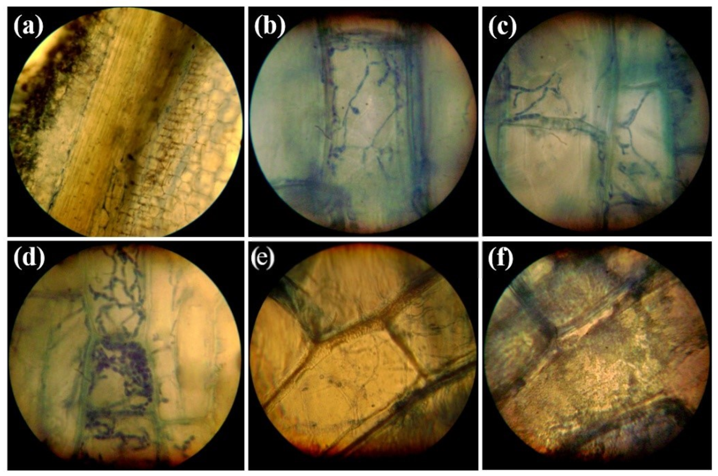
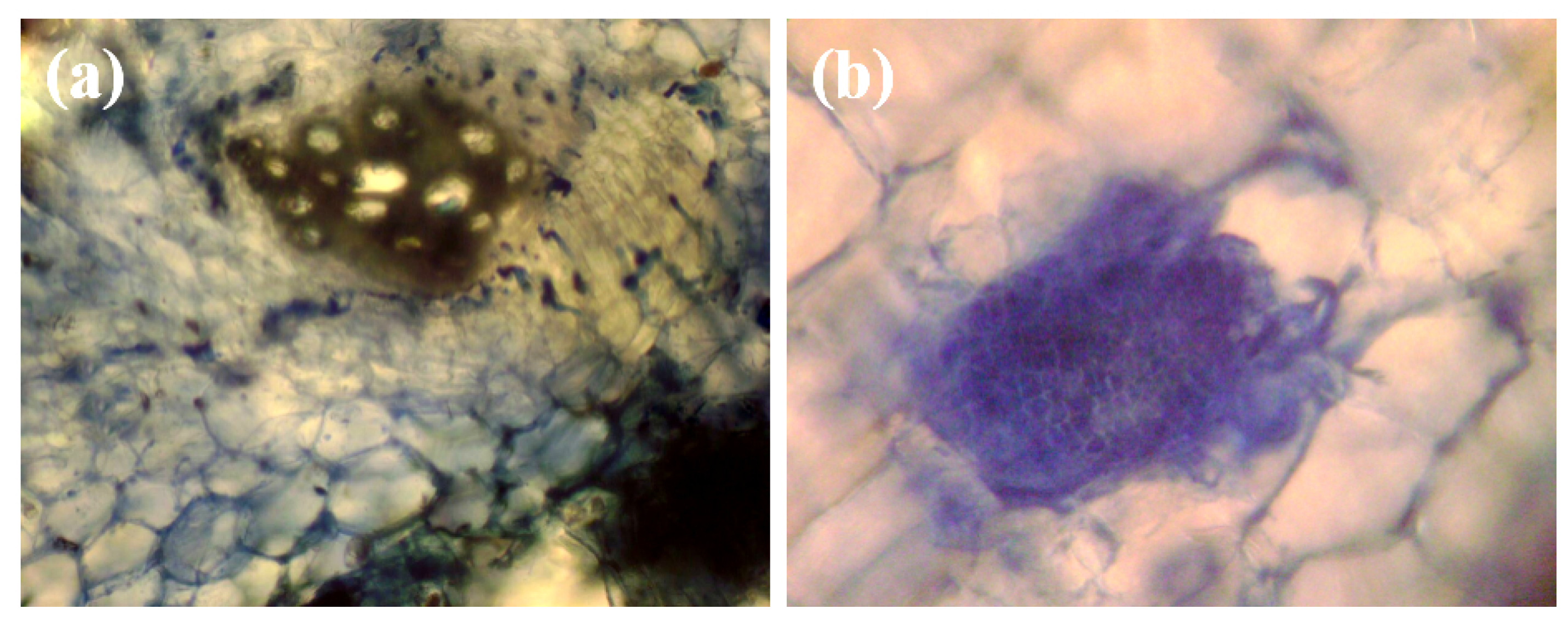
3.1. Histopathology of Infections Caused by P. melonis 502 on the Root Tissues of Cucumber

After 6 weeks of growing the plants on infected soil, the cortex cells had a distorted appearance (Figure 1a). P. melonis 502 densely colonized the parenchymal and epidermal tissues of the root. Hyphae were of different thicknesses and were observed not only in the intercellular space but also inside the cells, forming intracellular mycelium (Figure 1b–d). Figure 1f shows the hyphae of the fungus inside the cell, as well as the necrotic cell walls of root cells, which indicates the synthesis of hydrolytic enzymes by the fungus, providing the degradation of cells (Figure 1f).
The leading tissues (xylem and phloem) did not colonize (Figure 1a), which indicates the impossibility of plant vessel clogging and shows the fungus’s biochemical effects on plants, which causes the process of pathogenesis, but in xylem there were tiloses (Figure 2a). Tiloses were also present in the roots of plants affected by the fungi Phaemoniella chlamydospora and Phaeoacremonium sp. [37]. Our results confirm the results of Alfaro-Fernández and García-Luis [3], who also recorded the presence of tiloses in leading root tissues in the defeat of A. cucurbitacearum (syn. P. melonis). The etiology of tiloses formation is quite ambiguous and debatable, but it is known that these structures reduce water supply to the plant under the conditions of high demand for moisture, which may be the reason for their depletion [3,38]. In addition, we found microsclerotia in the affected tissues of the roots of cucumber plants (Figure 2b), which completely fill the cell of the host plant. Microsclerotia are known to increase the survival of a micro-organism in soil under adverse environmental conditions [39].
We previously reported that P. melonis 502 can synthesize the phytohormone ethylene (ET) in vitro and regulate plant growth [19]. Phytohormones can promote the penetration of the pathogen into the tissues of host plants, create a niche for the pathogen and/or trigger other disease processes [40]. ET activates and accelerates the aging process of plant tissues [41]. At the same time, that tissue shows increased activity to cellulase, which leads to the destruction of intercellular connections [42]. ET modulates plant susceptibility and fungal growth in plant tissue also [43].

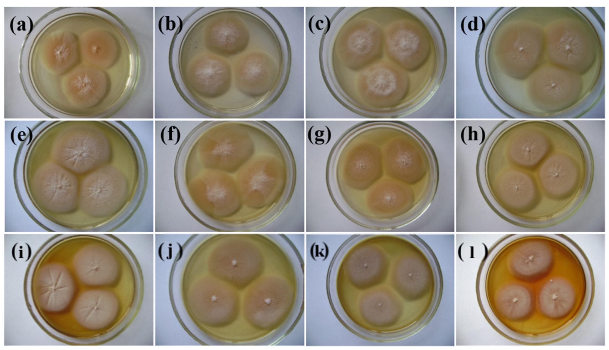
3.2. pH Growth of P. melonis 502

P. melonis 502 on BMA grows in the pH ranges from 6.0 to 12.0 (Figure 3, Table 1) and refers to acid-tolerant micro-organisms, that is, those that are able to grow and develop within wide pH ranges. The pH-optimum is slightly alkaline (pH 8.5), with the colony diameter on the 10th day being 24.8 ± 0.1 mm (Table 1) and with the radial growth rate being 0.103 mm per h. The ability of the Plectosphaerellaceae fungi to grow in alkaline conditions was shown earlier [44]. Bondarenko et al. [45] isolated the Plectosphaerella spp. from acidic sod-podzolic and neutral soils by sowing on alkaline agar (pH 10.0–10.5). The ability of these fungi to grow on nutrient media with pH 4.0–10.0 is shown. Our results confirm that P. melonis 502 is alkalophilic.

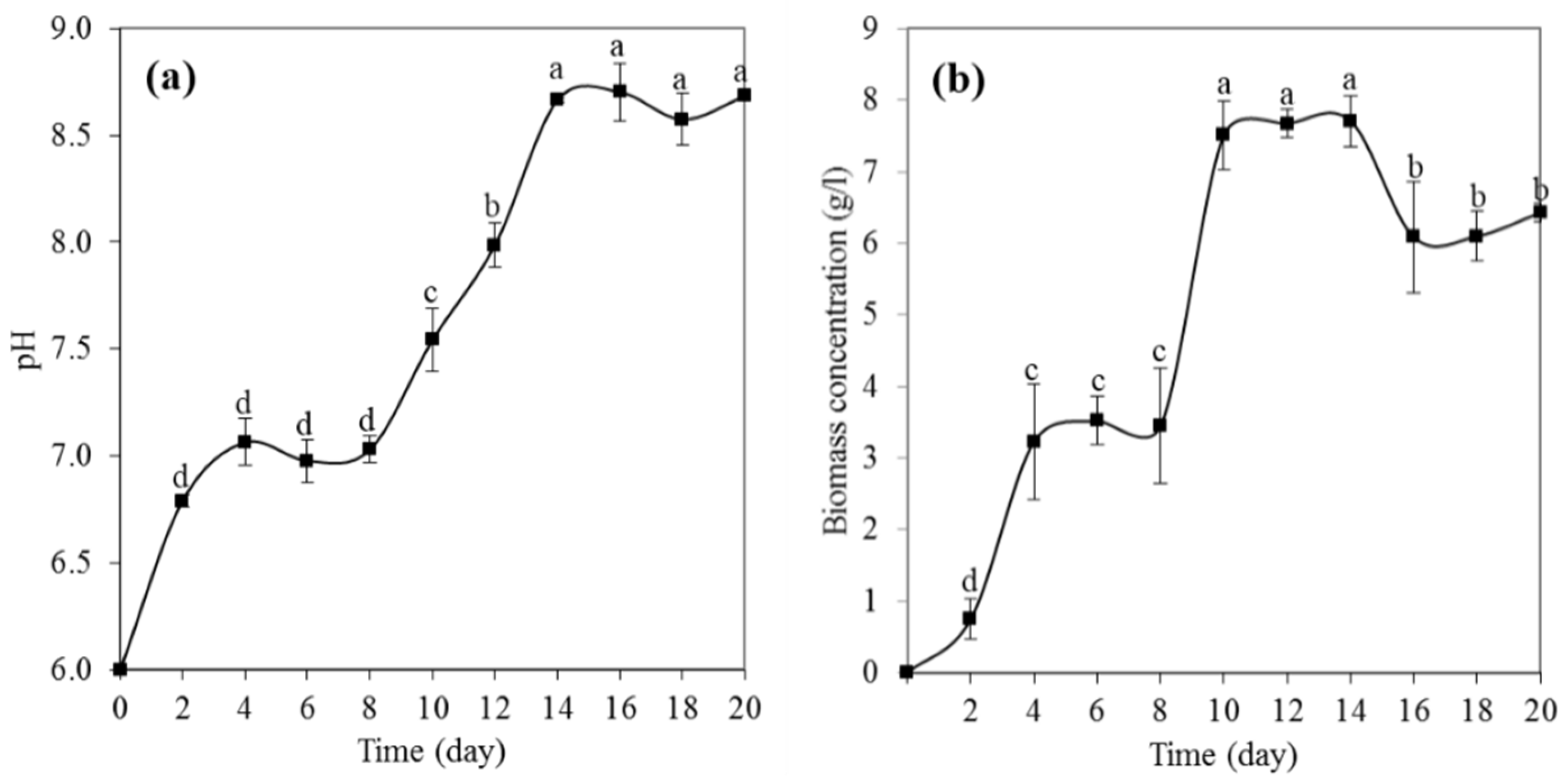
3.3. Physiological Growth Phases of P. melonis 502

The stationary phase of P. melonis 502 occurred after 10 days (Figure 4b). We also observed that the alkalescencing of the nutrient medium occurred during the stationary phase (up to 14 days) until a pH of 8.5 was reached (Figure 4a). Upon further cultivation of P. melonis 502, the pH of the medium in the deep culture remained unchanged.
Pathogens can adapt their broad pathogenic factors arsenal to a particular host during the evolution process and are able to affect and regulate the pH environment, since this indicator influences the activity of many enzymes, including enzymes of degradation of the cellular walls of plants [46,47]. We have shown that P. melonis 502 alkalizes the nutrient medium to the optimum pH for growth (pH 8.5). Jennings [48] believes that environmental leaching happens due to the active ammonia formation by amino acid deamination. Some fungi, such as Polyporus tricholoma, increased the pH of the nutrient medium with a different pH level to 5.5, regardless of the initial pH and other factors [49]. This ability to increase the pH of the nutrient medium was shown for some pathogenic fungi, in particular Aspergillus nidulans [47], Candida albicans [50] and Metarhizium anisopliae [51]. The phytopathogen of persimmon fruit A. alternata increased the pH of the nutrient medium from 3.8 to 6.0, which in turn induced the expression of the AaK1 gene and synthesis of endo-1,4-β-glucanase, which plays an important role in the development of the disease [52]. Adjusting the pH of the environment by Colletotrichum gloeosporioid in the host plant tissues would depend on the buffering power of the cytoplasm, and therefore, each specific pathogen would have different effects on different host plants [53].

3.4. Cellulase Assay

Maximum total (Table 2), exoglucanase (Table 3), endoglucanase (Table 4) and β-glucosidase (Table 5) activities developed after 6 weeks of cultivation. At the same time, pH 8.5 of the medium was the most optimal for the synthesis of cellulase enzymes. The maximum total cellulase activity was 0.326 U/mL−1, exoglucanase—0.539 U/mL−1, endoglucanase—0.950 U/mL−1 and β-glucosidase was 0.795 U/mL−1.
Phytopathogenic and entomopathogenic fungi of the genus Colletotrichum showed a dependence on the pH of the nutrient medium, in particular, the synthesis of cellulases at pH 8.0 was higher than at 5.0 and 6.8 [54]. For phytopathogen F. oxysporum, the optimal pH was 6.0 [55]. Hemibiotrophic pathogen of the anthracnose of beans C. lindemuthianum synthesized a complex of cellulase enzymes at an optimal pH of 5.5 [56]. Furthermore, a correlation between cellulase activity and the virulence of Sclerotinia sclerotiorum isolates was shown [24]. The biosynthesis of cellulase and pectinase plays an important role in the pathogenesis of Alternaria brassicae [26] and A. alternata [25]. Tomato phytopathogens A. tenuissima, A. arborescens, A. mimicula, A. interrupta and A. infctoria synthetized pectinase, cellulase, amylase, protease and lipase; however, only cellulase and pectinase production have correlated with isolate pathogenicity [57]. The role of cellulase enzymes in the pathogenesis of the Cephalosporium acremonium, which causes root rot of corn plants, proved to be important. For example, the pathogenicity of the fungus and crop loss correlated with the synthesis of hydrolytic enzymes, including cellulose [58].
Some fungi have exoglucosidase activity. For example, endophytic fungus Chaetomium cochliodes 3250 was characterized by exoglucanase activity along with endoglucanase and β-glucosidase [59]. However, the specified activities are much lower than those of P. Melonis 502, even with a pH of 5.5 and 7.0, which are not optimal for P. melonis 502’s growth and development. It should be noted that the exoglucosidase activity curve of P. melonis 502 is wave-like. We have also registered an increase in exocellulase activity between weeks 3 and 6 of cultivation. This, in turn, proves that fungi use reducing sugars as a carbon source [60]. When Ramanathan et al. [55] studied the cellulase activity of phytopathogen F. oxysporum, they also showed two peaks of cellulase activity—at days 4 and 8 of cultivation on a rotary shaker. In addition, in vitro synthesis of enzymes varies depending on the method of cultivation. High cellulase and β-glucosidase activity of the phytopathogen of sugar beet Sclerotium rolfsii was shown, the peaks of which occurred on days 9 and 10, respectively, while in a dynamic culture, the activity of enzymes was almost five times higher than in a static culture [27,61,62]. Regarding Plectosphaerella, it is known that the pH-optimum of P. cucumerina for the xylanase is 6.0 [63]. Chitinases Plectosphaerella sp. strain MF-1 is most active at pH 3–4 [64].
In general, it is now known that the P. melonis 502 is capable of the synthesis of ethylene [19] and cellulase enzymes in vitro. We also found that the culture fluid can regulate plant growth [19] and the fungus itself can penetrate into the tissues of plant roots. However, additional research is needed to reveal the mechanism of pathogenesis and to establish pathogenicity and virulence factors.

4. Conclusions

Thus, we have shown that P. melonis 502 is able to penetrate the root tissues of plants and form intracellular mycelium. It affects mainly cortex tissues (epidermal and parenchymal). Leading tissues (xylem and phloem) are not colonized, which indicates the impossibility of plant vessel blockage and indicates the effect of fungal metabolites on plants, which determine the process of pathogenesis. P. melonis 502 is able to develop in a wide range of pH, while the pH-optimum is 8.5. P. melonis 502 is able to adjust the pH of the medium to the optimal value—8.5. We also showed that the synthesis of cellulase enzymes depends on the pH. We studied the exo-, endo- and β-glucosidase activities of P. melonis 502 and found that the highest enzyme activity was shown on pH 8.5. We noted cells with intracellular mycelium and destroyed cell walls. Cellulase enzymes may be virulence factors and can degrade the plant cell wall.

Author Contributions

Conceptualization, V.P., H.T., Y.K., A.K. (Anna Kyslynska) and A.K. (Antonina Kalinichenko); Methodology, V.P., H.T., Y.K., A.K. (Anna Kyslynska), M.S., J.S. and A.K. (Antonina Kalinichenko); Investigation, H.T., Y.K. and A.K. (Anna Kyslynska); Resources, V.P., H.T., Y.K., M.S., J.S., A.K. (Anna Kyslynska) and A.K. (Antonina Kalinichenko); Writing—Original Draft Preparation, V.P., H.T., Y.K., A.K. (Anna Kyslynska), M.S., J.S. and A.K. (Antonina Kalinichenko); Writing—Review and Editing, V.P., H.T., Y.K., A.K. (Anna Kyslynska), M.S., J.S. and A.K. (Antonina Kalinichenko); Visualization, H.T., Y.K. and A.K. (Anna Kyslynska); Supervision, V.P. and Y.K.; Project Administration, Y.K.; Funding Acquisition, Y.K. and J.S. All authors have read and agreed to the published version of the manuscript.

Funding

This work was financially supported by the National Academy of Agrarian Science of Ukraine (No. 0116U003070) and VEGA Grant No. 1/20629/20 (Slovakia).

Institutional Review Board Statement

Not applicable.

Informed Consent Statement

Not applicable.

Data Availability Statement

Not applicable.

Conflicts of Interest

The authors declare that there is no conflict of interest.

References

  1. Bruton, B.D. Soilborne diseases in cucurbitaceae: Pathogen virulence and host resistance. In Cucurbitaceae 98: Evaluation and Enhancement of Cucurbit Germplasm; McCreight, J.D., Ed.; ASHS Press: Alexandria, VA, USA, 1998; pp. 143–166. ISBN 978-0961502799. [Google Scholar]
  2. Aegerter, B.J.; Gordon, T.R.; Davis, R.M. Occurrence and pathogenicity of fungi associated with melon root rot and vine decline in California. Plant Dis. 2000, 84, 224–230. [Google Scholar] [CrossRef] [PubMed] [Green Version]
  3. Alfaro-Fernández, A.; García-Luis, A. Colonisation and histological changes in muskmelon and autumn squash tissues infected by Acremonium cucurbitacearum or Monosporascus cannonballus. Eur. J. Plant Pathol. 2009, 125, 73–85. [Google Scholar] [CrossRef]
  4. Alfaro-García, A.; Armengol, J.; Bruton, B.D.; Gams, W.; García-Jiménez, J.; Martínez-Ferrer, G. The taxonomic position of the causal agent of Acremonium collapse. Mycologia 1996, 88, 804–808. [Google Scholar] [CrossRef]
  5. Forti, J. Aspectos Patológicos, Epidemiológicos y Culturales de Acremonium cucurbitacearum Alfaro-Garcia, w. Gams et j. Garcia-Jiménez. Ph.D. Thesis. Universidad Politecnica de Valencia, Valencia, Spain, 1997. Available online: https://dialnet.unirioja.es/servlet/tesis?codigo=245756 (accessed on 7 April 2022).
  6. Armengol, J.; Sanz, E.; Martínez-Ferrer, G.; Sales, R.; Bruton, B.D.; García-Jiménez, J. Host range of acremonium cucurbitacearum, cause of acremonium collapse of muskmelon. Plant Pathol. 1998, 47, 29–35. [Google Scholar] [CrossRef]
  7. Bruton, B.D.; Davis, R.M.; Gordon, T.R. Occurrence of Acremonium sp. and Monosporascus cannonballus in the major cantaloupe and watermelon growing areas of California. Plant Dis. 1995, 79, 754. [Google Scholar] [CrossRef]
  8. Bruton, B.D.; Garcia-Jimenez, J.; Armengol, J. Analysis of the relationship between temperature and vine declines caused by Acremonium cucurbitacearum and Monosporascus cannonballus on Muskmelon. Subtrop. Plant Sci. 1999, 51, 23–28. Available online: http://www.subplantsci.org/wp-content/uploads/2016/03/SPSJ-51-23-28-Bruton-et-al.pdf (accessed on 7 April 2022).
  9. Bruton, B.D.; Garcia-Jimenez, J.; Armengol, J.; Popham, T.W. Assessment of virulence of Acremonium cucurbitacearum and Monosporascus cannonballus on Cucumis melo. Plant Dis. 2000, 84, 907–913. [Google Scholar] [CrossRef] [Green Version]
  10. Bruton, B.D.; Popham, T.W.; García-Jiménez, J.; Armengol, J.; Miller, M.E. Disease reaction among selected Cucurbitaceae to an Acremonium cucurbitacearum isolate from Texas. Hortscience 2000, 35, 677–680. [Google Scholar] [CrossRef] [Green Version]
  11. Carlucci, A.; Raimondo, M.L.; Santos, J.; Phillips, A.J.L. Plectosphaerella species associated with root and collar rots of horticultural crops in southern Italy. Pers. Mol. Phylogeny Evol. Fungi 2012, 28, 34–48. [Google Scholar] [CrossRef] [Green Version]
  12. Chilosi, G.; Reda, R.; Aleandri, M.P.; Camele, I.; Altieri, L.; Montuschi, C.; Languasco, L.; Rossi, V.; Agosteo, G.E.; Macrì, C.; et al. Fungi associated with root rot and collapse of melon in Italy. OEPP/EPPO Bull. 2008, 38, 147–154. [Google Scholar] [CrossRef]
  13. Garsia-Jimenez, J.; Velazquez, M.T.; Jorda, C.; Alfaro-Garcia, A. Acremonium species as the causal agent of muskmelon collapse in Spain. Plant Dis. 1994, 78, 416–419. [Google Scholar] [CrossRef]
  14. Kopilov, E.; Tsekhmister, H.; Nadkernychna, O.; Kyslynska, A. Identification of Plectosphaerella melonis from cucumber plants in Ukraine. Phytopathol. Mediterr. 2021, 60, 259–263. [Google Scholar] [CrossRef]
  15. Martinez-Culebras, P.V.; Abad-Campos, P.; Garcia-Jimenez, J. Molecular characterization and PCR detection of the melon pathogen Acremonium cucurbitacearum. Eur. J. Plant Pathol. 2004, 110, 801–809. [Google Scholar] [CrossRef]
  16. Mostafa, M.A.; Attia, M.F.; Merghany, M.M.; Salama, R.M. Association of Plectosphaerella melonis with cantaloupe decline for the first time in Egypt. Plant Arch. 2019, 19, 2565–2573. Available online: http://plantarchives.org/19-2/2565-2573%20(5261).pdf (accessed on 7 April 2022).
  17. Raimondo, M.L.; Carlucci, A. Characterization and pathogenicity assessment of Plectosphaerella species associated with stunting disease on tomato and pepper crops in Italy. Plant Pathol. 2018, 67, 626–641. [Google Scholar] [CrossRef]
  18. Raimondo, M.L.; Carlucci, A. Characterization and pathogenicity of Plectosphaerella spp. collected from basil and parsley in Italy. Phytopathol. Mediterr. 2018, 57, 284–295. [Google Scholar] [CrossRef]
  19. Tsekhmister, H.V.; Kyslynska, A.S.; Kopilov, E.P.; Nadkernychna, O.V. Plant growth regulatory activity in the phytopathogenic fungus Plectosphaerella melonis strain 502. Agric. Sci. Pract. 2021, 8, 13–24. [Google Scholar] [CrossRef]
  20. Schulz, B.; Boyle, C. The endophytic continuum. Mycol. Res. 2006, 109, 661–686. [Google Scholar] [CrossRef] [Green Version]
  21. Selim, K.A.; El-Beih, A.A.; AbdEl-Rahman, T.M.; El-Diwany, A.I. Biology of endophytic Fungi. Curr. Res. Environ. Appl. Mycol. J. Fungal Biol. 2012, 2, 31–82. [Google Scholar] [CrossRef]
  22. Blagoveshchenskaya, E.Y. Endophyte-plant as a complex dynamic system. Mycol. Today 2011, 2, 126–134. Available online: https://istina.msu.ru/media/publications/article/d02/ec8/1497555/Blagoveshchenskaya2011_endoph_system.pdf (accessed on 7 April 2022). (In Russian).
  23. Thines, E.; Aguirre, J.; Foster, A.J.; Deising, H.B. Genetics of phytopathology: Secondary metabolites as virulence determinants of fungal plant pathogens. Prog. Bot. 2006, 67, 134–161. [Google Scholar] [CrossRef]
  24. Asoufi, H.; Hameed, K.M.; Mahasneh, A. The cellulase and pectinase activities associated with the virulence of indigenous Sclerotinia sclerotiorum isolates in Jordan Valley. Plant Pathol. J. 2007, 23, 233–238. [Google Scholar] [CrossRef] [Green Version]
  25. Hubballi, M.; Sornakili, A.; Nakkeeran, S.; Anand, T.; Raguchander, T. Virulence of Alternaria alternata infecting noni associated with production of cell wall degrading enzymes. J. Plant Prot. Res. 2011, 51, 87–92. [Google Scholar] [CrossRef]
  26. Jain, V.; Dhawan, K. Major cell wall degrading enzymes in two contrasting cultivars of Brassica juncea infected with Alternaria brassicae. Crucif. Newslett. 2008, 27, 20–21. Available online: https://www.brassica.info/info/publications/cn/CruciferaeNewsletter_vol27_2008.pdf#page=20 (accessed on 7 April 2022).
  27. Moussa, T.A.A.; Tharwat, N.A. Optimization of cellulase and -glucosidase induction by sugarbeet pathogen Sclerotium rolfsii. Afr. J. Biotechnol. 2007, 6, 1048–1054. Available online: https://academicjournals.org/journal/AJB/article-full-text-pdf/1F42A327000 (accessed on 7 April 2022).
  28. Hassan, A.G. Formulations as biocontrol and role of pectinase and cellulose in pathogenicity. J. Agric. Food Environ. 2020, 1, 1–5. [Google Scholar] [CrossRef]
  29. Ziedan, E.S.H.; Saad, M.M.; Hemida, K.A.; El-Naggar, M.A.E.A.; Mostafa, M.H.; El-Samman, M.G.E.R. In vitro: Stem cutting a simple technique for determining aggressive potential of fungal isolates causing root rot disease of grapevine. GSC Biol. Pharm. Sci. 2020, 11, 141–147. [Google Scholar] [CrossRef]
  30. Lalaoui, F.; Halama, P.; Dumortier, V.; Paul, B. Cell wall-degrading enzymes produced in vitro by isolates of Phaeosphaeria nodorum differing in aggressiveness. Plant Pathol. 2000, 49, 727–733. [Google Scholar] [CrossRef]
  31. Ogórek, R. Enzymatic activity of potential fungal plant pathogens and the effect of their culture filtrate on seed germination and seedling growth of garden cress (Lepidium sativum L.). Eur J. Plant. Pathol 2016, 145, 469–481. [Google Scholar] [CrossRef]
  32. Methods of Experimental Mycology. Moscow, ISBN 978-5-458-27224-7. 2014. Available online: https://www.bookvoed.ru/files/3515/10/62/35.pdf (accessed on 7 April 2022). (In Russian).
  33. Pirt, J. Principles of Microbe and Cell Cultivation; Blackwell Scientific: Oxford, UK, 1975; ISBN 0632081503. [Google Scholar]
  34. Kurchenko, I.M.; Pavlichenko, A.K.; Yurieva, E.M. Growth characteristics of strains of Fusarium poae (Peck) Wollenw and Penicillium funiculosum Thom. Mikrobiolohichnyi Zhurnal 2013, 75, 47–51. (In Russian) [Google Scholar]
  35. Somogyi, M. Notes on sugar determination. J. Biol. Chem. 1952, 195, 19–23. [Google Scholar] [CrossRef]
  36. Nelson, N. A photometric adaptation of the Somogyi method for the determination of glucose. J. Biochem. 1944, 153, 375–380. [Google Scholar] [CrossRef]
  37. Del Rio, J.A.; Gonzalez, A.; Fuster, M.D.; Botia, J.M.; Gomez, P.; Frias, V.; Ortuño, A. Tylose formation and changes in phenolic compounds of grape roots infected with Phaeomoniella chlamydospora and Phaeoacremonium species. Phytopathol. Mediterr. 2001, 40, 394–399. Available online: http://0-www-jstor-org.brum.beds.ac.uk/stable/44981647 (accessed on 7 April 2022).
  38. Pivonia, S.; Cohen, R.; Kafkafi, U.; Ben Ze’ev, I.S.; Katan, J. Sudden wilt of melons in Southern Israel: Fungal agents and relationship with plant development. Plant Dis. 1997, 81, 1264–1268. [Google Scholar] [CrossRef] [PubMed] [Green Version]
  39. Jumpponen, A.; Trappe, J.M. Dark septate endophytes: A review of facultative biotrophic root-colonizing fungi. New Phytol. 1998, 140, 295–310. [Google Scholar] [CrossRef]
  40. Kazan, K.; Lyons, R. Intervention of phytohormone pathways by pathogen effectors. Plant Cell 2014, 26, 2285–2309. [Google Scholar] [CrossRef] [Green Version]
  41. Mattoo, A.K.; Handa, A.K. Ethylene signaling in plant cell death. In Plant Cell Death Processes; Nooden, L., Ed.; Academic Press: New York, NY, USA, 2004; pp. 125–142. [Google Scholar]
  42. Lutova, L.A.; Provorov, N.A. Plant Development Genetics; SPb. Nauka: Saint Petersburg, Russia, 2000; 539p., Available online: https://www.twirpx.com/file/327348/ (accessed on 15 February 2022). (In Russian)
  43. Wang, S.; Park, Y.S.; Yang, Y.; Borrego, E.J.; Isakeit, T.; Gao, X.; Kolomiets, M.V. Seed-derived ethylene facilitates colonization but not aflatoxin production by Aspergillus flavus in maize. Front. Plant Sci. 2017, 8, 415. [Google Scholar] [CrossRef] [Green Version]
  44. Grum-Grzhimaylo, A.A.; Debets, A.J.M.; van Diepeningen, A.D.; Georgieva, M.L.; Bilanenko, E.N. Sodiomyces alkalinus, a new alkaliphilic ascomycete within the Plectosphaerellaceae. Pers. Mol. Phylogeny Evol. Fungi 2013, 31, 147–158. [Google Scholar] [CrossRef] [Green Version]
  45. Bondarenko, S.A.; Georgieva, M.L.; Bilanenko, E.N. Alkalitolerant micromycetes in acidic and neutral soils of the temperate zone. Microbiology 2016, 85, 737–744. [Google Scholar] [CrossRef]
  46. Idnurm, A.; Howlett, B.J. Pathogenicity genes of phytopathogenic fungi. Mol. Plant Pathol. 2001, 2, 241–255. [Google Scholar] [CrossRef]
  47. Prusky, D.; Yakoby, N. Pathogenic fungi: Leading or led by ambient pH? Mol. Plant Pathol. 2003, 4, 509–516. [Google Scholar] [CrossRef] [PubMed]
  48. Jennings, D.H. Some perspectives on nitrogen and phosphorus metabolism in fungi. In Nitrogen, Phosphorus and Sulphur Utilization by Fungi; Boddy, L., Machant, R., Read, D.J., Eds.; Cambridge University Press: Cambridge, UK, 1989; pp. 1–31. ISBN 9780521106245. [Google Scholar]
  49. Vieira, G.R.T.; Liebl, M.; Tavares, L.B.; Paulert, R.; Smania, A.J. Submerged culture conditions for the production of mycelial biomass and antimicrobial metabolites by polyporus tricholoma mont. Braz. J. Microbiol. 2008, 39, 561–568. [Google Scholar] [CrossRef] [PubMed] [Green Version]
  50. De Bernardis, F.; Muhlschlegel, F.A.; Cassone, A. The pH of the host niche controls gene expression in and virulence of Candida albicans. Infect. Immun. 1998, 66, 3317–3325. [Google Scholar] [CrossRef] [PubMed] [Green Version]
  51. St Leger, R.J.; Joshi, L.; Roberts, D. Ambient pH is a major determinant in the expression of cuticle-degrading enzymes and hydrophobin by Metarhizium anisopliae. Appl. Environ. Microbiol. 1998, 64, 709–713. [Google Scholar] [CrossRef] [PubMed] [Green Version]
  52. Eshel, D.; Miyara, I.; Tong, A.; Dinoor, A.; Prusky, D. pH Regulates endoglucanase expression and virulence of Alternaria alternata in persimmon fruit. Mol. Plant-Microbe Interact. (MPMI) 2002, 15, 774–779. [Google Scholar] [CrossRef] [PubMed] [Green Version]
  53. Prusky, D.; McEvoy, J.L.; Leverentz, B.; Conway, W.S. Local modulation of host pH by Colletotrichum species as a mechanism to increase virulence. Mol. Plant-Microbe Interact. 2001, 14, 105–113. [Google Scholar] [CrossRef] [Green Version]
  54. Maccheroni, W., Jr.; Araújo, W.L.; Azevedo, J.L. Ambient pH-regulated enzime secretion in endophytic and pathogenic isolates of the fungal genus Colletotrichum. Sci. Agric. 2004, 61, 298–302. [Google Scholar] [CrossRef] [Green Version]
  55. Ramanathan, G.; Banupriya, S.; Abirami, D. Production and optimization of cellulase from Fusarium oxysporum by submerged fermentation. J. Sci. Ind. Res. 2010, 69, 454–459. Available online: http://nopr.niscair.res.in/bitstream/123456789/9685/1/JSIR%2069(6)%20454-459.pdf (accessed on 7 April 2022).
  56. Acosta-Rodríguez, I.; Piñón-Escobedo, C.; Zavala-Páramo, M.G.; López-Romero, E.; Cano-Camacho, H. Degradation of cellulose by the bean-pathogenic fungus Colletotrichum lindemuthianum. Production of extracellular cellulolytic enzymes by cellulose induction. Antonie Van Leeuwenhoek 2005, 87, 301–310. [Google Scholar] [CrossRef]
  57. Ramezani, Y.; Taheri, P.; Mamarabadi, M. Identification of Alternaria spp. associated with tomato early blight in Iran and investigating some of their virulence factors. J. Plant Pathol. 2019, 101, 647–659. [Google Scholar] [CrossRef]
  58. Yasmin, A.; Hameed, A.; Ghaffar, A. Enzymatic activity of fungal pathogens in corn. Pak. J. Bot. 2006, 38, 1305–1316. Available online: http://www.pakbs.org/pjbot/PDFs/38(4)/PJB38(4)1305.pdf (accessed on 7 April 2022).
  59. Kopilov, E.; Kyslynska, A.; Nadkernychna, O.; Tsekhmister, H. Formation and functioning of Chaetomium cochliodes/Fagopyrum esculentum endophytic association. J. Microbiol. Biotechnol. Food Sci. 2020, 10, 190–196. [Google Scholar] [CrossRef]
  60. Bilay, V.I.; Bilay, T.I.; Musich, E.G. Transformatsiya Tsellyulozyi Gribami; Naukova Dumka: Kyiv, Ukraine, 1982; 295p, Available online: https://koha.lib.tsu.ru/cgi-bin/koha/opac-detail.pl?biblionumber=232959 (accessed on 7 April 2022). (In Russian)
  61. Dankevych, L.; Leonova, N.; Dragovoz, I.; Patyka, V.; Kalinichenko, A.; Wlodarczyk, P.; Wlodarczyk, B. The synthesis of plant growth stimulators by phytopathogenic bacteria as factor of pathogenicity. Appl. Ecol. Environ. Res. 2019, 16, 1581–1593. [Google Scholar] [CrossRef]
  62. Kolomiiets, Y.V.; Grygoryuk, I.P.; Butsenko, L.M.; Kalinichenko, A.V. Biotechnological control methods against phytopathogenic bacteria in tomatoes. Appl. Ecol. Environ. Res. 2019, 17, 3215–3230. [Google Scholar] [CrossRef]
  63. Zhang, G.M.; Huang, J.; Huang, G.R.; Ma, L.X.; Zhang, X. Molecular cloning and heterologous expression of a new xylanase gene from Plectosphaerella cucumerina. Appl. Microbiol. Biotech. 2007, 74, 339–346. [Google Scholar] [CrossRef]
  64. Velmurugan, N.; Kalpana, D.; Han, J.H.; Cha, H.J.; Lee, Y.S. A novel low temperature chitinase from the marine fungus Plectosphaerella sp. strain MF-1. Bot. Mar. 2011, 54, 75–81. [Google Scholar] [CrossRef]
Figure 1. Longitudinal section of the main roots of cucumber plants affected by P. melonis 502 (a). Cross section of the main cucumber roots infected with P. melonis 502 (bf). Colonization by the fungus P. melonis 502 of the roots of cucumber plants, which leads to necrosis of the cell wall (e). Lysis of root cells of cucumber plants affected by P. melonis 502 (f). 900× magnification.
Applsci 12 05085 g001
Figure 2. Cross section of cucumber root tissue affected by P. melonis 502. (a) Tiloses formed on the inner surface of xylem vessels, 200× magnification. (b) Microsclerotia of P. melonis in root tissues of cucumber plants, 400× magnification.
Applsci 12 05085 g002
Figure 3. P. melonis 502 colonies on BMA with different pH values: 6.0 (a), 6.5 (b), 7.0 (c), 7.5 (d), 8.0 (e), 8.5 (f), 9.0 (g), 9.5 (h), 10.0 (i), 10.5 (j), 11.0 (k) and 11.5 (l).
Applsci 12 05085 g003
Figure 4. pH of the P. melonis 502 cultural fluid (a) and biomass concentration (b) in shake cultures. The values represent the average ± SD of three replicates. Mean growth rates followed by different letters are significantly different according to Duncan’s multiple range test at p ≤ 0.05.
Applsci 12 05085 g004
Table 1. Growth rate of P. melonis 502 colonies at different pH values.
pHColony Diameter, mm (10 days)Radial Growth Rate, mm per h
6.020.3 ± 0.2 e0.084 ± 0.000 e
6.521.0 ± 0.0 e0.088 ± 0.000 e
7.023.1 ± 0.1 c0.096 ± 0.000 c
7.523.6 ± 0.2 b0.098 ± 0.001 b
8.024.0 ± 0.0 b0.100 ± 0.000 b
8.524.8 ± 0.1 a0.103 ± 0.001 a
9.023.9 ± 0.1 b0.100 ± 0.000 b
9.523.8 ± 0.2 b0.099 ± 0.001 b
10.023.1 ± 0.1 c0.096 ± 0.001 c
10.523.2 ± 0.1 bc0.097 ± 0.001 bc
11.021.9 ± 0.1 d0.091 ± 0.000 d
11.520.7 ± 0.2 e0.086 ± 0.001 e
12.014.7 ± 0.3 f0.061 ± 0.001 f
The values represent the average ± SD of three replicates. Mean growth rates in the same column followed by different letters are significantly different according to Duncan’s multiple range test at p ≤ 0.05.
Table 2. Total cellulase activity of P. melonis 502 under different pH values.
Cultivation Period,
Week
Activity (U mL−1) ± SD
pH
5.57.08.5
10.031 ± 0.002 aa0.047 ± 0.002 aa0.077 ± 0.005 ab
20.020 ± 0.001 aa0.025 ± 0.000 aa0.053 ± 0.005 ab
30.046 ± 0.004 aa0.065 ± 0.006 ba0.118 ± 0.004 bb
40.048 ± 0.002 aa0.071 ± 0.002 ba0.132 ± 0.006 bb
50.066 ± 0.005 aa0.072 ± 0.003 ba0.163 ± 0.006 cb
60.050 ± 0.005 aa0.068 ± 0.010 ba0.326 ± 0.019 db
70.066 ± 0.023 aa0.039 ± 0.013 ca0.145 ± 0.011 eb
80.021 ± 0.001 ba0.048 ± 0.006 cb0.149 ± 0.007 ec
The values represent the average ± SD of three replicates. Mean growth rates in the same column followed by different letters are significantly different according to DMRT test at p ≤ 0.05. The first letter shows the differences between the weeks of cultivation, and the second letter shows the differences between the pH values of the medium.
Table 3. Exoglucanase activity of P. melonis 502 under different pH values.
Cultivation Period,
Week
Activity (U mL−1) ± SD
pH
5.57.08.5
10.121 ± 0.003 aa0.128 ± 0.002 aa0.135 ± 0.010 aa
20.080 ± 0.000 ba0.090 ± 0.002 ba0.141 ± 0.017 ab
30.101 ± 0.013 ba0.161 ± 0.003 cb0.253 ± 0.002 bc
40.068 ± 0.003 ca0.084 ± 0.001 da0.151 ± 0.001 cb
50.115 ± 0.005 da0.142 ± 0.003 ea0.248 ± 0.010 db
60.148 ± 0.015 ea0.225 ± 0.005 fa0.539 ± 0.019 ec
70.136 ± 0.006 ea0.097 ± 0.012 gb0.156 ± 0.003 fd
80.063 ± 0.004 fa0.095 ± 0.004 gb0.196 ± 0.010 gc
The values represent the average ± SD of three replicates. Mean growth rates in the same column followed by different letters are significantly different according to DMRT test at p ≤ 0.05. The first letter shows the differences between the weeks of cultivation, and the second letter shows the differences between the pH values of the medium.
Table 4. Endoglucanase activity of P. melonis 502 under different pH values.
Cultivation Period,
Week
Activity (U mL−1) ± SD
pH
5.57.08.5
10.061 ± 0.001 aa0.059 ± 0.001 aa0.069 ± 0.002 aa
20.037 ± 0.001 aa0.041 ± 0.001 aa0.067 ± 0.003 aa
30.066 ± 0.000 aa0.139 ± 0.019 bb0.272 ± 0.016 bc
40.084 ± 0.000 aa0.127 ± 0.022 ba0.337 ± 0.011 cb
50.024 ± 0.001 aa0.090 ± 0.008 ba0.404 ± 0.005 db
60.000 ± 0.000 aa0.016 ± 0.022 bb0.950 ± 0.071 eb
70.000 ± 0.000 aa0.025 ± 0.009 ba0.233 ± 0.004 fb
80.039 ± 0.002 aa0.049 ± 0.003 ba0.421 ± 0.043 gb
The values represent the average ± SD of three replicates. Mean growth rates in the same column followed by different letters are significantly different according to DMRT test at p ≤ 0.05. The first letter shows the differences between the weeks of cultivation, and the second letter shows the differences between the pH values of the medium.
Table 5. β-glucosidase activity of P. melonis 502 under different pH values.
Cultivation Period,
Week
Activity (U mL−1) ± SD
pH
5.57.08.0
10.059 ± 0.001 aa0.078 ± 0.014 aa0.212 ± 0.006 ab
20.036 ± 0.002 aa0.043 ± 0.005 aa0.174 ± 0.005 ab
30.066 ± 0.000 aa0.161 ± 0.006 bb0.244 ± 0.016 bc
40.090 ± 0.002 aa0.195 ± 0.010 bb0.293 ± 0.004 bc
50.168 ± 0.007 ba0.245 ± 0.009 bb0.421 ± 0.024 cc
60.363 ± 0.042 ca0.283 ± 0.006 bb0.795 ± 0.034 dc
70.203 ± 0.019 da0.127 ± 0.018 cb0.398 ± 0.064 ec
80.058 ± 0.013 ea0.106 ± 0.003 ca0.389 ± 0.012 eb
The values represent the average ± SD of three replicates. Mean growth rates in the same column followed by different letters are significantly different according to DMRT test at p ≤ 0.05. The first letter shows the differences between the weeks of cultivation, and the second letter shows the differences between the pH values of the medium.
Publisher’s Note: MDPI stays neutral with regard to jurisdictional claims in published maps and institutional affiliations.

