Article

Biological Evaluation of Alkyl Triphenylphosphonium Ostruthin Derivatives as Potential Anti-Inflammatory Agents Targeting the Nuclear Factor κB Signaling Pathway in Human Lung Adenocarcinoma A549 Cells

1
Department of Applied Biology, Kyoto Institute of Technology, Matsugasaki, Sakyo-ku, Kyoto 606-8585, Japan
2
Division of Molecular and Cellular Immunoscience, Department of Biomolecular Sciences, Faculty of Medicine, Saga University, Saga 849-8501, Japan
3
Faculty of Chemistry, University of Science, 227 Nguyen Van Cu Street, District 5, Ho Chi Minh City 72711, Vietnam
4
Cancer Research Laboratory, University of Science, 227 Nguyen Van Cu Street, District 5, Ho Chi Minh City 72711, Vietnam
5
Linh Trung Ward, Vietnam National University, Ho Chi Minh City 71300, Vietnam
6
The Center for Advanced Insect Research Promotion (CAIRP), Kyoto Institute of Technology, Matsugasaki, Sakyo-ku, Kyoto 606-8585, Japan
*
Author to whom correspondence should be addressed.
Submission received: 30 July 2021 / Revised: 29 August 2021 / Accepted: 30 August 2021 / Published: 2 September 2021

Abstract

:
Ostruthin (6-geranyl-7-hydroxycoumarin) is one of the constituents isolated from Paramignya trimera and has been classified as a simple coumarin. We recently reported the synthesis of alkyl triphenylphosphonium (TPP) derivatives from ostruthin and evaluated their anticancer activities. In the present study, we demonstrated that alkyl TPP ostruthin derivatives inhibited the up-regulation of cell-surface intercellular adhesion molecule-1 (ICAM-1) in human lung adenocarcinoma A549 cells stimulated with tumor necrosis factor-α (TNF-α) without affecting cell viability, while ostruthin itself exerted cytotoxicity against A549 cells. The heptyl TPP ostruthin derivative (termed OS8) attenuated the up-regulation of ICAM-1 mRNA expression at concentrations higher than 40 µM in TNF-α-stimulated A549 cells. OS8 inhibited TNF-α-induced nuclear factor κB (NF-κB)-responsive luciferase reporter activity at concentrations higher than 40 µM, but did not affect the translocation of the NF-κB subunit RelA in response to the TNF-α stimulation at concentrations up to 100 µM. A chromatin immunoprecipitation assay showed that OS8 at 100 µM prevented the binding of RelA to the ICAM-1 promoter. We also showed that OS8 at 100 µM inhibited the TNF-α-induced phosphorylation of RelA at Ser 536. Moreover, the TNF-α-induced phosphorylation of an inhibitor of NF-κB α and extracellular signal-regulated kinase was reduced by OS8. These results indicate that OS8 has potential as an anti-inflammatory agent that targets the NF-κB signaling pathway.

1. Introduction

Cell adhesion molecules, such as intercellular adhesion molecule-1 (ICAM-1; also known as CD54), play an essential role in the recruitment of leukocytes in blood vessels and their migration to local inflammation sites [1,2]. The up-regulation of ICAM-1 on endothelial cells is stimulated by proinflammatory cytokines, such as tumor necrosis factor-α (TNF-α) and interleukin-1 [3]. These proinflammatory cytokines primarily induce the nuclear factor κB (NF-κB) signaling pathway [4,5]. In response to TNF-α, the NF-κB subunit RelA (also known as p65) is translocated from the cytoplasm to the nucleus and then binds to the κB sites of target genes [6,7]. The NF-κB signaling pathway contributes to immune responses, inflammation, and survival by promoting the expression of various intrinsic genes [8].
The coumarin ring (benzopyran-2-one) system is present in numerous plant-derived compounds. Coumarins display diverse pharmacological properties and biological activities and have attracted medical researchers for decades to investigate their applicability as drugs [9,10]. To date, coumarin analogs have been proven to exert various therapeutic effects, including anti-inflammatory, anticancer, antioxidant, and antibacterial activities [9,10]. Ostruthin (6-geranyl-7-hydroxycoumarin) (Figure 1) is classified as a simple coumarin based on its chemical structure [10]. We previously reported the isolation of ostruthin from the CHCl3 extract of Paramignya trimera and showed that it exhibited strong inhibitory activity against α-glucosidase [11]. Ostruthin has so far been reported to exert diverse biological effects, such as antimycobacterial, antimalarial, anticancer, and anti-inflammatory activities [10,12,13,14,15,16].
We recently reported the synthesis and biological evaluation of alkyl triphenylphosphonium (TPP) ostruthin derivatives to enhance the pharmacokinetic properties of ostruthin [17]. The modification of small molecules with TPP cations has been shown to promote mitochondrial delivery [18]. Ostruthin exhibited cytotoxic activities against human pancreatic cancer PANC-1 cells, human cervical cancer HeLa cells, and human hepatic cancer HepG2 cells [17]. Ostruthin TPP derivatives with different lengths of alkyl linkers were found to exert stronger or weaker cytotoxic activities against PANC-1, HeLa, and HepG2 cells [17]. Collectively, these findings clearly showed that the covalent linking of ostruthin with a TPP moiety and alkyl linkers influenced its cytotoxic activity.
Regarding anti-inflammatory activity, ostruthin has been shown to suppress the lipopolysaccharide-induced expression of inducible NO synthase and cyclooxygenase-2 in mouse microglia BV-2 cells [15]. Ostruthin was recently identified in an extract of Peucedanum ostruthium (L.) Koch as an active constituent that inhibits TNF-α-induced NF-κB reporter activity in human embryonic kidney 293 cells [16]. In the present study, we evaluated the anti-inflammatory activities of ostruthin and its TPP derivatives with different lengths of alkyl linkers in human lung adenocarcinoma A549 cells (Figure 1). The results obtained showed that the alkyl TPP derivatives of ostruthin exhibited anti-inflammatory activity without affecting cell viability, while ostruthin itself showed cytotoxic activity.

2. Materials and Methods

2.1. Cells

Human lung adenocarcinoma A549 cells (JCRB0076) and human fibrosarcoma HT-1080 cells (JCRB9113) were originally obtained from the National Institutes of Biomedical Innovation, Health and Nutrition JCRB Cell Bank (Osaka, Japan). A549 cells and HT-1080 cells were maintained in RPMI 1640 medium supplemented with heat-inactivated fetal calf serum (Sigma-Aldrich, St. Louis, MO, USA) and penicillin-streptomycin mixed solution (Nacalai Tesque, Kyoto, Japan), as described in our previous studies [19,20].

2.2. Reagents

Ostruthin was purified from the CHCl3 extract of the roots of P. trimera as previously reported [11]. The TPP derivatives of ostruthin with a butyl linker (OS5), pentyl linker (OS6), hexyl linker (OS7), and heptyl linker (OS8) were prepared as described in our previous study (Figure 1) [17]. Recombinant human TNF-α was provided by Dainippon Pharmaceuticals (Osaka, Japan).

2.3. Cell-ELISA

The cell-surface expression of ICAM-1 was assessed by cell-enzyme-linked immunosorbent assay (cell-ELISA) according to our previous studies [19,20]. Cells were washed with phosphate-buffered saline (PBS) and fixed with 1% paraformaldehyde–PBS for 15 min, followed by blocking with 1% bovine serum albumin (BSA)–PBS overnight. Cell-ELISA was performed using a mouse anti-human ICAM-1 antibody (clone 15.2; Leinco Technologies, St. Louis, MO, USA) and peroxidase-conjugated goat anti-mouse IgG (H + L) antibody (Jackson ImmunoResearch, West Grove, PA, USA). After the colorimetric reaction with o-phenylene diamine and H2O2, absorbance at 450 nm was measured with iMarkTM microplate reader (Bio-Rad Laboratories, Hercules, CA, USA).

2.4. Cell Viability Assay

The 3-(4,5-dimethylthiazol-2-yl)-2,5-diphenyltetrazolium bromide (MTT) assay was performed as described in our previous study [19,20]. Cells were incubated with MTT (500 µg/mL) for the last 2 h, and MTT formazan was solubilized with 5% SDS overnight. A crystal violet assay was conducted as previously reported [21]. Cells were washed with PBS and then stained with 0.25% crystal violet–methanol, followed by thorough washing with water. Absorbance at 570 nm was measured with iMarkTM microplate reader.

2.5. Reporter Assay

The ICAM-1 promoter (−1604 to +40) was inserted into the KpnI and HindIII sites of the pGL4.22 [luc2CP/Puro] vector (Promega, Madison, WI, USA). The internal HindIII site of the ICAM-1 promoter was mutated by the replacement of −273 T to A. The NF-κB-responsive firefly luciferase reporter plasmid was previously described [22]. A549 cells were transiently transfected with firefly luciferase reporter plasmids, together with the cytomegalovirus promoter-driven Renilla luciferase reporter plasmid by using HilyMax transfection reagent (Dojindo, Kumamoto, Japan), and used in the reporter assay as previously described [23]. Relative light units were measured with Lumitester C-110 (Kikkoman Biochemifa, Tokyo, Japan).

2.6. Flow Cytometry

The amount of cell-surface ICAM-1 was assessed by flow cytometry as previously described [24]. A mouse anti-human ICAM-1 antibody (clone 15.2) and mouse IgG1 isotype control antibody (MOPC-21; BioLegend, San Diego, CA, USA) together with a phycoerythrin-labeled anti-mouse IgG antibody (Jackson ImmunoResearch) were used. Cells were stained serially with primary and secondary antibodies, and washed twice with PBS containing 2% BSA and 2 mM EDTA. Stained cells were measured by FACSCalibur (BD Biosciences, San Jose, CA, USA), followed by data analysis using FlowJo software (Tomy Digital Biology, Tokyo, Japan).

2.7. Real-Time PCR

The mRNA expression of ICAM-1 and β-actin was assessed by total RNA extraction using Sepasol®-RNA I Super G (Nacalai Tesque), reverse transcription using ReverTra Ace® (TOYOBO, Osaka, Japan), and real-time PCR using TB Green® Premix Ex TaqTM II (Tli RNaseH Plus) (Takara Bio, Kusatsu, Japan) and Thermal Cycler Dice® Real Time System Lite (Takara Bio), as previously described [25]. Primer-specific standard curves for the ICAM-1 148-bp fragment [26] and β-actin 143-bp fragment [27] were used for the quantitation of mRNA levels.

2.8. Western Blotting

Nuclear and cytoplasmic fractions were prepared as described in our previous study [28]. Cells were washed with PBS and then lyzed with Triton X-100 lysis buffer (1% Triton X-100, 50 mM Tris-HCl (pH 7.4) and 2 mM dithiothreitol) containing cOmpleteTM (Sigma-Aldrich) as protease inhibitor cocktail. Cell lysates were centrifuged (15,300× g, 5 min) to separate pellets and supernatants as cytoplasmic fractions. Pellets were washed with 1% Triton X-100 lysis buffer, then solubilized with sonication and collected as nuclear factions. Proteins were separated by SDS-PAGE and transferred to ClearTrans® nitrocellulose membrane (0.22 µm; FUJIFILM Wako Pure Chemical Corporation, Osaka, Japan). After blocking overnight, membranes were incubated with primary and secondary antibodies. Primary antibodies to inhibitor of NF-κB α (IκBα) (25; BD Biosciences), phospho-IκBα (Ser32/36) (5A5; Cell Signaling Technology, Danvers, MA, USA), extracellular signal-regulated kinase (ERK) 1/ERK2 (137F5; Cell Signaling Technology), phospho-ERK1/ERK2 (Thr202/Tyr204) (#9101; Cell Signaling Technology), RelA (C-20; Santa Cruz Biotechnology, Dallas, TX, USA), RelA (2A12A7; Thermo Fisher Scientific, Waltham, MA, USA), phospho-RelA (Ser536) (93H1; Cell Signaling Technology), β-actin (AC-15; Sigma-Aldrich, St. Louis, MO, USA), γ1-actin (2F3; FUJIFILM Wako Pure Chemical Corporation), lamin A/C (E-1; Santa Cruz Biotechnology), and glyceraldehyde-3-phosphate dehydrogenase (GAPDH) (6C5; Santa Cruz Biotechnology), and peroxidase-conjugated goat anti-mouse IgG(H + L) antibody and peroxidase-conjugated goat anti-rabbit IgG(H + L) antibody (Jackson ImmunoResearch) as secondary antibodies were used for Western blotting. The blots for target proteins were reprobed with the γ1-actin protein, lamin A/C protein, and GAPDH protein. Protein bands were analyzed by ImageQuant LAS 4000 mini (GE Healthcare Japan, Tokyo, Japan). The expression of γ1-actin was similar to that of β-actin under our experimental conditions.

2.9. ChIP Assay

A chromatin immunoprecipitation (ChIP) assay was performed as described in our previous studies [25,29]. Cells were fixed with 1% formaldehyde and washed with PBS. The fixed cells were suspended serially in three lysis buffers, followed by sonication to shear chromatin DNA. The anti-RelA antibody (C-20) and DynabeadsTM protein A (Thermo Fisher Scientific) were used for immunoprecipitation. Beads were washed with different buffers, including elution buffer. Immunoprecipitated and input DNA were treated with ribonuclease A and then proteinase K, followed by purification with the QIAquick PCR purification kit (QIAGEN, Hilden, Germany). Operated primer pairs for the amplification of the ICAM-1 promoter regions (−286 to −90 and −625 to −446) [30] were used for real-time PCR. Primer-specific standard curves were used for the quantitation of initial DNA levels.

2.10. Statistical Analysis

Data were evaluated by a one-way ANOVA and Tukey’s test for multiple comparisons.

3. Results

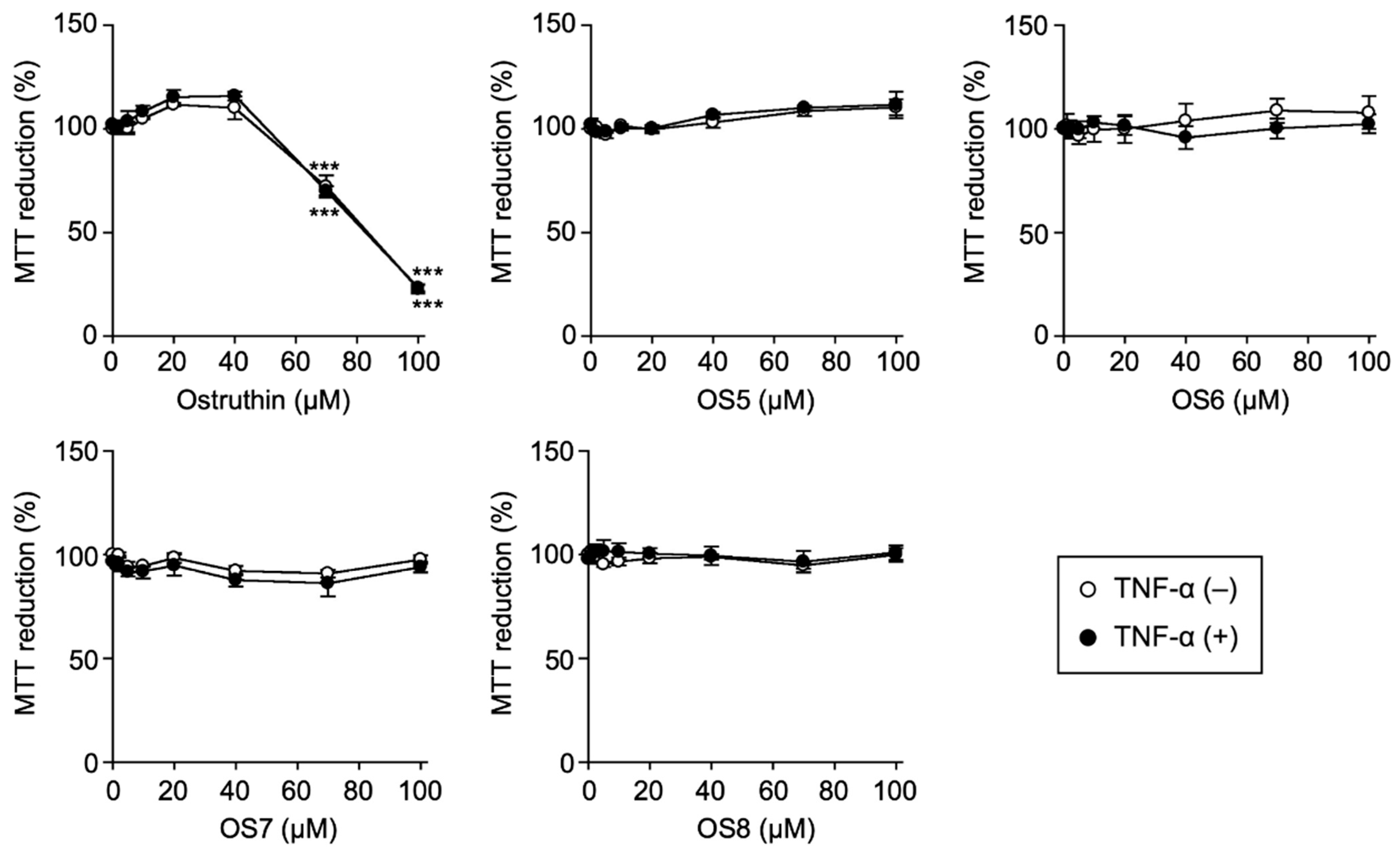
3.1. Biological Evaluation of Ostruthin and Its Alkyl TPP Derivatives on the Viability of A549 Cells

Ostruthin was purified from the CHCl3 extract of P. trimera [11]. Four TPP derivatives were synthesized from ostruthin as a scaffold (Figure 1) [17]. As the first step, MTT assay was used to examine the effects of the test compounds on the viability of A549 cells. The MTT assay revealed that ostruthin inhibited MTT-reducing activity at IC50 values of 83.2 ± 2.5 and 82.8 ± 0.9 µM (n = 3, calculated from three independent experiments) in the absence and presence of TNF-α, respectively (Figure 2). In contrast, the four alkyl TPP derivatives of ostruthin at concentrations up to 100 μM did not significantly decrease MTT-reducing activity (Figure 2).
Crystal violet staining was also used to assess cell viability because it mainly stains adherent cells. Ostruthin decreased the number of crystal violet-stained cells at IC50 values of 63.4 ± 3.1 and 68.6 ± 4.4 μM (n = 3) in the absence and presence of TNF-α, respectively (Figure S1). The number of crystal violet-stained cells was not affected by OS5 or OS8, but was slightly reduced by OS6 and OS7 (Figure S1). These results confirmed that OS5, OS6, OS7, and OS8 did not markedly affect the viability of A549 cells at concentrations up to 100 µM.

3.2. Biological Evaluation of Ostruthin and Its Alkyl TPP Derivatives on the TNF-α-Induced Expression of Cell-Surface ICAM-1 in A549 Cells

In A549 cells, the TNF-α stimulation up-regulates the expression of cell-surface ICAM-1 within 6 h at sufficient levels, and this may be quantitatively measured using a cell-ELISA [19]. The treatment with TNF-α significantly increased the cell-surface expression of ICAM-1 (Figure 3). The expression level of cell-surface ICAM-1 was reduced by all of the test compounds in a dose-dependent manner (Figure 3). Among the test compounds examined, ostruthin exerted the strongest inhibitory effects and attenuated the up-regulation of cell-surface ICAM-1 induced by TNF-α at an IC50 value of 55.3 ± 2.5 µM (n = 3) and completely at concentrations higher than 70 μM (Figure 3). OS5, OS6, OS7, and OS8 interfered with cell-surface ICAM-1 expression at IC50 values of >100, 88.6 ± 5.9, 65.1 ± 14.9, and 68.6 ± 8.3 µM (n = 3), respectively (Figure 3). These results suggested that the strong inhibition of cell-surface ICAM-1 expression by ostruthin was at least partly due to reductions in cell viability and attached cells. In contrast, the alkyl TPP derivatives of ostruthin exerted inhibitory effects against the TNF-α-dependent up-regulation of cell-surface ICAM-1 without significantly affecting cell viability. The flow cytometric assay showed that OS8 inhibited the TNF-α-induced expression of cell-surface ICAM-1 in a dose-dependent manner with an IC50 value of 62.0 ± 3.4 µM (n = 3) (Figure S2), which confirmed cell-ELISA-based results (Figure 3).

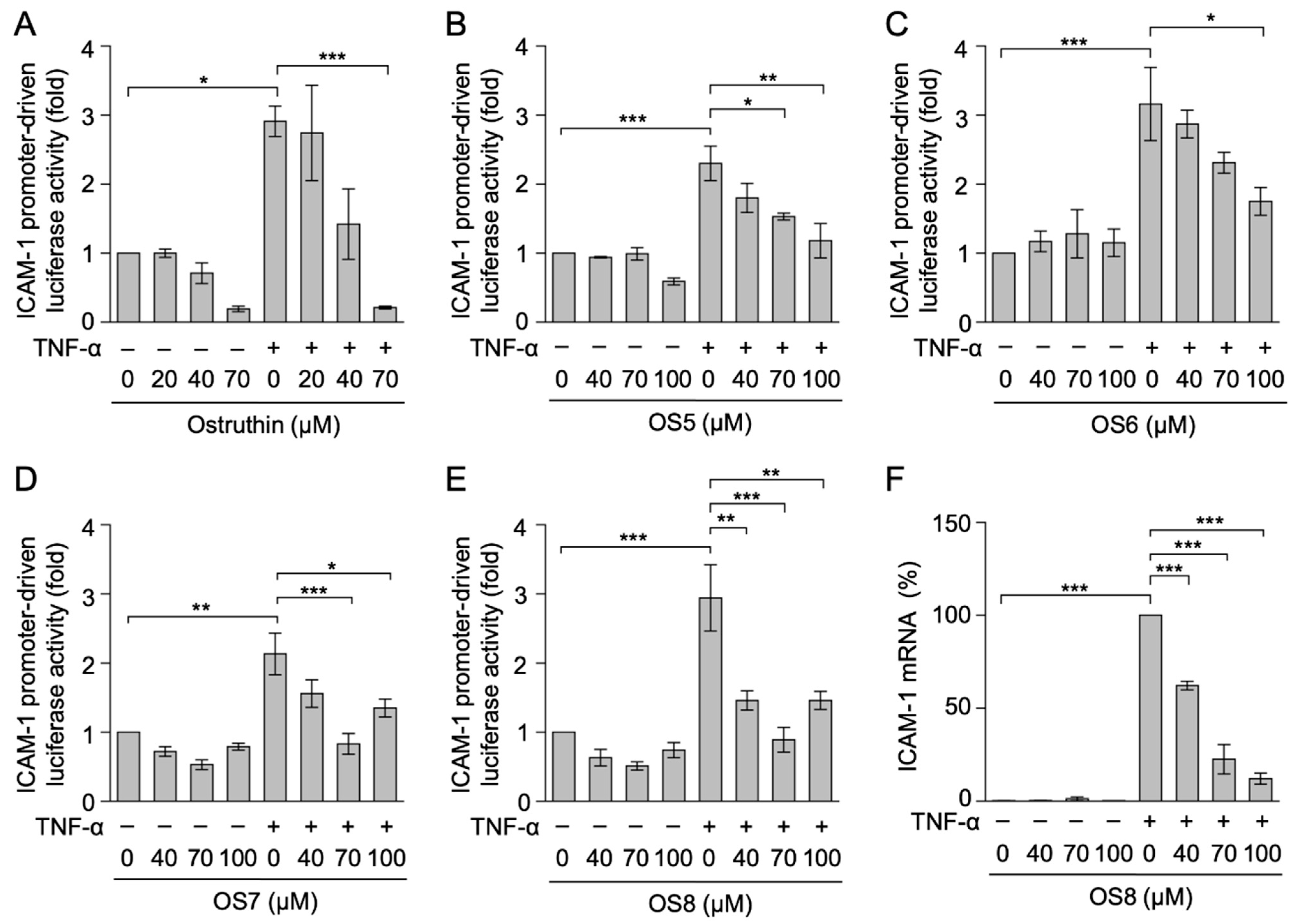
3.3. Biological Evaluation of Ostruthin and Its Alkyl TPP Derivatives on TNF-α-Induced ICAM-1 Promoter-Driven Luciferase Activity in A549 Cells

We investigated the effects of ostruthin alkyl TPP derivatives on ICAM-1 promoter-driven luciferase reporter activity induced by TNF-α. A 2.5-h incubation with TNF-α increased luciferase activity by approximately 3-fold from that in unstimulated cells (Figure 4A–E). All test compounds reduced TNF-α-induced luciferase reporter activity (Figure 4A–E). Among the alkyl TPP derivatives of ostruthin, OS8 significantly inhibited ICAM-1 promoter-luciferase reporter activity at concentrations higher than 40 μM (Figure 4E). Therefore, OS8 was considered to be the most potent anti-inflammatory agent without any influence on cell viability.
We further examined the effects of OS8 on TNF-α-induced ICAM-1 transcription using quantitative PCR. In response to TNF-α, ICAM-1 mRNA levels were approximately 400-fold higher than those in untreated cells (Figure 4F). OS8 inhibited TNF-α-induced ICAM-1 mRNA expression in a dose-dependent manner and strongly at concentrations of 70 and 100 µM (Figure 4F). These results demonstrated that OS8 inhibited TNF-α-induced ICAM-1 transcription.

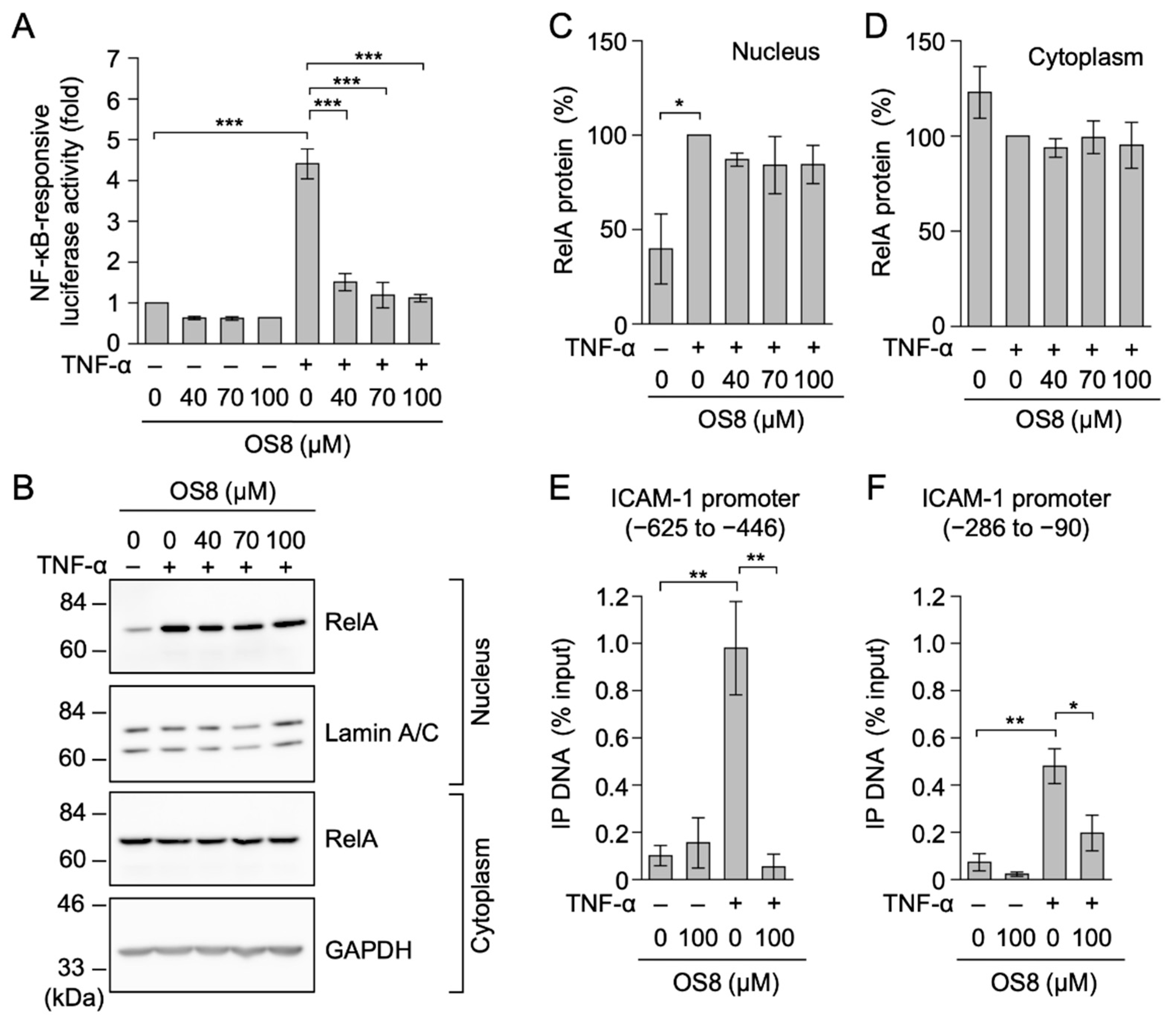
3.4. OS8 Prevented TNF-α-Induced ICAM-1 Expression and NF-κB-Responsive Luciferase Activity in HT-1080 Cells

Human fibrosarcoma HT-1080 cells were employed to generalize the biological activity of OS8. The MTT assay showed that OS8 slightly decreased the viability of HT-1080 cells by approximately 20–30% at concentrations higher than 70 µM during a 7-h incubation (Figure 5A). Under these conditions, OS8 inhibited TNF-α-induced ICAM-1 expression by 60–80% at concentrations higher than 70 µM (Figure 5B).
The transcriptional activation of the ICAM-1 gene is primarily mediated by NF-κB transcription factors in response to TNF-α [3,4,5]. Although OS8 slightly affected cell viability during a 3.5-h incubation (Figure 5C), TNF-α-induced NF-κB-responsive luciferase reporter activity was more strongly inhibited by OS8 at concentrations of 40–100 µM (Figure 5D). These results indicate that OS8 inhibited TNF-α-induced gene expression, at least in two different cell lines.

3.5. OS8 Prevented the Binding of RelA to the ICAM-1 Promoter in TNF-α-Stimulated A549 Cells

In A549 cells, TNF-α increased NF-κB-responsive luciferase activity by approximately 4-fold from that in unstimulated cells (Figure 6A). OS8 reduced NF-κB-responsive luciferase reporter activity at concentrations higher than 40 µM (Figure 6A).
The translocation of the NF-κB subunit RelA from the cytoplasm to the nucleus was observed in A549 cells within 30 min of the TNF-α stimulation [28,31]. The TNF-α stimulation induced an approximately 2-fold increase in nuclear RelA levels (Figure 6B–D). OS8 did not decrease the amount of nuclear RelA at concentrations up to 100 μM (Figure 6B,C). These results revealed that OS8 did not affect TNF-α-induced nuclear RelA translocation.
Based on the above results, we speculated that OS8 may prevent the binding of RelA to the ICAM-1 promoter. Multiple NF-κB-responsive sites were previously reported to be located in the ICAM-1 promoter [30,32]. A ChIP assay was performed to investigate the direct binding of RelA to these NF-κB-responsive sites. The binding of RelA to the ICAM-1 promoter was markedly increased by the TNF-α stimulation (Figure 6E,F). OS8 at 100 µM significantly suppressed the increase in RelA binding (Figure 6E,F). These results indicated that OS8 prevented the binding of RelA to the ICAM-1 promoter, which accounted for the reduction in ICAM-1 mRNA expression.

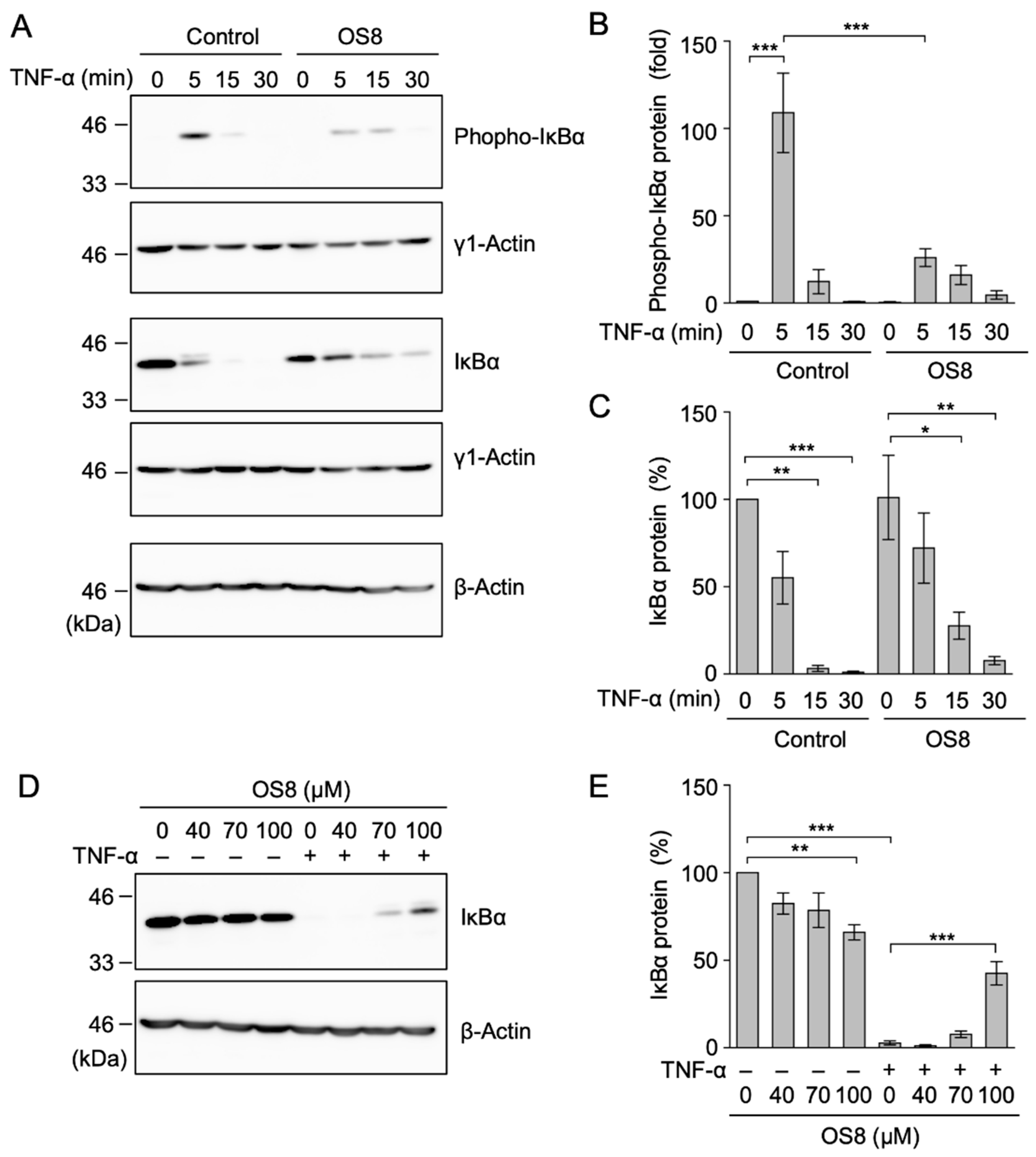
3.6. OS8 Inhibited the Phosphorylation of IκBα in TNF-α-Stimulated A549 Cells

To elucidate the mechanisms by which OS8 inhibits the binding of RelA to the ICAM-1 promoter, we examined the phosphorylation of components of the NF-κB signaling pathway. In A549 cells, the results of the time-course experiment showed that the TNF-α stimulation induced the phosphorylation of IκBα within 5 min, which was followed by the degradation of IκBα within 15 min (Figure 7A–C). OS8 at 100 µM markedly diminished TNF-α-induced IκBα phosphorylation (Figure 7A,B). In contrast, TNF-α-induced IκBα degradation was delayed, but proceeded for 30 min in the presence of OS8 (Figure 7A,C). This result is consistent with OS8 not markedly affecting the nuclear translocation of RelA 30 min after a stimulation with TNF-α (Figure 6B,C). The results of the dose-dependent experiment showed that OS8 at 100 µM suppressed the degradation of IκBα to some extent 15 min after the TNF-α stimulation (Figure 7D,E).

3.7. OS8 Inhibited the Phosphorylation of RelA and ERK1/ERK2 in TNF-α-Stimulated A549 Cells

RelA is phosphorylated at Ser 536, which is required for transcriptional activation, nuclear localization, and protein stability [33,34,35]. The TNF-α stimulation induced the Ser 536 phosphorylation of RelA in A549 cells (Figure 8A,B). OS8 at 100 µM inhibited TNF-α-induced RelA phosphorylation at Ser 536 without affecting the amount of total RelA (Figure 8A–C).
ERK1 and ERK2 were phosphorylated in A549 cells stimulated with TNF-α (Figure 8D,E). The TNF-α-induced phosphorylation of ERK1/ERK2 was inhibited by OS8 without affecting total ERK1/ERK2 levels (Figure 8D–F). Collectively, the present results indicate that OS8 inhibited the TNF-α-induced phosphorylation of RelA and ERK1/ERK2.

4. Discussion

Ostruthin inhibited TNF-α-induced ICAM-1 expression as anti-inflammatory activity in A549 cells, but also exhibited cytotoxic activity in the same cell line. However, in the present study, the synthetic alkyl TPP derivatives of ostruthin inhibited the TNF-α-induced expression of ICAM-1 without affecting the viability of A549 cells. We selected OS8 as the most potent derivative and showed that it inhibited TNF-α-induced ICAM-1 mRNA expression and NF-κB-dependent reporter activity. In TNF-α-stimulated A549 cells, OS8 did not affect RelA nuclear translocation, but prevented the subsequent binding of RelA to the ICAM-1 promoter. The present results also demonstrated that OS8 inhibited the TNF-α-induced phosphorylation of IκBα, RelA, and ERK. Based on similarities in the chemical structures of the derivatives, other compounds (OS5, OS6, and OS7) may inhibit TNF-α-induced ICAM-1 expression by a similar mechanism to that of OS8.
We showed that ostruthin markedly reduced TNF-α-induced ICAM-1 expression in A549 cells. A previous study reported that ostruthin prevented the lipopolysaccharide-induced expression of inducible NO synthase and cyclooxygenase-2 in mouse BV-2 microglial cell line [15]. Ostruthin was recently shown to inhibit TNF-α-induced NF-κB reporter activity in human embryonic kidney 293 cells [16]. Consistent with this finding, daphnetin (7,8-dihydroxycoumarin), a coumarin derivative closely related to ostruthin, was reported to block the translocation of NF-κB subunits in concanavalin A-stimulated mouse T cells [36]. It also decreased the level of phosphorylated RelA in high glucose-stimulated human glomerular mesangial cells [37]. These findings suggest the potential of ostruthin as an agent that targets the NF-κB signaling pathway. However, ostruthin exhibited cytotoxicity against A549 cells at concentrations that decreased ICAM-1 expression. Among the OS derivatives, OS8 inhibited TNF-α-induced ICAM-1 expression at an IC50 value of 68.6 ± 8.3 µM, which was a slightly higher concentration than that of ostruthin (55.3 ± 2.5 µM). In contrast, OS8 did not affect the viability of A549 cells at concentrations up to 100 µM. Therefore, the alkyl TPP modification of ostruthin reduced its cytotoxic activity and improved its potential for anti-inflammatory activity.
We previously demonstrated that ostruthin exhibited cytotoxic activity against three human cancer cell lines, i.e., pancreatic PANC-1 cells, cervical HeLa cells, and hepatic HepG2 cells [17]. We also showed that ostruthin reduced the viability of human lung adenocarcinoma A549 cells. Consistent with these findings, ostruthin exhibited cytotoxic activities against human pancreatic cancer cell lines (BcPc3, CFPAC1, PANC-1, and PSN-1) and rat neural PC12 cells [14,38,39]. Ostruthin also reduced the proliferation of normal cells, i.e., rat vascular smooth muscle cells and human pancreatic ductal epithelial cells [40,41,42]. Although sensitivity to ostruthin may vary in different types of cells and/or be influenced by experimental conditions (e.g., incubation time), these findings collectively indicated that ostruthin exhibits cytotoxic activities against cancerous and normal cells. OS7 and OS8 exhibited stronger cytotoxicity against PANC-1 cells and HeLa cells than ostruthin, but weaker cytotoxic activity against HepG2 cells [17]. In the present study, all TPP derivatives (OS5, OS6, OS7, and OS8) exhibited weaker cytotoxic activities against A549 cells. These results suggest that ostruthin and ostruthin derivatives target multiple proteins that are essential for cell survival and proliferation, thereby interfering with different target proteins in a cell context-dependent manner. Moreover, the selectivity of ostruthin and its derivatives among target proteins in each cell line is altered by the modification of a TPP moiety and/or alkyl linkers because TPP modifications are utilized conceptionally for mitochondrial delivery [18].
In addition to cell-based biological activities, previous studies reported that ostruthin directly inhibited the enzymatic activities of ethoxyresorufin O-dealkylase and pentoxyresorufin O-dealkylase (cytochrome P450 family members) [43], acetylcholine esterase [44], and α-glucosidase [11,45]. Furthermore, ostruthin has been identified as an activator of TWIK-related K+ channels [46]. These biological activities of ostruthin do not appear to be directly linked to the inhibition of the NF-κB signaling pathway or cytotoxic activity. Nevertheless, these findings suggest that ostruthin and its derivatives have the ability to target multiple proteins in the cell.
Regarding its mechanism of action, we revealed that OS8 did not affect the nuclear translocation of RelA, but inhibited the binding of RelA to the ICAM-1 promoter in A549 cells. Moreover, we showed that OS8 inhibited the TNF-α-induced phosphorylation of IκBα, RelA, and ERK. OS8 delayed TNF-α-induced IκBα degradation, but did not markedly affect the nuclear translocation of RelA at later time points. Previous studies demonstrated that the phosphorylation of Ser 536 is mediated by multiple protein kinases, such as IκB kinase β, and is involved in transcriptional activation, nuclear localization, and protein stability [33,34,35]. Therefore, the inhibition of RelA phosphorylation by OS8 appears to be responsible for the inhibition of TNF-α-induced NF-κB activation, including RelA binding to the ICAM-1 promoter. Moreover, the activation of ERK was required for the TNF-α-induced RelA phosphorylation at Ser 536 in mouse epidermal JB cells [47]. It is also possible that the inhibition of TNF-α-induced ERK activation by OS8 is involved in the inhibition of RelA phosphorylation. Further experiments are needed to elucidate the molecular mechanisms by which OS8 inhibits the TNF-α-induced phosphorylation of IκBα, RelA, and ERK.

5. Conclusions

Ostruthin exhibited anti-inflammatory and cytotoxic activities in A549 cells. The present results demonstrated that the alkyl TPP modification reduced the cytotoxic activity of ostruthin while maintaining its anti-inflammatory activity. OS8 inhibited TNF-α-induced ICAM-1 expression without affecting cell viability. OS8 prevented the NF-κB signaling pathway and inhibited the binding of the NF-κB subunit RelA to the ICAM-1 promoter. OS8 inhibited the TNF-α-induced phosphorylation of IκBα, RelA, and ERK. In conclusion, the present results revealed that OS8 has potential as an anti-inflammatory agent that targets the NF-κB signaling pathway.

Supplementary Materials

The following are available online at https://0-www-mdpi-com.brum.beds.ac.uk/article/10.3390/biochem1020010/s1, Figure S1: Ostruthin decreased cell viability, whereas OS5, OS6, OS7, and OS8 did not, Figure S2: OS8 inhibited TNF-α-induced cell surface ICAM-1 expression in a dose-dependent manner.

Author Contributions

Conceptualization, N.T.V. and T.K.; formal analysis, N.T.V., E.K., K.N., C.M., Y.M., and T.K.; investigation, N.T.V., E.K., K.N., C.M., Y.M., S.H., and S.F.; resources, N.T.N., P.H.D., and M.T.T.N.; writing—original draft preparation, N.T.V.; writing—review and editing, N.T.V., Y.M., P.H.D., and T.K.; visualization, N.T.V., E.K., K.N., C.M., and T.K.; supervision, T.K.; funding acquisition, T.K. All authors have read and agreed to the published version of the manuscript.

Funding

This work was partly supported by the Japan Society for the Promotion of Science (JSPS) KAKENHI Grant number 19H02885 (to T.K.) and Grants-in-Aid from the JSPS Core-to-Core program, B Asia-Africa Science Platforms.

Institutional Review Board Statement

Not applicable.

Informed Consent Statement

Not applicable.

Data Availability Statement

The data presented in the present study are available upon request from the corresponding author.

Conflicts of Interest

The authors declare no conflict of interest.

References

  1. Gerhardt, T.; Ley, K. Monocyte trafficking across the vessel wall. Cardiovasc. Res. 2015, 107, 321–330. [Google Scholar] [CrossRef] [PubMed] [Green Version]
  2. Vestweber, D. How leukocytes cross the vascular endothelium. Nat. Rev. Immunol. 2015, 15, 692–704. [Google Scholar] [CrossRef] [PubMed]
  3. Van de Stolpe, A.; Van der Saag, P.T. Intercellular adhesion molecule-1. J. Mol. Med. 1996, 74, 13–33. [Google Scholar] [CrossRef] [PubMed]
  4. Collins, T.; Read, M.A.; Neish, A.S.; Whitley, M.Z.; Thanos, D.; Maniatis, T. Transcriptional regulation of endothelial cell adhesion molecules: NF-κB and cytokine-inducible enhancers. FASEB J. 1995, 9, 899–909. [Google Scholar] [CrossRef] [Green Version]
  5. Roebuck, K.A.; Finnegan, A. Regulation of intercellular adhesion molecule-1 (CD54) gene expression. J. Leukoc. Biol. 1999, 66, 876–888. [Google Scholar] [CrossRef]
  6. Bhoj, V.G.; Chen, Z.J. Ubiquitylation in innate and adaptive immunity. Nature 2009, 458, 430–437. [Google Scholar] [CrossRef]
  7. Hayden, M.S.; Ghosh, S. Regulation of NF-κB by TNF family cytokines. Semin. Immunol. 2014, 26, 253–266. [Google Scholar] [CrossRef] [Green Version]
  8. Karin, M.; Greten, F.R. NF-κB: Linking inflammation and immunity to cancer development and progression. Nat. Rev. Immunol. 2005, 5, 749–759. [Google Scholar] [CrossRef]
  9. Stefanachi, A.; Leonetti, F.; Pisani, L.; Catto, M.; Carotti, A. Coumarin: A natural, privileged and versatile scaffold for bioactive compounds. Molecules 2018, 23, 250. [Google Scholar] [CrossRef] [Green Version]
  10. Venugopala, K.N.; Rashmi, V.; Odhav, B. Review on natural coumarin lead compounds for their pharmacological activity. BioMed Res. Int. 2013, 2013, 963248. [Google Scholar] [CrossRef] [Green Version]
  11. Dang, P.H.; Le, T.H.; Phan, P.K.T.; Le, P.T.T.; Nguyen, M.T.T.; Nguyen, N.T. Two acridones and two coumarins from the roots of Paramignya trimera. Tetrahedron Lett. 2017, 58, 1553–1557. [Google Scholar] [CrossRef]
  12. Khalid, S.A.; Farouk, A.; Geary, T.G.; Jensen, J.B. Potential antimalarial candidates from African plants: An in vitro approach using Plasmodium falciparum. J. Ethnopharmacol. 1986, 15, 201–209. [Google Scholar] [CrossRef]
  13. Schinkovitz, A.; Gibbons, S.; Stavri, M.; Cocksedge, M.J.; Bucar, F. Ostruthin: An antimycobacterial coumarin from the roots of Peucedanum ostruthium. Planta Med. 2003, 69, 369–371. [Google Scholar] [CrossRef] [Green Version]
  14. Li, F.; Okamura, Y.; Dibwe, D.F.; Awale, S.; Kadota, S.; Tezuka, Y. Anti-austerity agents from Rhizoma et Radix Notopterygii (Qianghuo). Planta Med. 2012, 78, 796–799. [Google Scholar] [CrossRef] [Green Version]
  15. Tuan Anh, H.L.; Kim, D.C.; Ko, W.; Ha, T.M.; Nhiem, N.X.; Yen, P.H.; Tai, B.H.; Truong, L.H.; Long, V.N.; Gioi, T.; et al. Anti-inflammatory coumarins from Paramignya trimera. Pharm. Biol. 2017, 55, 1195–1201. [Google Scholar] [CrossRef] [Green Version]
  16. Zwirchmayr, J.; Grienke, U.; Hummelbrunner, S.; Seigner, J.; de Martin, R.; Dirsch, V.M.; Rollinger, J.M. A Biochemometric approach for the identification of in vitro anti-inflammatory constituents in masterwort. Biomolecules 2020, 10, 679. [Google Scholar] [CrossRef] [PubMed]
  17. Dang, P.H.; Dao, T.H.X.; Le, V.T.; Nguyen, C.M.; Ly, T.T.; Nguyen, H.X.; Le, T.H.; Do, T.N.V.; Nguyen, M.T.T.; Sun, S.; et al. Synthesis of alkyl triphenylphosphonium ostruthin derivatives as potential cytotoxic candidates. ChemistrySelect 2020, 5, 12636–12640. [Google Scholar] [CrossRef]
  18. Zielonka, J.; Joseph, J.; Sikora, A.; Hardy, M.; Ouari, O.; Vasquez-Vivar, J.; Cheng, G.; Lopez, M.; Kalyanaraman, B. Mitochondrial-targeted triphenylphosphonium-based compounds: Syntheses, mechanisms of action, and therapeutic and diagnostic applications. Chem. Rev. 2017, 117, 10043–10120. [Google Scholar] [CrossRef] [PubMed]
  19. Yamada, Y.; Taketani, S.; Osada, H.; Kataoka, T. Cytotrienin A, a translation inhibitor that induces ectodomain shedding of TNF receptor 1 via activation of ERK and p38 MAP kinase. Eur. J. Pharmacol. 2011, 667, 113–119. [Google Scholar] [CrossRef]
  20. Mitsuda, S.; Yokomichi, T.; Yokoigawa, J.; Kataoka, T. Ursolic acid, a natural pentacyclic triterpenoid, inhibits intracellular trafficking of proteins and induces accumulation of intercellular adhesion molecule-1 linked to high-mannose-type glycans in the endoplasmic reticulum. FEBS Open Bio 2014, 4, 229–239. [Google Scholar] [CrossRef] [PubMed] [Green Version]
  21. Fukuhara, S.; Tanigaki, R.; Kimura, K.I.; Kataoka, T. Kujigamberol interferes with pro-inflammatory cytokine-induced expression of and N-glycan modifications to cell adhesion molecules at different stages in human umbilical vein endothelial cells. Int. Immunopharmacol. 2018, 62, 313–325. [Google Scholar] [CrossRef] [PubMed]
  22. Dohrman, A.; Kataoka, T.; Cuenin, S.; Russell, J.Q.; Tschopp, J.; Budd, R.C. Cellular FLIP (long form) regulates CD8+ T cell activation through caspase-8-dependent NF-κB activation. J. Immunol. 2005, 174, 5270–5278. [Google Scholar] [CrossRef] [Green Version]
  23. Matsuda, I.; Matsuo, K.; Matsushita, Y.; Haruna, Y.; Niwa, M.; Kataoka, T. The C-terminal domain of the long form of cellular FLICE-inhibitory protein (c-FLIPL) inhibits the interaction of the caspase 8 prodomain with the receptor-interacting protein 1 (RIP1) death domain and regulates caspase 8-dependent nuclear factor κB (NF-κB) activation. J. Biol. Chem. 2014, 289, 3876–3887. [Google Scholar] [PubMed] [Green Version]
  24. Vo, T.N.; Sasaki, S.; Miyake, Y.; Nguyen, N.T.; Dang, P.H.; Nguyen, M.T.T.; Kataoka, T. α-Conidendrin inhibits the expression of intercellular adhesion molecule-1 induced by tumor necrosis factor-α in human lung adenocarcinoma A549 cells. Eur. J. Pharmacol. 2021, 890, 173651. [Google Scholar] [CrossRef]
  25. Harada, M.; Morimoto, K.; Kondo, T.; Hiramatsu, R.; Okina, Y.; Muko, R.; Matsuda, I.; Kataoka, T. Quinacrine inhibits ICAM-1 transcription by blocking DNA binding of the NF-κB subunit p65 and sensitizes human lung adenocarcinoma A549 cells to TNF-α and the Fas ligand. Int. J. Mol. Sci. 2017, 18, 2603. [Google Scholar] [CrossRef] [PubMed] [Green Version]
  26. Wan, M.; Liu, J.; Ouyang, X. Nucleotide-binding oligomerization domain 1 regulates Porphyromonas gingivalis-induced vascular cell adhesion molecule 1 and intercellular adhesion molecule 1 expression in endothelial cell through NF-κB pathway. J. Periodont. Res. 2015, 50, 189–196. [Google Scholar] [CrossRef] [PubMed]
  27. Zhang, Y.; Lian, F.; Zhu, Y.; Xia, M.; Wang, Q.; Ling, W.; Wang, X.D. Cyanidin-3-O-β-glucoside inhibits LPS-induced expression of inflammatory mediators through decreasing IκBα phosphorylation in THP-1 cells. Inflamm. Res. 2010, 59, 723–730. [Google Scholar] [CrossRef]
  28. Hirano, S.; Kataoka, T. Deoxynivalenol induces ectodomain shedding of TNF receptor 1 and thereby inhibits the TNF-α-induced NF-κB signaling pathway. Eur. J. Pharmacol. 2013, 701, 144–151. [Google Scholar] [CrossRef]
  29. Fukuoka, N.; Harada, M.; Nishida, A.; Ito, Y.; Shiota, H.; Kataoka, T. Eomesodermin promotes interferon-γ expression and binds to multiple conserved noncoding sequences across the Ifng locus in mouse thymoma cell lines. Genes Cells 2016, 21, 146–162. [Google Scholar] [CrossRef]
  30. Xue, J.; Thippegowda, P.B.; Hu, G.; Bachmaier, K.; Christman, J.W.; Malik, A.B.; Tiruppathi, C. NF-κB regulates thrombin-induced ICAM-1 gene expression in cooperation with NFAT by binding to the intronic NF-κB site in the ICAM-1 gene. Physiol. Genom. 2009, 38, 42–53. [Google Scholar] [CrossRef] [Green Version]
  31. Tanigaki, R.; Takahashi, R.; Nguyen, M.T.T.; Nguyen, N.T.; Do, T.V.N.; Nguyen, H.X.; Kataoka, T. 4-Hydroxypanduratin A and isopanduratin A inhibit tumor necrosis factor α-stimulated gene expression and the nuclear factor κB-dependent signaling pathway in human lung adenocarcinoma A549 cells. Biol. Pharm. Bull. 2019, 42, 26–33. [Google Scholar] [CrossRef] [PubMed] [Green Version]
  32. Ren, Z.; Kang, W.; Wang, L.; Sun, B.; Ma, J.; Zheng, C.; Sun, J.; Tian, Z.; Yang, X.; Xiao, W. E2F1 renders prostate cancer cell resistant to ICAM-1 mediated antitumor immunity by NF-κB modulation. Mol. Cancer 2014, 13, 84. [Google Scholar] [CrossRef] [Green Version]
  33. Perkins, N.D. Post-translational modifications regulating the activity and function of the nuclear factor kappa B pathway. Oncogene 2006, 25, 6717–6730. [Google Scholar] [CrossRef] [PubMed] [Green Version]
  34. Hoesel, B.; Schmid, J.A. The complexity of NF-κB signaling in inflammation and cancer. Mol. Cancer 2013, 12, 86. [Google Scholar] [CrossRef] [PubMed] [Green Version]
  35. Christian, F.; Smith, E.L.; Carmody, R.J. The regulation of NF-κB subunits by phosphorylation. Cells 2016, 5, 12. [Google Scholar] [CrossRef] [PubMed] [Green Version]
  36. Song, B.; Wang, Z.; Liu, Y.; Xu, S.; Huang, G.; Xiong, Y.; Zhang, S.; Xu, L.; Deng, X.; Guan, S. Immunosuppressive activity of daphnetin, one of coumarin derivatives, is mediated through suppression of NF-κB and NFAT signaling pathways in mouse T cells. PLoS ONE 2014, 9, e96502. [Google Scholar] [CrossRef]
  37. Xu, K.; Guo, L.; Bu, H.; Wang, H. Daphnetin inhibits high glucose-induced extracellular matrix accumulation, oxidative stress and inflammation in human glomerular mesangial cells. J. Pharm. Sci. 2019, 139, 91–97. [Google Scholar] [CrossRef]
  38. Qi, J.; Ojika, M.; Sakagami, Y. Differentiation in a rat PC12 cell line induced by ostruthin and (–)-bornyl ferulate, constituents of a Chinese herbal medicine. Biosci. Biotechnol. Biochem. 1999, 63, 1501–1502. [Google Scholar] [CrossRef]
  39. Nguyen, V.T.; Scarlett, C.J. Cytotoxic activity of extracts and fractions from Paramignya trimera root and Phyllanthus amarus against pancreatic cancer cell lines. J. Cancer Res. Ther. 2019, 15, 245–249. [Google Scholar] [CrossRef]
  40. Joa, H.; Vogl, S.; Atanasov, A.G.; Zehl, M.; Nakel, T.; Fakhrudin, N.; Heiss, E.K.; Picker, P.; Urban, E.; Wawrosch, C.; et al. Identification of ostruthin from Peucedanum ostruthium rhizomes as an inhibitor of vascular smooth muscle cell proliferation. J. Nat. Prod. 2011, 74, 1513–1516. [Google Scholar] [CrossRef]
  41. Nguyen, V.T.; Sakoff, J.A.; Scarlett, C.J. Physicochemical, antioxidant, and cytotoxic properties of Xao tam phan (Paramignya trimera) root extract and its fractions. Chem. Divers. 2017, 14, e1600396. [Google Scholar] [CrossRef] [PubMed]
  42. Nguyen, V.T.; Sakoff, J.A.; Scarlett, C.J. Physicochemical properties, antioxidant, and anti-proliferative capacities of dried leaf and its extract from Xao tam phan (Paramignya trimera). Chem. Biodivers. 2017, 14, e1600498. [Google Scholar] [CrossRef]
  43. Cai, Y.; Bennett, D.; Nair, R.V.; Ceska, O.; Ashwood-Smith, M.J.; DiGiovanni, J. Inhibition and inactivation of murine hepatic ethoxy- and petoxyresorufin O-dealkylase by naturally occurring coumarins. Chem. Res. Toxicol. 1993, 6, 872–879. [Google Scholar] [CrossRef]
  44. Urbain, A.; Marston, A.; Hostettmann, K. Coumarins from Peucedanum ostruthium as inhibitors of acetylcholinesterase. Pharm. Biol. 2005, 43, 647–650. [Google Scholar] [CrossRef] [Green Version]
  45. Trinh, D.H.; Tran, P.H.; Trinh, B.T.D.; Nguyen, H.T.; Nguyen, H.D.; Ha, L.D.; Nguyen, L.H.D. Coumarins and acridone alkaloids with α-glucosidase inhibitory and antioxidant activity from the roots of Paramignya trimera. Phytochem. Lett. 2020, 35, 94–98. [Google Scholar] [CrossRef]
  46. Joseph, A.; Thuy, T.T.T.; Thanh, L.T.; Okada, M. Antidepressive and anxiolytic effects of ostruthin, a TREK-1 channel activator. PLoS ONE 2018, 13, e0201092. [Google Scholar] [CrossRef] [PubMed] [Green Version]
  47. Hu, J.; Nakano, H.; Sakurai, H.; Colburn, N.H. Insufficient p65 phosphorylation at S536 specifically contributes to the lack of NF-κB activation and transformation in resistant JB6 cells. Carcinogenesis 2004, 25, 1991–2003. [Google Scholar] [CrossRef] [PubMed] [Green Version]
Figure 1. Structures of ostruthin and its alkyl TPP derivatives.
Biochem 01 00010 g001
Figure 2. Ostruthin decreased cell viability, whereas OS5, OS6, OS7, and OS8 did not. A549 cells were preincubated with serial dilutions of the test compounds for 1 h, and then incubated without (open circles) or with (filled circles) TNF-α (2.5 ng/mL) for 6 h in the presence of test compounds at the final concentrations (0–100 µM). MTT reduction (%) represents the mean ± S.E. of three independent experiments. *** p < 0.001.
Biochem 01 00010 g002
Figure 3. Ostruthin, OS5, OS6, OS7, and OS8 inhibited cell-surface ICAM-1 expression. A549 cells were treated as described in Figure 2. Cell-surface ICAM-1 levels were evaluated by cell-ELISA. ICAM-1 expression (%) represents the mean ± S.E. of three independent experiments. * p < 0.05 and *** p < 0.001.
Biochem 01 00010 g003
Figure 4. Ostruthin, OS5, OS6, OS7, and OS8 inhibited TNF-α-induced ICAM-1-promoter-driven luciferase activity. (AE) A549 cells were transfected with ICAM-1 promoter-driven and control cytomegalovirus promoter-driven luciferase reporter plasmids. Transfected A549 cells were treated with serial dilutions of ostruthin (A), OS5 (B), OS6 (C), OS7 (D), or OS8 (E) for 1 h, and were then incubated without (−) or with (+) TNF-α (2.5 ng/mL) for 2.5 h in the presence of test compounds at the final concentrations (0–100 µM). Luciferase activity (fold) represents the mean ± S.E. of three independent experiments. * p < 0.05, ** p < 0.01, and *** p < 0.001. (F) A549 cells were preincubated with different concentrations of OS8 for 1 h, and were then incubated without (−) or with (+) TNF-α (2.5 ng/mL) for 2 h in the presence of OS8 at the final concentrations (0–100 µM). The amount of ICAM-1 mRNA was analyzed by real-time PCR. ICAM-1 mRNA (%) represents the mean ± S.E. of three independent experiments. *** p < 0.001.
Biochem 01 00010 g004
Figure 5. OS8 inhibited ICAM-1 expression and NF-κB-responsive luciferase activity in TNF-α-stimulated HT-1080 cells. (AC) HT-1080 cells were preincubated with serial dilutions of OS8 for 1 h, and then incubated without (open circles) or with (filled circles) TNF-α (2.5 ng/mL) for 6 h (A,B) and 2.5 h (C) in the presence of OS8 at the final concentrations (0–100 µM). MTT reduction (%) represents the mean ± S.E. of three independent experiments (A,C). ICAM-1 expression (%) represents the mean ± S.E. of three independent experiments (B). * p < 0.05, ** p < 0.01, and *** p < 0.001. (D) HT-1080 cells were transfected with NF-κB-responsive and control cytomegalovirus promoter-driven luciferase reporter plasmids. Transfected HT-1080 cells were preincubated with serial dilutions of OS8 for 1 h, and then incubated without (−) or with (+) TNF-α (2.5 ng/mL) for 2.5 h in the presence of OS8 at the final concentrations (0–100 µM). Luciferase activity (fold) represents the mean ± S.E. of three independent experiments. * p < 0.05 and ** p < 0.01.
Biochem 01 00010 g005
Figure 6. OS8 did not affect the nuclear translocation of RelA, but inhibited NF-κB-responsive luciferase activity and RelA binding to the ICAM-1 promoter in TNF-α-stimulated A549 cells. (A) A549 cells were transfected with NF-κB-responsive and control cytomegalovirus promoter-driven luciferase reporter plasmids. Transfected A549 cells were treated as described in Figure 5D. Luciferase activity (fold) represents the mean ± S.E. of three independent experiments. *** p < 0.001. (BD) A549 cells were treated as described in Figure 4F, except for an incubation without (−) or with (+) TNF-α for 30 min. Nuclear and cytoplasmic lysates were analyzed by Western blotting. Representative blots from three independent experiments are shown (B). The protein level of RelA in the nucleus (C) and cytoplasm (D) was standardized by that of lamin A/C and GAPDH, respectively. The RelA protein (%) represents the mean ± S.E. of three independent experiments. * p < 0.05. (E,F) A549 cells were treated as described in Figure 6B–D. A ChIP assay was performed with an anti-RelA antibody. IP DNA (% input) for ICAM-1 promoter regions at −625 to −446 (E) and −286 to −90 (F) represents the mean ± S.E. of three independent experiments. * p < 0.05 and ** p < 0.01.
Biochem 01 00010 g006
Figure 7. OS8 inhibited the phosphorylation of IκBα in TNF-α-stimulated A549 cells. (AC) A549 cells were preincubated with OS8 for 1 h and then incubated without (−) or with (+) TNF-α (2.5 ng/mL) for the indicated times in the presence or absence of OS8 (100 µM). Cytoplasmic lysates were analyzed by Western blotting. Representative blots from three independent experiments are shown (A). The protein levels of phospho-IκBα and total IκBα were standardized by that of γ1-actin (B,C). * p < 0.05, ** p < 0.01, and *** p < 0.001. (D,E) A549 cells were treated as described in Figure 4F, except for an incubation without (−) or with (+) TNF-α for 15 min. Cytoplasmic lysates were analyzed by Western blotting. Representative blots from three independent experiments are shown (D). The protein level of IκBα was standardized by that of β-actin. ** p < 0.01 and *** p < 0.001.
Biochem 01 00010 g007
Figure 8. OS8 inhibited the phosphorylation of RelA and ERK in TNF-α-stimulated A549 cells. (AF) A549 cells were treated as described in Figure 4F, except for an incubation without (−) or with (+) TNF-α for 30 min. In (DF), A549 cells were treated with fetal calf serum-free medium. Whole cell lysates were analyzed by Western blotting. Representative blots from three independent experiments are shown (A,D). The protein levels of phospho-RelA (B), RelA (C), phospho-ERK1/ERK2 (E), and ERK1/ERK2 (F) were standardized by that of γ1-actin. * p < 0.05, ** p < 0.01, and *** p < 0.001.
Biochem 01 00010 g008
Publisher’s Note: MDPI stays neutral with regard to jurisdictional claims in published maps and institutional affiliations.

