Article

Identification and Accumulation of Phenolic Compounds in the Leaves and Bark of Salix alba (L.) and Their Biological Potential

by
Ewelina Piątczak
1,
Monika Dybowska
2,
Elżbieta Płuciennik
3,
Katarzyna Kośla
3,
Joanna Kolniak-Ostek
4 and
Urszula Kalinowska-Lis
2,*
1
Department of Biology and Pharmaceutical Botany, Faculty of Pharmacy, Medical University of Lodz, Muszyńskiego 1, 90-151 Łódź, Poland
2
Department of Cosmetic Raw Materials Chemistry, Faculty of Pharmacy, Medical University of Lodz, Muszyńskiego 1, 90-151 Łódź, Poland
3
Department of Molecular Carcinogenesis, Medical University of Lodz, Żeligowskiego 7/9, 90-752 Łódź, Poland
4
Department of Fruit, Vegetable and Plant Nutraceuticals Technology, Wroclaw University of Environmental and Life Sciences, Chełmońskiego 37, 51-630 Wrocław, Poland
*
Author to whom correspondence should be addressed.
Submission received: 31 August 2020 / Revised: 16 September 2020 / Accepted: 28 September 2020 / Published: 29 September 2020
(This article belongs to the Collection Pharmacology of Medicinal Plants)

Abstract

:
The study examines the phenolic compounds in hydromethanolic extracts of Salix alba (L.) leaves and bark as well as their antioxidant activity and cytotoxic potential. UPLC-PDA-Q/TOF-MS analysis showed a total of 29 phenolic compounds in leaves and 34 in bark. Total phenolic compound content was 5575.96 mg/100 g of dry weight (DW) in leaves and 2330.31 mg/100 g DW in bark. The compounds were identified as derivatives of phenolic acids (seven in leaves and five in bark), flavanols and procyanidins (eight in leaves and 26 in bark) and flavonols (14 in leaves and three in bark). Both extracts exhibited strong antioxidant potential, assessed by radical scavenging activity against 1,1-diphenyl-2-picrylhydrazyl (DPPH) and 2,2′-azinobis (3-ethylbenzothiazoline-6-sulfonic acid (ABTS), but the bark extract was even stronger than the ascorbic acid used as a standard. The cytotoxicity of both extracts was evaluated against human skin fibroblasts and human epidermal keratinocytes cell lines using the Presto Blue cell viability assay. The keratinocytes were more resistant to tested extracts than fibroblasts. The leaf and bark extracts at concentrations which exhibited antioxidant activity were also not toxic against the keratinocyte cell line. Thus, S. alba extracts, especially the leaf extract, offer promise as a nontoxic natural antioxidant, in cosmetic products or herbal medicines, and as a source of bioactive secondary metabolites.

1. Introduction

The last few years have seen a growth in the need for natural drugs obtained from medicinal plants as an alternative to synthetic products [1]. A number of these bioactive compounds serve as starting points for the development of new drugs [2]. Clinical studies have shown that some herbal medicines are better tolerated by patients and show fewer side effects than synthetic derivatives [3].
Willow bark extract has been used for thousands of years as an analgesic, antipyretic and anti-inflammatory agent. Records suggest that, as long as 6000 years ago, white willow was used in Mesopotamia. Hippocrates recommended chewing willow bark for patients suffering from fever, inflammation and pain [4]. Currently, willow bark is used to treat many different types of pain, such as rheumatic pain, back pain, toothache, as well as menstrual cramps. It is also used to relieve fever, sore throat, headache and flu, mainly due to the presence of salicin, which is a natural nonselective COX-1 and COX-2 inhibitor [5].
Although willow bark extracts are generally standardized to salicin, other compounds in the extracts, including polyphenols, may also play prominent roles in areas such as cosmetology [6,7]. Recently, barks of different vascular woody plants are considered as a valuable source of bioactive compounds, exhibiting wide biological activities, including, among others, antioxidant, anti-inflammatory, and antitumor properties. Many studies confirm that the wide spectrum of bioactivities of bark extracts is connected mainly with the presence of (poly)phenolic compounds [8,9]. Polyphenolic compounds demonstrate antimutagenic, anticarcinogenic, and antiaging properties; they also demonstrate very strong antioxidant properties by capturing reactive oxygen species [10] and can hence protect biological systems against the oxidative stress that can cause cancer, atherosclerosis, diabetes, accelerated aging and Alzheimer’s disease, among others [11,12].
White willow (Salix alba L.)—Salicaceae is a small tree that naturally occurs in Europe, Asia and North Africa. The species is common in cold and low-temperature regions of the northern hemisphere, and in Poland, where it mainly grows on riverbanks on moist soils [13].
The three species: Salix purpurea, S. daphnoides and S. fragilis are the main sources of the drug willow bark—Salicis Cortex and are widely described in the literature [14]. Reports on the polyphenolic compounds present in other Salix species are incomplete and limited. Only a few compounds have been reported so far in the bark and/or leaves of other than S. alba willow species, e.g., S. purpurea, S. daphnoides, S. phylicifolia, S. myrsinites, S. glauca, S. fragilis, S. lapponum, S. borealis and S. reticulata [15,16,17,18,19]. Data on the chemical composition and biological activity of S. alba are limited [6,20,21]. Karl et al. [20] detected several flavonoid derivatives in S. alba. Zaiter et al. [21] quantified the main compounds (lipids, protein, ashes, carbohydrates) from hydromethanolic extracts of S. alba bark powders obtained after grinding and sieving. Sulaiman et al. [6] determined the total phenolic content in ethanolic extract from S. alba bark by the Folin–Ciocalteu method, and assayed various biological activities, including radical scavenging activity by DPPH assay.
However, the phytochemical profile of white willow leaf extract has not been investigated so far. Therefore, the aim of the present study is to perform a qualitative and quantitative analysis of the polyphenolic profile of leaf and bark extracts of S. alba using UPLC-PDA-Q/TOF-MS. The paper also compares the biological potentials of the two analyzed extracts. It examines their radical scavenging activity using 1,1-diphenyl-2-picrylhydrazyl (DPPH) and 2,2′-azinobis(3-ethylbenzothiazoline-6-sulfonic acid (ABTS) assays. In addition, in this study, we also investigated, for the first time, the cytotoxic activity of the two extracts against two lines of human skin cell lines (human skin fibroblast and human epidermal keratinocytes) in Presto Blue cell viability assay.

2. Materials and Methods

2.1. Reagents and Standards

TPTZ (2,4,6-tris(2-pyridyl)-s-triazine), ABTS (2,2′azino-bis(3-ethylbenzothiazoline-6-sulphonic acid), potassium persulfate, DPPH (2,2-diphenyl-1-picrylhydrazyl), phosphate buffer solution at pH 7.4, FeCl3, acetate buffer at pH 3.6, methanol, were supplied by Sigma-Aldrich (Merck, Darmstadt, Germany). HCl, ascorbic acid and chloroform were supplied by POCH (Avantor, Gliwice, Poland).
Formic acid, methanol, 1-caffeoylquinic, 3-caffeoylquinic and 5-caffeoylquinic acid were purchased from Sigma-Aldrich (Steinheim, Germany). Acetonitrile was purchased from Merck (Darmstadt, Germany). Isorhamnetin 3-O-rutinoside, isorhamnetin 3-O-galactoside, quercetin 3-O-galactoside, quercetin 3-O-glucoside, quercetin 3-O-rutinoside, (+)-catechin and procyanidin B2 were purchased from Extrasynthese (Lyon, France).

2.2. Plant Material

Bark and leaves of Salix alba (L.) were collected in Lębork, Poland (54°32′ N 17°45″ E) (22 April 2019). The plant was identified by E. Piątczak according to Rutkowski [22]. The voucher specimen was deposited at the Department of Biology and Pharmaceutical Botany, Medical University of Łódź, Poland (No. EP/S.a./2019). The plant material was air dried at room temperature for two weeks protected from light.

2.3. Preparation of Extracts

Dry plant material (1 g was used for antioxidant experiments and 100 mg was weighted for UPLC analysis) was pre-extracted with chloroform overnight to remove chlorophyll. After filtration, the remaining plant material was extracted three times with a 30 mL methanol/water mixture 7:3 (v/v) for 30 min in an ultrasonic bath at 40 °C. Then, the mixture was filtered through a paper filter, the extracts were combined and evaporated to dryness under reduced pressure. The yields of the extracts were as follows: 32.8% (leaf extract) and 34.6% (bark extract).

2.4. Phytochemical Profiling

UPLC-PDA-Q/TOF-MS Analysis of Polyphenols

The polyphenols in the S. alba extracts were identified using an ACQUITY Ultra Performance LC system equipped with a photodiode array detector with a binary solvent manager (Waters Corporation, Milford, MA, USA) with a mass detector G2 Q-TOF micro mass spectrometer (Waters, Manchester, UK) equipped with an electrospray ionization (ESI) source operating in negative mode. Individual polyphenols were separated using a UPLC BEH C18 column (1.7 mm, 2.1 × 100 mm, Waters) at 30 °C. A 10 µL volume of samples were injected, and the elution was completed in 15 min with a sequence of linear gradients and constant flow rates of 0.42 mL/min. The mobile phase consisted of solvent A (0.1% formic acid, v/v) and solvent B (100% acetonitrile). The linear gradient was as follows: 0.0–1.0 min, 99% A, 0.42 mL/min (isocratic), 1.0–12.0 min, 65.0% A, 0.42 mL/min (linear), 12.0–12.5 min, 99% A, 0.42 mL/min (linear), 12.5–13.5 min, 99% A, 0.42 mL/min (isocratic). The analysis was carried out using full scan, data-dependent MS scanning from m/z 100–1500. Leucine-enkephalin was used as the reference compound at a concentration of 500 pg/mL, and the [M-H] ion at 554.2615 Da was detected. The [M-H] ions were detected during a 15 min analysis performed within ESI–MS accurate mass experiments, which were permanently introduced via the LockSpray channel using a Hamilton pump. The lock mass correction was 1000 for the mass window. The mass spectrometer was operated in negative-ion mode, set to the base peak intensity (BPI) chromatograms, and scaled to 12,400 counts per second (cps) (100%). The optimized MS conditions were as follows: capillary voltage of 2500 V, cone voltage of 30 V, source temperature of 100 °C, desolvation temperature of 400 °C and desolvation gas (nitrogen) flow rate of 600 L/h.
Collision-induced fragmentation experiments were performed using argon as the collision gas, with voltage ramping cycles from 0.3 to 2 V. Characterization of the single components was carried out based on the retention time and the accurate molecular masses. Each compound was optimized to its estimated molecular mass in the negative mode, before and after fragmentation. The data obtained from UPLC–MS were subsequently entered into the MassLynx 4.0 ChromaLynx Application Manager software (Waters).
The runs were monitored at the following wavelengths: flavan-3-ols and procyanidins at 280 nm, phenolic acids at 320 nm and flavonols at 360 nm. The PDA spectra were measured over the wavelength range of 200–600 nm in steps of 2 nm. The retention times and spectra were compared to those of the authentic standards.
The quantification of phenolic compounds was performed by external calibration curves, using reference compounds selected based on the principle of structure-related target analyte/standard (chemical structure or functional group). The calibration curve for 3-caffeoylquinic acid was used to quantify caffeoylquinic acid derivatives. The calibration curve for caffeic acid was used to quantify caffeoylhexose derivatives. Then, 1-caffeoylquinic and 5-caffeoylquinic acids were quantified with their own standards. The calibration curve for procyanidin B2 was used to quantify all B-type procyanidins. (+)-catechin and (−)-epicatechin were quantified with (+)-catechin standard. The calibration curves of quercetin rutinoside, 3-O-glucoside and 3-O-galactoside were used to quantify quercetin derivatives. For isorhamnetin quantification, isorhamnetin 3-O-rutinoside and 3-O-glucoside were used.
All determinations were done in triplicate (n = 3).

2.5. Antioxidant Activity

2.5.1. ABTS Assay

The antioxidant activity was determined using the ABTS radical cation decolorization test according to Re et al. [23] and Guss et al. [24] using a Shimadzu UV-1800 UV/Vis spectrophotometer. An ABTS stock solution was prepared by 19.5 mg ABTS (Sigma-Aldrich Sp. z o.o., Poznań, Poland) mixed with 50 mM phosphate buffer at pH 7.4 and 3.3 mg potassium persulfate (Sigma-Aldrich Sp. z o.o., Poznań, Poland). This mixture was kept in the dark at room temperature for 14 h before use. The ABTS stock solution was diluted with phosphate buffer at pH 7.4 to give an absorbance of the negative control (0.9 ± 0.05) at 734 nm. The assay was performed by adding 0.5 mL of each willow extract (from leaves and bark, separately) at concentrations of 7.5, 10.0, 15.0, 25.0 and 50.0 µg/mL to 0.5 mL of ABTS solution. The absorbance was measured after 10 min at 734 nm. The results were expressed as EC50, the concentration of the sample (in µg of dry extract/mL) at which 50% of maximum scavenging activity was recorded. Ascorbic acid was used as an antioxidant standard.

2.5.2. DPPH Assay

Radical scavenging activity of samples was evaluated by spectrophotometry by monitoring the decrease of absorbance at 517 nm of methanolic solution of the radical DPPH (2,2-diphenyl-1-picrylhydrazyl) incubated for 30 min in the darkness in the absence or in the presence of plant extracts [25,26]. The DPPH solution was diluted with methanol/water mixture 7:3 (v/v) to give an absorbance of the negative control (0.9 ± 0.05) at 517 nm. The assays were performed by adding 0.5 mL of each plant extract at concentrations of 1.0, 5.0, 10.0, 15.0, 20.0, 25.0, 50.0, 100.0 (µg/mL) to 0.5 mL of DPPH solution. The concentration of the extract (µg of dry extract/mL), necessary to reduce the initial concentration of DPPH by 50% (EC50) under the experimental conditions was calculated. Ascorbic acid was used as a positive control.

2.6. Cytotoxic Activity

2.6.1. Cell Lines and Culture Medium

Cytotoxicity was measured against two human cell lines: 1BR.3.N (human skin fibroblasts, ECACC, Co. 90020508) and human epidermal keratinocytes, neonatal cells (Millipore, co. SCCE020).
The human skin fibroblast (1BR.3.N) cells were maintained in EMEM (EBSS) culture medium supplemented with 2 mM glutamine, 1% nonessential amino acids (NEAA), 10% fetal bovine serum (FBS), 1% antibiotic–antimycotic.
Human epidermal keratinocytes were cultured in EpiGRO™ human epidermal keratinocyte complete medium with 6 mM of L-glutamine, 0.40% EpiFactor P, 1.0 μM epinephrine, 0.5 ng/mL of TGF-α, 100 ng/mL hydrocortisone hemisuccinate, 5 μg/mL of insulin and apo-transferrin.
The cells were cultured at 37 °C in a humidified 5% CO2 incubator.

2.6.2. Assay for Cytotoxic Activity

The extract of S. alba leaf and bark were tested for in vitro cytotoxicity against 1BR.3.N and human epidermal keratinocytes, Neonatal cells by Presto Blue cell viability assay [27] according to the manufacturer’s instructions.
The stock solution (1 mg/mL) of plant extracts were dissolved in a mixture of methanol/water (7:3 v/v). The final dilution of the stock solutions was made in culture medium. Cells were also treated with vehicle control (i.e., equivalent amounts of a 7:3 methanol/water solution mixture without extracts).
Keratinocytes and fibroblasts were seeded into 96-well plates in the full medium at a density of 104 cells/well. Twenty-four hours after seeding, the cells were treated with different concentrations of plant extracts (from 5 to 200 μg/mL) or with vehicle control in either full medium (for keratinocytes) or medium without FBS (for fibroblasts). After 24 h of incubation, cytotoxicity was evaluated using the Presto Blue cell viability assay on a VICTOR™ ×4 Multilabel Plate Reader (Perkin Elmer, London, UK). IC50 values, which means the extract concentration exhibited 50% cell growth inhibition, were calculated using the GraphPad Prism 5.01 software tool.

2.7. Statistical Analysis

Each experiment was performed in triplicate and repeated at least three times. All data are presented as means ± standard deviation (SD). EC50 values (antioxidant activity) were calculated using Microsoft Excel software. IC50 values (cytotoxic activity) were calculated using the GraphPad Prism 5.01 software tool.
The statistical significance between both extracts was determined using a nonparametric, distribution-free Mann–Whitney U-test or the Kruskal–Wallis test. Differences between means were considered significant at p ≤ 0.05 for both tests. All the statistical analyses were calculated using Statistica software (version 13.1, 2016, STATSoft, Cracow, Poland).

3. Results and Discussion

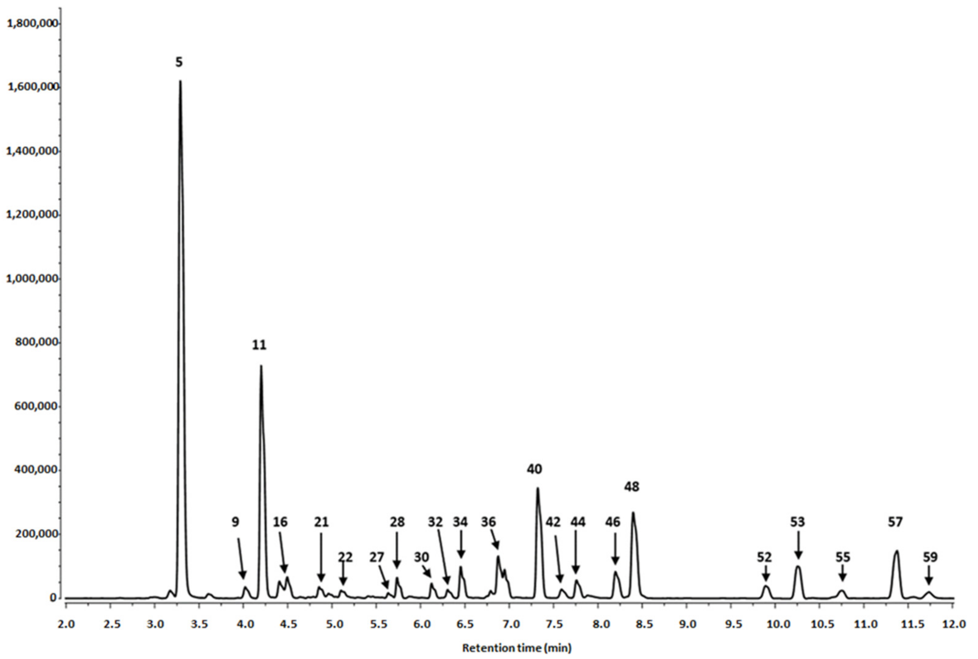
3.1. Phytochemical Profiling

3.1.1. Identification of Phenolics in Salix alba Extracts

In the present study, the phytochemical profile of hydromethanolic extracts of S. alba leaves and bark was analyzed using UPLC-PDA-Q/TOF-MS. The results of the qualitative and quantitative analyses are summarized in Table 1 and Figure 1, Figure 2 and Figure 3.
Twenty-nine phenolic compounds were identified in the leaf extracts and 35 in the bark extracts. They included derivatives of phenolic acids, flavanols, procyanidins and flavonols.

Phenolic Acid Derivatives

Seven derivatives of caffeic acid (peaks 1, 5, 9, 11, 15, 21 and 24) were detected in S. alba leaf extract and five (peaks 1–3, 11 and 13) in the bark extract. Among them, caffeic acid hexoses, caffeoyl hexose-deoxyhexosides, caffeoylquinic acid isomers and dimers were identified. Three isomeric compounds found in leaves, viz. 1-O-caffeoylquinic acid, 3-O-caffeoylquinic acid (neochlorogenic acid) and 5-O-caffeoylquinic acid (chlorogenic acid), peaks 9, 11 and 24 (tr = 4.04, 4.12 and 5.14 min), were identified by comparison with authentic standards (Table 1). They exhibited a pseudomolecular [M-H] ions at m/z 353 and fragmentation ions at m/z 191 and m/z 179, corresponding to quinic acid and caffeic acid residues, respectively. One of the mentioned derivatives, i.e., 3-O-caffeoylquinic acid, peak 11 (tr = 4.12), was also present in the bark.
The further derivatives, two isomeric caffeoylquinic acid dimers, were identified only in leaves. Compounds 15 and 21 (tr = 4.24 and 4.77 min) were represented by a pseudomolecular [M-H] ions at m/z 707. The presence of fragmentation ions at m/z 353 due to the loss of caffeoylquinic acids (−353 u) confirmed their dimeric structure.
Caffeoylquinic acid derivatives (1- and/or 3-, 4-, 5-caffeoylquinnic acids) have previously been detected in bud extracts of Salix pyrolifolia [28] and in the leaves of other species, for example Morus alba, Morus rubra [29], Salvia verticillata [30], Gaultheria procumbens [31], Sorbus domestica [32].
Caffeic acid hexoses were detected in both the bark and leaves. Compounds 1, 2 and 13 (tr = 2.51, 2.72 and 4.17 min) found in the bark and the compound 1 (tr = 2.51 min) detected in leaves had main pseudomolecular ions [M-H] at m/z 341 and the fragmentation ions at m/z 179 which correspond to the loss of hexose residues (162 u).
Compounds 3 (tr = 2.76 min) and 5 (tr = 3.26 min), identified as a caffeoyl hexose-deoxyhexosides, were detected in both extracts, bark and leaves, respectively. They exhibited pseudomolecular ions [M-H] at m/z 487. The fragmentation pattern showed ions at m/z 308 [M-H-179] and at m/z 179 [M-H-162-146]. The first was obtained by the loss of the caffeic acid aglicone (179 u). The residual part of the compound was composed of a hexose residue (162 u) and a deoxyhexoside residue (146 u).

Flavanols and Procyanidins

Twenty-six compounds from the flavanols and procyanidins group were found in the S. alba bark extract. These compounds consisted mainly (epi)catechin derivatives such as A-type and B-type procyanidins (dimers, trimers and tetramers) and their digallates; (epi)catechin-(epi)gallocatechins or (epi)catechin methyl-hexosides. Much fewer compounds from this group of derivatives were found in leaves that included two epigallocatechins, A-type procyanidin dimer and five B-type procyanidins (one dimer, three trimers and one pentamer).
Free (+)-catechin and (−)-epicatechin were present only in the bark extract. Peaks 19 and 20 (tr = 4.55 and 4.74 min), were detected with pesudomolecular ions [M-H] at m/z 289. Two epigallocatechins, peaks 22 (tr = 5.08) and 28 (tr = 5.75), contained in leaves had the main ion [M-H] at m/z 305. Three isomers of (epi)catechin-(epi)gallocatechins, i.e., peaks 4, 6 and 7 (tr = 3.12, 3.45 and 3.69 min) were present only in the bark; they were identified by pseudomolecular ions [M-H] at m/z 593 and fragmentation ions at m/z 289, attributed to (epi)catechin. (+)-Catechin and (−)-epicatechin are found in a range of fruits, beans and chocolate [33]. The compounds have been also widely distributed in leaf extracts of several Salix species [15], including S. alba, S. subserrata, S. fragilis, S. daphnoides, S. caprea, S. cinerea [19,34]. Catechin was also a predominant compound in water extracts of S. aegyptiaca leaves [35] and buds of S. pyrolifolia [28].
B-type procyanidins (dimer, trimers, tetramers and pentamer) were detected in both the bark and leaves. The B-type procyanidin dimer (peak 36 of tr = 6.79), trimers (peaks 16, 26, 27 and 30 of tr = 4.30, 5.45, 5.64 and 6.10 min), tetramers (peaks 29 and 31 of tr = 6.07 and 6.20 min) and pentamer (peak 34 of tr = 6.47 min) from leaves and bark showed pseudomolecular ions at m/z 575 (dimer), m/z 865 (trimers), m/z 1153 (tetramers) and m/z 1441 (pentamer), respectively. Each of these five B-type procyanidins was represented by fragmentation ion at m/z 287 typical for the monomer of catechin and its multiple.
In the bark and leaves a multitude of isomeric A-type procyanidins dimers (peaks 10, 12, 14, 18, 33 and 45 of tr = 4.09, 4.16, 4.20, 4.48, 6.45 and 8.08 min, respectively) showed ions at m/z 577 and 289; trimers (peaks 17, 25 and 47 of tr = 4.39, 5.30 and 8.17 min) at m/z 865, 577 and 289 and tetramer (peak 43 of tr = 7.60 min) at m/z 1152, 863, 577 and 289. All these mentioned values of peaks were a multiplication factor of the mass of the monomeric (epi)catechin equals to 287.
Proanthocyanidins are oligomers or polymers of flavan-3-ols, which are commonly distributed in plants, for example in fruits (apple, grape, cranberry), red wine, cinnamon and the hawthorn inflorescences [36]. The metabolites exhibit various biological activities, including antihypertensive, antioxidative, and anti-inflammatory properties [15]. The possible mechanism of anti-inflammatory action of proanthocyanidin dimers, B1 and B2, may be connected with the inhibition of transcription of nuclear factor-kappa B (NF-κB) [37]. The B-type procyanidin identified in white willow leaves in the present study were proanthocyanidins consisting of catechin and/or epicatechin units. Earlier, the presence of procyanidins (oligomers and polymers) (B and C-type) has been confirmed in the leaves of three willow species, including S. alba bark extracts [19,21,38]. Among other species, several procyanidin A and B-type derivatives have been identified in Gaultheria procumbens leaf extracts [39] and six different dimers and three different trimers of B-type procyanidin have been identified in Sorbus demestica leaf extract [32].
Peaks 38, 41, 49 and 50 (tr = 7.14, 7.28, 8.68 and 9.56 min) detected only in the bark, were identified as (epi)catechin methyl-hexosides. They showed fragmentation ions at m/z 289, corresponding to (epi)catechin aglycon resulting from a loss of methyl-hexoside [M-H-176] moiety from a pseudomolecular ion at m/z 465 [M-H].
A-type procyanidin dimer digallate (peak 8, tr = 3.87 min) and procyanidin trimer digallate (peak 23, tr = 5.12 min) demonstrated pseudomolecular ions at m/z 881 and 1169, respectively. Each compound demonstrated fragmentation ions at m/z 289 assigned to epichatechin. The fragmentation patterns of trimeric compound (peak 23) at m/z 289, 577 and 865 confirmed the presence of monomeric, dimeric and trimeric epicatechin, respectively, and the loss of digallate residue (304 u) from a main pseudomolecular ion at m/z 1169.
The last compound present in the willow bark—(epi)catechin-ethyl trimer (peak 56, tr = 11.09 min) with the main ion at m/z 893 and fragmentation ions at m/z 603 and 289 revealed that the compound fragmentized into a (epi)catechin-ethyl (317 u) moiety and two (epi)catechins (289 u).

Flavonols

The S. alba leaf extract was particularly rich in flavonol derivatives, while the bark extract had a negligible amount of the compounds, including only three derivatives.
Fourteen flavonols were detected in the leaf extracts: eight quercetin derivatives (peaks 32, 35, 37, 39, 42, 44, 46 and 48) and six isorhamnetin derivatives (peaks 40, 52, 53, 55, 57 and 59) (Table 1). None of them were contained in the bark extract.
Quercetin derivatives were represented by quercetin 3-O-rutinoside (rutin), -3-O-galactoside, -3-O-glucoside, methyl-pentoside and -acylated-hexosides. Each of the derivatives demonstrated the typical quercetin fragment at m/z 301. Peak 32 (tr = 6.39 min) was identified as quercetin 3-O-rutinoside (rutin). The rutin exhibited a pseudomolecular ion [M-H] at m/z 609 and a fragmentation ion at m/z 301 obtained by the loss of a rutinoside residue [M-H-308]. The compound is commonly present in plants from various taxa, for example in aerial parts of Salvia verticillata [30], Gaultheria procumbens [31] and Morus alba [29] as well as in the leaves of white willow [20].
Peaks 42 and 44 (tr = 7.54 and 7.70 min), assigned to quercetin -3-O-galactoside and quercetin -3-O-glucoside had main ions at m/z 463 and fragmentation ions attributed to the loss of a hexoside moiety (−162 u) at m/z 301 [M-H-hexoside]. Both the mentioned glycosides of quercetin have been earlier detected in leaf extracts from six different northern willow species [18] as well as in buds of Salix pyrolifolia [28]. Quercetin -3-O-glucoside, together with another quercetin derivative, quercetin7′,3-dimethylether 3-O-glucoside, has been earlier detected in the leaves of S. alba [20]. Quercetin -3-O-glucoside, together with several other quercetin and kaempferol glycosides have been also identified in the leaves of Morus alba, M. rubra, Gaultheria procumbens [29,31].
Other quercetin derivatives, four isomeric acylated-hexosides, peaks 37, 39, 46, 48 (tr = 7.13, 7.18, 8.14, 8.43 min), and methyl-pentoside, peak 35 (tr = 6.62 min), had the typical quercetin ion fragment at m/z 301 and main ions [M-H] at m/z 651, 651, 505, 505 and 447, respectively. Two quercetin-acylated-deoxyhexosides (peaks 37 and 39) of the main ions at m/z 651 gave fragmentation ions at m/z 446 and 301 suggesting that the compounds initially lost some hexoside residue together with an acetyl group (−162 u and −42 u) and subsequently a deoxyhexoside residue, e.g., rhamnose moiety (−146 u). Two quercetin-acylated-hexosides (peaks 46 and 48) had pseudomolecular ions at m/z 505 and fragmentation ions at m/z 301 and one of them additionally had an ion at m/z 463, which revealed both compounds lost acetyl group (−42 u) and some hexoside moiety (−162 u). The last quercetin derivative, methyl-pentoside (peak 35), gave ions at m/z 447 and at 301, which confirmed the loss of methyl-pentoside residue (−146 u).
The isorhamnetin derivatives included -3-O-rutinoside, peak 40 (tr = 7.22 min); -3-O-galactoside, peak 53 (tr = 10.20 min); and –acylated-hexosides, peaks 52, 55, 57, 59 (tr = 9.80, 10.63, 11.24, 11.63 min), that had a [M-H] m/z at 623, 477 and 519 [477 + 42 (acetyl group –C(O)CH3)], respectively. All of them possess the typical isorhamnetin (or deprotonated isorhamnetin) fragment at m/z 315 (or at m/z 314) formed by the cleavage of hexoside residues, i.e., rutinoside (−308 u), -galactoside (−162 u) and –acylated-hexosides (−204 u [−162 u and −42 u]), from the isorhamnetin glycosides. Additionally, the -acylated-hexoside derivatives possess a fragment at m/z 299 corresponding to demethylated isorhamnetin. Isorhamnetin derivatives were often present in the leaves of various species of willows. Isorhamnetin-3-O-glucoside and isorhamnetin-3-O-rutinoside have been earlier detected in the leaves of S. alba [20]. Isorhamnetin-3-O-glucoside has been earlier found in extracts of leaves of six Salix species grown in Finnish Lapland [18] and in the leaves of S. viminalis [40]. A different derivative of isorhamnetin (isorhamnetin-3-O-rhamnoside) has been also identified in buds of S. pyrolifolia [28].
As for the content of flavonols in the bark extract, only three derivatives were detected, i.e., quercetin 3-O-hexoside, kaempferol pentoside and kaempferol 3-O-galactoside.
Quercetin 3-O-hexoside, peak 51 (tr = 9.69 min), had a typical quercetin ion fragment at m/z 301 obtained by a cleavage of a hexoside moiety (162 u) from the main ion [M-H] at m/z 463.
Two kaempferol derivatives, peaks 54 and 58 (tr = 10.27 and 11.33 min), displayed pseudomolecular ions [M-H] at m/z 423 and 447, respectively. The fragmentation ions at m/z 287 confirmed the presence of a characteristic protonated kaempferol in the mentioned derivatives and the cleavage of a pentoside (−132 u) and galactoside (−162 u) moieties, respectively, from the main ions.

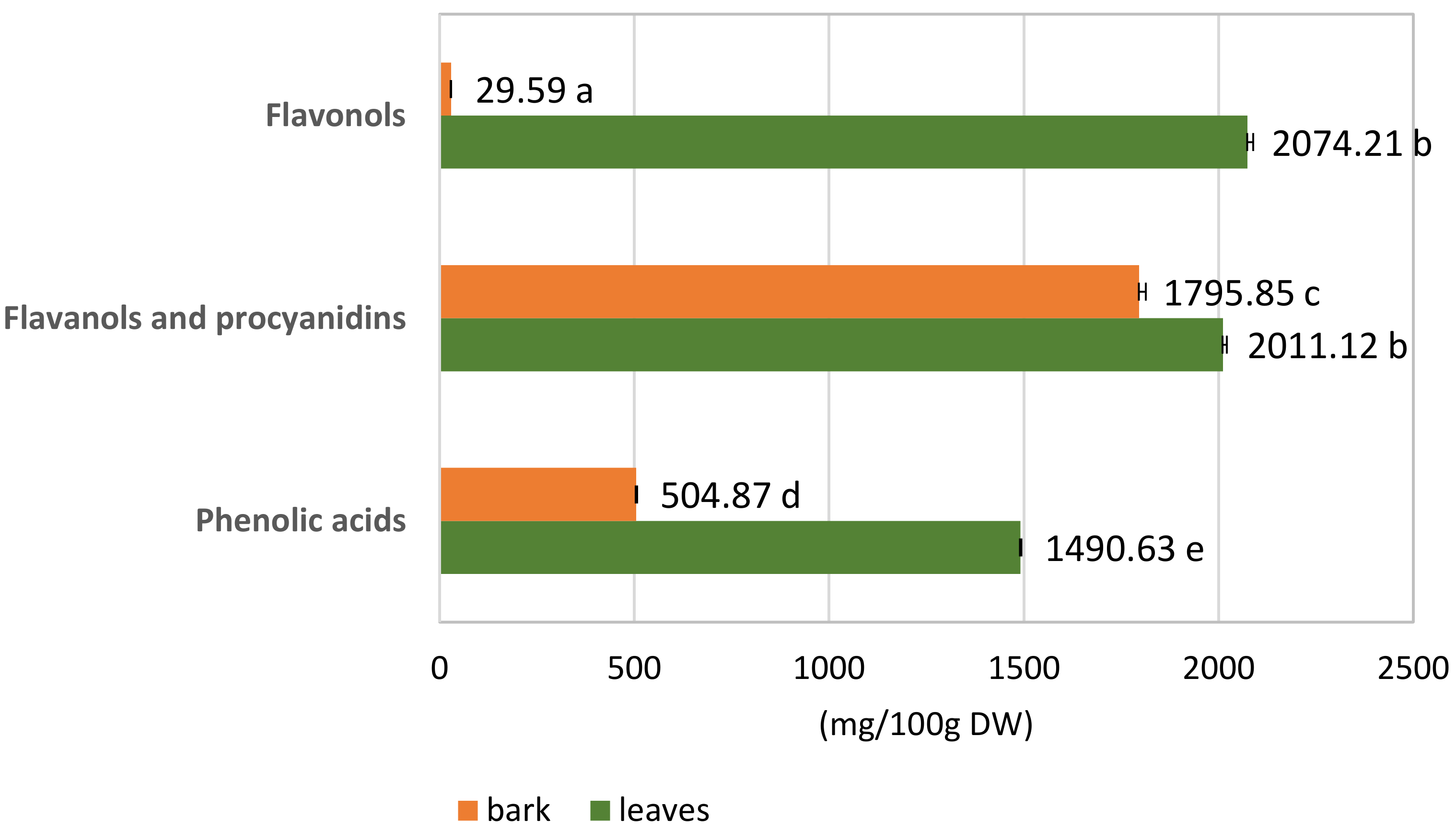
3.1.2. Quantitative Analysis of the S. alba Leaf and Bark Extracts

Quantitative analysis of dry extracts of S. alba leaves and bark was conducted for the presence of polyphenolic compounds, such as phenolic acids, flavanols and procyanidins, and flavonols (Table 1; Figure 3). The analyses were performed based on external calibration curves using selected reference compounds (Materials and Methods: Section 2.4). The concentration of the individual substances was expressed in mg/100 g DW.
The two extracts had different phenolic compound contents. S. alba leaf extract was a richer source of total active phenolic ingredients than bark extract: 5575.96 mg/100 g DW in the leaves compared to 2330.31 mg/100 g DW in the bark, i.e., 58.2% higher content.
Flavonol content was up to 98.5% higher in the leaves than the bark, with only trace amounts in the bark. Similarly, the leaves also had 66% more phenolic acids than the bark. Total flavanols and procyanidins content was also 11% higher in the leaves than in the bark.
In the leaves, the number of flavonols was 2074.21 mg/100 g DW, similarly as the content of the flavanols and procyanidins, which was equal to 2011.12 mg/100 g DW. Among the flavonols, quercetin derivatives accounted for 56.3% (1168.33 mg/100 g DW); the remaining 43.7% comprised isorhamnetin derivatives (905.88 mg/100 g DW). Phenolic acids constituted a significant amount of this extract and were equal to 1490.63 mg/100 g DW.
In the bark, the flavanols and procyanidins constituted as much as 77.1% of all considered phenolic compounds (1795.85 mg/100 g DW). Phenolic acid content was about 3.5 fold lower (504.87 mg/100 g DW) than in leaves. The content of flavonols in the bark was negligible (29.59 mg/100 g DW) (Table 1).

3.2. The Antioxidant Activity

The antioxidant activity of the hydromethanolic leaf and bark extracts was determined by DPPH and ABTS radical scavenging assays (Table 2).
The bark extract of S. alba exhibited stronger antioxidant activity (48% in DPPH and 33% in ABTS), than the leaf extract. However, leaf extract activity was also very strong, and similar (at p ≤ 0.05) to the natural reference antioxidant: ascorbic acid (EC50 = 28 μg/mL in DPPH assay and 65 μg/mL in ABTS assay) (Table 2). The bark extract demonstrated lower EC50 values (EC50 = 13.5 μg/mL in DPPH assay and 21.5 μg/mL in ABTS assay) than the reference standard, indicating even stronger antioxidant activity than ascorbic acid. A previous DPPH measurement of antioxidant activity in S. alba bark extract showed slightly lower activity (EC50 = 19.1 μg/mL) [41]. Another study reported even lower antioxidant activity in DPPH (58–65 μg/mL) and ABTS (47–53 μg/mL) assays, depending on the granulometry classes of the powdered bark extract [21]. Among other Salix species, S. atrocinerea and S. viminalis bark extracts exhibited similar activities in the DPPH test (EC50 = 10.98 and 14.06 μg/mL, respectively) as in the present study. On the other hand, the bark extract of S. fragilis demonstrated lower activity (EC50 = 23.62 μg/mL) [42]. The results of the ABTS assay are comparable with those achieved earlier for other willow species, including S. purpurea barks (EC50 = 20 μg/mL) [43].
Our paper is the first to describe the antioxidant potential of leaf extract of white willow. The extracts from leaves of other Salix species, for example S. subserrata and S. aegyptiaca [34,35], exhibited lower antioxidant activity in DPPH assay than leaf extract of S. alba in our study. The differences in the antioxidant capacity observed between even the same species, in the same assay, could be caused by differences in phytochemical profile, time of harvesting or even extraction method of the plant material.
The greatest influence on the antioxidant properties of the willow extracts was probably exerted by phenolic compounds, present in each extract. These are mainly phenolic acid derivatives and flavonols (quercetin and isorhamnetin derivatives) in leaves. In the bark extract, catechin, epicatechin and A and B-type procyanidin derivatives are probably responsible mainly for the properties.

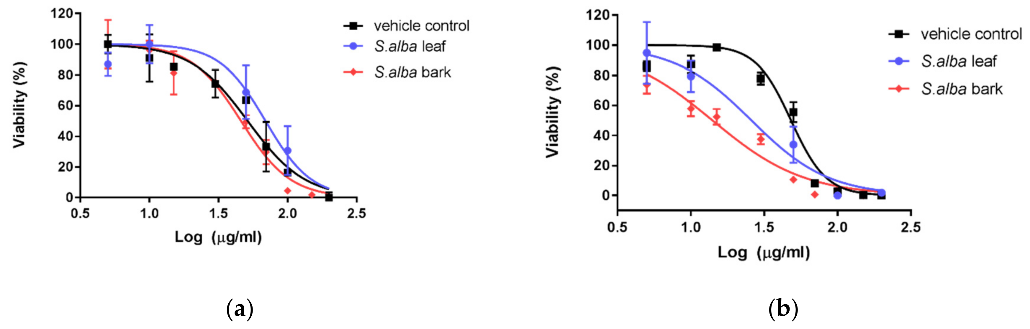
3.3. Cytotoxic Activity

The hydromethanolic extracts of S. alba leaves and bark were tested for in vitro cytotoxicity, using two human skin cell lines: human skin fibroblasts (1BR.3.N) and human epidermal keratinocytes, neonatal cells. These two cell lines were chosen to check the cytotoxic effect of tested extracts (in the range of concentrations from 5 to 200 µg/mL) on skin cells and check the possibility of external use of the extracts, for example in cosmetology.
The S. alba bark and leaf extracts demonstrated different cytotoxic activity against fibroblasts and keratinocytes (Figure 4a,b). The obtained IC50 values were higher for keratinocytes than for fibroblasts, which means that both extracts were less cytotoxic to keratinocytes than fibroblasts. The cytotoxic effect of both S. alba bark and leaves was similar as the vehicle control, i.e., equivalent amounts of methanol/water solution (7:3), for keratinocytes (IC50 values of 46.08 and 69.33 µg/mL, respectively, versus 51.51 µg/mL for vehicle control). In the case of fibroblasts, the cytotoxic activity of S. alba bark was almost twice that of leaves (IC50 values of 14.06 and 25.92 µg/mL, respectively, versus 48.22 µg/mL for vehicle control).
It can be concluded that keratinocytes were more resistant to the cytotoxic effect of both S. alba extracts (Figure 4a). However, in the case of fibroblasts, the leaf extract demonstrated lower cytotoxic potential than bark extract (Figure 4b).
However, it is important to note that both extracts exhibited significant antioxidant activity at concentrations that were not toxic to the keratinocytes (EC50 = 13.5 μg/mL in DPPH assay and 21.5 μg/mL in ABTS assay). Therefore, the extracts may be active ingredients of products used externally, which are safe for the epidermis. However, lower concentrations should be used in cosmetics acting deeper in the skin, because of the higher toxicity of the tested extracts, especially bark extract, to the fibroblast cells of the dermis.
In earlier studies, S. alba bark extract has demonstrated cytotoxic and genotoxic potential against different cell lines, for example the human leukemia (HL-60) cell line [6], human peripheral leukocyte cells and the human hepatoma cell line, with doses from 50 and 200 µg/mL [44]. However, other extracts from bark of S. atrocinerea and S. fragilis did not exert a cytotoxic effect (at 625 and 1250 µg/mL) against immortalized human keratinocytes (HaCaT) and mouse fibroblast (L929) cells [42]. The differences between cytotoxicity dose could be connected with differences in the solvent or cell line model. Our paper is the first investigation of the cytotoxic potential of S. alba extracts on skin cell lines.

4. Conclusions

Although extracts from Salix sp. are commonly used for treatment, mainly due to salicin content, the phytochemical profiles of polyphenol compounds in white willow leaves and bark extracts appear to have a potentially beneficial influence on human health due to the presence of quercetin glycosides, monomeric, dimeric and trimeric flavan-3-ol derivatives, including B-type procyanidins, as well as caffeoylquinic pseudodepsides. These specialized secondary plant metabolites are known to exhibit a wide range of biological activities. Moreover, both extracts of white willow described in the present study exhibited strong antioxidant activity at concentrations that were not toxic to the keratinocytes. Therefore, the extracts, especially leaf extract, may be used as a potential new source of bioactive polyphenols with possible applications in some antiaging cosmetics, which would be safe for the epidermis. Bark extracts, also rich in polyphenolic compounds, should be used at lower concentrations than leaf extracts, especially in cosmetics that penetrate deeper into the skin, because of their higher toxicity to fibroblasts. However, further studies are needed, particularly those connected with the potential for utilization and extract standardization.

Author Contributions

Conceptualization, U.K.-L., M.D.; methodology, M.D., J.K.-O., E.P. (Elżbieta Płuciennik), K.K.; software, E.P. (Ewelina Piątczak), E.P. (Elżbieta Płuciennik), M.D., J.K.-O.; validation, J.K.-O., M.D.; formal analysis, U.K.-L.; investigation, K.K., J.K.-O., M.D.; resources, E.P. (Ewelina Piątczak), M.D.; data curation, E.P. (Ewelina Piątczak), M.D., J.K.-O.; writing—original draft preparation, E.P. (Ewelina Piątczak), M.D., U.K.-L.; writing—review and editing, U.K.-L., E.P. (Ewelina Piątczak); visualization, J.K.-O., K.K., U.K.-L., E.P. (Elżbieta Płuciennik), E.P. (Ewelina Piątczak); supervision, U.K.-L.; project administration, U.K.-L.; funding acquisition, U.K.-L. All authors have read and agreed to the published version of the manuscript.

Funding

This research was funded by Medical University of Lodz (grant No. 503/3-066-02/503-31-001).

Conflicts of Interest

The authors declare no conflict of interest.

References

  1. Braithwaite, M.C.; Tyagi, C.; Tomar, L.K.; Kumar, P.; Choonara, Y.E.; Pillay, V. Nutraceutical-based therapeutics and formulation strategies augmenting their efficiency to complement modern medicine: An overview. J. Funct. Foods 2014, 6, 82–99. [Google Scholar] [CrossRef]
  2. Rates, S.M. Plants as source of drugs. Toxicon 2001, 39, 603–613. [Google Scholar] [CrossRef]
  3. Schmid, B.; Lüdtke, R.; Selbmann, H.K.; Kotter, R.; Tschirdewahn, B.; Schaffner, W.; Heide, L. Efficacy and tolerability of a standardized willow bark extract in patients with osteoarthritis: Randomized placebo-controlled, double blind clinical trial. Phytother. Res. 2001, 15, 344–350. [Google Scholar] [CrossRef] [PubMed]
  4. Mahdi, J.G.; Mahdi, A.J.; Mahdi, A.J.; Bowen, I.D. The historical analysis of aspirin discovery, its relation to the willow tree and antiproliferative and anticancer potential. Cell Prolif. 2006, 39, 147–155. [Google Scholar] [CrossRef]
  5. Bonaterra, G.A.; Kelber, O.; Weiser, D.; Metz, J.; Kinscherf, R. In vitro anti-proliferative effects of the willow bark extract STW 33-I. Arzneimittelforschung 2010, 60, 330–335. [Google Scholar] [CrossRef]
  6. Sulaiman, G.M.; Hussien, N.N.; Marzoog, T.R.; Awad, H.A. Phenolic content, antioxidant, antimicrobial and cytotoxic activities of ethanolic extract Salix alba. Am. J. Biochem. Biotechnol. 2013, 9, 41–46. [Google Scholar] [CrossRef]
  7. Ishikado, A.; Sono, Y.; Matsumoto, M.; Robida-Stubbs, S.; Okuno, A.; Goto, M.; King, G.L.; Blackwell, T.K.; Makino, T. Willow bark extract increases antioxidant enzymes and reduces oxidative stress through activation of Nrf2 in vascular endothelial cells and Caenorhabditis elegans. Free Radic. Biol. Med. 2013, 65, 1506–1515. [Google Scholar] [CrossRef] [Green Version]
  8. Tanase, C.; Coșarcă, S.; Muntean, D.-L. A Critical review of phenolic compounds extracted from the bark of woody vascular plants and their potential biological activity. Molecules 2019, 24, 1182. [Google Scholar] [CrossRef] [Green Version]
  9. Tanase, C.; Mocan, A.; Coșarcă, S.; Gavan, A.; Nicolescu, A.; Gheldiu, A.-M.; Vodnar, D.C.; Muntean, D.-L.; Crișan, O. Biological and chemical insights of beech (Fagus sylvatica L.) Bark: A source of bioactive compounds with functional properties. Antioxidants 2019, 8, 417. [Google Scholar] [CrossRef] [Green Version]
  10. Arnao, M.B.; Cano, A.; Acosta, M. The hydrophilic and lipophilic contribution to total antioxidant activity. Food Chem. 2001, 73, 239–244. [Google Scholar] [CrossRef]
  11. Ingkaninan, K.; Temkitthawon, P.; Chuenchom, K.; Yuyaem, T.; Thongnoi, W. Screening for acetylcholinesterase inhibitory activity in plants used in Thai traditional rejuvenating and neurotonic remedies. J. Ethnopharmacol. 2003, 89, 261–264. [Google Scholar] [CrossRef] [PubMed]
  12. Valko, M.; Leibfritz, D.; Moncol, J.; Cronin, M.T.; Mazur, M.; Telser, J. Free radicals and antioxidants in normal physiological functions and human disease. Int. J. Biochem. Cell Biol. 2007, 39, 44–84. [Google Scholar] [CrossRef] [PubMed]
  13. Lauron-Moreau, A.; Pitre, F.E.; Argus, G.W.; Labrecque, M.; Brouillet, L. Phylogenetic relationships of american willows (Salix L., Salicaceae). PLoS ONE 2015, 10, e0138963. [Google Scholar] [CrossRef]
  14. Mohr, R. Weidenrinde: Salicis cortex. In Europaisches Arzneibuch 8. Ausgabe, 2. Nachtrag (Ph.Eur. 8.2); Amtliche deutsche Ausgabe, 1st ed.; Deutscher Apotheker Verlag: Stuttgart, Germany, 2015; pp. 2135–2136. [Google Scholar]
  15. Jürgenliemk, G.; Petereit, F.; Nahrstedt, A. flavan-3-ols and procyanidins from the bark of Salix purpurea L. Pharmazie 2007, 62, 231–234. [Google Scholar]
  16. Esatbeyoglu, T.; Wray, V.; Winterhalter, P. Dimeric procyanidins: Screening for B1 to B8 and semisynthetic preparation of B3, B4, B6, and B8 from a polymeric procyanidin fraction of white willow bark (Salix alba). J. Agric. Food Chem. 2010, 58, 7820–7830. [Google Scholar] [CrossRef] [PubMed]
  17. Wiesneth, S.; Petereit, F.; Jürgenliemk, G. Salix daphnoides: A screening for oligomeric and polymeric proanthocyanidins. Molecules 2015, 20, 13764–13779. [Google Scholar] [CrossRef] [PubMed] [Green Version]
  18. Nyman, T.; Julkunen-Tiitto, R. Chemical variation within and among six northern willow species. Phytochemistry 2005, 66, 2836–2843. [Google Scholar] [CrossRef]
  19. Wiesneth, S.; Aas, G.; Heilmann, J.; Jürgenliemk, G. Investigation of the flavan-3-ol patterns in willow species during one growing-season. Phytochemistry 2018, 145, 26–39. [Google Scholar] [CrossRef]
  20. Karl, C.H.; Müller, G.; Pedersen, P.A. Flavonoide aus Salix alba. Die struktur des Terniflorins und eines weiteren acylflavonoides. Phytochemistry 1976, 15, 1084–1085. [Google Scholar] [CrossRef]
  21. Zaiter, A.; Becker, L.; Petit, J.; Zimmer, D.; Karam, M.-C.; Baudelaire, É; Scher, J.; Dicko, A. Antioxidant and antiacetylcholinesterase activities of different granulometric classes of Salix alba (L) bark powders. Powder Technol. 2016, 301, 649–656. [Google Scholar] [CrossRef]
  22. Rutkowski, L. Klucz do Oznaczania Roślin Naczyniowych Polski Niżowej; Wydawnictwo Naukowe PWN: Warszawa, Poland, 2007; pp. 86, 88, 91. (In Polish) [Google Scholar]
  23. Re, R.; Pellegrini, N.; Proteggente, A.; Pannala, A.; Yang, M.; Rice-Evans, C. Antioxidant activity applying an improved ABTS radical cation decolorization assay. Free Radic. Biol. Med. 1999, 26, 1231–1237. [Google Scholar] [CrossRef]
  24. Guss, K.L.; Pavanni, S.; Prati, B.; Dazzi, L.; de Oliveira, J.P.; Nogueira, B.V.; Pereira, T.M.C.; Fronza, M.; Endringer, D.C.; Scherer, R. Ultrasound-assisted extraction of Achyrocline satureioides prevents contrast-induced nephropathy in mice. Ultrason. Sonochem. 2017, 37, 368–374. [Google Scholar] [CrossRef] [PubMed]
  25. Scherer, R.; Godoy, H.T. Antioxidant activity index (AAI) by the 2, 2-diphenyl-1-picrylhydrazyl method. Food Chem. 2009, 112, 654–658. [Google Scholar] [CrossRef]
  26. Santos, S.A.O.; Vilela, C.; Domingues, R.M.A.; Oliveira, C.S.D.; Villaverde, J.J.; Freire, C.S.R.; Neto, C.P.; Silvestre, A.J.D. Secondary metabolites from Eucalyptus grandis wood cultivated in Portugal, Brazil and South Africa. Ind. Crop. Prod. 2017, 95, 357–364. [Google Scholar] [CrossRef]
  27. Varbanov, H.P.; Kuttler, F.; Banfi, D.; Turcatti, G.; Dyson, P.J. Repositioning approved drugs for the treatmentof problematic cancers using a screening approach. PLoS ONE 2017, 12, e0171052. [Google Scholar] [CrossRef] [PubMed] [Green Version]
  28. Lavola, A.; Maukonen, M.; Julkunen-Tiitto, R. Variability in the composition of phenolic compounds in winter-dormant Salix pyrolifolia in relation to plant part and age. Phytochemistry 2018, 153, 102–110. [Google Scholar] [CrossRef]
  29. Thabti, I.; Elfalleh, W.; Hannachi, H.; Ferchichi, A.; Da Graca Campos, M. Identification and quantification of phenolic acids and flavonol glycosides in Tunisian Morus species by HPLC-DAD and HPLC–MS. J. Funct. Foods 2012, 4, 367–374. [Google Scholar] [CrossRef]
  30. Katanić Stanković, J.S.; Srećković, N.; Mišić, D.; Gašić, U.; Imbimbo, P.; Monti, D.M.; Mihailović, V. Bioactivity, biocompatibility and phytochemical assessment of lilac sage, Salvia verticillata L. (Lamiaceae)-A plant rich in rosmarinic acid. Ind. Crops Prod. 2020, 143, 111932. [Google Scholar] [CrossRef]
  31. Michel, P.; Owczarek, A.; Kosno, M.; Gontarek, D.; Matczak, M.; Olszewska, M.A. Variation in polyphenolic profile and in vitro antioxidant activity of eastern teaberry (Gaultheria procumbens L.) leaves following foliar development. Phytochem. Lett. 2017, 20, 356–364. [Google Scholar] [CrossRef]
  32. Matczak, M.; Marchelak, A.; Michel, P.; Owczarek, A.; Piszczan, A.; Kolodziejczyk-Czepas, J.; Nowak, P.; Olszewska, M.A. Sorbus domestica L. leaf extracts as functional products: Phytochemical profiling, cellular safety, pro-inflammatory enzymes inhibition and protective effects against oxidative stress in vitro. J. Funct. Foods 2018, 40, 207–218. [Google Scholar] [CrossRef]
  33. Williamson, G.; Manach, C. Bioavailability and bioefficacy of polyphenols in humans. II. Review of 93 intervention studies. Am. J. Clin. Nutr. 2005, 81, 243S–255S. [Google Scholar] [CrossRef] [PubMed]
  34. Tawfeek, N.; Sobeh, M.; Hamdan, D.I.; Farrag, N.; Roxo, M.; El-Shazly, A.M.; Wink, M. Phenolic compounds from Populus alba L. and Salix subserrata Willd. (Salicaceae) counteract oxidative stress in Caenorhabditis elegans. Molecules 2019, 24, 1999. [Google Scholar] [CrossRef] [PubMed] [Green Version]
  35. Enayat, S.; Banerjee, S. Comparative antioxidant activity of extracts from leaves, bark and catkins of Salix aegyptiaca sp. Food Chem. 2009, 116, 23–28. [Google Scholar] [CrossRef]
  36. Cos, P.; De Bruyne, T.; Hermans, N.; Apers, S.; Berghe, D.V.; Vlietinck, A.J. Proanthocyanidins in health care: Current and new trends. Curr. Med. Chem. 2004, 11, 1345–1359. [Google Scholar] [CrossRef]
  37. Park, Y.C.; Rimbach, G.; Saliou, C.; Valacchi, G.; Packer, L. Activity of monomeric, dimeric, and trimeric flavonoids on NO production, TNF-alpha secretion, and NF-kappaB-dependent gene expression in RAW 264.7 macrophages. FEBS Lett. 2000, 465, 93–97. [Google Scholar] [CrossRef] [Green Version]
  38. Pobłocka-Olech, L.; Krauze-Baranowska, M. SPE-HPTLC of procyanidins from the barks of different species and clones of Salix. J. Pharmaceut. Biomed. 2008, 48, 965–968. [Google Scholar] [CrossRef]
  39. Michel, P.; Dobrowolska, A.; Kicel, A.; Owczarek, A.; Bazylko, A.; Granica, S.; Piwowarski, J.P.; Olszewska, M.A. Polyphenolic profile, antioxidant and anti-inflammatory activity of eastern teaberry (Gaultheria procumbens L.) leaf extracts. Molecules 2014, 19, 20498–20520. [Google Scholar] [CrossRef] [Green Version]
  40. Karl, C.H.; Pedersen, P.A.; Schwarz, C.H. Ein neues flavonolacetylglucosid aus Salix viminalis. Phytochemistry 1977, 16, 1117. (In German) [Google Scholar] [CrossRef]
  41. Amel Zabihi, N.; Mahmoudabady, M.; Soukhtanloo, M.; Hayatdavoudi, P.; Beheshti, F.; Niazmand, S. Salix alba attenuated oxidative stress in the heart and kidney of hypercholesterolemic rabbits. Avicenna J. Phytomed. 2018, 8, 63–72. [Google Scholar]
  42. Ramos, P.A.B.; Moreirinha, C.; Silva, S.; Costa, E.M.; Veiga, M.; Coscueta, E.; Santos, S.A.O.; Almeida, A.; Pintado, M.M.; Freier, C.S.R.; et al. The health-promoting potential of Salix spp. bark polar extracts: Key insights on phenolic composition and in vitro bioactivity and biocompatibility. Antioxidants 2019, 8, 609. [Google Scholar] [CrossRef] [Green Version]
  43. Durak, A.; Gawlik-Dziki, U.; Sugier, D. Coffee enriched with willow (Salix purpurea and Salix myrsinifolia) bark preparation- interactions of antioxidative phytochemicals in a model system. J. Funct. Foods 2015, 18, 1106–1116. [Google Scholar] [CrossRef]
  44. Maistro, E.L.; Terrazzas, P.M.; Perazzo, F.F.; Gaivão, I.O.M.; Sawaya, A.C.H.F.; Rosa, P.C.P. Salix alba (white willow) medicinal plant presents genotoxic effects in human cultured leukocytes. J. Toxicol. Environ. Health 2019, 82, 1223–1234. [Google Scholar] [CrossRef] [PubMed]
Figure 1. Segment from 2.00 to 12.00 min of LC-DAD chromatogram at 340 nm of S. alba leaves. Peak number identities are displayed in Table 1.
Biomolecules 10 01391 g001
Figure 2. Segment from 2.00 to 11.50 min of LC-DAD chromatogram at 280 nm of S. alba bark. Peak number identities are displayed in Table 1.
Biomolecules 10 01391 g002
Figure 3. Contents (mg/100 g DW) ± SD of phenolic compounds in hydromethanolic extracts of leaves and bark of S. alba. Means with the same letter are not statistically different at p ≤ 0.05 in the Kruskal–Wallis test.
Biomolecules 10 01391 g003
Figure 4. Effects of S. alba bark and leaf hydromethanolic extracts on keratinocyte (a) and fibroblast (b) cell line viability in Presto Blue cell viability assay.
Biomolecules 10 01391 g004
Table 1. Characteristic ions and phenolic compounds content in Salix alba hydromethanolic leaf and bark extracts (mg/100 g DW) by UPLC-PDA-Q/TOF-MS.
Peak NoRetention Time (tr)λmax (nm)[M-H]
(m/z) a
MS/MS Fragments (m/z) aTentative IdentificationContent (mg/100 g DW) c ± SD
LeavesBark
Phenolic Acids
12.51324341179Caffeoylhexose I2.98 ± 0.0757.71 ± 0.09 *
22.72324341179Caffeoylhexose IIn.d.24.71 ± 0.10 *
32.76320487308/179Caffeoyl hexose-deoxyhexoside In.d.140.00 ± 0.22 *
53.26320487308/179Caffeoyl hexose-deoxyhexoside II875.32 ± 1.18 *n.d.
94.04324353191/1791-O-Caffeoylquinic acid b117.43 ± 0.88 *n.d.
114.12324353191/1793-O-Caffeoylquinic acid b386.31 ± 1.2641.96 ± 2.53 *
134.17324341179Caffeoylhexose IIIn.d.240.49 ± 0.46 *
154.24325707353/191/179Caffeoylquinic acid dimer I60.63 ± 1.83 *n.d.
214.77325707353/191/179Caffeoylquinic acid dimer II10.11 ± 0.75 *n.d.
245.143243531915-O-Caffeoylquinic acid b37.85 ± 0.59 *n.d.
Sum 1490.63 ± 2.86Sum 504.87 ± 2.58 *
Flavanols and Procyanidins
43.12280593407/289(Epi)catechin-(Epi)gallocatechin In.d.130.67 ± 1.16 *
63.45280593407/289(Epi)catechin-(Epi)gallocatechin IIn.d.10.09 ± 0.19 *
73.69280593407/289(Epi)catechin-(Epi)gallocatechin IIIn.d.59.79 ± 5.79 *
83.87279881407/289A-type procyanidin dimer digallaten.d.48.66 ± 0.50 *
104.09280577407/289A-type procyanidin dimer b In.d.224.33 ± 1.58 *
124.16281577407/289A-type procyanidin dimer b II241.31 ± 1.0322.33 ± 0.57 *
144.20280577289A-type procyanidin dimer b IIIn.d.9.22 ± 0.90 *
164.30277865577/287B-type procyanidin trimer I241.43 ± 1.1622.90 ± 0.46 *
174.39277865577/289A-type procyanidin trimer In.d.25.87 ± 0.18 *
184.48281577407/289A-type procyanidin dimer b IVn.d.14.27 ± 5.63 *
194.55280289 (+)-Catechinn.d63.79 ± 2.60 *
204.74280289245(−)-Epicatechin bn.d.176.49 ± 14.02 *
225.08281305221/179Epigallocatechin b I319.52 ± 1.62 *n.d.
235.122791169865/577/289A-type procyanidin trimer digallaten.d.99.25 ± 0.04 *
255.30277865577/289A-type procyanidin trimer IIn.d.15.07 ± 0.08 *
265.45277865577/287B-type procyanidin trimer IIn.d.40.79 ± 1.01 *
275.64280865577/287B-type procyanidin trimer III124.37 ± 9.44 *n.d.
285.75280305221/219/179Epigallocatechin b II324.69 ± 0.88 *n.d.
296.072811153865/577/287B-type procyanidin tetramer In.d.25.54 ± 0.04 *
306.10277865577/287B-type procyanidin trimer IV246.46 ± 1.69 *n.d.
316.202811154577/287B-type procyanidin tetramer IIn.d.56.07 ± 1.51 *
336.45280577289A-type procyanidin dimer b Vn.d.198.24 ± 0.06 *
346.472811441864/575/287B-type procyanidin pentamer118.11 ± 0.13 *n.d.
366.79279575287B-type procyanidin dimer b I395.23 ± 0.73 *n.d.
387.14280465289(Epi)catechin methyl-hexoside In.d.68.55 ± 1.12 *
417.28280465289(Epi)catechin methyl-hexoside IIn.d.56.78 ± 0.99 *
437.602801152863/577/289A-type procyanidin tetramern.d33.45 ± 0.11 *
458.08280577407/289A-type procyanidin dimer b VIn.d.94.91 ± 0.08 *
478.17280865577/289A-type procyanidin trimer IIIn.d.103.61 ± 0.10 *
498.68280465289(Epi)catechin methyl-hexoside IIIn.d.96.21 ± 1.10 *
509.56280465289(Epi)catechin methyl-hexoside IVn.d.28.52 ± 0.07 *
5611.09280893603/289(Epi)catechin-ethyl trimern.d.70.45 ± 0.9 *
Sum 2011.12 ± 9.92Sum 1795.85 ± 16.78
Flavonols
326.39350609301Quercetin 3-O-rutinoside b92.91 ± 2.05 *n.d.
356.62340447301Quercetin methyl-pentoside9.11 ± 0.39 *n.d.
377.13340651446/301Quercetin acylated-deoxyhexoside I166.51 ± 1.79 *n.d.
397.18340651447/301Quercetin acylated-deoxyhexoside II148.93 ± 1.02 *n.d.
407.22351623315Isorhamnetin 3-O-rutinoside b459.80 ± 5.61 *n.d.
427.54355463301Quercetin 3-O-galactoside b59.27 ± 0.24 *n.d.
447.70355463301Quercetin 3-O-glucoside b98.43 ± 1.88 *n.d.
468.14340505301Quercetin-acylated-hexoside I180.03 ± 0.99 *n.d.
488.43340505463/301Quercetin-acylated-hexoside II413.14 ± 11.86 *n.d.
519.69355463301Quercetin 3-O-hexosiden.d.10.47 ± 0.06 *
529.80353519314/299Isorhamnetin-acylated-hexoside I4.65 ± 0.12 *n.d.
5310.20350477314Isoramnetin 3-O-galactoside b145.89 ± 2.11 *n.d.
5410.27350423287Kaempferol pentosiden.d.6.58 ± 0.09 *
5510.63350519314/299Isorhamnetin acylated-hexoside II32.62 ± 0.88 *n.d.
5711.24353519314/299Isorhamnetin-acylated-hexoside III234.36 ± 4.29 *n.d.
5811.33346447287Kaempferol 3-O-galactoside bn.d.12.54 ± 0.07 *
5911.63353519314/299Isorhamnetin-acylated-hexoside IV28.56 ± 1.18 *n.d.
Sum2074.21 ± 14.50Sum29.59 ± 0.13 *
Sum of phenolic compounds5575.96 ± 17.802330.31 ± 16.98 *
a Experimental data; b Identified using corresponding authentic standards; c Values are means ± standard deviation (SD) n = 3. Means indicated with an asterisk (*) within lines are different at p ≤ 0.05 in Mann–Whitney U-test. Amounts of phenolic acids, flavanols, procyanidins, flavonols and flavanones, were converted into 3-O-caffeoylquinic acid (caffeoylquinic acid derivatives), caffeic acid (caffeic acid derivatives), (+)-catechin ((+)-catechin and (−)-epicatechin), epi)catechin methyl-hexoside, (epi)catechin-ethyl trimer), procyanidin B2 (B-type polymeric procyanidins), procyanidin A2 (A-type polymeric procyanidins), quercetin 3-O-galactoside (quercetin derivatives), kaempferol 3-O-galactoside (kaempferol derivatives), isorhamnetin 3-O-glucoside (isorhamnetin derivatives), hesperidin (hesperetin derivatives). Epigallocatechin and quercetin 3-O-rutinoside were converted on the basis of the corresponding authentic standards.
Table 2. In vitro antioxidant activities in DPPH and ABTS assays of S. alba hydromethanolic leaf and bark extracts.
Plant MaterialDPPH *ABTS *
Bark13.51 ± 0.2 a21.50 ± 0.32 a
Leaf28.23 ± 0.6 b65.41 ± 0.27 b
Ascorbic acid
(antioxidant standard)
28.88± 0.21 b65.43± 0.22 b
The means with the same letter within the columns do not differ significantly according to the Kruskal–Wallis test (p ≤ 0.05). The values are means of six replicates ± SD; * EC50 the concentration of sample (μg/mL) showing 50% of maximal radical scavenging activity.

