
Effect of Silver Nanoparticles on Blue Light Phototoxicity against Fusobacterium nucleatum


Department of Prosthodontics, Goldschleger School of Dental Medicine, Tel Aviv University, Tel Aviv-Yafo 69978, Israel
*
Author to whom correspondence should be addressed.
Biophysica 2021, 1(4), 405-412; https://0-doi-org.brum.beds.ac.uk/10.3390/biophysica1040029
Submission received: 5 September 2021
/
Revised: 26 September 2021
/
Accepted: 30 September 2021
/
Published: 5 October 2021
Abstract
:A previous study showed that sub-lethal exposure of blue light caused cell membrane damage in Fusobacterium nucleatum (Fn). The aim of the present study was to test the combined effect of blue light and silver nanoparticles against Fn. Bacterial suspensions were exposed to blue light (400–500 nm) with or without silver nanoparticles (10 nm). Exposed and non-exposed samples were studied for malodor production (Odor judge scores), VSC levels (Halimeter), reactive oxygen species (ROS) production (fluorimeter), and bacterial cell membrane damage (fluorescence microscopy). The results showed that combining blue light exposure and silver nanoparticles significantly reduced malodor and VSC production by Fn concomitant with increased ROS levels and bacterial cell membrane damage. These results suggest that silver nanoparticles may increase blue light phototoxicity against Fn.
1. Introduction
The proteolytic activity of anaerobic Gram-negative oral bacteria such as Fusobacterium nucleatum has been associated with oral malodor and gum disease [1]. These bacteria break down oral proteins and glycoproteins into their amino acid building blocks. These are further metabolized, yielding malodorous compounds such as the volatile sulfide compounds (VSC) [2] that are felt during exhalation and speech.
Silver nanoparticles are considered a promising antimicrobial agent due to the multi-target action of silver [3]. Due to their antimicrobial activity [4], low cytotoxicity, and low immunogenicity [5] silver nanoparticles have multiple biomedical applications. Although their antibacterial mechanism is not yet clear, several mechanisms have been proposed. These include the release of silver ions [6], cell wall and cytoplasmic membrane damage [7], and reactive oxygen species (ROS) induction [8]. The antimicrobial effect of silver nanoparticles combined with blue light against antibiotic resistant Pseudomonas aeruginosa was also shown [9]. However, the mechanism for this effect is still unclear.
Previous studies have demonstrated the phototoxic effect of blue light against malodor producing bacteria [10] with or without the added effect of photosensitizers [11] or photoactivators [12]. Furthermore, the main antibacterial mechanism of blue light against Fusobacterium nucleatum was shown to be membrane damage [13] that could be leveraged to increase the efficacy of other antibacterial agents [14]. The aim of the present study was to test the added effect of silver nanoparticles on blue light phototoxicity against Fusobacterium nucleatum.
2. Materials and Methods
2.1. Light Source
A high intensity non-coherent visible light, known in dentistry as the plasma-arc, i.e., a xenon light source supplemented with a filter (wavelengths, 400–500 nm) (Sapphire® Supreme, Den-Mat®, Lompoc, CA, USA) fitted with a 9 mm diameter light guiding tip was applied. The average light power (1500 mW/cm2, ‘SC’ mode) was measured with the unit’s built in power meter prior to each experiment.
2.2. Bacterial Strain and Growth Conditions
Fusobacterium nucleatum (PK1594) was cultured in brain heart infusion (BHI) broth at 37 °C for 72 h under anaerobic conditions using an anaerobic jar and kit (GasPack® EZ, BD, Oxford, UK).
2.3. Experimental Protocol
Bacterial suspensions were spun down (5000× g, 5 min), the supernatant was discarded and replaced with 200 μL of phosphate-buffered saline (PBS) with or without silver nanoparticles (0.02% w/v, 10 nm, Sigma), and samples were placed in 48-well plate (Nunc) with a two well gap between samples and exposed to intermittent blue light (5 s) from a constant distance (5 mm) for a total exposure time of 60 s equivalent to light fluence of 82 J/cm2 as indicated. Following light exposure, the samples were studied for cell membrane damage, ROS levels, and malodor and VSC production abilities, as described below.
2.4. Malodor Scores
Test tubes containing 2 mL BHI medium were supplemented with 100 μL of the above treated bacterial suspensions and incubated at 37 °C for 48 h under anaerobic conditions. Following incubation, the test tubes were randomized, and malodor production levels were blindly scored by a trained odor judge. Malodor levels were measured organoleptically immediately after opening each test tube. The scores were recorded according to a semi-integer scale of 0 to 5, as follows: 0, no appreciable odor; 1, barely noticeable odor; 2, slight, but clearly noticeable odor; 3, moderate odor; 4, strong odor; and 5, extremely strong odor.
2.5. VSC Levels
Volatile sulfide compounds (VSC) levels were measured in the test tubes’ headspace using a sulfide monitor (HalimeterTM, Interscan Corp., Simi Valley, CA, USA). The monitor was zeroed on ambient air, and a one quarter inch diameter disposable plastic straw was attached to the air inlet of the monitor. VSC levels were measured by inserting the other end of the straw 2 cm into the gas headspace of each test tube immediately after removing the cap and recording the maximal reading in ppb sulfide equivalents.
2.6. Reactive Oxygen Species (ROS) Levels
ROS levels were quantified using 2′,7′-dichlorodihydrofluorescein diacetate (H2DCF-DA) fluorescent probe [15]. Aliquots of 50 μL treated bacterial samples, and controls were added to 150 μL phosphate-buffered saline (PBS) in a 96-well dark plate. Then, 5 μL of 1mM H2DCF-DA were added to each well and incubated for 1 h in the dark, at 37 °C. The fluorescence density was measured using a fluorescence microplate reader (Infinite F-200, TECAN, Männedorf, Switzerland) with excitation at 480 nm and emission at 530 nm.
2.7. Membrane Integrity
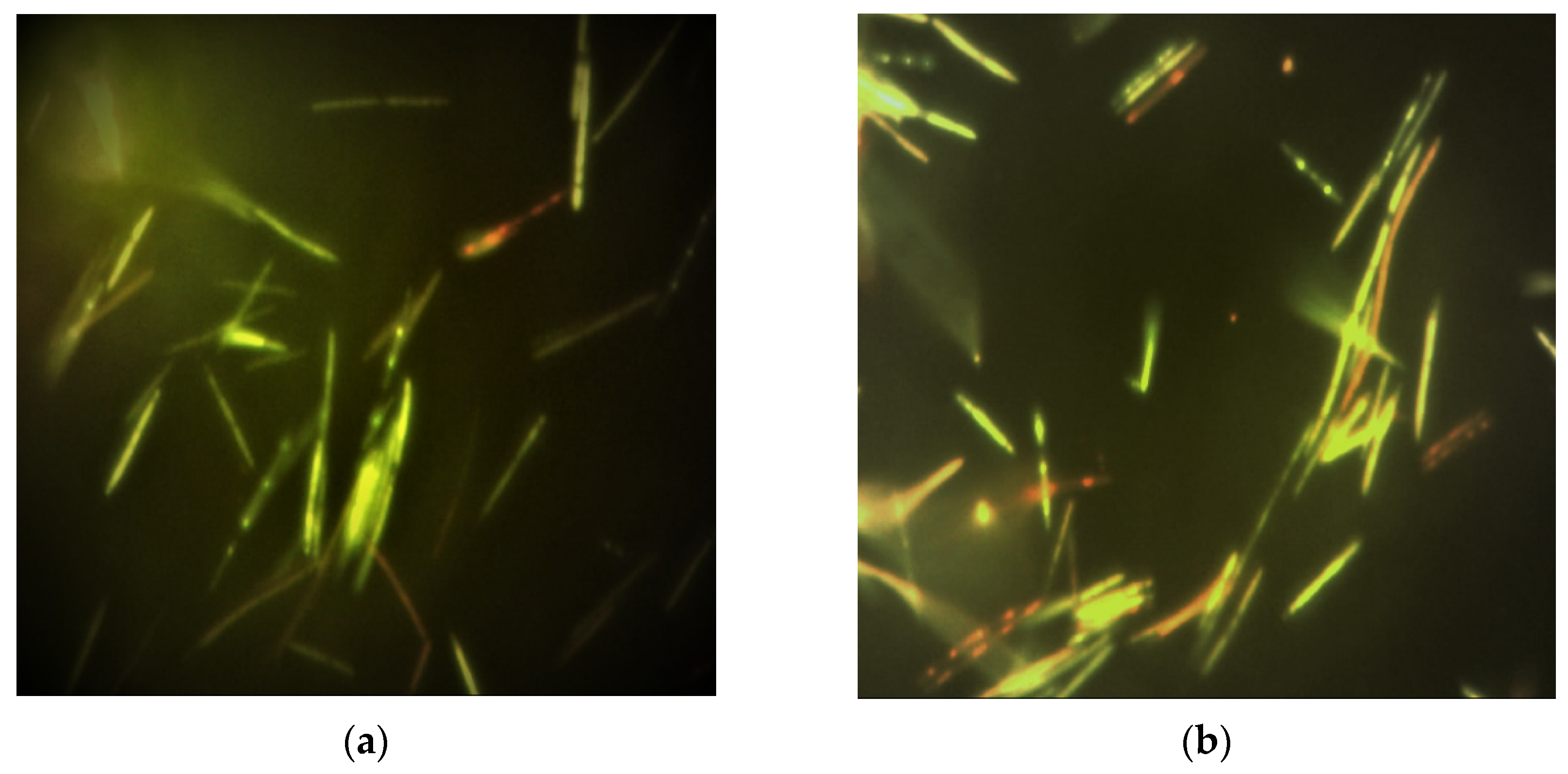
Membrane damage was determined [16] using a Bacteria Live/Dead Staining Kit® (Promokine, Heidelberg, Germany), which comprised of DMAO, a green-fluorescent dye that binds nucleic acids of both membrane intact and membrane damaged bacterial cells, and Ethidium homodimer-III (EthD-III), a red-fluorescent dye that binds only to nucleic acids of membrane damaged bacterial cells. Bacterial samples (100 µL) were added with 1 µL of the dye mixture and incubated in the dark for 15 min at room temperature. The samples (10 µL) were wet mounted on slides, covered with cover slips, and studied under a fluorescent microscope (×1000, L3201LED, MRC), using an excitation light of 460–470 nm and a blue LED filter with cutoff of 500 nm. Digital images of six random fields were taken using a mounted camera (AM7023, DinoEye®, Anmo, Taiwan). Results were recorded as percentage of red fluorescent dyed bacteria of total counts.
2.8. Statistical Analysis
To compare the effect of the various treatments on measured parameters, ANOVA was applied with post hoc pairwise comparisons, according to Dunnet and Scheffe. The tests applied were two-tailed, and p ≤ 0.05 was considered statistically significant. Experiments were conducted in six replicates.
3. Results
3.1. Malodor and VSC Levels
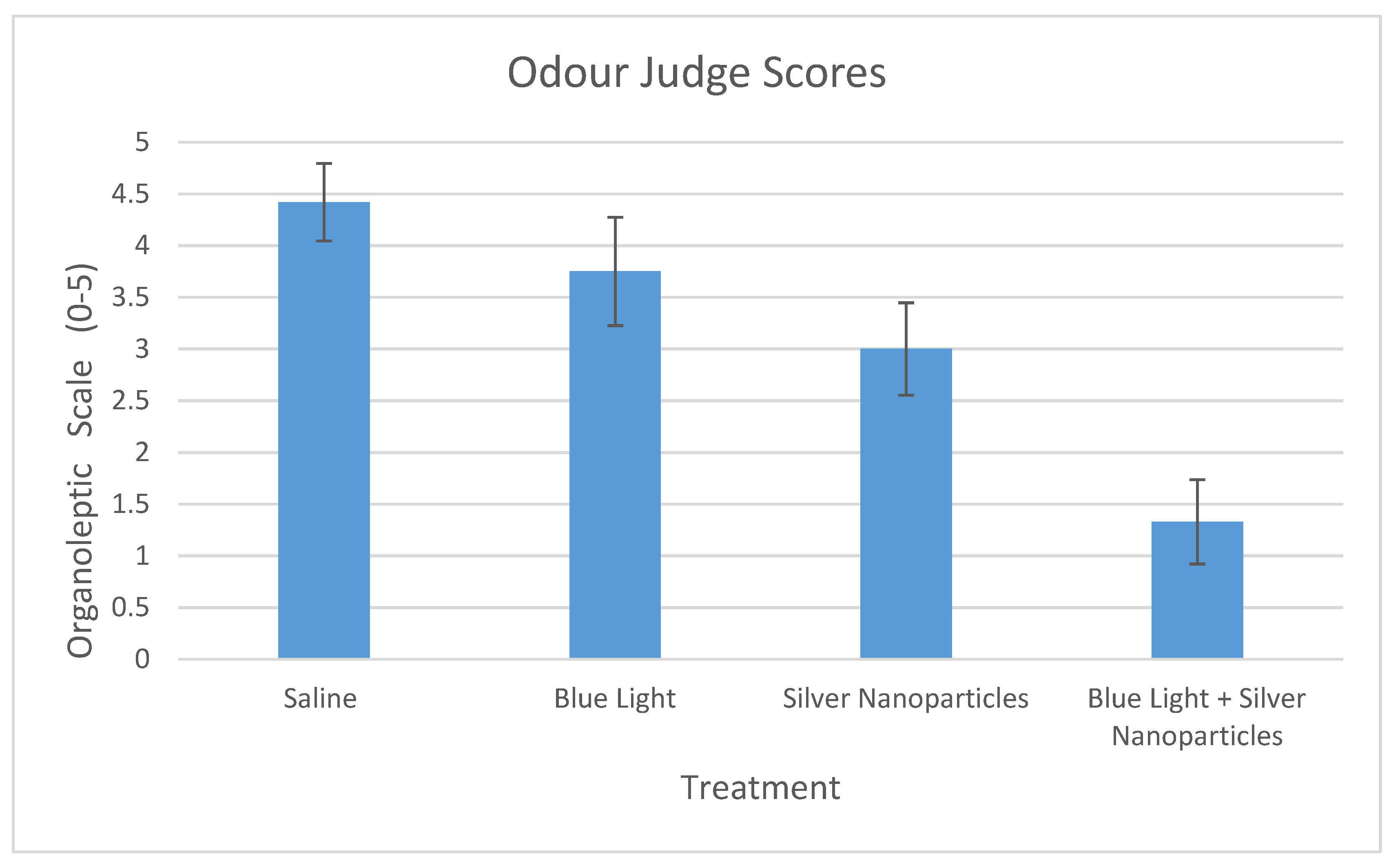
The effect of the different treatments and controls on malodor scores and VSC production are presented in Figure 1 and Figure 2. Although silver nanoparticles did show a statistically significant effect on the malodor related parameters (p < 0.001), only the combined treatment of blue light and silver nanoparticles was effective in reducing malodor and VSC levels below detection threshold (OJ scores < 2 and VSC < 110 ppb).
3.2. Reactive Oxygen Species (ROS) Levels
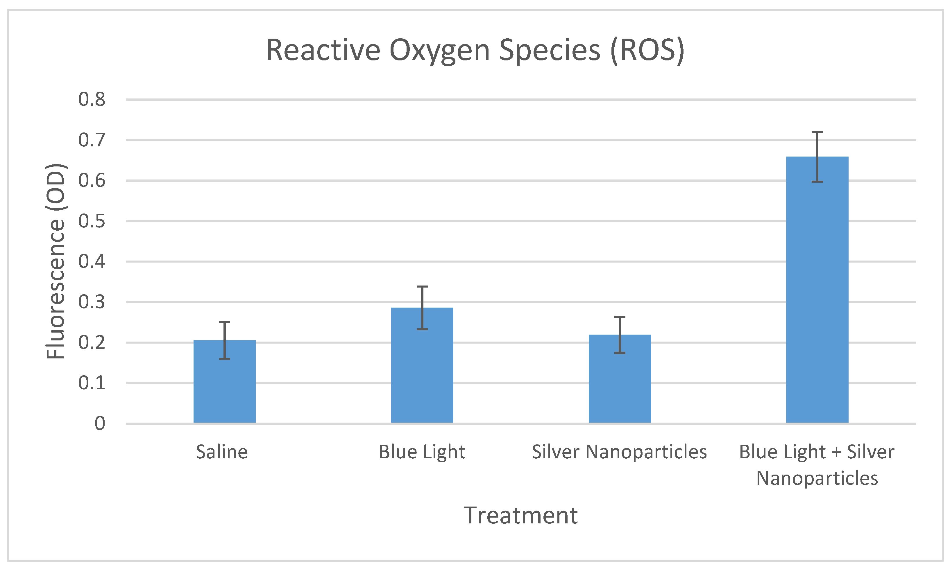
The effects of the different treatments on reactive oxygen species (ROS) production are presented in Figure 3. Although blue light showed slight elevation in ROS levels, only the combined effect of blue light and silver nanoparticles resulted in a significant increase in ROS levels (p < 0.001).
3.3. Membrane Integrity
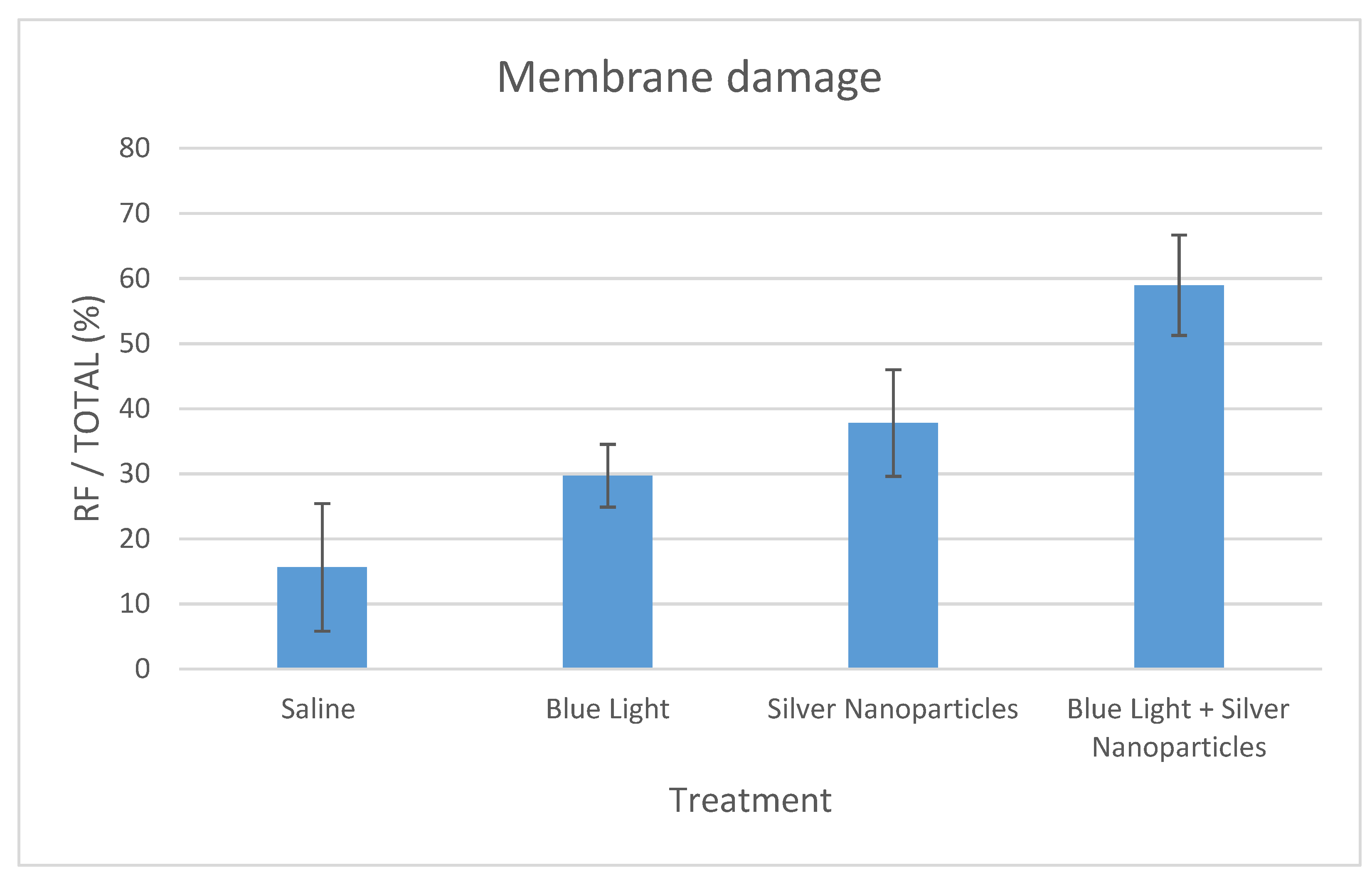
The effects of the different treatments on cell membrane damage are presented in Figure 4 and Figure 5. The combined treatment of blue light and silver nanoparticles was most effective, resulting in approximately 60% of bacterial cells with damaged cell membrane. This effect was statistically significant (p < 0.001) as compared with the individual treatments and control.
4. Discussion
Previous studies showed that sub-lethal dosage of blue light can increase cell membrane permeability in Fusobacterium nucleatum [13,14] and suggested that this effect was mediated through the production of reactive oxygen species [11]. In the present study, we tested the combined effect of sub-lethal blue light and silver nanoparticles on the ability of Fusobacterium nucleatum to produce malodor, its cell membrane integrity, and measured the levels of ROS produced following this treatment in order to understand the mechanism for the combined phototoxic effect. The results showed that the combined treatment caused a significant reduction in malodor related parameters concomitant with a rise in ROS levels and cell membrane damage as compared with the individual treatments and control.
Other researchers have also shown that ROS induced sub-lethal damage can be induced in bacteria using a low dosage of blue light exposure [17], resulting in increased membrane permeability. Silver nanoparticles, on the other hand, were reported to have several potential antibacterial mechanisms that include direct membrane damage [7] and ROS production [8]. The results of the present study show that treating Fusobacterium nucleatum with silver nanoparticle alone did cause increased cell membrane damage but did not affect ROS levels, whereas combining it with sub-lethal blue light resulted in increased membrane damage concomitant with significant increase in ROS levels, suggesting that silver nanoparticles may also serve as a potent photosensitizer.
The rising problem of bacterial resistant strains to antimicrobial therapy, as well as the risk of floral shift that may give rise to opportunistic infections, has stressed the importance of developing new approaches towards treating bacterial infections. For example, crippling strategies known as anti-virulence treatments [18] that target the bacteria’s virulence factors rather than their vitality and growth may eliminate the selective stress that promotes bacterial resistance while decreasing their pathogenicity.
Volatile sulfide compounds (VSC) are considered a key element in oral malodor production as well as an important virulence factor for bacteria, such as Fusobacterium nucleatum [19]. VSCs have been shown to induce the progression of periodontal disease and to promote DNA damage and gingival cells death [20].
Within the limitations of an in vitro study, the results of the present study suggest that using sub-lethal blue light exposure combined with silver nanoparticles may increase the efficacy of the antibacterial phototoxic effect against Fusobacterium nucleatum, even at a sub-lethal dosage. However, in addition to its beneficial antibacterial phototoxic effect, blue light may also have disruptive effects on mammalian cells [21], possibly mediated by light-induced ROS. Therefore, in any prospective clinical trial exposure, time should be minimized to reduce any risk to the oral soft tissues. Furthermore, the size and concentration of silver nanoparticles is a complex and controversial issue, since smaller size particles are most effective but show greater risk for mammalian cells toxicity [22]. These points merit further study.
5. Conclusions
The results of the present study suggest that the antibacterial phototoxic effect of blue light against Fusobacterium nucleatum may be enhanced by silver nanoparticles.
Author Contributions
Conceptualization, N.S.; methodology, N.S. and U.J.; formal analysis, U.J. and A.R.; investigation, N.S., U.J. and S.L.; resources, U.J.; data curation, N.S., A.R. and S.L.; writing—original draft preparation, N.S.; writing—review and editing S.L.; supervision, N.S.; project administration, S.L. All authors have read and agreed to the published version of the manuscript.
Funding
This research received no external funding.
Data Availability Statement
Not applicable.
Conflicts of Interest
The authors declare no conflict of interest.
References
- Liu, P.-F.; Haake, S.K.; Gallo, R.L.; Huang, C.-M. A novel vaccine targeting Fusobacterium nucleatum against abscesses and halitosis. Vaccine 2009, 27, 1589–1595. [Google Scholar] [CrossRef] [PubMed] [Green Version]
- Kang, Y.-B.; Kim, B.-G.; Chung, J.; Lee, H.-C.; Oh, J.-S. Inhibitory effect of Weissella cibaria isolates on the production of volatile sulphur compounds. J. Clin. Periodontol. 2006, 33, 226–232. [Google Scholar] [CrossRef] [PubMed]
- Mesquita, Q.M.; Dias, J.C.; Neves, M.G.P.M.S.; Almeida, A.; Faustino, M.A.F. Revisiting current photoactive materials for antimicrobial photodynamic therapy. Molecules 2018, 23, 2424. [Google Scholar] [CrossRef] [Green Version]
- Oves, M.; Aslam, M.; Rauf, M.A.; Qayyum, S.; Qari, H.A.; Khan, M.S.; Alam, M.Z.; Tabrez, S.; Pugazhendhi, A.; Ismail, I.M. Antimicrobial and anticancer activities of silver nanoparticles synthesized from the root hair extract of Phoenix dactylifera. Mater. Sci. Eng. C 2018, 89, 429–443. [Google Scholar] [CrossRef]
- Samuel, M.S.; Jose, S.; Selvarajan, E.; Mathimani, T.; Pugazhendhi, A. Biosynthesized silver nanoparticles using Bacillus amyloliquefaciens; Application for cytotoxicity effect on A549 cell line and photocatalytic degradation of p-nitrophenol. J. Photochem. Photobiol. B Biol. 2020, 202, 111642. [Google Scholar] [CrossRef]
- Bapat, R.A.; Chaubal, T.V.; Joshi, C.P.; Bapat, P.R.; Choudhury, H.; Pandey, M.; Gorain, B.; Kesharwani, P. An overview of application of silver nanoparticles for biomaterials in dentistry. Mater. Sci. Eng. C 2018, 91, 881–898. [Google Scholar] [CrossRef]
- Khorrami, S.; Zarrabi, A.; Khaleghi, M.; Danaei, M.; Mozafari, M.R. Selective cytotoxicity of green synthesized silver nanoparticles against the MCF-7 tumor cell line and their enhanced antioxidant and antimicrobial properties. Int. J. Nanomed. 2018, 13, 8013–8024. [Google Scholar] [CrossRef] [PubMed] [Green Version]
- Ramkumar, V.S.; Pugazhendhi, A.; Gopalakrishnan, K.; Sivagurunathan, P.; Saratale, G.D.; Dung, T.N.B.; Kannapiran, E. Biofabrication and characterization of silver nanoparticles using aqueous extract of seaweed Enteromorpha compressa and its biomedical properties. Biotechnol. Rep. 2017, 14, 1–7. [Google Scholar] [CrossRef]
- El-Azizi, M.M.; El Din, S.N.; El-Tayeb, T.A.; Aisha, K.A. In Vitro and In Vivo antimicrobial activity of combined therapy of silver nanoparticles and visible blue light against Pseudomonas aeruginosa. Int. J. Nanomed. 2016, 11, 1749–1758. [Google Scholar] [CrossRef] [Green Version]
- Sterer, N.; Feuerstein, O. Effect of visible light on malodour production by mixed oral microflora. J. Med. Microbiol. 2005, 54, 1225–1229. [Google Scholar] [CrossRef] [PubMed]
- Jeffet, U.; Nasrallah, R.; Sterer, N. Effect of red dyes on blue light phototoxicity against VSC producing bacteria in an experimental oral biofilm. J. Breath Res. 2016, 10, 046011. [Google Scholar] [CrossRef] [PubMed]
- Sterer, N.; Jeffet, U.; Dadoun, A.; Greenstein, R.B.-N.; Kohavi, D. Zinc enhances the phototoxic effect of blue light against malodour-producing bacteria in an experimental oral biofilm. J. Med. Microbiol. 2014, 63, 1071–1075. [Google Scholar] [CrossRef] [Green Version]
- Jeffet, U.; Shimon, R.; Sterer, N. Effect of high intensity blue light on Fusobacterium nucleatum membrane integrity. Photochem. Photobiol. 2019, 96, 178–181. [Google Scholar] [CrossRef]
- Jeffet, U.; Dagan, N.; Sterer, N. Effect of sublethal blue light on herbal extract activity against volatile sulfide compound production by Fusobacterium nucleatum. Photochem. Photobiol. 2021, 97, 443–447. [Google Scholar] [CrossRef]
- Rosseti, I.B.; Chagas, L.R.; Costa, M.S. Photodynamic antimicrobial chemotherapy (PACT) inhibits biofilm formation by Candida albicans, increasing both ROS production and membrane permeability. Lasers Med. Sci. 2014, 29, 1059–1064. [Google Scholar] [CrossRef]
- Kim, M.-J.; Mikš-Krajnik, M.; Kumar, A.; Ghate, V.; Yuk, H.-G. Antibacterial effect and mechanism of high-intensity 405 ± 5 nm light emitting diode on Bacillus cereus, Listeria monocytogenes, and Staphylococcus aureus under refrigerated condition. J. Photochem. Photobiol. B Biol. 2015, 153, 33–39. [Google Scholar] [CrossRef] [PubMed]
- McKenzie, K.; Maclean, M.; Grant, M.H.; Ramakrishnan, P.; MacGregor, S.J.; Anderson, J.G. The effects of 405 nm light on bacterial membrane integrity determined by salt and bile tolerance assays, leakage of UV-absorbing material and SYTOX green labelling. Microbiology 2016, 162, 1680–1688. [Google Scholar] [CrossRef] [PubMed] [Green Version]
- Fleitas Martínez, O.; Cardoso, M.M.; Ribeiro, S.M.; Franco, O.L. Recent advances in anti-virulence therapeutic strategies with a focus on dismantling bacterial membrane microdomains, toxin neutralization, quorum-sensing interference and biofilm inhibition. Front. Cell. Infect. Microbiol. 2019, 9, 74. [Google Scholar] [CrossRef]
- Mothersole, R.G.; Wolthers, K.R. Structural and kinetic insight into the biosynthesis of H2S and L-lanthionine from L-cysteine by a pyridoxal L-phosphate-dependent enzyme from Fusobacterium nucleatum. Biochemistry 2019, 58, 3592–3603. [Google Scholar] [CrossRef]
- Yaegaki, K.; Qian, W.; Murata, T.; Imai, T.; Sato, T.; Tanaka, T.; Kamoda, T. Oral malodorous compound causes apoptosis and genomic DNA damage in human gingival fibroblasts. J. Periodontal Res. 2008, 43, 391–399. [Google Scholar] [CrossRef]
- Wataha, J.C.; Lockwood, P.E.; Lewis, J.B.; Rueggeberg, F.A.; Messer, R.L. Biological effects of blue light from dental curing units. Dent. Mater. 2004, 20, 150–157. [Google Scholar] [CrossRef]
- Cho, Y.-M.; Mizuta, Y.; Akagi, J.-I.; Toyoda, T.; Sone, M.; Ogawa, K. Size-dependent acute toxicity of silver nanoparticles in mice. J. Toxicol. Pathol. 2018, 31, 73–80. [Google Scholar] [CrossRef] [PubMed] [Green Version]
Figure 1.
The mean results (±SD) of the odor judge scores given using an organoleptic scale of 0–5 for the various treatments, as indicated.
Figure 1.
The mean results (±SD) of the odor judge scores given using an organoleptic scale of 0–5 for the various treatments, as indicated.

Figure 2.
The mean results (±SD) of the volatile sulfide compounds (VSC) levels measured using a sulfide monitor (Halimeter) for the various treatments, as indicated.
Figure 2.
The mean results (±SD) of the volatile sulfide compounds (VSC) levels measured using a sulfide monitor (Halimeter) for the various treatments, as indicated.

Figure 3.
The mean results (±SD) of the reactive oxygen species (ROS) levels measured using the fluorescent probe 2′,7′-dichlorodihydrofluorescein diacetate (H2DCF-DA) for the various treatments, as indicated.
Figure 3.
The mean results (±SD) of the reactive oxygen species (ROS) levels measured using the fluorescent probe 2′,7′-dichlorodihydrofluorescein diacetate (H2DCF-DA) for the various treatments, as indicated.

Figure 4.
The mean results (±SD) of the percentage of membrane damaged bacterial cells measured using digital image analysis from fluorescent microscopy for the various treatments, as indicated. Membrane damaged bacterial cells are stained with ethidium homodimer-III (red fluorescence) and non-damaged ones are stained with DMAO (green fluorescence).

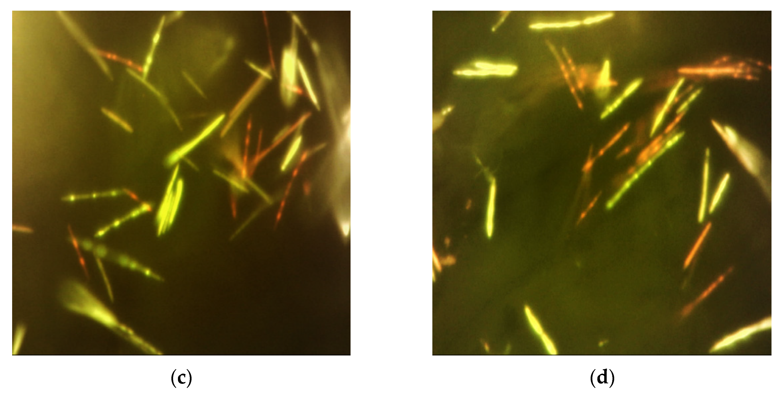
Figure 5.
Fluorescent microscopy (FM) images (magnification × 1000) showing membrane damaged (stained red) and undamaged (stained green) bacteria for the various treatments: (a) saline (negative) control; (b) blue light; (c) silver nanoparticles; and (d) blue light combined with silver nanoparticles.


Publisher’s Note: MDPI stays neutral with regard to jurisdictional claims in published maps and institutional affiliations. |
© 2021 by the authors. Licensee MDPI, Basel, Switzerland. This article is an open access article distributed under the terms and conditions of the Creative Commons Attribution (CC BY) license (https://creativecommons.org/licenses/by/4.0/).
Share and Cite
MDPI and ACS Style
Jeffet, U.; Livne, S.; Rahmanov, A.; Sterer, N. Effect of Silver Nanoparticles on Blue Light Phototoxicity against Fusobacterium nucleatum. Biophysica 2021, 1, 405-412. https://0-doi-org.brum.beds.ac.uk/10.3390/biophysica1040029
AMA Style
Jeffet U, Livne S, Rahmanov A, Sterer N. Effect of Silver Nanoparticles on Blue Light Phototoxicity against Fusobacterium nucleatum. Biophysica. 2021; 1(4):405-412. https://0-doi-org.brum.beds.ac.uk/10.3390/biophysica1040029
Chicago/Turabian StyleJeffet, Uziel, Shiri Livne, Arkadi Rahmanov, and Nir Sterer. 2021. "Effect of Silver Nanoparticles on Blue Light Phototoxicity against Fusobacterium nucleatum" Biophysica 1, no. 4: 405-412. https://0-doi-org.brum.beds.ac.uk/10.3390/biophysica1040029