Article

Duplex On-Site Detection of Vibrio cholerae and Vibrio vulnificus by Recombinase Polymerase Amplification and Three-Segment Lateral Flow Strips

1
Key Laboratory of Molecular Biophysics of Ministry of Education, Department of Biomedical Engineering, College of Life Science and Technology, Center for Human Genome Research, Huazhong University of Science and Technology, Wuhan 430074, China
2
Jiangsu Key Laboratory of Marine Pharmaceutical Compound Screening, Jiangsu Key Laboratory of Marine Biological Resources and Environment, Co-Innovation Center of Jiangsu Marine Bio-Industry Technology, School of Pharmacy, Jiangsu Ocean University, Lianyungang 222005, China
3
Ustar Biotechnologies (Hangzhou) Ltd., Hangzhou 310053, China
4
Jiangsu Institute of Oceanology and Marine Fisheries, Nantong 226007, China
*
Authors to whom correspondence should be addressed.
Submission received: 20 April 2021 / Revised: 7 May 2021 / Accepted: 10 May 2021 / Published: 12 May 2021

Abstract

:
Vibrio cholerae and Vibrio vulnificus are two most reported foodborne Vibrio pathogens related to seafood. Due to global ocean warming and an increase in seafood consumption worldwide, foodborne illnesses related to infection of these two bacteria are growing, leading to food safety issues and economic consequences. Molecular detection methods targeting species-specific genes are effective tools in the fight against bacterial infections for food safety. In this study, a duplex detection biosensor based on isothermal recombinase polymerase amplification (RPA) and a three-segment lateral flow strip (LFS) has been established. The biosensor used lolB gene of Vibrio cholerae and empV gene of Vibrio vulnificus as the detection markers based on previous reports. A duplex RPA reaction for both targets were constructed, and two chemical labels, FITC and DIG, of the amplification products were carefully tested for effective and accurate visualization on the strip. The biosensor demonstrated good specificity and achieved a sensitivity of 101 copies per reaction or one colony forming unit (CFU)/10 g of spiked food for both bacteria. Validation with clinical samples showed results consistent with that of real-time polymerase chain reaction. The detection process was simple and fast with a 30-min reaction at 37 °C and visualization on the strip within 5 min. With little dependence on laboratory settings, this biosensor was suitable for on-site detection, and the duplex system enabled simultaneous detection of the two important foodborne bacteria. Moreover, the principle can be extended to healthcare and food safety applications for other pathogens.

1. Introduction

The ubiquitous, Gram-negative halophilic bacteria of the Vibrio genus are capable of infecting animals including humans [1]. Many of them are related to marine animals such as shrimp, fish, and shellfish, and have posted threats to public health by causing foodborne illnesses through contamination of seafood [2]. The rising seawater temperature in recent years has benefited the growth of Vibrio species, meanwhile the seafood consumption is increasing worldwide [3]. These factors have heightened the concern of Vibrio pathogens on food safety. Vibrio cholerae and Vibrio vulnificus are two of those most reported foodborne Vibrio pathogens related to seafood [4]. Accurate and reliable detection methods of them are important for both food safety controls and epidemiological monitoring.
Molecular detection methods targeting species-specific genes are effective tools in fighting against bacterial infections for food safety [5]. These methods are based on DNA amplification principles, including polymerase chain reaction (PCR), real-time PCR (qPCR), loop-mediated isothermal amplification (LAMP) [6], and recombinase polymerase amplification (RPA) [7]. To give some examples, PCR- and qPCR- based methods targeting genes rpoA (genus-specific RNA polymerase subunit A), ctxA (cholera toxin subunit A), toxR (global regulatory gene), etc. have been developed for the detection of V. cholerae [8,9]. Genes vvhA (cytolysin), pilF (pilus-type IV assembly protein), hly (hemolysin), etc. have been selected as the targets of PCR- and qPCR- based detection methods for V. vulnificus [4,10,11]. To reduce the dependence on sophisticated thermocyclers for on-site detections, LAMP-based methods have been developed targeting the ompW (outer membrane protein) gene of V. cholerae, and rpoS (RNA polymerase subunit sigma factor S) and vvhA genes of V. vulnificus [12,13,14]. RPA reactions can amplify DNA targets isothermally under relatively low temperatures and are more convenient for on-site detections than LAMP [15,16]. For more on-site detection choices, RPA assays targeting lolB (outer membrane lipoprotein) gene of V. cholerae and empV (extracellular metalloproteinase) gene of V. vulnificus have been developed [17,18]. These molecular detection methods have improved the rapidity, sensitivity, and accuracy of the detection practice of these two important Vibrio pathogens, and have made good contributions to food safety and public health.
Because identifying various pathogenic Vibrio species in food contaminations is a practical requirement, multiplex approaches have been made with the aim of targeting multiple species in a single assay. These were mainly qPCR approaches, TaqMan or SYBR Green-based, with which multiple Vibrio species, including V. cholerae and V. vulnificus, could be simultaneously detected in a single run [4,19]. To date, isothermal amplification-based multiplex detection assays targeting multiple Vibrio species have not been reported.
In this study, a duplex detection biosensor for V. cholerae and V. vulnificus based on RPA and a 3-segment lateral flow strip (LFS) has been established. The biosensor had a good specificity and achieved a sensitivity of 101 copies per reaction or 1 colony forming unit (CFU)/10 g of spiked food for both bacteria. The detection included a 30-min reaction at 37 °C and visualization on the strip within 5 min. The biosensor was suitable for on-site detection, and the duplex system enabled simultaneous detection of the two important foodborne bacteria, which provided more convenience. In addition, the principle can be extended to healthcare and food safety applications for other pathogens.

2. Materials and Methods

2.1. Bacterial Strains

The 16 Vibrio species reference strains including Vibrio cholerae, Vibrio vulnificus, Vibrio parahemolyticus, Vibrio alginolyticus, Vibrio harveyi, Vibrio mediterranei, Vibrio shilonii, Vibrio splendidus, Vibrio mimicus, Vibrio ichthyoenteri, Vibrio campbellii, Vibrio chagasii, Vibrio fluvialis, Vibrio natriegens, Vibrio ponticus and Vibrio rotiferianus were obtained from the Jiangsu Institute of Oceanology and Marine Fisheries (Nantong, China). The 3 common foodborne pathogenic species reference strains Salmonella Typhimurium, Listeria monocytogenes, and Staphylococcus aureus were purchased from American Type Culture Collection, Manassas, VA, USA. Among these species, V. cholerae and V. vulnificus were the detection targets of this study, and other species were included as controls. All strains were confirmed by 16S rRNA sequencing [20]. Information of the strains are listed in Table 1. For template preparation, genomic DNA of the bacterial strains were extracted with the TIANamp Bacteria DNA Kit (Tiangen Biotech Co Ltd., Beijing, China) and quantified by a Qubit 4 fluorometer (Thermo Fisher Scientific Inc., Wilmington, DE, USA).

2.2. Primers and Probes

The primer and probe sequences used in this study were derived from previously reported RPA-based individual assays for V. cholerae and V. vulnificus detections (Table 2) [17,18]. The target genes for V. cholerae and V. vulnificus were the outer membrane lipoprotein gene lolB (GenBank accession number AP014524.1) and the metalloprotease gene empV (GenBank accession number U50548.1), respectively. The primer and probe design followed the manufacturer’s instructions of the RAA-nfo Nucleic Acid Amplification Reagent (Hangzhou ZC Bio-Sci & Tech Co Ltd., Hangzhou, China). Chemical modifications [fluorescein isothiocyanate (FITC), digoxigenin (DIG) or biotin] were made to the 5′ ends of the probes and the reverse primers to facilitate differential visualization on the strips. For each probe, a base in the middle was replaced with a tetrahydrofuran (THF), and the 3′ end was blocked with a Spacer C3 (SpC3). This probe design could reduce primer-dependent artifacts when using the RAA-nfo Nucleic Acid Amplification Reagent [7]. All the primers and probes were synthesized by General Biosystems Co Ltd., Anhui, China.

2.3. RPA-LFS

RPA reactions were performed according to the manufacturer’s instructions of the RAA-nfo Nucleic Acid Amplification Reagent (Hangzhou ZC Bio-Sci & Tech Co Ltd.). The reactions were performed at 37 °C for 30 min. After the reaction, 5 μL of the reaction mixture was applied to the sample pad of the LFS, and the starting end of the LFS was put into 100 μL of the strip solvent (Ustar Biotechnologies Ltd., Hangzhou, China). The test and control lines were visualized in 5 min.

2.4. Preparation of Spiked Food Samples

Spiked food samples were used to determine the sensitivity of the biosensor in food matrix. Shrimp was selected as the representative food sample. After purchased from a local supermarket, the shrimp samples were confirmed to have no contamination of V. cholerae or V. vulnificus using qPCR [21,22]. Homogenization of the shrimp samples were conducted with the handheld 3rd Gen. TGrinder (Tiangen Biotech Co Ltd.). To 100 mL of alkaline peptone broth (Sinopharm Chemical Reagent Co Ltd., Beijing, China), 10 g of the homogenate was added and suspended thoroughly, and the suspension was spiked with 100, 101, or 102 CFU of the mix of V. cholerae and V. vulnificus. For enrichment, the suspension was put in a shaker and incubated with 200 rpm shaking under 30 °C. At each planned time points, 1 mL of the enrichment suspension was collected and centrifuged at 5000× g for 5 min. The pelleted bacterial cells were resuspended with 200 µL of water. DNA was released by boiling at 100 °C for 10 min, and 1 µL of the boiled resuspension was used as the template for RPA-LFS.

2.5. Clinical Samples

Clinical samples were used to validate the biosensor. The clinical samples were kindly provided by the Jiangsu Institute of Oceanology and Marine Fisheries (Nantong, China). There were 20 shrimp samples (Litopenaeus vannamei, sampling tissue: hepatopancreas), 10 fish samples (Carassius auratus, sampling tissue: liver), and 10 shellfish samples (ostrea gigas thunberg, sampling tissue: meat). The samples were disinfected with ethanol, and 1 g of each sample was put into 9 mL of PBS and homogenized by the handheld grinder. One milliliter of the sample homogenate was boiled at 100 °C for 10 min, and centrifuged at 8000× g for 2 min. The supernatant (1 µL) was used as the template for RPA-LFS or qPCR.

2.6. qPCR

Specific primers (Table 2) described in previous reports for the detections of V. cholerae and V. vulnificus were used in qPCR assays of this study as reference methods [21,22]. The targeted genes were lolB gene of V. cholerae and gyrB (DNA gyrase subunit B) gene of V. vulnificus. The qPCR reaction mixtures (20 μL) contained 1 μl of the DNA template, 0.4 μL of each primer (10 μM), and 10 μL of the 2X SYBR Green qPCR Mix (Vazyme Biotech Co Ltd., Nanjing, China). The cycling programs followed the reported procedures [21,22]. The qPCR assays were performed on an Applied Biosystems 7900HT Fast Real-Time PCR System. A cycle threshold (Ct) less than 32 was considered positive.

3. Results

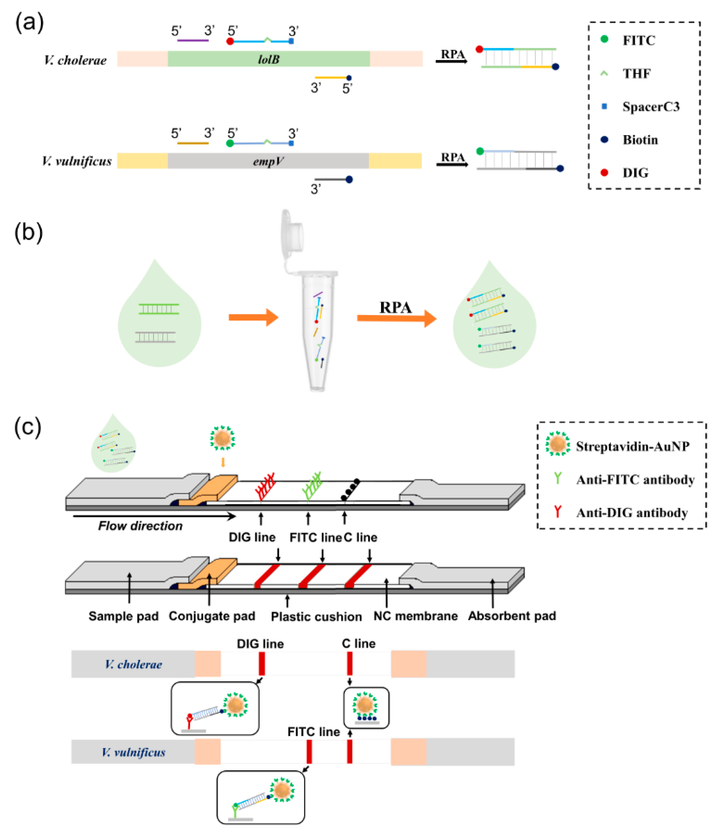
3.1. Principle of the Biosensor

The principle of the duplex detection biosensor for V. cholerae and V. vulnificus was based on duplex RPA amplification of the target gene fragments followed by visualization of the amplification products on a three-segment LFS (Figure 1). The lolB gene of V. cholerae and empV gene of V. vulnificus had been used as molecular markers for detection of these two bacteria in previously reported assays [17,18]. In this study, these target gene fragments were specifically amplified in RPA reactions with chemically labeled primers and probes (Figure 1a). The use of FITC- or DIG- labeled probes and biotin-labeled reverse primers could generate amplification products with FITC or DIG at one end and biotin at the other with the RAA-nfo Nucleic Acid Amplification Reagent (Hangzhou ZC Bio-Sci & Tech Co Ltd.). Efficient one-tube duplex amplification for the two detection markers was achieved by reaction optimization (Figure 1b), and the amplification products were applied to a three-segment LFS for simultaneous detection of the two bacteria (Figure 1c). The strip had the sample pad, the conjugate pad, the three detection segments and the absorbent pad on the solvent migration route. The conjugate pad was soaked with streptavidin-bound gold nanoparticles (streptavidin-AuNPs). The three detection segments were DIG line (anti-DIG antibody coated), FITC line (anti-FITC antibody coated) and C line (biotin coated control line). Amplification products of the lolB gene fragment of V. cholerae had the DIG labeling and should be visualized at the DIG line, while amplification products of the empV gene fragment of V. vulnificus had the FITC labeling and should be visualized at the FITC line. Visualization of the C line should indicate the proper completion of the strip assay. Selection of FITC labeling for V. vulnificus and DIG labeling for V. cholerae, and the order of DIG-, FITC-, and C-lines on the solvent migration route of the strip, were determined in this study (see below).

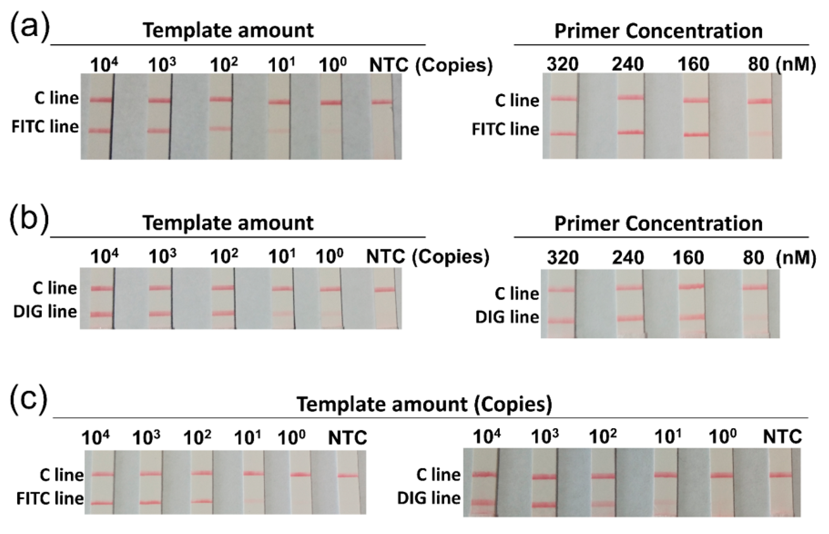
3.2. Optimization of the Duplex RPA Reaction

In the RPA reaction, chemical labels of the probes remain on one end of the amplification products (Figure 1a). FITC and DIG are chemical groups usually used for 5′-end labeling of nucleic acids [23]. In this study, they were selected as the labeling groups of the probes to facilitate visualization of the RPA amplification products on the strip through antibody-antigen interactions (Figure 1c). We designed primers and probes according to previously established RPA detection assays [17,18] (Table 2), and selected FITC labeling for V. vulnificus (probe VV-P1) and DIG labeling for V. cholerae (probe VC-P1) to start with.
To build the duplex RPA reaction, the optimal concentration range of the primers and probes in the single reaction was firstly determined. Different template amounts were used to assess the limit of detection (LOD) of the single reactions for V. vulnificus and V. cholerae, and the LOD of 101 gene copies per reaction was observed for both bacteria (Figure 2a,b, left panels). Then the concentrations of the primers and probes were decreased proportionally to determine the lowest primer/probe concentrations that could be used (Figure 2a,b, right panels). The results showed that, in the single reactions for both bacteria, the primer/probe concentrations at 50% of what the manufacturer recommended ((primers) = 160 nM; (probe) = 48 nM) had no obvious effect on the signal intensity. Thus, we determined to use 160 nM of each primer and 48 nM of each probe in the duplex RPA reaction. Validation of the duplex reaction showed unaffected LOD comparing to the single reactions (Figure 2c).

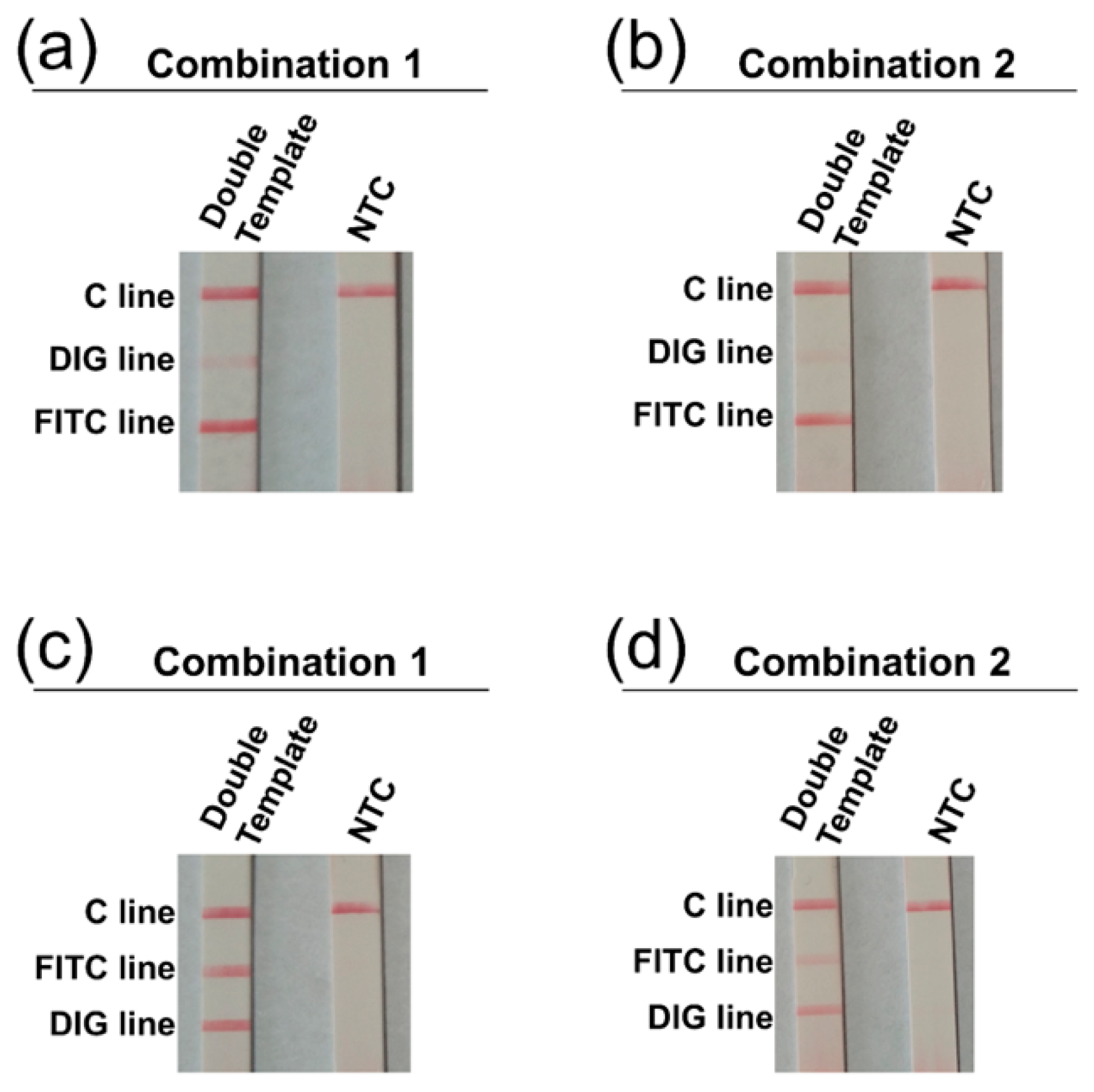
3.3. Determination of Visualization Lines on the 3-Segment Strip

The amplicons from the duplex reactions were applied to the 3-segment strips. In the first 3-segment design of the strip, the 3 lines on the solvent migration route were FITC-, DIG-, and C-lines. The initial amplicon labeling design was FITC labeling for V. vulnificus and DIG labeling for V. cholerae (probe combination 1: probes VC-P1 and VV-P1 used). Surprisingly, the visualization on the DIG line was very weak (Figure 3a). Then we tried to change the amplicon labeling to DIG for V. vulnificus and FITC for V. cholerae (probe combination 2: probes VC-P2 and VV-P2 used) (Table 2). However, the visualization on the DIG line did not improve (Figure 3b). Then, another three-segment design of the strip was used, of which the three lines on the solvent migration route were DIG-, FITC-, and C-lines. With this three-segment design, amplicons with the probe combination 1 visualized normally, while amplicons with the probe combination 2 had weaker visualizations (Figure 3c,d). Thus, the three-segment design of the strip should be DIG-, FITC-, and C-lines in order on the solvent migration route, and the amplicon labeling should be FITC for V. vulnificus and DIG for V. cholerae (probe combination 1).

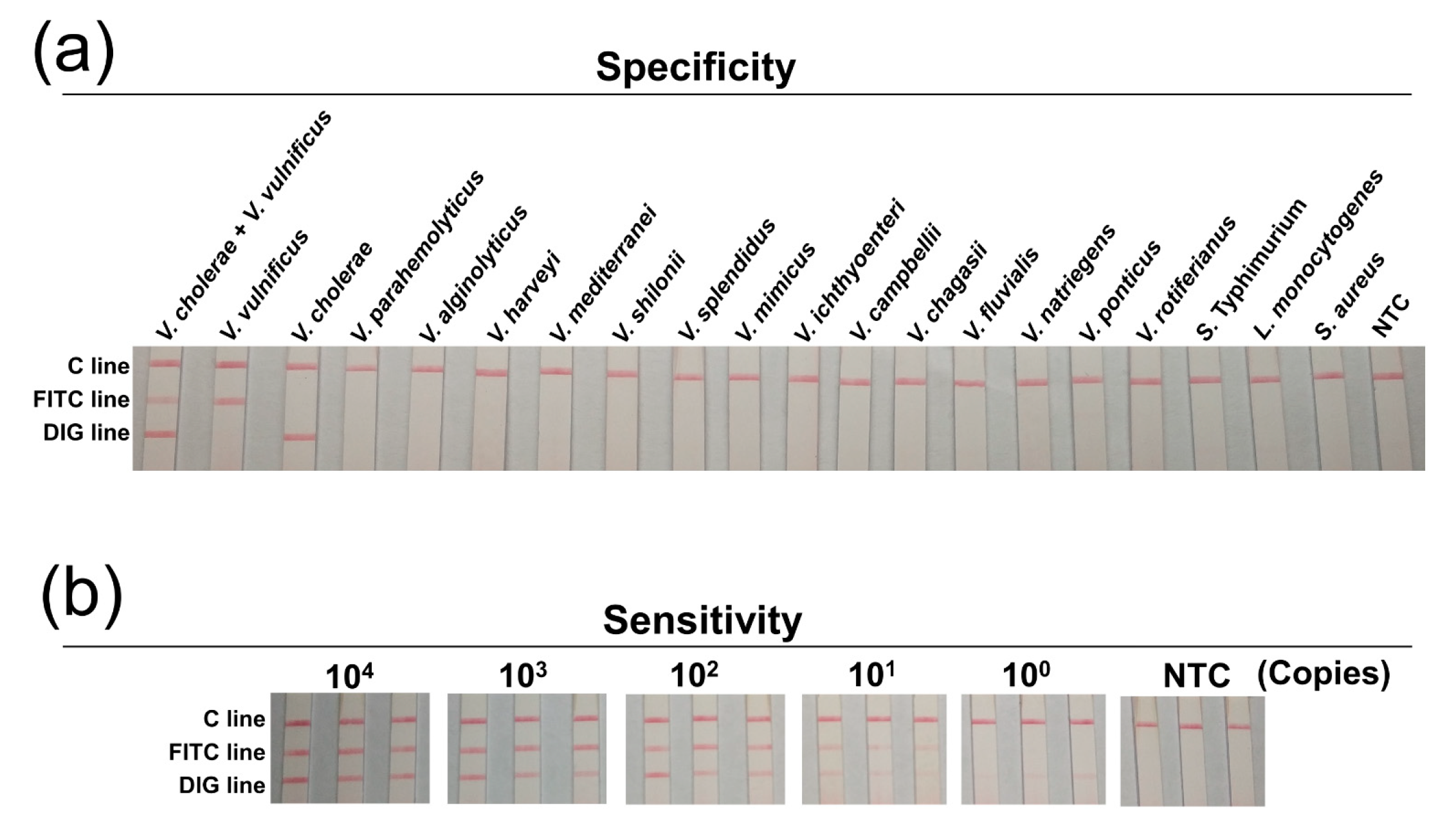
3.4. Specificity and Sensitivity

The specificity of this biosensor was established with a series of Vibrio species and several commonly seen foodborne pathogens. The results showed that V. vulnificus and V. cholerae templates gave positive signals at the corresponding lines, and all other templates gave negative signals (Figure 4a). This suggested good specificity of the biosensor.
The detection sensitivity was tested with serial dilutions of the bacteria DNAs. 100–104 copies of the genomic DNAs of V. vulnificus and V. cholerae were mixed and served as the template for the duplex amplification. The amplicons were applied to the 3-segment strip and showed a sensitivity of 101 copies for both bacteria (Figure 4b). Additionally, the detection sensitivity was tested in food matrix. 100–102 CFU of V. vulnificus and V. cholerae were spiked in 10 g of shrimp meat, and the spiked samples were enriched for 0-8 hr followed by sample processing and duplex amplification. Application of the amplicons on the strips showed that as low as 1 CFU/10 g of V. vulnificus and V. cholerae could be detected after 4 h of enrichment (Figure 5). Thus, the sensitivity of the method was 101 copies per reaction or 1 CFU/10 g of spiked food for both V. vulnificus and V. cholerae.

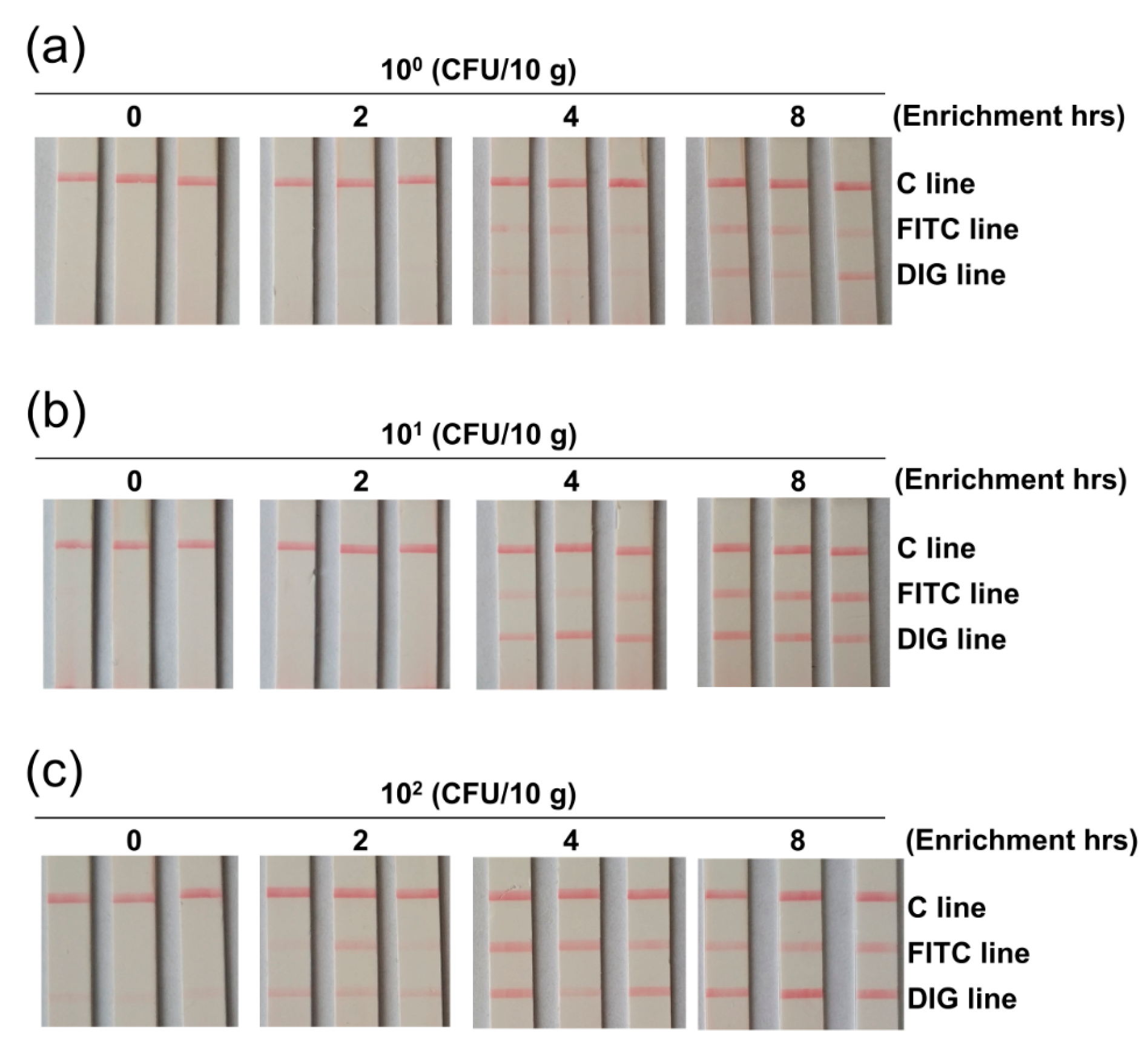
3.5. Clinical Sample Applications

A total of 40 clinical samples including shrimp (20 samples), fish (10 samples) and shellfish (10 samples) were collected from different areas of China and tested for V. cholerae and V. vulnificus by this duplex detection biosensor. The detection results were compared with qPCR (Table 3). Three shrimp samples, two fish samples and two shellfish samples were positive for V. cholerae. Three shrimp samples and one shellfish sample were positive for V. vulnificus. There was one sample positive for both bacteria. The detection results of the biosensor were consistent with that of qPCR.

4. Discussion

Detection of pathogenic Vibrio species in seafood contaminations is important for food safety and public health [2,24]. For the two important Vibrio pathogens, V. cholerae and V. vulnificus, molecular detection methods based on PCR, qPCR, LAMP, and RPA have been developed [8,9,12,13,17,18,25,26]. LAMP and RPA using isothermal amplification principles have provided greater convenience for on-site detections [6,7]. Efforts for multiplexing have been made using PCR and qPCR methods to detect multiple species in a single assay [8,9,25,26]. This study developed a duplex detection biosensor for V. cholerae and V. vulnificus based on RPA and a three-segment LFS, giving the on-site detection practice a more convenient choice.
To construct the duplex RPA reaction, one potential problem was the amplification preference (or bias) that could lead to nonuniformity of the amplicons [16,27]. This was essential because biased amplification of one target would affect the sensitivity of the reaction to the other. It has been reported that the amplification preference of RPA reactions could come from the size-dependent amplification rate of different targets [16,28]. In this study, we selected amplification targets with similar amplicon sizes for the two Vibrio species to prevent biased amplification in the duplex RPA reaction (Table 2). Moreover, the primers and probes were designed based on previously reported detection assays in which the specificity had been established [17,18], to prevent cross amplifications in the duplex reaction. Specifically, the primer/probe set for V. cholerae targeted the lolB gene, which was highly conserved in all serogroups of V. cholerae, but not conserved in other enteric bacterial species [29]. The primer/probe set for V. vulnificus targeted the empV gene, which also showed good specificity and evolutionary conservation [18].
We reduced the primer and probe concentrations to a point that the amplification efficiencies were not affected in reactions with single primer/probe sets, and then combined the two primer/probe sets at 1:1 molar ratio in one reaction mixture. The sensitivity was not affected in the duplex reaction when tested with double templates (V. cholerae and V. vulnificus) (Figure 2).
Surprisingly, when we applied the amplification products of the duplex RPA reaction to a three-segment strip on which the test lines were ordered as FITC-DIG-control, the visualization on the DIG line was very weak. This should not be a result of biased amplification, because when we exchanged the chemical labels of the amplicons (probe combination 1 changed to probe combination 2), the visualization of the test lines was the same. Moreover, there was no sign of biased amplification when using the 2-segment strips. Good visualization was obtained when a three-segment strip with the test lines ordered as DIG-FITC-control was used. The results indicated that the two test lines on the strip had to be placed in a particular order. A possible reason for this could be that there was a cross reaction between the DIG labeling on the amplicon and the anti-FITC antibody on the test line. The specificity of antibody–antigen reactions is usually considered high, but not to the extent of complete elimination of non-specific interactions [30]. For the design of three-segment strip in this study, it was important to determine the visualization lines by experimental testing.
After the duplex RPA reaction was built and the three-segment strip was determined, the duplex detection biosensor was established. This was for the first time an RPA-LFS-based multiplex detection assay targeting multiple Vibrio species was developed. Only a few similar assays had been developed for multiplex detection of microbial species, such as Giardia/Cryptosporidium/Entamoeba triplex detection [31], and Streptococcus pneumoniae/Legionella pneumophila duplex detection [32], as examples. Multiplexing with single-tube RPA is challenging because of the amplification bias as mentioned above, possibly caused by competition for the recombinase proteins from primers [16,27]. Careful optimizations of the RPA reaction thus are important for any multiplex assay. In addition, experimental testing for single-strip visualization of the amplicons is also essential as indicated by this study. Multiplexing on RPA-LFS detection requires sophisticated customization indeed.
The duplex biosensor from this study showed satisfactory performance. The specificity for V. cholerae and V. vulnificus was as good as the previously reported RPA-based individual assays [17,18]. The sensitivity was 101 copies per reaction or 1 CFU/10 g of spiked food with enrichment for both the Vibrio pathogens. This sensitivity was also as good as the previously reported qPCR-, LAMP-, and RPA- based individual assays [12,13,17,18,21,33]. The biosensor could simultaneously detect two important foodborne Vibrio pathogens within 35 min (30 min of amplification and 5 min of visualization) under a convenient isothermal temperature of 37 °C. The template preparation was easy by sample boiling. Validation with clinical samples showed results consistent with that of qPCR. The simple and fast procedure made the duplex biosensor very suitable for on-site detection, providing a more convenient choice for the detection practice of V. cholerae and V. vulnificus. Moreover, the principle can be extended to healthcare and food safety applications for other pathogens.

Author Contributions

Conceptualization, S.G.; methodology, S.G.; formal analysis, P.W., X.L., S.G., H.S. and Q.L.; investigation, P.W., L.L., C.M., X.Z., J.Y., L.Y. and S.G.; writing—original draft preparation, P.W. and S.G.; writing—review and editing, P.W. and S.G.; supervision, S.G. and Q.L.; funding acquisition, S.G. and H.S. All authors have read and agreed to the published version of the manuscript.

Funding

This work was supported by grants from the National Natural Science Foundation of China (31470275), the Key Natural Science Research Project of the Jiangsu Higher Education Institutions of China (20KJA416002), the Fishery Science and Technology Innovation Program of China (Y2018-14), the Nantong Municipal Science and Technology Plan of China (MS12020059), the Lianyungang Science and Technology Project of China (SF2003), the Science and Technology Project of Lianyungang High-tech Zone of China (HZ201901), the Open-end Funds of Jiangsu Key Laboratory of Marine Pharmaceutical Compound Screening (HY202004), and the Priority Academic Program Development of Jiangsu Higher Education Institutions of China.

Institutional Review Board Statement

Not applicable.

Informed Consent Statement

Not applicable.

Data Availability Statement

The data presented in this study are available in the article, further inquiries can be directed to the corresponding author.

Conflicts of Interest

The authors declare no conflict of interest.

References

  1. Austin, B. Vibrios as causal agents of zoonoses. Vet. Microbiol. 2010, 140, 310–317. [Google Scholar] [CrossRef] [Green Version]
  2. Daniels, N.A.; MacKinnon, L.; Bishop, R.; Altekruse, S.; Ray, B.; Hammond, R.M.; Thompson, S.; Wilson, S.; Bean, N.H.; Griffin, P.M.; et al. Vibrio parahaemolyticus infections in the United States, 1973–1998. J. Infect Dis. 2000, 181, 1661–1666. [Google Scholar] [CrossRef] [Green Version]
  3. Vezzulli, L.; Colwell, R.R.; Pruzzo, C. Ocean warming and spread of pathogenic vibrios in the aquatic environment. Microbial ecol. 2013, 65, 817–825. [Google Scholar] [CrossRef]
  4. Robert-Pillot, A.; Copin, S.; Himber, C.; Gay, M.; Quilici, M.-L. Occurrence of the three major Vibrio species pathogenic for human in seafood products consumed in France using real-time PCR. Int. J. Food Microbio. 2014, 189, 75–81. [Google Scholar] [CrossRef]
  5. Bonnin-Jusserand, M.; Copin, S.; Le Bris, C.; Brauge, T.; Gay, M.; Brisabois, A.; Grard, T.; Midelet-Bourdin, G. Vibrio species involved in seafood-borne outbreaks (Vibrio cholerae, V. parahaemolyticus and V. vulnificus): Review of microbiological versus recent molecular detection methods in seafood products. Crit. Rev. Food Sci. Nutr. 2019, 59, 597–610. [Google Scholar] [CrossRef]
  6. Notomi, T.; Okayama, H.; Masubuchi, H.; Yonekawa, T.; Watanabe, K.; Amino, N.; Hase, T. Loop-mediated isothermal amplification of DNA. Nucleic Acids Res. 2000, 28, E63. [Google Scholar] [CrossRef] [PubMed] [Green Version]
  7. Piepenburg, O.; Williams, C.H.; Stemple, D.L.; Armes, N.A. DNA Detection Using Recombination Proteins. PLoS Biol. 2006, 4, e204. [Google Scholar] [CrossRef]
  8. Jeyasekaran, G.; Raj, K.T.; Shakila, R.J.; Thangarani, A.J.; Sukumar, D. Multiplex polymerase chain reaction-based assay for the specific detection of toxin-producing Vibrio cholerae in fish and fishery products. Appl. Microbiol. Biot. 2011, 90, 1111–1118. [Google Scholar] [CrossRef]
  9. Chapela, M.J.; Fajardo, P.; Garrido, A.; Cabado, A.G.; Ferreira, M.; Lago, J.; Vieites, J.M. Comparison between a TaqMan polymerase chain reaction assay and a culture method for ctx-positive Vibrio cholerae detection. J. Agric. Food Chem. 2010, 58, 4051–4055. [Google Scholar] [CrossRef] [PubMed]
  10. Messelhäusser, U.; Colditz, J.; Thärigen, D.; Kleih, W.; Höller, C.; Busch, U. Detection and differentiation of Vibrio spp. in seafood and fish samples with cultural and molecular methods. Int. J. Food. Microbiol. 2010, 142, 360–364. [Google Scholar] [CrossRef] [PubMed]
  11. Baker-Austin, C.; Lemm, E.; Hartnell, R.; Lowther, J.; Onley, R.; Amaro, C.; Oliver, J.D.; Lees, D. pilF polymorphism-based real-time PCR to distinguish Vibrio vulnificus strains of human health relevance. Food Microbiol. 2012, 30, 17–23. [Google Scholar] [CrossRef] [Green Version]
  12. Srisuk, C.; Chaivisuthangkura, P.; Rukpratanporn, S.; Longyant, S.; Sridulyakul, P.; Sithigorngul, P. Rapid and sensitive detection of Vibrio cholerae by loop-mediated isothermal amplification targeted to the gene of outer membrane protein ompW. Lett. Appl. Microbio. 2010, 50, 36–42. [Google Scholar] [CrossRef]
  13. Surasilp, T.; Longyant, S.; Rukpratanporn, S.; Sridulyakul, P.; Sithigorngul, P.; Chaivisuthangkura, P. Rapid and sensitive detection of Vibrio vulnificus by loop-mediated isothermal amplification combined with lateral flow dipstick targeted to rpoS gene. Mol. Cell. Probes. 2011, 25, 158–163. [Google Scholar] [CrossRef]
  14. Han, F.; Ge, B. Quantitative detection of Vibrio vulnificus in raw oysters by real-time loop-mediated isothermal amplification. Int. J. Food Microbiol. 2010, 142, 60–66. [Google Scholar] [CrossRef]
  15. Li, J.; Macdonald, J.; von Stetten, F. Review: A comprehensive summary of a decade development of the recombinase polymerase amplification. Analyst 2018, 144, 31–67. [Google Scholar] [CrossRef] [PubMed] [Green Version]
  16. Lobato, I.M.; O’Sullivan, C.K. Recombinase polymerase amplification: Basics, applications and recent advances. Trends Analyst Chem. 2018, 98, 19–35. [Google Scholar] [CrossRef] [PubMed]
  17. Tang, Y.; Cao, Y.; Yu, Y.; Yan, S.; Wang, Y.; Pan, Y.; Zhang, W. Real-Time Recombinase Polymerase Amplification Assay for the Detection of Vibrio cholerae in Seafood. Food Anal Methods 2017, 10, 2657–2666. [Google Scholar] [CrossRef]
  18. Yang, X.; Zhang, X.; Wang, Y.; Shen, H.; Jiang, G.; Dong, J.; Zhao, P.; Gao, S. A Real-Time Recombinase Polymerase Amplification Method for Rapid Detection of Vibrio vulnificus in Seafood. Front. Microbiol. 2020, 11, 586981. [Google Scholar] [CrossRef] [PubMed]
  19. Kim, H.-J.; Lee, H.-J.; Lee, K.-H.; Cho, J.-C. Simultaneous detection of Pathogenic Vibrio species using multiplex real-time PCR. Food Control. 2012, 23, 491–498. [Google Scholar] [CrossRef]
  20. Hiergeist, A.; Reischl, U.; Gessner, A. Multicenter quality assessment of 16S ribosomal DNA-sequencing for microbiome analyses reveals high inter-center variability. Int. J. Med. Microbiol. 2016, 306, 334–342. [Google Scholar] [CrossRef] [PubMed] [Green Version]
  21. Cho, M.S.; Ahn, T.Y.; Joh, K.; Paik, S.Y.; Kwon, O.S.; Jheong, W.H.; Joung, Y.; Park, D.S. A novel marker for the species-specific detection and quantitation of Vibrio cholerae by targeting an outer membrane lipoprotein lolB gene. J. Microbiol. Biotechnol. 2013, 23, 555–559. [Google Scholar] [CrossRef] [Green Version]
  22. Kumar, H.S.; Parvathi, A.; Karunasagar, I.; Karunasagar, I. A gyrB-based PCR for the detection of Vibrio vulnificus and its application for direct detection of this pathogen in oyster enrichment broths. Int. J. Food Microbiol. 2006, 111, 216–220. [Google Scholar] [CrossRef]
  23. Li, J.; Pollak, N.M.; Macdonald, J. Multiplex Detection of Nucleic Acids Using Recombinase Polymerase Amplification and a Molecular Colorimetric 7-Segment Display. ACS Omega 2019, 4, 11388–11396. [Google Scholar] [CrossRef] [Green Version]
  24. FAO. The State of World Fisheries and Aquaculture. 2014. Available online: http://www.fao.org/3/i3720e/i3720e.pdf (accessed on 12 May 2021).
  25. Espiñeira, M.; Atanassova, M.; Vieites, J.M.; Santaclara, F.J. Validation of a method for the detection of five species, serogroups, biotypes and virulence factors of Vibrio by multiplex PCR in fish and seafood. Food Microbiol. 2010, 27, 122–131. [Google Scholar] [CrossRef] [PubMed]
  26. Taminiau, B.; Korsak, N.; Lemaire, C.; Delcenserie, V.; Daube, G. Validation of real-time PCR for detection of six major pathogens in seafood products. Food Control. 2014, 44, 130–137. [Google Scholar] [CrossRef]
  27. Kim, J.Y.; Lee, J.-L. Development of a multiplex real-time recombinase polymerase amplification (RPA) assay for rapid quantitative detection of Campylobacter coli and jejuni from eggs and chicken products. Food Control 2017, 73, 1247–1255. [Google Scholar] [CrossRef] [Green Version]
  28. Kersting, S.; Rausch, V.; Bier, F.F.; von Nickisch-Rosenegk, M. Multiplex isothermal solid-phase recombinase polymerase amplification for the specific and fast DNA-based detection of three bacterial pathogens. Microchim. Acta 2014, 181, 1715–1723. [Google Scholar] [CrossRef] [Green Version]
  29. Lalitha, P.; Siti Suraiya, M.N.; Lim, K.L.; Lee, S.Y.; Nur Haslindawaty, A.R.; Chan, Y.Y.; Ismail, A.; Zainuddin, Z.F.; Ravichandran, M. Analysis of lolB gene sequence and its use in the development of a PCR assay for the detection of Vibrio cholerae. J. Microbiol. Methods 2008, 75, 142–144. [Google Scholar] [CrossRef]
  30. Ko Ferrigno, P. Non-antibody protein-based biosensors. Essays Biochem. 2016, 60, 19–25. [Google Scholar] [CrossRef] [Green Version]
  31. Crannell, Z.; Castellanos-Gonzalez, A.; Nair, G.; Mejia, R.; White, A.C.; Richards-Kortum, R. Multiplexed Recombinase Polymerase Amplification Assay To Detect Intestinal Protozoa. Anal. Chem. 2016, 88, 1610–1616. [Google Scholar] [CrossRef]
  32. Kersting, S.; Rausch, V.; Bier, F.F.; von Nickisch-Rosenegk, M. A recombinase polymerase amplification assay for the diagnosis of atypical pneumonia. Anal. Biochem. 2018, 550, 54–60. [Google Scholar] [CrossRef] [PubMed]
  33. Panicker, G.; Bej, A.K. Real-time PCR detection of Vibrio vulnificus in oysters: Comparison of oligonucleotide primers and probes targeting vvhA. Appl. Environ. Microbiol. 2005, 71, 5702–5709. [Google Scholar] [CrossRef] [PubMed] [Green Version]
Figure 1. Schematic diagram of the duplex RPA-LFS biosensor. (a) Target amplification by RPA of the two Vibrio species. The target genes for V. cholerae and V. vulnificus were shown as horizontal strips. Primers and probes were shown as horizontal lines. Labels and modifications on the probes, the reverse primers and the RPA products were indicated. (b) Target amplification in the duplex RPA reaction. The duplex RPA system contained primer/probe sets for both detection markers. Both targets could be amplified with appropriate labels on the amplicons. (c) Simultaneous differential detection by visualization lines on the 3-segment strip: DIG, FITC and C (control) lines. The structure of the strip was shown with the coating materials indicated. RPA products from V. cholerae gave signal at the DIG line and that from V. vulnificus gave signal at the FITC line because of the different labels. Signal at the C line indicated proper finish of the lateral flow detection.
Biosensors 11 00151 g001
Figure 2. Optimization of the duplex RPA reaction. The concentration range of the primer/probe set in the single detection assay was determined for V. vulnificus (a) or V. cholerae (b). Different template amounts were tested in the single detection assays with the primer/probe concentrations set as recommended by the manufacturer of the reagent (the left panels of a and b). The final concentrations were: [forward primer] = 320 nM, [reverse primer] = 320 nM, and [probe] = 96 nM. The template amount was indicated on top of each strip. Using 102 copies of the templates (the right panels of a and b), the primer/probe concentrations were decreased proportionally (forward primer:reverse primer:probe =1:1:0.3) to determine the concentration range. Concentration of each primer was indicated on top of the strip run amplicons of the corresponding reaction. Then the sensitivity of the duplex RPA reaction was confirmed (c). Mixed templates (double template of V. vulnificus and V. cholerae) of different amounts were tested and separately visualized for V. vulnificus (left panel of c) and V. cholerae (right panel of c). Final concentrations of the primers and probes in the duplex RPA reaction: [each primer] = 160 nM, and [each probe] = 48 nM. The NTC strips were the no-template controls. The 2-segment strips were used to visualize amplicons with corresponding labels. The images represent results from three independent experiments.
Biosensors 11 00151 g002
Figure 3. Determination of visualization lines on the 3-segment strip. Two orders of the visualization lines on the 3-segment strip were tested: FITC-DIG-C, and DIG-FITC-C. Two amplicon labeling designs were tested on each of the strip types: (1) FITC for V. vulnificus and DIG for V. cholerae (Combination 1: probes VC-P1 and VV-P1 used); (2) DIG for V. vulnificus and FITC for V. cholerae (Combination 2: probes VC-P2 and VV-P2 used). The duplex RPA products from probe Combination 1 and Combination 2 were visualized on the strips with the two orders of visualization lines: FITC-DIG-C (a,b), and DIG-FITC-C (c,d). “Double Template” meant the RPA reactions had both V. vulnificus and V. cholerae as the templates (104 copies of each, mixed). The NTC strips were the no-template controls. The images represent results from three independent experiments.
Biosensors 11 00151 g003
Figure 4. Specificity and sensitivity of the duplex RPA-LFS biosensor. (a) Specificity of the biosensor. Templates used for testing were indicated on top of each strip. The DNA concentrations of the templates were normalized to 10 ng/μL and 1 μL was used for each reaction. The image represented results from three independent experiments. (b) Sensitivity of the biosensor. Mixed templates (double template of V. vulnificus and V. cholerae) of different amounts were used for testing. The amount (in copies) was shown at the top of the strips. The triplicate strips represented results from three independent experiments. The NTC strips were the no-template controls.
Biosensors 11 00151 g004
Figure 5. Limit of detection in spiked food samples. The image shows the detection results of the duplex RPA-LFS biosensor for spiked food samples. The spiking amounts were 100 (a), 101 (b), and 102 (c) CFU of the mix of V. cholerae and V. vulnificus per 10 g of shrimp. The enrichment time (in hours) after spiking was indicated on top of the strips. The triplicate strips represented results from three independent experiments.
Biosensors 11 00151 g005
Table 1. Bacteria strains used in this study.
SpeciesStrain TypeDesignation
Vibrio choleraeReference strainATCC 14100
Vibrio vulnificusReference strainATCC 27562
Vibrio parahemolyticusReference strainATCC 17802
Vibrio alginolyticusReference strainATCC 17749
Vibrio harveyiReference strainATCC 43516
Vibrio mediterraneiReference strainATCC 43341
Vibrio shiloniiReference strainATCC BAA-91
Vibrio splendidusReference strainMCCC 1A04096
Vibrio mimicusReference strainMCCC 1A02602
Vibrio ichthyoenteriReference strainMCCC 1A00057
Vibrio campbelliiReference strainMCCC 1A02605
Vibrio chagasiiReference strainMCCC 1B00386
Vibrio fluvialisReference strainMCCC 1A02761
Vibrio natriegensReference strainMCCC 1D00129
Vibrio ponticusReference strainMCCC 1H00061
Vibrio rotiferianusReference strainMCCC 1B00068
Salmonella TyphimuriumReference strainATCC 14028
Listeria monocytogenesReference strainATCC 19115
Staphylococcus aureusReference strainATCC 6538
Table 2. Primer and probe sequences.
MethodTargetPrimer/Probe NameSequence (5′-3′)Length (bp)Amplicon Size (bp)
RPAV. choleraeVC-FATCTTCAAGCTGTTCAACGGGAATATCTAA30218
VC-RBiotin-ATCAGCGACAATCGTTCAACTTTCAATGGC30
VC-P1DIG-ATCAGGCTTTGTGCATCTTGGTCGCGGTAGA [THF] TTGATCATCATAAGTTTCG-SpC351
VC-P2FITC-ATCAGGCTTTGTGCATCTTGGTCGCGGTAGA [THF] TTGATCATCATAAGTTTCG-SpC351
V. vulnificusVV-FGAGATGGATTCTTTGTATAACATTGCGT28214
VV-RBiotin-ACGATGACGTTGGTTGTGTTTCATTATC28
VV-P1FITC-GGTGAAGTTGGCTGGTGGTTATTTTCTGAA [THF] CATGGTTGTTGAGCTC-SpC347
VV-P2DIG-GGTGAAGTTGGCTGGTGGTTATTTTCTGAA [THF] CATGGTTGTTGAGCTC-SpC347
qPCRV. choleraeVC195FCCGTTGAGGCGAGTTTGGTGAGA23195
VC195RGTGCGCGGGTCGAAACTTATGAT23
V. vulnificusgyr-vv1GTCCGCAGTGGAATCCTTCA20285
gyr-vv2TGGTTCTTACGGTTACGGCC20
Table 3. Detection of V. cholerae and V. vulnificus in clinical samples.
No.Food TypeSample SourceDetection Results for V. choleraeDetection Results for V. vulnificus
RPA-LFSqPCRRPA-LFSqPCR
1ShrimpQingdao, China----
2ShrimpQingdao, China----
3ShrimpQingdao, China----
4ShrimpQingdao, China----
5ShrimpQingdao, China++--
6ShrimpQingdao, China----
7ShrimpQingdao, China--++
8ShrimpQingdao, China++++
9ShrimpQingdao, China----
10ShrimpQingdao, China----
11ShrimpLianyungang, China----
12ShrimpLianyungang, China----
13ShrimpLianyungang, China----
14ShrimpLianyungang, China----
15ShrimpLianyungang, China----
16ShrimpLianyungang, China--++
17ShrimpLianyungang, China----
18ShrimpLianyungang, China++--
19ShrimpLianyungang, China----
20ShrimpLianyungang, China----
21FishQingdao, China----
22FishQingdao, China----
23FishQingdao, China----
24FishQingdao, China----
25FishQingdao, China----
26FishYancheng, China++--
27FishYancheng, China++--
28FishYancheng, China----
29FishYancheng, China----
30FishYancheng, China----
31ShellfishQingdao, China----
32ShellfishQingdao, China++--
33ShellfishQingdao, China----
34ShellfishQingdao, China++--
35ShellfishQingdao, China----
36ShellfishLianyungang, China--++
37ShellfishLianyungang, China----
38ShellfishLianyungang, China----
39ShellfishLianyungang, China----
40ShellfishLianyungang, China----
(+: positive result; -: negative result).
Publisher’s Note: MDPI stays neutral with regard to jurisdictional claims in published maps and institutional affiliations.

