Hot-Band-Absorption-Induced Anti-Stokes Fluorescence of Aggregation-Induced Emission Dots and the Influence on the Nonlinear Optical Effect

 , and
, and 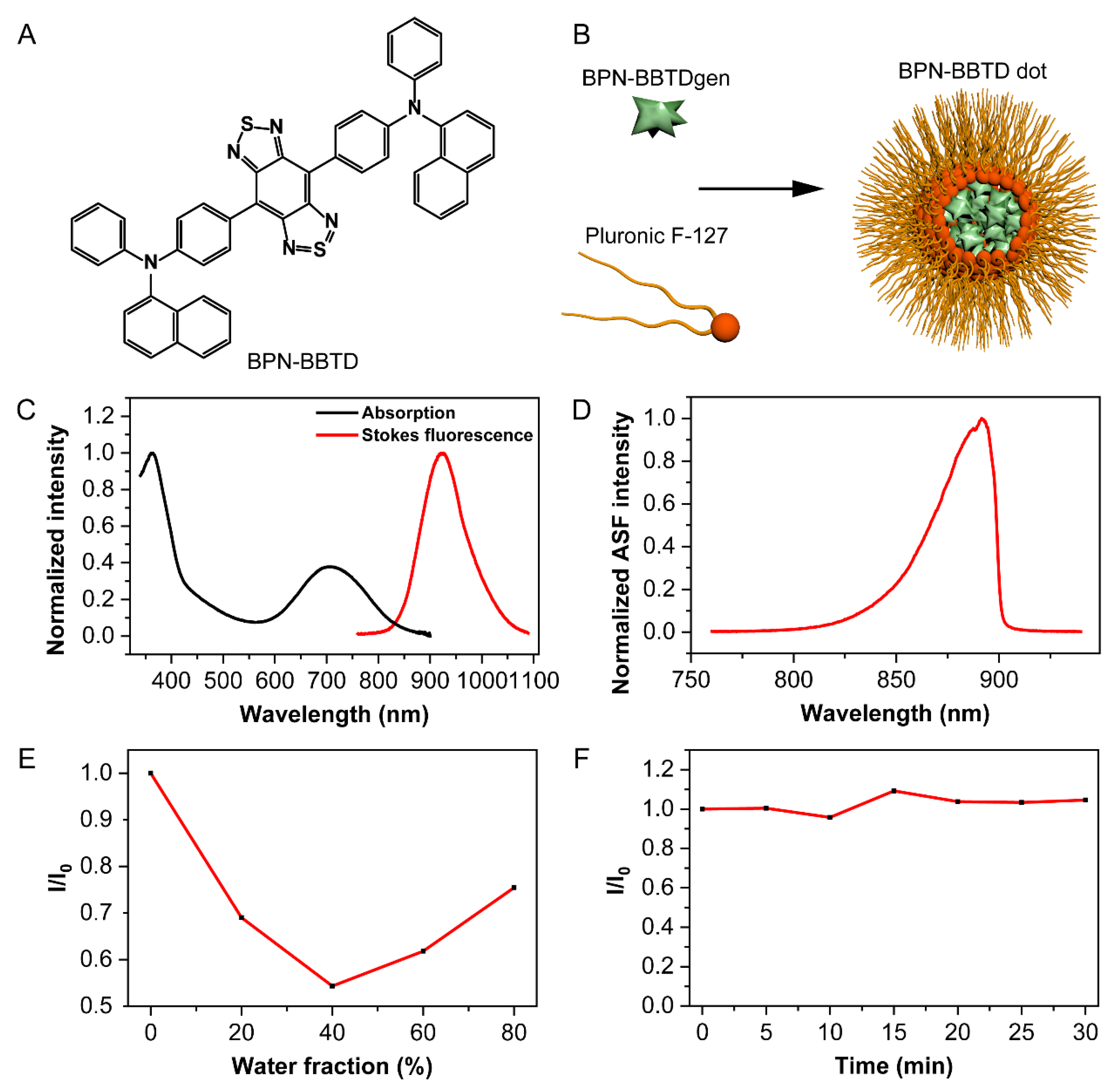
Abstract
:1. Introduction
2. Materials and Methods
2.1. Chemicals and Materials
2.2. Fabrication of BPN-BBTD Dots
2.3. Absorption and Fluorescence Spectra Measurement
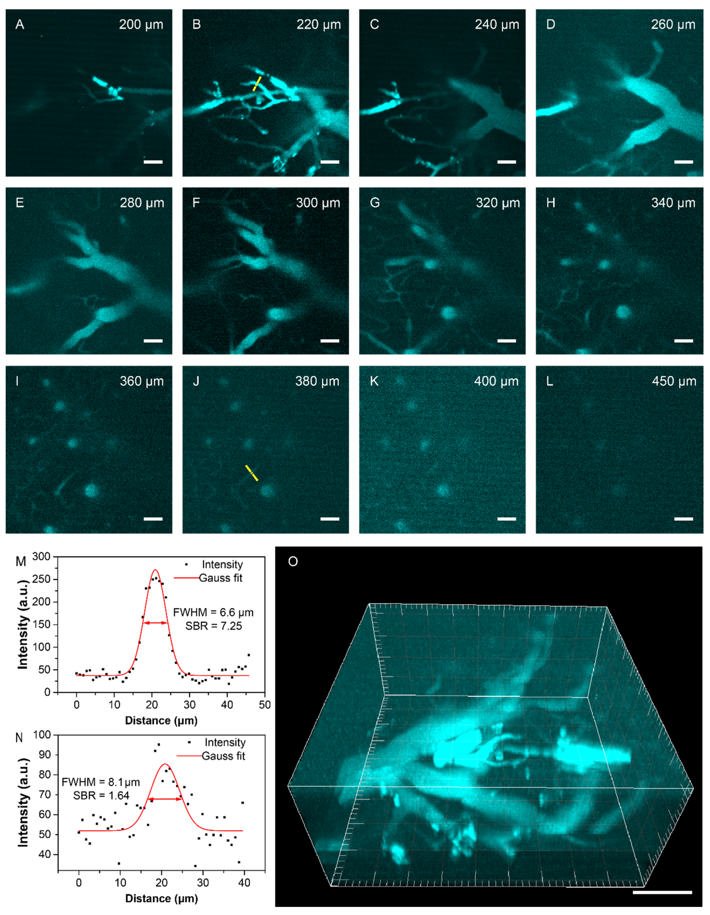
2.4. Animal Preparation for Cerebrovascular Microscopic Imaging
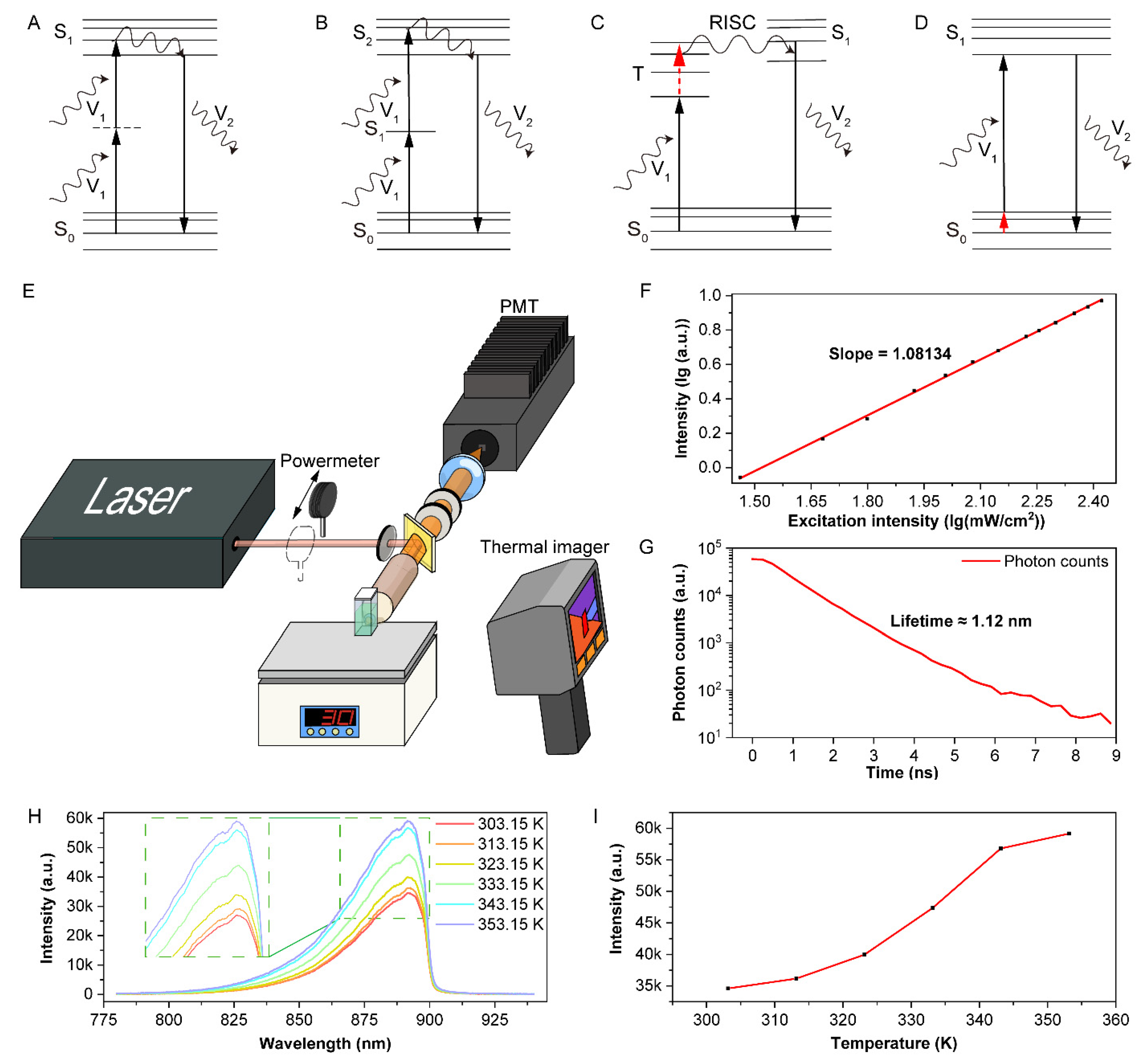
2.5. Optical Setup of the First Near-Infrared (NIR-I, 760–900 nm) Anti-Stokes Fluorescence Confocal Microscopy
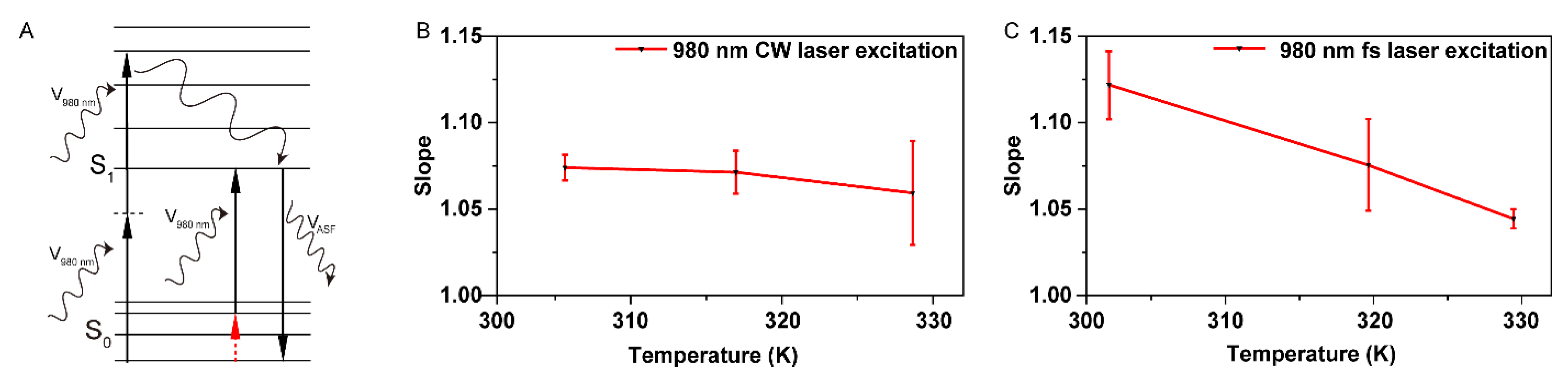
2.6. Power Dependence Measurement at Different Temperatures
2.7. Anti-Stokes Fluorescence Lifetime Measurement
2.8. Optical Setup for Photobleaching Test
3. Results and Discussion
3.1. Characterizations of BPN-BBTD
3.2. Mechanism of Anti-Stokes Fluorescence in BPN-BBTD Dots and Its Effect on Nonlinear Optics
3.3. In Vivo Anti-Stokes Fluorescence Confocal Microscopic Imaging
4. Conclusions
Supplementary Materials
Author Contributions
Funding
Institutional Review Board Statement
Informed Consent Statement
Data Availability Statement
Acknowledgments
Conflicts of Interest
References
- Feng, Z.; Bai, S.; Qi, J.; Sun, C.; Zhang, Y.; Yu, X.; Ni, H.; Wu, D.; Fan, X.; Xue, D.; et al. Biologically Excretable Aggregation-Induced Emission Dots for Visualizing Through the Marmosets Intravitally: Horizons in Future Clinical Nanomedicine. Adv. Mater. 2021, 33, 2008123. [Google Scholar] [CrossRef] [PubMed]
- Fan, X.; Xia, Q.; Zhang, Y.; Li, Y.; Feng, Z.; Zhou, J.; Qi, J.; Tang, B.Z.; Qian, J.; Lin, H. Aggregation-Induced Emission (AIE) Nanoparticles-Assisted NIR-II Fluorescence Imaging-Guided Diagnosis and Surgery for Inflammatory Bowel Disease (IBD). Adv. Healthc. Mater. 2021, 2101043. [Google Scholar] [CrossRef] [PubMed]
- Yu, W.; Guo, B.; Zhang, H.; Zhou, J.; Yu, X.; Zhu, L.; Xue, D.; Liu, W.; Sun, X.; Qian, J. NIR-II fluorescence in vivo confocal microscopy with aggregation-induced emission dots. Sci. Bull. 2019, 64, 410–416. [Google Scholar] [CrossRef] [Green Version]
- Chen, G.; Qiu, H.; Prasad, P.N.; Chen, X. Upconversion nanoparticles: Design, nanochemistry, and applications in theranostics. Chem. Rev. 2014, 114, 5161–5214. [Google Scholar] [CrossRef] [PubMed]
- Liu, Q.; Feng, W.; Yang, T.; Yi, T.; Li, F. Upconversion luminescence imaging of cells and small animals. Nat. Protoc. 2013, 8, 2033–2044. [Google Scholar] [CrossRef]
- Liu, Y.; Su, Q.; Zou, X.; Chen, M.; Feng, W.; Shi, Y.; Li, F. Near-infrared in vivo bioimaging using a molecular upconversion probe. Chem. Commun. 2016, 52, 7466–7469. [Google Scholar] [CrossRef]
- Guo, B.; Feng, Z.; Hu, D.; Xu, S.; Middha, E.; Pan, Y.; Liu, C.; Zheng, H.; Qian, J.; Sheng, Z.; et al. Precise Deciphering of Brain Vasculatures and Microscopic Tumors with Dual NIR-II Fluorescence and Photoacoustic Imaging. Adv. Mater. 2019, 31, 1902504. [Google Scholar] [CrossRef]
- Feng, Z.; Tang, T.; Wu, T.; Yu, X.; Zhang, Y.; Wang, M.; Zheng, J.; Ying, Y.; Chen, S.; Zhou, J.; et al. Perfecting and extending the near-infrared imaging window. Light Sci. Appl. 2021, 10, 197. [Google Scholar] [CrossRef]
- Li, D.; Zhang, H.; Streich, L.L.; Wang, Y.; Lu, P.; Wang, L.; Prevedel, R.; Qian, J. AIE-nanoparticle assisted ultra-deep three-photon microscopy in the in vivo mouse brain under 1300 nm excitation. Mater. Chem. Front. 2021, 5, 3201–3208. [Google Scholar] [CrossRef]
- Yu, X.; Ying, Y.; Feng, Z.; Qi, J.; Zheng, J.; Zhang, Y.; Liu, J.; Qian, J.; Tang, B.Z.; Zhang, D. Aggregation-induced emission dots assisted non-invasive fluorescence hysterography in near-infrared IIb window. Nano Today 2021, 39, 101235. [Google Scholar] [CrossRef]
- Yu, X.; Feng, Z.; Cai, Z.; Jiang, M.; Xue, D.; Zhu, L.; Zhang, Y.; Liu, J.; Que, B.; Yang, W.; et al. Deciphering of cerebrovasculatures via ICG-assisted NIR-II fluorescence microscopy. J. Mater. Chem. B 2019, 7, 6623–6629. [Google Scholar] [CrossRef] [PubMed]
- Feng, Z.; Yu, X.; Jiang, M.; Zhu, L.; Zhang, Y.; Yang, W.; Xi, W.; Li, G.; Qian, J. Excretable IR-820 for in vivo NIR-II fluorescence cerebrovascular imaging and photothermal therapy of subcutaneous tumor. Theranostics 2019, 9, 5706–5719. [Google Scholar] [CrossRef] [PubMed]
- Zhu, X.; Su, Q.; Feng, W.; Li, F. Anti-Stokes shift luminescent materials for bio-applications. Chem. Soc. Rev. 2017, 46, 1025–1039. [Google Scholar] [CrossRef] [PubMed]
- Zhou, J.; Fan, X.; Wu, D.; Liu, J.; Zhang, Y.; Ye, Z.; Xue, D.; He, M.; Zhu, L.; Feng, Z.; et al. Hot-band absorption of indocyanine green for advanced anti-stokes fluorescence bioimaging. Light Sci. Appl. 2021, 10, 182. [Google Scholar] [CrossRef]
- Wang, T.; Xu, C. Three-photon neuronal imaging in deep mouse brain. Optica 2020, 7, 947–960. [Google Scholar] [CrossRef]
- Zhou, J.; Wen, S.; Liao, J.; Clarke, C.; Tawfik, S.A.; Ren, W.; Mi, C.; Wang, F.; Jin, D. Activation of the surface dark-layer to enhance upconversion in a thermal field. Nat. Photonics 2018, 12, 154–158. [Google Scholar] [CrossRef] [Green Version]
- Di, X.; Wang, D.; Zhou, J.; Zhang, L.; Stenzel, M.H.; Su, Q.P.; Jin, D. Quantitatively Monitoring In Situ Mitochondrial Thermal Dynamics by Upconversion Nanoparticles. Nano Lett. 2021, 21, 1651–1658. [Google Scholar] [CrossRef]
- Uoyama, H.; Goushi, K.; Shizu, K.; Nomura, H.; Adachi, C. Highly efficient organic light-emitting diodes from delayed fluorescence. Nature 2012, 492, 234–238. [Google Scholar] [CrossRef]
- Fontenot, R.; Mathur, V.; Barkyoumb, J.; Mungan, C.; Tran, T. Measuring the Anti-Stokes Luminescence of CdSe/ZnS Quantum Dots for Laser Cooling Applications. Proc. SPIE 2016, 9821, 1–7. [Google Scholar] [CrossRef]
- Larson, D.R.; Zipfel, W.R.; Williams, R.M.; Clark, S.W.; Bruchez, M.P.; Wise, F.W.; Webb, W.W. Water-Soluble Quantum Dots for Multiphoton Fluorescence Imaging in Vivo. Science 2003, 300, 1434–1436. [Google Scholar] [CrossRef]
- Zhang, Y.; Jiang, D.; Yang, W.; Wang, D.; Zheng, H.; Du, Y.; Li, X.; Li, Q. Near-infrared-emitting colloidal Ag2S quantum dots exhibiting upconversion luminescence. Superlattices Microstruct. 2017, 102, 512–516. [Google Scholar] [CrossRef]
- Liu, Q.; Yang, T.; Feng, W.; Li, F. Blue-Emissive Upconversion Nanoparticles for Low-Power-Excited Bioimaging in Vivo. J. Am. Chem. Soc. 2012, 134, 5390–5397. [Google Scholar] [CrossRef] [PubMed]
- Xiong, X.; Song, F.; Wang, J.; Zhang, Y.; Xue, Y.; Sun, L.; Jiang, N.; Gao, P.; Tian, L.; Peng, X. Thermally Activated Delayed Fluorescence of Fluorescein Derivative for Time-Resolved and Confocal Fluorescence Imaging. J. Am. Chem. Soc. 2014, 136, 9590–9597. [Google Scholar] [CrossRef] [PubMed]
- Chen, Z.; Wu, Z.; Ni, F.; Zhong, C.; Zeng, W.; Wei, D.; An, K.; Ma, D.; Yang, C. Emitters with a pyridine-3,5-dicarbonitrile core and short delayed fluorescence lifetimes of about 1.5 μs: Orange-Red TADF-based OLEDs with very slow efficiency roll-offs at high luminance. J. Mater. Chem. C 2018, 6, 6543–6548. [Google Scholar] [CrossRef]
- Skaisgiris, R.; Serevičius, T.; Kazlauskas, K.; Geng, Y.; Adachi, C.; Juršėnas, S. Origin of dual emission in σ-bridged donor–acceptor TADF compounds. J. Mater. Chem. C 2019, 7, 12601–12609. [Google Scholar] [CrossRef] [Green Version]
- Yuan, J.; Tang, Y.; Xu, S.; Chen, R.; Huang, W. Purely organic optoelectronic materials with ultralong-lived excited states under ambient conditions. Sci. Bull. 2015, 60, 1631–1637. [Google Scholar] [CrossRef] [Green Version]
- Chen, J.-X.; Tao, W.-W.; Chen, W.-C.; Xiao, Y.-F.; Wang, K.; Cao, C.; Yu, J.; Li, S.; Geng, F.-X.; Adachi, C.; et al. Red/Near-Infrared Thermally Activated Delayed Fluorescence OLEDs with Near 100 % Internal Quantum Efficiency. Angew. Chem. Int. Ed. 2019, 58, 14660–14665. [Google Scholar] [CrossRef]
- Redmond, R.W.; Gamlin, J.N. A Compilation of Singlet Oxygen Yields from Biologically Relevant Molecules. Photochem. Photobiol. 1999, 70, 391–475. [Google Scholar] [CrossRef]
- Kachynski, A.V.; Kuzmin, A.N.; Pudavar, H.E.; Prasad, P.N. Three-dimensional confocal thermal imaging using anti-Stokes luminescence. Appl. Phys. Lett. 2005, 87, 023901. [Google Scholar] [CrossRef]
- Luo, J.; Xie, Z.; Lam, J.W.Y.; Cheng, L.; Chen, H.; Qiu, C.; Kwok, H.S.; Zhan, X.; Liu, Y.; Zhu, D.; et al. Aggregation-induced emission of 1-methyl-1,2,3,4,5-pentaphenylsilole. Chem. Commun. 2001, 18, 1740–1741. [Google Scholar] [CrossRef]
- Liu, W.; Zhang, Y.; Qi, J.; Qian, J.; Tang, B.Z. NIR-II Excitation and NIR-I Emission Based Two-photon Fluorescence Lifetime Microscopic Imaging Using Aggregation-induced Emission Dots. Chem. Res. Chin. Univ. 2021, 37, 171–176. [Google Scholar] [CrossRef]
- Alifu, N.; Zebibula, A.; Qi, J.; Zhang, H.; Sun, C.; Yu, X.; Xue, D.; Lam, J.W.Y.; Li, G.; Qian, J.; et al. Single-Molecular Near-Infrared-II Theranostic Systems: Ultrastable Aggregation-Induced Emission Nanoparticles for Long-Term Tracing and Efficient Photothermal Therapy. ACS Nano 2018, 12, 11282–11293. [Google Scholar] [CrossRef]
- Tian, L.; Xu, Z.; Zhao, S.; Cui, Y.; Liang, Z.; Zhang, J.; Xu, X. The Upconversion Luminescence of Er3+/Yb3+/Nd3+ Triply-Doped β-NaYF4 Nanocrystals under 808-nm Excitation. Materials 2014, 7, 7289. [Google Scholar] [CrossRef] [PubMed] [Green Version]
- Han, M.; Zhu, Z.; Ouyang, M.; Liu, Y.; Shu, X. Highly Efficient Triplet-Triplet-Annihilation Upconversion Sensitized by a Thermally Activated Delayed Fluorescence Molecule in Optical Microcavities. Adv. Funct. Mater. 2021, 2104044. [Google Scholar] [CrossRef]
- Jia, T.; Wang, Q.; Xu, M.; Yuan, W.; Feng, W.; Li, F. Highly efficient BODIPY-doped upconversion nanoparticles for deep-red luminescence bioimaging in vivo. Chem. Commun. 2021, 57, 1518–1521. [Google Scholar] [CrossRef]
- Treanor, C.E.; Rich, J.W.; Rehm, R.G. Vibrational Relaxation of Anharmonic Oscillators with Exchange-Dominated Collisions. J. Chem. Phys. 1968, 48, 1798–1807. [Google Scholar] [CrossRef]


Publisher’s Note: MDPI stays neutral with regard to jurisdictional claims in published maps and institutional affiliations. |
© 2021 by the authors. Licensee MDPI, Basel, Switzerland. This article is an open access article distributed under the terms and conditions of the Creative Commons Attribution (CC BY) license (https://creativecommons.org/licenses/by/4.0/).
Share and Cite
Zhang, Y.; Zhou, J.; Peng, S.; Yu, W.; Fan, X.; Liu, W.; Ye, Z.; Qi, J.; Feng, Z.; Qian, J. Hot-Band-Absorption-Induced Anti-Stokes Fluorescence of Aggregation-Induced Emission Dots and the Influence on the Nonlinear Optical Effect. Biosensors 2021, 11, 468. https://0-doi-org.brum.beds.ac.uk/10.3390/bios11110468
Zhang Y, Zhou J, Peng S, Yu W, Fan X, Liu W, Ye Z, Qi J, Feng Z, Qian J. Hot-Band-Absorption-Induced Anti-Stokes Fluorescence of Aggregation-Induced Emission Dots and the Influence on the Nonlinear Optical Effect. Biosensors. 2021; 11(11):468. https://0-doi-org.brum.beds.ac.uk/10.3390/bios11110468
Chicago/Turabian StyleZhang, Yuhuang, Jing Zhou, Shiyi Peng, Wenbin Yu, Xiaoxiao Fan, Wen Liu, Zikang Ye, Ji Qi, Zhe Feng, and Jun Qian. 2021. "Hot-Band-Absorption-Induced Anti-Stokes Fluorescence of Aggregation-Induced Emission Dots and the Influence on the Nonlinear Optical Effect" Biosensors 11, no. 11: 468. https://0-doi-org.brum.beds.ac.uk/10.3390/bios11110468