
Neurocognitive Performance Improvement after Obstructive Sleep Apnea Treatment: State of the Art


 , , , , , , , , ,
Abstract
:1. Introduction
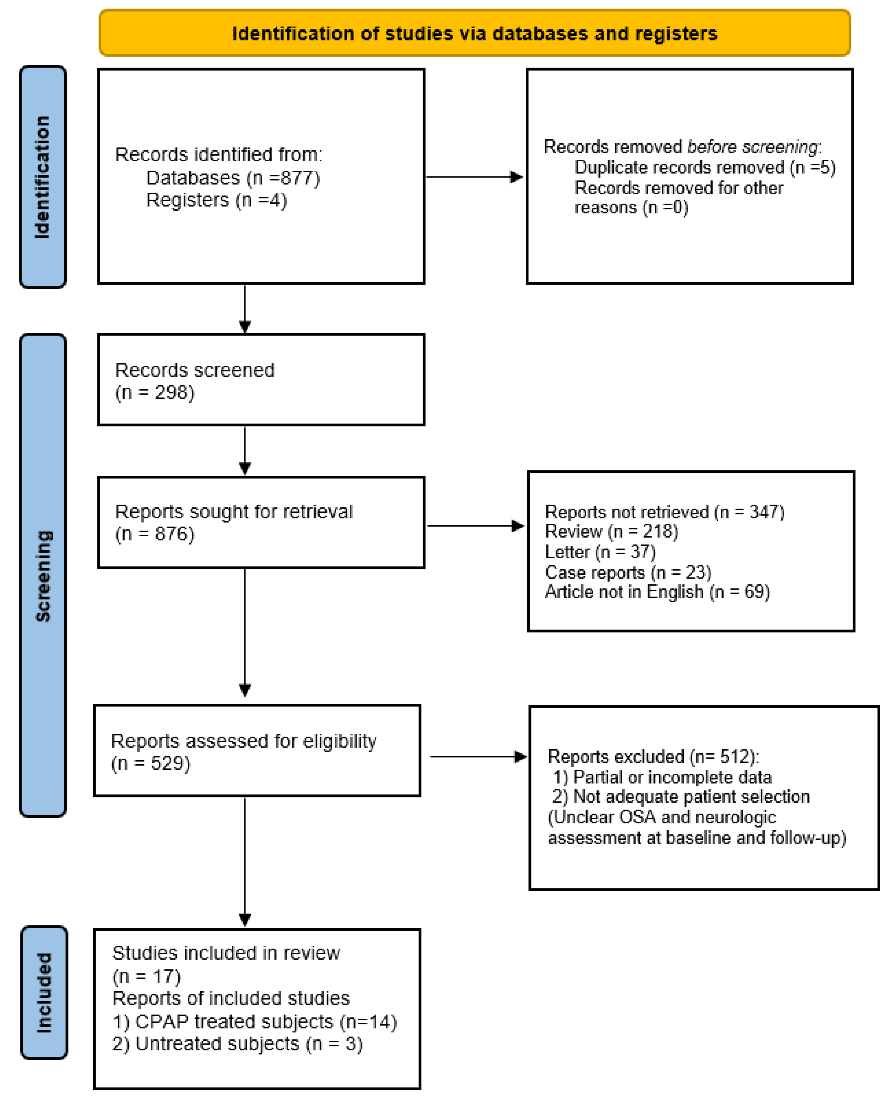
2. Materials and Methods
2.1. Protocol Data Extraction and Outcomes
- Original articles;
- The article was published in the English language;
- The studies included patients with impaired neurocognitive performance undergoing treatment for OSAS;
- The studies reported detailed information on pre-and post-treatment OSAS outcomes, validated questionnaires on neurocognitive performance at baseline and after treatment, and patients’ comorbidities;
- We excluded case reports, editorials, letters to the editor, or reviews from the study.
2.2. Electronic Database Search
3. Results
3.1. Attention and Executive Functions
3.2. Memory and Learning
3.3. General Cognitive Function
3.4. Psychomotor Function
3.5. Patients’ Comorbidities
4. Discussion
Study’s Limitations
5. Conclusions
Author Contributions
Funding
Institutional Review Board Statement
Informed Consent Statement
Conflicts of Interest
References
- Liguori, C.; Mercuri, N.B.; Izzi, F.; Romigi, A.; Cordella, A.; Sancesario, G.; Placidi, F. bstructive Sleep Apnea is Associated With Early but Possibly Modifiable Alzheimer’s Disease Biomarkers Changes. Sleep 2017, 40, zsx011. [Google Scholar] [CrossRef] [Green Version]
- Iannella, G.; Maniaci, A.; Magliulo, G.; Cocuzza, S.; La Mantia, I.; Cammaroto, G.; Greco, A.; Vicini, C. Current challenges in the diagnosis and treatment of obstructive sleep apnea syndrome in the elderly. Pol. Arch. Intern. Med. 2020, 130, 649–654. [Google Scholar] [CrossRef] [Green Version]
- Franklin, K.A.; Lindberg, E. Obstructive sleep apnea is a common disorder in the population—A review on the epidemiology of sleep apnea. J. Thorac. Dis. 2015, 7, 1311–1322. [Google Scholar]
- Durán, J.; Esnaola, S.; Rubio, R.; Iztueta, A. Obstructive sleep apnea-hypopnea and related clinical features in a population-based sample of subjects aged 30 to 70 yr. Am. J. Respir. Crit. Care Med. 2001, 163 Pt 1, 685–689. [Google Scholar] [CrossRef]
- Devita, M.; Montemurro, S.; Argentieri, M.; Dinoi, G.; Malvina, M.; Rusconi, M.L.; Mondini, S. Cognitive and motor reaction times in obstructive sleep apnea syndrome: A study based on computerized measures. Brain Cogn. 2017, 117, 26–32. [Google Scholar] [CrossRef] [PubMed]
- Maniaci, A.; Iannella, G.; Cocuzza, S.; Vicini, C.; Magliulo, G.; Ferlito, S.; Cammaroto, G.; Meccariello, G.; DE Vito, A.; Nicolai, A.; et al. Oxidative Stress and Inflammation Biomarker Expression in Obstructive Sleep Apnea Patients. J. Clin. Med. 2021, 10, 277. [Google Scholar] [CrossRef]
- Iannella, G.; Magliulo, G.; Maniaci, A.; Meccariello, G.; Cocuzza, S.; Cammaroto, G.; Gobbi, R.; Sgarzani, R.; Firinu, F.; Corso, R.M.; et al. Olfactory function in patients with obstructive sleep apnea: A meta-analysis study. Eur. Arch. Otorhinolaryngol. 2021, 278, 883–891. [Google Scholar] [CrossRef]
- Lau, E.Y.Y.; Eskes, G.A.; Morrison, D.L.; Rajda, M.; Spurr, K.F. Executive function in patients with obstructive sleep apnea treated with continuous positive airway pressure. J. Int. Neuropsychol. Soc. 2010, 16, 1077–1088. [Google Scholar] [CrossRef] [Green Version]
- Canessa, N.; Castronovo, V.; Cappa, S.F.; Aloia, M.S.; Mrelli, S.; Falini, A.; Alemanno, F.; Ferini-Strambi, L. Obstructive Sleep Apnea: Brain Structural Changes and Neurocognitive Function before and after Treatment. Am. J. Respir. Crit. Care Med. 2011, 183, 1419–1426. [Google Scholar] [CrossRef]
- Xu, T.; You, D.; Chen, X. Non-surgical treatment of obstructive sleep apnea syndrome. Eur. Arch. Oto-Rhino-Laryngol. 2018, 275, 335–346. [Google Scholar]
- Turner, K.; Zambrelli, E.; Lavolpe, S.; Baldi, C.; Furia, F.; Canevini, M.P. Obstructive sleep apnea: Neurocognitive and behavioral functions before and after treatment. Funct. Neurol. 2019, 34, 71–78. [Google Scholar]
- Joo, E.Y.; Tae, W.S.; Lee, M.J.; Kang, J.W.; Park, H.S.; Lee, J.Y.; Suh, M.; Hong, S.B. Reduced brain gray matter concentration in patients with obstructive sleep apnea syndrome. Sleep 2010, 33, 235–241. [Google Scholar] [CrossRef] [Green Version]
- Iannella, G.; Magliulo, G.; Di Luca, M.; De Vito, A.; Meccariello, G.; Cammaroto, G.; Pelucchi, S.; Bonsembiante, A.; Maniaci, A.; Vicini, C. Lateral pharyngoplasty techniques for obstructive sleep apnea syndrome: A comparative experimental stress test of two different techniques. Eur. Arch. Otorhinolaryngol. 2020, 277, 1793–1800. [Google Scholar] [CrossRef] [PubMed]
- Di Luca, M.; Iannella, G.; Montevecchi, F.; Magliulo, G.; De Vito, A.; Cocuzza, S.; Maniaci, A.; Meccariello, G.; Cammaroto, G.; Sgarzani, R.; et al. Use of the transoral robotic surgery to treat patients with recurrent lingual tonsillitis. Int. J. Med. Robot. 2020, 16, e2106. [Google Scholar] [CrossRef] [PubMed]
- Carvalho, F.R.; Lentini-Oliveira, D.A.; Prado, L.B.; Prado, G.F.; Carvalho, L.B. Oral appliances and functional orthopaedic appliances for obstructive sleep apnoea in children. Cochrane Database Syst. Rev. 2016, 10, CD005520. [Google Scholar] [CrossRef] [PubMed]
- Venekamp, R.P.; Hearne, B.J.; Chandrasekharan, D.; Blackshaw, H.; Lim, J.; Schilder, A.G. Tonsillectomy or adenotonsillectomy versus non-surgical management for obstructive sleep-disordered breathing in children. Cochrane Database Syst Rev. 2015, 10, CD011165. [Google Scholar] [CrossRef]
- Rosenzweig, I.; Glasser, M.; Crum, W.R.; Kempton, M.J.; Milosevic, M.; McMillian, A.; Leschziner, G.D.; Kumari, V.; Goadsby, P.; Sinonds, A.K..; et al. Changes in Neurocognitive Architecture in Patients with Obstructive Sleep Apnea Treated with Continuous Positive Airway Pressure. EBioMedicine 2016, 7, 221–229. [Google Scholar] [CrossRef] [PubMed] [Green Version]
- Lim, W.; Bardwell, W.A.; Loredo, J.S.; Kim, E.J.; Ancoli-Israel, S.; Morgan, E.E.; Heaton, R.K.; Dimsdale, J.E. Neuropsychological effects of 2-week continuous positive airway pressure treatment and supplemental oxygen in patients with obstructive sleep apnea: A randomized placebo-controlled study. J. Clin. Sleep Med. 2007, 3, 380–386. [Google Scholar] [CrossRef] [Green Version]
- Thompson, M.; Tiwari, A.; Fu, R.; Moe, E.; Buckley, D.I. A Framework to Facilitate the Use of Systematic Reviews and Meta-Analyses in the Design of Primary Research Studies; Agency for Healthcare Research and Quality (US): Rockville, MD, USA, 2012.
- Castronovo, V.; Scifo, P.; Castellano, A.; Aloia, M.S.; Iadanza, A.; Marelli, S.; Cappa, S.F.; Strambi, L.F.; Falini, A. White Matter Integrity in Obstructive Sleep Apnea before and after treatment. Sleep 2014, 37, 1465–1475. [Google Scholar] [CrossRef] [Green Version]
- Kanbay, A.; Demir, N.C.; Tutar, N.; Kostek, O.; Simsek, Z.O.; Buyucoglan, H.; Demir, R.; Parrino, L. The effect of CPAP therapy on insulin-like growth factor and cognitive functions in obstructive sleep apnea patients: CPAP therapy and cognitive function. Clin. Respir. J. 2017, 11, 506–513. [Google Scholar] [CrossRef] [PubMed]
- Ng, S.S.S.; Chan, T.; To, K.; Chan, K.K.P.; Ngai, J.; Tung, A.; Ko, F.W.S.; Hui, D.S.C. Prevalence of Obstructive Sleep Apnea Syndrome and CPAP Adherence in the Elderly Chinese Population. PLoS ONE 2015, 10, e0119829. [Google Scholar] [CrossRef] [PubMed]
- Barnes, M.; McEvoy, R.D.; Banks, S.; Tarquinio, N.; Murray, C.G.; Vowles, N.; Pierce, R.J. Efficacy of Positive Airway Pressure and Oral Appliance in Mild to Moderate Obstructive Sleep Apnea. Am. J. Respir. Crit. Care Med. 2004, 170, 656–664. [Google Scholar] [CrossRef]
- Torelli, F.; Moscufo, N.; Garreffa, G.; Placidi, F.; Romigi, A.; Zannino, S.; Bozzali, M.; Fasano, F.; Giulietti, G.; Djonlagic, I.; et al. Cognitive profile and brain morphological changes in obstructive sleep apnea. NeuroImage 2011, 54, 787–793. [Google Scholar] [CrossRef] [PubMed] [Green Version]
- Lusic Kalcina, L.; Pavlinac Dodig, I.; Pecotic, R.; Valic, M.; Dogas, Z. Psychomotor Performance in Patients with Obstructive Sleep Apnea Syndrome. Nat. Sci. Sleep 2020, 12, 183–195. [Google Scholar] [CrossRef] [PubMed] [Green Version]
- Kushida, C.A.; Nichols, D.A.; Holmes, T.H.; Quan, S.F.; Walsh, J.K.; Gottlieb, D.J.; Simo, R.D., Jr.; Guilleminault, C.; White, D.P.; Goodwin, J.L.; et al. Effects of Continuous Positive Airway Pressure on Neurocognitive Function in Obstructive Sleep Apnea Patients: The Apnea Positive Pressure Long-term Efficacy Study (APPLES). Sleep 2012, 35, 1593–1602. [Google Scholar] [PubMed]
- Buratti, L.; Viticchi, G.; Falsetti, L.; Cagnetti, C.; Luzzi, S.; Bartolini, M.; Provinciali, L.; Silvestrini, M. Vascular impairment in Alzheimer’s disease: The role of obstructive sleep apnea. J. Alzheimers Dis. 2014, 38, 445–453. [Google Scholar] [CrossRef] [PubMed] [Green Version]
- Kheirandish-Gozal, L.; Gozal, D. Obstructive Sleep Apnea and Inflammation: Proof of Concept Based on Two Illustrative Cytokines. Int. J. Mol. Sci. 2019, 20, 459. [Google Scholar] [CrossRef] [Green Version]
- Testa, D.; Carotenuto, M.; Precenzano, F.; Russo, A.; Donadio, A.; Marcuccio, G.; Motta, G. Evaluation of neurocognitive abilities in children affected by obstructive sleep apnea syndrome before and after adenotonsillectomy. Acta Otorhinolaryngol. Ital. 2020, 40, 122–132. [Google Scholar] [CrossRef] [PubMed]
- Aoki, K.; Matsuo, M.; Takahashi, M.; Murakami, J.; Aoki, Y.; Aoki, N.; Mizumoto, H.; Namikawa, A.; Hara, H.; Miyagawa, M.; et al. Association of sleep-disordered breathing with decreased cognitive function among patients with dementia. J. Sleep Res. 2014, 23, 517–523. [Google Scholar] [CrossRef] [Green Version]
- O’Hara, R.; Schröder, C.M.; Kraemer, H.C.; Kryla, N.; Cao, C.; Miller, E.; Schatzberg, A.F.; Yesavage, J.A.; Murphy, G.M. Nocturnal sleep apnea/hypopnea is associated with lower memory performance in APOE epsilon4 carriers. Neurology 2005, 65, 642–644. [Google Scholar] [CrossRef]
- Bardwell, W.A.; Berry, C.C.; Ancoli-Israel, S.; Dimsdale, J.E. Psychological correlates of sleep apnea. J. Psychosom. Res. 1999, 47, 583–596. [Google Scholar] [CrossRef]
- Beebe, D.W.; Gozal, D. Obstructive sleep apnea and the prefrontal cortex: Towards a comprehensive model linking nocturnal upper airway obstruction to daytime cognitive and behavioral deficits. J. Sleep Res. 2002, 11, 1–16. [Google Scholar] [CrossRef] [PubMed]
- Rouleau, I.; Décary, A.; Chicoine, A.J.; Montplaisir, J. Procedural skill learning in obstructive sleep apnea syndrome. Sleep 2002, 25, 401–411. [Google Scholar] [CrossRef] [PubMed]
- Salorio, C.F.; White, D.A.; Piccirillo, J.; Duntley, S.P.; Uhles, M.L. Learning, memory, and executive control in individuals with obstructive sleep apnea syndrome. J. Clin. Exp. Neuropsychol. 2002, 24, 93–100. [Google Scholar] [CrossRef] [PubMed]
- Gozal, D. CrossTalk proposal: The intermittent hypoxia attending severe obstructive sleep apnoea does lead to alterations in brain structure and function. J. Physiol. 2013, 591, 379–381. [Google Scholar] [CrossRef] [PubMed]
- Wang, W.H.; He, G.P.; Xiao, X.P.; Gu, C.; Chen, H.Y. Relationship between brain-derived neurotrophic factor and cognitive function of obstructive sleep apnea/hypopnea syndrome patients. Asian Pac. J. Trop. Med. 2012, 5, 906–910. [Google Scholar] [CrossRef] [Green Version]
- Werli, K.S.; Otuyama, L.J.; Bertolucchi, P.H.; Rizzi, C.F.; Guillemiault, C.; Tufik, S.; Poyares, D. Neurocognitive function in patients with residual excessive sleepiness from obstructive sleep apnea: A prospective, controlled study. Sleep Med. 2016, 26, 6–11. [Google Scholar] [CrossRef]
- Pace, A.; Iannella, G.; Rossetti, V.; Visconti, I.C.; Gulotta, G.; Cavaliere, C.; De Vito, A.; Maniaci, A.; Cocuzza, S.; Magliulo, G.; et al. Diagnosis of Obstructive Sleep Apnea in Patients with Allergic and Non-Allergic Rhinitis. Medicina 2020, 56, 454. [Google Scholar] [CrossRef]
- Cocuzza, S.; Marino, S.; Gulino, A.; Pustorino, E.; Murabito, P.; Maniaci, A.; Sabino, L.; Taibi, R.; Di Luca, M.; Falsaperla, R. ENT involvement and orobuccal movements’ disorders in Pandas patients: Assessment and rehabilitations tools. Eur. Rev. Med. Pharmacol. Sci. 2019, 23, 4110–4117. [Google Scholar] [CrossRef]
- Lal, C.; Di Bartolo, M.M.; Kumbhare, S.; Strange, C.; Joseph, J.E. Impact of obstructive sleep apnea syndrome on cognition in early postmenopausal women. Sleep Breath. 2016, 20, 621–626. [Google Scholar] [CrossRef]


| Authors | Study Design | Sample | Control Group | Age | Gender | Treatment | Sleep Parameters Pre vs. Post Treatment (mean ± SD) | Comorbidities | Questionnaire | Outcomes Pre | Outcomes Post | p -Value | Follow Up |
|---|---|---|---|---|---|---|---|---|---|---|---|---|---|
| Ng, S.S. et al., 2015 | Prospective controlled | 30 OSA | - | 73.9 ± 7.5 | 109 M, 125 F | CPAP | AHI: 16.8 ± 14.2 Nadir: 79 ± 14 | Hypertension, diabete mellitus, cardiovascular disorders | Digit span | 15.2 ± 3.08 | 15.4 ± 4.02 | p = 0.285 | 12 months |
| Digit Symbol | 30.4 ± 12.2 | 35.7 ± 15.1 | p < 0.001 | ||||||||||
| Stroop colour | 60.2 ± 17.7 | 65 ± 17.6 | p = 0.001 | ||||||||||
| Kanbay, A. et al., 2017 | Prospective controlled | 33 OSA | 17 * | 51 ± 9 vs. 47 ± 6 | 26 M 25 F | CPAP | AHI: 45.3 ± 30.9 | Hypertension, cardiovascular disorders, asthma | MMSE | 23.5 ± 3.6 vs. 28.1 ± 1.4 | 28.1 ± 1.6 vs. 28.1 ± 1.6 | p = 0.001 | 3 months |
| Werli, K.S. et al., 2016 | Prospective controlled | 15 OSA RES | 15 ** | 51.0 ± 8.4 vs. 51.8 ± 8.2 | 19 M 11 F | CPAP | ESS: 15 ± 2.5 AHI: 56.18 ± 27.55 meanSO2: 93.58 ± 2.93 | - | WCST categories | - | 1.6 ± 1.4 vs. 3 ± 1.4 | p = 0.04 | 12 months |
| FAS | - | 25.5 ± 5.3 vs. 30.7 ± 7.32 | p = 0.04 | ||||||||||
| Torelli, F. et al., 2011 | Prospective controlled | 16 OSA | 14 * | 55.8 ± 6.7 vs. 57.6 ± 5.1 | 22 M 8 F | untreated | AHI: 52.2 ± 2.6 meanSO2: 92.0 ± 3.1 | Hypertension, Diabetes, Hypercholesterolemia, Smoking | MMSE | 29.5 ± 0.8 vs. 29.6 ± 0.6 | -------------- | p = 0.60 | 12 months |
| RAVL | 40.9 ± 5.4 vs. 45.9 ± 6.4 | - | p = 0.026 | ||||||||||
| Digit span | 5.6 ± 0.6 vs. 5.9 ± 0.4 | - | p = 0.23 | ||||||||||
| Visual memory | 20.4 ± 1.2 vs. 19.7 ± 1.7 | - | p = 0.22 | ||||||||||
| Copy drawings | 9.9 ± 2.1 vs. 10.1 ± 1.2 | - | p = 0.72 | ||||||||||
| ROCF | 30.4 ± 6.1 vs. 33.9 ± 3.0 | - | p = 0.06 | ||||||||||
| RAPM | 29.7 ± 3.9 vs. 31.7 ± 2.6 | - | p = 0.11 | ||||||||||
| SVFT | 39.7 ± 0.5 vs. 39.9 ± 0.3 | - | p = 0.11 | ||||||||||
| PVFT | 26.7 ± 8.8 vs. 28.5 ± 6.6 | - | p = 0.56 | ||||||||||
| Stroop Test | 40.3 ± 13.1 vs. 33.9 ± 5.0 | - | p = 0.10 | ||||||||||
| Lau, E.Y. et al., 2010 | Prospective controlled | 37 OSA | 27 * | 57.9 ± 9.5 vs. 56.7c10.5 | 22 M 15 F | CPAP | ESS: 14.4 ± 5.2 vs. 8.3 ± 4.5 RDI: 42.2 ± 2.9 vs. 1.7 ± 1.5 meanSO2: 93.7 ± 3.5 vs. 95.7 ± 1.6 | - | WAIS-R Vocabulary | - | 56.6 ± 9.4 vs. 61.3 ± 5.9 | p = 0.017 | 3 months |
| WAIS-R Block Design | - | 30.8 ± 8.3 vs. 33.8 ± 10.1 | p = 0.193 | ||||||||||
| Digit Span | - | 16.1 ± 4.0 vs. 16.9 ± 0.2 | p = 0.474 | ||||||||||
| Stroop Color-Word | - | 39.0 ± 7.7 vs. 42.5 ± 7.8 | p = 0.082 | ||||||||||
| WCTS | - | 4.8 ± 1.6 vs. 5.6 ± 1.1 | p = 0.028 | ||||||||||
| Rey-O Recall | - | 17.3 ± 6.1 vs. 19.2 ± 6.4 | p = 0.229 | ||||||||||
| Akmal, M.K. et al., 2013 | Cross-sectional study | 20 OSA | - | 43.6 ± 4.12 | 14 M vs. 6 F | CPAP | - | Psychiatric comorbidities | EXIT25 | 24.5 ± 5.82 | 39.8 ± 5.41 | p < 0.001 | 1 months |
| Edwards, C. et al., 2015 | Cross-sectional study | 228 OSA | - | 52 ± 15.57 | 243 M vs. 183 F | CPAP | AHI: 46.7 ± 27.4 vs. 6.5 ± 1.6 | - | PHQ-9 Depression Scale | 11.3 ± 6.1 | 3.7 ± 2.9 | p < 0.001 | 3 months |
| Barnes et al., 2004 | Randomised controlled trial | 114 OSA | - | 47 ± 0.9 | 91 M vs. 23 F | CPAP vs. MAS or Placebo | AHI: 22.2 ± 1.5 ESS: 10.2 ± 0.5 | Hypertension | Stroop color association test | - | 9.3 ± 0.9 vs. 10.3 ± 0.9 vs. 9.2 ± 0.9 | p < 0.001 | 3 months |
| Digit span backward | - | 4.6 ± 0.1 vs. 4.6 ± 0.1 vs. 4.8 ± 0.1 | p < 0.05 | ||||||||||
| Digit symbol substitution task | - | 47.3 ± 0.4 vs. 47.5 ± 0.4 vs. 46.8 ± 0.4 | p < 0.05 | ||||||||||
| Castronovo et al., 2009 | Prospective controlled | 17 OSA | 15 * | 43.93 ± 7.8 | 32 M | CPA | AHI: 61.35 ± 97.7 vs. 9.8 ± 1.6 NadirSO2: 72.45 ± 7.69 vs. 91.2 ± 1.3 ESS: 12.0 ± 5.18 vs. 3.08 ± 2.24 | - | Rey’s List (learning) | 58.0 ± 7.1 vs. 48.54 ± 10.15 | 57.54 ± 8.36 vs. 48.54 ± 10.15 | p < 0.001 | 3 months |
| Corsi | 5.23 ± 1.09 vs. 6.53 ± 0.91 | 6.31 ± 0.85 vs. 6.53 ± 0.91 | p = 0.002 | ||||||||||
| TMB | 82.15 ± 26.16 vs. 59.4 ± 14.16 | 78.85 ± 23.42 vs. 59.4 ± 14.16 | p < 0.001 | ||||||||||
| Stroop | 5.08 ± 3.32 vs. 0.73 ± 1.03 | 0.83 ± 1.53 vs. 0.73 ± 1.03 | p < 0.001 | ||||||||||
| Wang, W.H. et al., 2012 | Randomised controlled trial | 28 OSA | 14 * | 44.93 ± 2.98 | 42 M | untreated | AHI: 49.63 ± 28.56 | - | MoCA | 24.04 ± 1.75 vs. 28.57 ± 1.09 | - | p < 0.01 | |
| Kushida, C.A. et al., 2012 | Randomised controlled trial | 1098 OSA | - | 52.2 ± 12.2 | 719 M 379 F | 442 activeCPAP vs. 401sham CPAP | AHI: 30.7 ± 24.9 NadirSO2: 81 ± 7.6 | - | PFN-TOTL | 23.32 vs. 23.08 | 23.48 vs. 23.01 | p = 0.2103 | 6 months |
| BSRT-SR | 49.72 vs. 48.86 | 54.09 vs. 54.28 | p = 0.7569 | ||||||||||
| SWMT-OMD | 0.035 vs. −0.074 | 0.072 vs. 0.018 | p = 0.2254 | ||||||||||
| Rosenzweig, I. et al., 2016 | Randomised controlled trial | 68 OSA | 35 * | 47.6 ± 11.1 | 80 M 23 F | CPAP | AHI: 36.58 ± 27.15 | - | ACE-R | 90.55 ± 1.11 vs. 94.91 ± 0.99 | 91.86 ± 2.44 vs. 90.70 ± 1.85 | p = 0.220 | 1months |
| Immediate LM | 36.36 ± 1.32 vs. 47.06 ± 1.84 | 44.43 ± 1.99 vs. 41.70 ± 2.28 | p = 0.338 | ||||||||||
| TMB | 62.02 ± 3.65 vs. 41.23 ± 2.0 | 51.05 ± 3.68 vs. 61.53 ± 4.81 | p = 0.0017 | ||||||||||
| TMA | 27.34 ± 1.04 vs. 24.12 ± 1.18 | 24.26 ± 1.28 vs. 28.02 ± 1.56 | p = 0.937 | ||||||||||
| Lim, W. et al., 2007 | Randomised controlled trial | 46 OSA | - | 46.7 ± 2.4 | - | CPAP-Oxygen-PlaceboCPAP | AHI: 63.5 ± 7.8 ESS: 11.2 ± 1.0 MeanSO2: 93.1 ± 1.1 | Hypertension | Letter/Number Sequencing | 11.0 vs. 11.1 vs. 11.7 | 11.9 vs. 11.7 vs. 12.9 | p = 0.005 | 2 weeks |
| Digit Span Total | 18.6 vs. 19.3 vs. 21.2 | 26.4 vs. 21.3 vs. 22.5 | p = 0.091 | ||||||||||
| Digit Vigilance | 5.6 vs. 8.9 vs. 14.1 | 7.2 vs. 7.3 vs. 10.6 | p = 0.196 | ||||||||||
| Stroop Color-Word | 37.7 vs. 40.1 vs. 37.9 | 37.3 vs. 44.2 vs. 41.9 | p = 0.007 | ||||||||||
| Turner et al., 2019 | Prospective cohort study | 16OSA | - | 36–80 | 15 M 1 F | CPAP | ESS: 9.31 ± 5.87 vs. 5.69 ± 3.44 | Epilepsy | Digit Span Forwad | 5.62 ± 1.02 | 5.81 ± 1.05 | p = 0.61 | 3 months |
| Digit Span Backward | 4.13 ± 0.7 | 05.12 ± 1.02 | p = 0.004 | ||||||||||
| ROCF | 19.08 ± 7.32 | 21.09 ± 7.69 | p = 0.48 | ||||||||||
| Corsi Span | 4.94 ± 0.9 | 5.6 ± 0.6 | p = 0.02 | ||||||||||
| Short story test | 10.19 ± 3.72 | 13.84 ± 2.89 | p = 0.004 | ||||||||||
| Attentional Matrices | 54.0 ± 6.0 | 55.06 ± 3.85 | p = 0.55 | ||||||||||
| Canessa, N. et al., 2010 | Prospective controlled | 17 OSA | 15 * | 44 ± 7.63 | 32 M | CPAP | AHI: 55.83 ± 19.08 vs. 2.5 ± 2.4 MeanSO2: 70.41 ± 9.13 vs. 91.4 ± 1.9 | Hypertension | MMSE | 29.35 ± 1.05 vs. 30.00 | 29.75 ± 0.57 | Ns | 3 months |
| Raven | 31.70 ± 3.90 vs. 34.6 ± 1.29 | 33.25 ± 2.46 | p = 0.03 | ||||||||||
| Digit Span forward | 5.58 ± 1.00 vs. 6.93 ± 0.70 | 6.56 ± 0.81 | p = 084 | ||||||||||
| Rey-list recall | 48.70 ± 9.67 vs. 13 ± 1.96 | 58.18 ± 7.92 | p < 0.001 | ||||||||||
| Liguori, C. et al., 2017 | Prospective controlled | 25 OSA vs. 10 OSA-CPAP | 15 * | 67.96 ± 7.92 | 26 F 14 M | CPAP | AHI: 36.34 ± 11.42 vs. 3.14 ± 1.54 MeanSO2: 92.37 ± 1.97 vs. 95 ± 0.82 | Hypertension | I-RAVL | - | 42.58 ± 2.50 vs. 46.7 ± 1.49 vs. 49.07 ± 3.22 | p < 0.001 | 12 months |
| Raven | - | 26.73 ± 69.33 vs. 33 ± 1.41 vs. 33.07 ± 0.80 | p < 0.0001 | ||||||||||
| Stroop color/word test | - | 33.96 ± 4.15 vs. 29.2 ± 1.5 vs. 26.57 ± 2.22 | p < 0.0001 | ||||||||||
| Lusic Kalcina, L. et al., 2020 | Prospective controlled | 103 OSA | 103 * | 57.14 ± 11.31 | 206 M | untreated | ESS: 8.65 ± 4.5 AHI: 45.02 ± 14.09 | Hypertension, diabetes, cardiovascular disorders, depression, arthritis, thyroid disease | CRD11-EB | 38.8 ± 19.3 vs. 33.3 ± 14.1 | - | p = 0.028 | |
| CRD311-EB | 5.2 ± 1.8 vs. 4.5 ± 1.3 | - | p = 0.003 | ||||||||||
| CRD411-EB | 25.1 ± 17.9 vs. 20.3 ± 11.1 | - | p = 0.038 | ||||||||||
| Questionnaire | Features |
|---|---|
| Digit Span | Measures cognitive attention abilities, working memory (central executive), and inhibition. Participants are presented with a random series of digits and are asked to repeat them in either the order presented (forward span) or in reverse order (backward span) |
| Digit Symbol | The test assesses brain damage, dementia, and depression, consisting of digit-symbol pairs followed by a list of digits. |
| Stroop tests | The test is used to examine the effects of interference on reading ability. Contains three parts: word page, color page, and word-color page, each with five columns containing 20 items. The subject’s task is to look at each sheet and move down the columns, reading words or naming the ink colors as quickly as possible, within a given time limit (45 s). |
| MMSE | The test included 11 questions in five categories as follows: orientation, registration memory, attention, and calculation, recall memory, and language. |
| WCSTcategories | This test evaluates the following functions: formation of concepts and problem solving, mental flexibility, abstraction-reasoning, and strategizing. |
| FAS | Test that evaluates the capacity of evoking words (under delimited conditions) and problem-solving strategies. The outcome variable is the number of words remembered. |
| RAVL | It is a list of 15 words read to the subject five times. Measures immediate memory, learning efficiency, interference effects, and recall after short and long periods. |
| Visual memory | Subjects are required to view a simple figure for 3 s and then recognize it in a multiple-choice condition to evaluate short memory. |
| Copy drawing | This task requires reproducing a geometrical figure both by freehand and by joining landmarks already traced on the sheet to evaluate construtional praxia. |
| ROCF | In this test, the subject is asked to reproduce a bidimensional complex figure from memory without forewarning, 15 min after copy, to evaluate short and long memory |
| RAPM | It is a set of 3 subtests (labeled A, Ab, and B) to evaluate non-verbal intelligence, visual processing speed, cognitive speed, and flexibility. It consists in choosing from a set of distractors the item logically missing in a given visual/spatial set. |
| SVFT | Subjects have to produce as many words as they can that fall into three semantic categories, in a time limit of 1 min per sub-test, to evaluate lenguage. |
| PVFT | Subjects have to produce as many words as they can, beginning with a given letter (A, F, S), in a time limit of 1 min per sub-test, to evaluate executive function. |
| WAIS-R vocabulary | Twelve vocabulary words were presented, and participants were asked to define this word. |
| WAIS-R block design | Determine the clinical value of measuring visuospatial abilities. The patient use hand movements to rearrange blocks with various color patterns on different sides to match a pattern. |
| Stroop color-word | The test measures selective and focused attention, cognitive flexibility, and inhibition. |
| WCTS | Test to examine the patient’s frontal functions; used to evaluate flexibility in the choice of problem-solving strategies and used to evaluate the inability to abstraction as well as perseveration. |
| Rey-O Recall | Examinees are asked to reproduce a complicated line drawing, first by copying it freehand (recognition) and then drawing from memory (recall). |
| EXIT 25 | The test consists of 25 items to assess executive functions in people with normal cognition or impairment to identify specific subtypes of mild cognitive impairment and the risk of dementia conversion. |
| PHQ9 depression scale | It is an instrument for making criteria-based diagnoses of depression. Higher PHQ-9 scores are associated with decreased functional status and increased symptom-related difficulties, sick days, and healthcare utilization. |
| Corsi | Test to assess visuospatial short-term memory. |
| TMB | Assesses executive abilities, including setting-shifting and mental flexibility. |
| MoCA | The test evaluated executive function, naming, attention, calculation, language, abstraction, memory, and orientation. |
| PFN-TOLT | The test assesses attention and psychomotor function and comprises the total time for the participant to scan, locate, and connect numbers in sequence. |
| BSRT-SR | The test assesses verbal learning and memory and consists of the total words recalled across six selective reminding trials. |
| SWMT-OMD | The test assesses an executive and frontal-lobe function component by requiring the participant to compare the spatial position of a stimulus with its position on a previous trial (n-backtest), pressing one button if the spatial position was the same as that on the previous trial or a second button if it differed. |
| ACE-R | Provides evaluation of six cognitive domains: orientation, attention, memory, verbal fluency, language, and visuospatial ability. It is useful for detecting dementia and mild cognitive impairment score. |
| Immediate LM | This test assessed the patient’s ability to remember two short stories presented orally, and it is a measure of verbal memory. |
| TMA | The test measures visual attention and processing speed. |
| Letter/number sequencing | It is a test that measures an individual’s short-term memory skills in processing and re-sequence information. |
| Digit Vigilance | It measures vigilance during rapid visual tracking and accurate selection of target stimuli. It focuses on alertness and vigilance while placing minimal demands on two other components of attention: selectivity and capacity. |
| Corsi span | It is a test to assess a visuospatial memory. |
| Short story test | It assesses long-term verbal memory: a short story is read to the subject with the instruction to repeat, immediately afterward, everything they remember; then, the story is read again. After 10 min, the patient is asked to repeat the story once again. |
| Attentional matrices | Test to assess attention involves the use of rows of numbers randomly interspersed with a designated target number or numbers; the patient is instructed to cross out all target numbers in three matrices («5» in I, «2–6» in II, «1–4–9» in III), arranged in a random sequence, within a time limit of 45 s. |
| Raven | It is a non-verbal test used to measure one’s ability to use reasoning and logical ability. |
| CRD TEST | The chronometric instrument can measure: speed of solving simple arithmetic operations (CDR11); the speed of perception to a visual stimulus (CRD311); the speed of complex psycho-motor limbs coordination (CRD411). |
Publisher’s Note: MDPI stays neutral with regard to jurisdictional claims in published maps and institutional affiliations. |
© 2021 by the authors. Licensee MDPI, Basel, Switzerland. This article is an open access article distributed under the terms and conditions of the Creative Commons Attribution (CC BY) license (https://creativecommons.org/licenses/by/4.0/).
Share and Cite
Pollicina, I.; Maniaci, A.; Lechien, J.R.; Iannella, G.; Vicini, C.; Cammaroto, G.; Cannavicci, A.; Magliulo, G.; Pace, A.; Cocuzza, S.; et al. Neurocognitive Performance Improvement after Obstructive Sleep Apnea Treatment: State of the Art. Behav. Sci. 2021, 11, 180. https://0-doi-org.brum.beds.ac.uk/10.3390/bs11120180
Pollicina I, Maniaci A, Lechien JR, Iannella G, Vicini C, Cammaroto G, Cannavicci A, Magliulo G, Pace A, Cocuzza S, et al. Neurocognitive Performance Improvement after Obstructive Sleep Apnea Treatment: State of the Art. Behavioral Sciences. 2021; 11(12):180. https://0-doi-org.brum.beds.ac.uk/10.3390/bs11120180
Chicago/Turabian StylePollicina, Isabella, Antonino Maniaci, Jerome R. Lechien, Giannicola Iannella, Claudio Vicini, Giovanni Cammaroto, Angelo Cannavicci, Giuseppe Magliulo, Annalisa Pace, Salvatore Cocuzza, and et al. 2021. "Neurocognitive Performance Improvement after Obstructive Sleep Apnea Treatment: State of the Art" Behavioral Sciences 11, no. 12: 180. https://0-doi-org.brum.beds.ac.uk/10.3390/bs11120180