Review

Influence of Fibroblasts on Mammary Gland Development, Breast Cancer Microenvironment Remodeling, and Cancer Cell Dissemination

1
Department of Public Health, University of Naples Federico II, 80131 Naples, Italy
2
Department of Experimental and Clinical Medicine, University “Magna Graecia” of Catanzaro, 88100 Catanzaro, Italy
3
Department of Molecular Medicine and Medical Biotechnology, University of Naples Federico II, 80131 Naples, Italy
4
Anatomic Pathology Unit, Department of Advanced Biomedical Sciences, School of Medicine, University of Naples Federico II, 80131 Naples, Italy
5
Department of General, Oncological, Bariatric and Endocrine-Metabolic Surgery, University of Naples Federico II, 80131 Naples, Italy
6
Department of Clinical Medicine and Surgery, University of Naples Federico II, 80131 Naples, Italy
*
Authors to whom correspondence should be addressed.
Submission received: 20 May 2020 / Revised: 20 June 2020 / Accepted: 23 June 2020 / Published: 26 June 2020
(This article belongs to the Special Issue A Special Issue on Tumor Stroma )

Abstract

:
The stromal microenvironment regulates mammary gland development and tumorigenesis. In normal mammary glands, the stromal microenvironment encompasses the ducts and contains fibroblasts, the main regulators of branching morphogenesis. Understanding the way fibroblast signaling pathways regulate mammary gland development may offer insights into the mechanisms of breast cancer (BC) biology. In fact, the unregulated mammary fibroblast signaling pathways, associated with alterations in extracellular matrix (ECM) remodeling and branching morphogenesis, drive breast cancer microenvironment (BCM) remodeling and cancer growth. The BCM comprises a very heterogeneous tissue containing non-cancer stromal cells, namely, breast cancer-associated fibroblasts (BCAFs), which represent most of the tumor mass. Moreover, the different components of the BCM highly interact with cancer cells, thereby generating a tightly intertwined network. In particular, BC cells activate recruited normal fibroblasts in BCAFs, which, in turn, promote BCM remodeling and metastasis. Thus, comparing the roles of normal fibroblasts and BCAFs in the physiological and metastatic processes, could provide a deeper understanding of the signaling pathways regulating BC dissemination. Here, we review the latest literature describing the structure of the mammary gland and the BCM and summarize the influence of epithelial-mesenchymal transition (EpMT) and autophagy in BC dissemination. Finally, we discuss the roles of fibroblasts and BCAFs in mammary gland development and BCM remodeling, respectively.

1. Introduction

The adult mammalian gland develops mainly after birth. It has a complex structure comprising a branching epithelium surrounded by a stromal microenvironment [1]. The interaction between these two compartments, together with a series of growth hormones and growth factors, modulates its development [2,3,4,5]. Notably, in breast cancer (BC), alterations in the signaling pathways regulating the development of the physiological mammary gland contribute to cancer growth [6,7,8,9]. Moreover, mammary stromal fibroblasts, which maintain extracellular matrix (ECM) homeostasis, also modulate morphogenesis in both normal and tumorigenic mammary glands when interacting with epithelial and cancer cells [9,10,11,12].
BCs are heterogeneous solid tumors that can be classified into distinctive histological and molecular subtypes, associated with different invasive capabilities, sites of metastasis, and clinical outcomes [12,13,14,15,16,17]. For instance, BCs can express estrogen receptor (ER), progesterone receptor (PR), and human epidermal growth factor receptor 2 (HER-2), whereas those with the worst prognosis, the so called triple negative breast cancers (TNBCs), lack the expression of the three receptors [12].
Like most solid tumors, BCs are very heterogeneous and abnormal tissues, characterized by a stromal tumor microenvironment (TME) that supports tumor development and dissemination [18,19,20,21]. Major players in the structure of the TME and in the behavior of both stromal and cancerous cells are breast cancer-associated fibroblasts (BCAFs). Indeed, these non-cancerous stromal cells represent up to 80% of the tumor mass [12,15]. Not surprisingly, several studies have demonstrated that the recruitment and activation of BCAFs induce deep changes to the TME, thereby sustaining cancer dissemination [15,18,22,23,24]. Of note, El-Ashry’s group [25] showed that BCAFs and cancer cell aggregates circulate in the peripheral blood of patients with metastatic BC. Consistently, additional evidence has demonstrated that they facilitate metastasis by contributing to the formation of metastatic niches in distant organs, thereby facilitating the metastatic process [25,26]. These findings clearly suggest using BCAFs as key diagnostic biomarkers in metastatic BC [25,26]. Considering that BCAFs originate from normal fibroblasts [12], and that both fibroblast types regulate normal and tumorigenic mammary gland development, we believe that a better understanding of the role of fibroblasts and BCAFs in mammary gland and breast cancer microenvironment (BCM) remodeling could contribute to the development of new therapeutic strategies targeting BC growth. Hence, the aim of this paper is twofold. Firstly, we review the most recent findings on the structure of the mammary gland and BCM, and on the influence of epithelial-mesenchymal transition (EpMT) and autophagy in BC dissemination. Secondly, we compare the roles of fibroblasts and BCAFs in regulating mammary gland development and microenvironment remodeling linked to cancer cell dissemination.

2. Mammary Gland Structure and Development

The parenchyma of the adult female mammary gland is composed of tree-like branching ducts spreading radially from the nipple and terminating in expanded alveolar aggregates, known as lobules [4,27]. The duct wall is constituted by an outer and inner layer (Figure 1A,C) [4,27].
The outer “basal” layer is composed of myoepithelial cells connected directly to the basement membrane and displays smooth muscle-like contractile properties. The inner “luminal” layer, instead, includes cells with significantly different, polarized epithelial features. The myoepithelial cell layer, whose contraction sustains milk secretion, regulates the homeostasis of the basement membrane, which is a thin and dense structure made up of collagen IV, laminin, and proteoglycans [28]. This tissue network is embedded in a stromal microenvironment, which includes the extracellular matrix (ECM), blood and lymphatic vessels, and multiple stromal cell types, including fibroblasts, endothelial cells, adipocytes, immune cells, and mesenchymal stem cells (MSCs) [1,3,27]. The ECM contains fibrillar collagen, proteoglycans, glycoproteins tenascin C (TNC), and fibronectin (FN) [29]. Its main function is to regulate mammary cell pathways through cell-cell interactions and paracrine and mechanical signals [28]. The key fibrillar protein in the ECM [3] is collagen I. Collagen I and collagen bundle alignments are important regulators of mammary gland development and duct outgrowth during puberty [30,31]. However, on mammography, a dense breast tissue is associated with abnormal accumulation of collagen I in the breast stroma—an event highly suggestive of cancer development. Indeed, studies have shown that BC cells frequently originates from dense breast tissues and extended beyond the basement membrane into adjacent tissue [28] (Figure 1B,D).
Although mouse and human mammary glands display structural and endocrine differences, the current knowledge of mammary gland development is based on studies in murine experimental models. The reason is that, being easy to handle, they are particularly suited for studying endocrine regulation and stromal epithelial cell crosstalk [1,3,8,27].
By and large, the mammary gland develops mainly postnatally in puberty, pregnancy, lactation, and involution—stages in which reproductive hormones and growth factors play a decisive role [3,4,5]. At the onset of puberty, the ovarian steroid hormones regulate branching morphogenesis by promoting the extension and branching of mammary ducts (Figure 1C) [5]. Then, at a later stage, the epithelium infiltrates the fat-pad stroma and builds the mammary gland. Thereafter, the gland continues to evolve by undergoing differentiation, and de-differentiation during menstruation, pregnancy, lactation, and involution [32]. Outgrowth of mammary ducts begins at the tips of the ducts, namely, the terminal end buds (TEBs). TEBs are mammary structures characterized by a bulb shape. They develop at the onset of puberty under the influence of ovarian estrogen and the pituitary growth hormone. It is in these structures that proliferating cells differentiate into luminal epithelial cells and basal myoepithelial cells, and luminal cell apoptosis generates the hollow ducts (Figure 1C) [33,34,35]. Their function is to modulate ductal growth throughout the fat pad, while undergoing regular bifurcation events, which ultimately lead to the formation of the mammary ductal tree (Figure 1C) [35]. During pregnancy, intense epithelial cell proliferation leads to further ductal branching and alveolar formation. Subsequently, after parturition, the luminal epithelial cells of the inner layer of the alveoli differentiate into secretory cells, thereby eliciting milk production and secretion through the ducts during lactation [4]. Throughout this process, the nuclear steroid receptors ERα and PR regulate the physiological development of the mammary gland [36]. Indeed, studies on ERα and PR knockout mice show that ERα expression in the mammary epithelium is essential for ductal branching and elongation, whereas PR expression in the epithelium is essential for normal secretory alveolar development [37,38].
Another essential player in milk production is the terminal duct lobular units (TDLUs), that is, clusters of multiple small acini located at the distal ends of the ducts [33,39] (Figure 1A). One of the least static structures within the mammary gland, TDLUs constitute a potential site of origin of BC [39,40]. More specifically, in a normal breast, with the conclusion of childbearing and advancing years, TDLUs undergo an involution process linked to reduced numbers of both acini/TDLU and TDLUs [41,42]. Such involution is correlated with age, menopausal status, and parity [43]. On the other hand, the absence of TDLU involution is correlated with an increased risk of BC [44]. Consistently, evidence shows that BC originates in the TDLUs and advances through a series of molecular events that ultimately lead to invasive and metastatic carcinomas. These include increased epithelial cell proliferation, atypical hyperplasia, and carcinoma in situ [33].

Influence of Fibroblasts on Mammary Gland Development

Research on the role of fibroblasts in the development of the mammary gland is facilitated in mice, because the epithelial branching of the mouse mammary gland continues to develop many weeks after birth [8,9,45]. Two different fibroblast cell populations have been identified in the stroma of the mammary gland: lobular and interlobular fibroblasts [40]. Lobular fibroblasts envelop TDLUs and express high levels of the CD105 marker. Their primary function is to sustain luminal epithelial growth and branching morphogenesis by producing ECM proteins, matrix remodeling enzymes, and several growth factors. Instead, the interlobular fibroblasts express high levels of the CD26 marker and are characterized by an immune-related signature [40]. Conversely, the intralobular fibroblasts are characterized by a myofibroblast-related signature that includes the expression of the transforming growth factor (TGF)-β1, TNC, and α-SMA [40]. Moreover, since their expression profile coincides with that of the tumor stoma, some authors have suggested that these fibroblasts represent a reservoir of BCAFs localized to the TDLU [40].
The presence of aberrantly activated stromal cells, especially myofibroblasts, can favor BC development and sometimes precedes the malignant transformation of epithelial cells in the mammary gland [12,46,47]. This process, named desmoplasia, not only generates a niche for cancer cells, but also produces a hypoxic and acidic microenvironment responsible for compromising chemotherapeutic treatments [21].
Interestingly, scientists have recently turned their attention to examining the underlying mechanisms by which obesity may predispose women to BC. Several studies suggest that obesity sustains myofibroblast differentiation within the mammary adipose tissue, thereby inducing fibrotic remodeling of the microenvironment and BC development [48,49,50]. Consistently, in mammary tissues of obese mice, myofibroblast differentiation increases the activity of the TGF-β1/SMAD3/miR-140 negative-feedback loop. In particular, the activation of TGF-β1 drives SMAD3 activation that, in turn, inhibits miR-140 transcription by binding to the miR-140 locus. This signaling network impedes SMAD3 degradation by miR-140, thus promoting myofibroblast differentiation and fibrosis [1]. Furthermore, mammary stromal vascular fraction from high-fat diet–fed mice has been shown to induce self-renewal and the invasive capability of the non-invasive BC cell line MCF10DCIS [1].
On the other hand, myofibroblasts also regulate the morphogenesis of normal breast epithelial cells. In fact, 3D-collagen I cocultures of non-malignant human breast epithelial cell line HMT-3522 and immortalized human mammary myofibroblasts lead to the formation of acinus-like structures characterized by a central lumen and cell growth arrest [11]. More specifically, myofibroblasts induce epithelial cell differentiation by affecting matrix tension, and by interacting with paracrine signaling activity [11]. Conversely, 3D-collagen I monocultures of HMT-3522 cells drive uncontrolled cell proliferation and morphological disorganization, resembling a pattern of BC in vivo [11].
Stromal fibroblasts also regulate branching morphogenesis and ECM remodeling during the different stages of mammary gland development [51,52,53,54] by interacting with several signaling pathways. For instance, during epithelial branching, normal fibroblasts work alongside paracrine signals, which are mediated by a series of growth factors, including fibroblast growth factors (FGFs), insulin-like growth factor (IGF), hepatocyte growth factor (HGF), and TGF-β1 [55,56] (Figure 2A).
Under normal conditions, the FGF or IGF signaling activity is reduced, stunting epithelial branching. Conversely, when FGF is upregulated, cell polarity is disrupted, thereby inducing cell proliferation, cell survival, and invasion capability. As shown in Figure 2B, this chain of events closely mirrors the molecular processes occurring during BC development [57,58,59]. Low concentrations of TGF-β1 also contribute to mammary ductal morphogenesis mediated by fibroblasts (Figure 2A) [56]. Moreover, FGF signaling, elicited by FGF2 and FGF9, triggers ERK1/ERK2 activation, cell proliferation, and migration in the murine mammary gland. FGF2 is also involved in fibroblast collagen remodeling and modulates fibroblast ECM synthesis. Evidence shows that FGF2 and FGF9 signaling in primary mammary fibroblasts, co-cultured with 3D epithelial organoids, sustains the branching of the mammary epithelium (Figure 2A) [60]. Moreover, the epithelial growth factor receptor (EGFR) pathway also influences mammary gland stroma (Figure 2A) [61]. In brief, the activation of the EGFR pathway in stromal cells, which are localized in front of the invading TEB, stimulates matrix metalloproteinase (MMP)-14 expression and MMP-2 activation—a process leading to ECM remodeling and epithelial branching [61].
Different members of the Sprouty/Spred gene family, which are considered tumor suppressor genes, also participate in mammary branching morphogenesis by interfering with EGFR signaling activity [9,62]. Among these is Spry 1. In vitro and in vivo analysis show that loss of Spry1 function in mammary fibroblasts drives upregulation of EGFR signaling, following amphiregulin and TGF-α stimulation, consequently increasing ECM remodeling and mammary epithelium branching in pubertal mice. Another member of this gene family participating in mammary branching morphogenesis is Spry2. Spry 2, expressed in the luminal epithelial, myoepithelial, and stromal cells of the mouse mammary gland, also affects FGF signaling associated with physiological epithelial morphogenesis (Figure 2) [63] Conversely, gain of Spry2 function in mammary fibroblasts reduces EGFR signaling associated with stunted epithelial branching [9]. Using quantitative phosphoproteomic analysis, Shi et al. showed that deletion of Spry1, 2, and 4 in triple-knockout (SpryKO) mouse mammary fibroblasts, triggers a BCAF-like phenotype associated with alteration in ErbB, insulin, vascular endothelial growth factor (VEGF), focal adhesion, and mTOR signaling pathways [64]. Therefore, the loss of fine regulation of Spry/EGFR signaling alters mammary gland development and drives BC development by increasing the invasive capability of tumor cells [9,64] (Figure 2B). This evidence clearly suggests that the components of the EGFR family constitute potential therapeutic targets [65].
Platelet derived growth factor receptor (PDGFR) α activity induces activation of quiescent fibroblasts and ECM remodeling [66]. Indeed, the constitutive activation of PDGFRα in mice mammary fibroblasts hinders the invasion and branching of the ductal epithelium during post-pubertal development. However, it does not alter the epithelial differentiation program, and elicits fibrosis and a pro-tumorigenic stromal microenvironment (Figure 2) [67]. In addition, PDGFRα+ fibroblasts from involuting mice mammary glands, display an activated phenotype, COX/prostaglandin E2 (PGE2) dependent activity, and pro-tumorigenic and immunosuppressive activity in an immunocompetent and orthotopic tumor transplant model [68]. Moreover, PDGFRα+ mammary fibroblasts from involuting mammary gland show increased expression levels of fibrillar collagens, TGF-β1, Lysyl oxidase (LOX), MMP-2, MMP-3, and CXCL12 genes, compared to mammary fibroblasts from nulliparous mouse mammary glands. Accordingly, PDGFRα+ mammary fibroblasts could be potential targets for the prevention and treatment of postpartum BC; notably, the nonsteroidal anti-inflammatory drug (NSAID) ibuprofen has been shown to suppress their pro-tumorigenic activity [68].
SHANK-associated RH domain interacting protein (SHARPIN) is a ubiquitin-related protein involved in several processes, including inflammatory responses, the development of both normal and cancer tissues [69], and inhibition of integrin activity in different cell types [70,71,72,73]. Under physiological conditions, stromal SHARPIN regulates ductal outgrowth during puberty [32]. In particular, SHARPIN modulates ECM remodeling during mammary gland branching morphogenesis [32]. Furthermore, the SHARPIN/α11β1 axis regulates mechano-transduction in mammary gland fibroblasts [74].
Notably, progress on the current understanding of the interaction between the epithelium and stroma during mammary gland, and BC development has been made possible thanks to the implementation of the cleared fat-pad transplantation experimental system [75]. Today, it is possible to mimic a normal or malignant human breast tissues in mice by resecting the epithelium from the fat pad of the mammary gland within just three weeks after birth, by inoculating human stromal fibroblasts, as well as a mixture of human mammary fibroblasts and epithelial cells [76,77,78]. Remarkably, the inoculated human fibroblasts sustain the engraftment of the subsequently injected mixture of human mammary fibroblasts and epithelial cells. Based on this evidence, it has been further confirmed that the mammary stroma provides the proper environment for the development of either normal mammary epithelium or benign and malignant breast lesions [76].

3. Hallmarks of Metastatic BCM

As opposed to breast carcinomas in situ, invasive breast carcinomas have the capability to invade the mammary stroma, vascularize, and metastasize [33,79]. The invasive and capability of cancer cells is dramatically influenced by the TME, which consists of non-cancer stromal cells, blood vessels, lymphatic tumor vessels, and ECM [12,14,18,21,26]. Non-cancer stromal cells include BCAFs, MSCs, adipocytes, T cells, natural killers, macrophages, endothelial cells, and pericytes, the last of which participate in blood and lymphatic vessel formation [15]. In particular, BCAFs are very heterogeneous and abundant non-cancerous cell types of the BC stroma, and dramatically contribute to cancer cell metastasis [12,15,80]. BCAFs mainly derive from normal resident fibroblasts, which are induced by cancer cells to enter into a constitutively activated state [12]. Approximately 80% of normal breast fibroblasts can be reprogrammed into BCAFs through a complex cascade of events [50,81,82,83]. Generally, by continuously interacting with cancer cells, normal fibroblasts enter into what is known as a reversible “primed state”. In this initial state, primed fibroblasts are already able to sustain the progression of cancer despite not yet expressing all the features of constitutively activated BCAFs [12,84]. Subsequently, they activate and enhance several autocrine signaling pathways, which enable them to preserve the myofibroblastic phenotype and acquire cancer-associated fibroblast (CAF) phenotype even in the absence of the paracrine signaling of BC cells [12].
In solid tumors, the crosstalk between cancer cells and fibroblasts is modulated by reactive oxygen species (ROS), which drive CAF differentiation and constitutive activation [21]. Among them, hydrogen peroxide (H2O2) is the main ROS regulating the signaling pathways of normal and cancer cells and supporting CAF differentiation [18,85,86]. However, ROS overproduction can inhibit tumor growth and lead to cancer cell death. At early stages of cancer progression, transformed epithelial cells generate a reductive tumor microenvironment by decreasing oxidative stress and by upregulating endogenous antioxidant defenses, thereby promoting tumorigenesis [87]. Therefore, targeting endogenous antioxidant defenses by using pro-oxidant molecules, like 1,25-dihydroxyvitamin D, could represent a promising therapeutic strategy for cancer prevention [88]. In support of this hypothesis, Wilmansk et al. demonstrated that 1,25-dihydroxyvitamin D enhances oxidative stress in early BC progression and decreases BC cell viability [88].
It is known that, when BCAFs interact with cancer cells, they produce high levels of TGF-β [84], which is involved in myofibroblast [89] and CAF differentiation [90,91,92]. BCAFs share similarities with activated fibroblasts, named myofibroblasts, i.e., cells involved in wound healing and inflammatory processes [12]. However, unlike normal myofibroblasts, BCAFs fail to undergo apoptosis [12]. They persist in the BC stroma by maintaining a constitutive myofibroblastic phenotype associated with α-SMA expression. This protein, which is the most significant marker of fibroblast activation and myofibroblast differentiation [93], is highly expressed in BCAFs. For this reason, it is widely used as a CAF marker (Figure 2B). Other important markers for the identification and characterization of BCAFs are vimentin, fibroblast specific protein (FSP), fibroblast activation protein (FAP), osteonectin, desmin, TNC, PDGFR α/β, and the loss of Cav-1 and/or CD34 [12,94,95].
BCAFs are also involved in ECM synthesis and remodeling, and support BC cell migration, invasion, and survival in blood circulation [15,25]. In this complicated network, breast cancer cells, by paracrine interactions, recruit and activate stromal cells that are involved in all steps of the metastatic process (Figure 3) [15,96,97].
The detachment of cancer cells from tumor nests and epithelial-mesenchymal transition (EpMT) constitute the first steps of the whole metastatic process (Figure 3) [98,99,100]. Practically, cancer cells invade the neighboring tissue, cross the vascular endothelial layer, and enter the blood and lymphatic vessels, thus becoming circulating BC cells. The metastatic process is completed by extravasation of metastatic cancer cells and formation of metastasis at organ sites distant from the site of the primary tumor (Figure 3) [100].
In this multistep-process, lymphatic and blood vessels play an essential role in supporting cancer cell dissemination and metastasis. Their formation occurs in the TME through the activation of paracrine signals deriving from both cancer and stromal cells [100,101,102]. Lymphatic vessels are more permeable than blood vessels, so they have long been considered the preferential route of BC cell dissemination [103]. However, some studies have contradicted this hypothesis. For instance, a recent work analyzing the blood and lymphatic vasculature in primary BC and lymph node metastases has demonstrated that, whereas blood vessels always localize within tumor cell nests (TCNs) and tumor-associated stroma (TAS), lymphatic vessels rarely do so. Indeed, they tend to localize mainly between cancer cells and stromal cells [104].
Other important players in the metastatic process are specific intravasation sites termed tumor microenvironment of metastasis (TMEM) [105]. In particular, these specific microanatomical structures act as “doorways”, through which migrating cells enter into the bloodstream. They include perivascular macrophages, vascular endothelial cells, as well as cancer cells expressing high levels of the mammalian-enabled (Mena) isoform. This actin-regulatory protein elicits cell movement, invasion, and EGF-induced metastasis. The concerted action of these cells forms a micro-anatomic structure that enables migrating cancer cells to enter into the circulation, eventually determining the severity of the metastatic outcome in BC patients [106,107,108]. Tumor cell intravasation happens at TMEM sites where VEGF, secreted by TMEM macrophages, triggers a momentary blood vessel permeability [109]. Importantly, since TMEM has been detected in primary human BCs, in both the regional lymph node and distant metastases [106], it constitutes an essential metastatic biomarker for predicting early recurrence within the first 5 years after diagnosis, as evidenced in a uniformly treated clinical trial cohort of patients with ER+/PR+/HER2 early stage BC [110].
The ECM plays a pivotal role in the invasive capability and metastatic cascade. Fundamentally, it acts as a functional scaffold that provides biophysical and biochemical cues to influence cell adhesion, migration, and dissemination [111]. The intertwined structure of the ECM is made of different molecules, such as collagen, laminin, FN, proteoglycans, and tenascin [15,21,112,113]. Additionally, it stores molecules and growth factors, such as MMPs, protease inhibitors, and VEGF, all of which regulate the metastatic process [21,100]. Kim et al. reported, for instance, that high levels of VEGF cause BC cells to form brain metastases [114]. In fact, by using a BC mouse model, they demonstrated that the VEGF-receptor tyrosine kinase inhibitor PTK787/Z 222,584 significantly reduces CD31+ vessels in brain lesions, enhances apoptosis within the tumor mass, thereby decreasing the tumor burden in the brain [114]. This is in line with evidence showing that CD31 expression is often upregulated in endothelial cells of brain metastases of BCs, cervical carcinomas, and non-small cell lung cancers [115,116].
Among the various molecular components of the ECM responsible for BC malignancy is collagen I. BC tissue is characterized by a desmoplastic microenvironment. Such a microenvironment is mainly due to collagen deposition and cross-linking associated with ECM stiffening, growth factor signaling, and, ultimately breast malignancy [117]. Indeed, studies have shown that increased expression of collagen I is associated with a high occurrence of metastasis [118].
Fundamentally, the passage from non-malignant breast tissue to invasive ductal carcinoma (IDC) is significantly associated with deposition, linearization, and bundling of collagen. In fact, accumulation of collagen fibers drives ECM stiffening that, in turn, activates mechanically sensitive signaling pathways regulating focal adhesion, mechanically-activated factor Yorkie Associated Protein (YAP), TGF−β signaling, and pro-tumorigenic macrophage infiltration [119]. Furthermore, cross-linking and collagen bundling, regulated by the cross-linking enzymatic activity of LOX, create the molecular tracks that pave the way for cancer cells to migrate beyond the primary tumor [117,120,121,122]. Indeed, studies have shown that high expression of LOX in BCAFs induces ECM remodeling, invasion and metastasis of MDA-MB-231 cells in vitro and vivo experiments [123]. The role of LOX in the early stages of metastasis has been well established in the literature. It is produced and secreted in the vascular network by the tumor hypoxic microenvironment by virtue of the generation of extending avascular regions, structural/functional vessels alterations, and upregulation of hypoxia-inducible factors (HIFs) [124,125,126,127,128,129]. When this cross-linking enzyme reaches the bones of BC patients, it alters tissue homeostasis through bone remodeling, thus generating pre-metastatic bone lesions in ER- BC patients. Subsequently, these lesions provide a scaffold for circulating BC cells to generate bone metastases [130]. In fact, the pre-metastatic niche is a favorable, permissive and hospitable microenvironment required for tumor engraftment (Figure 3). As shown in the Figure 3, the pre-metastatic niche evolves into a metastatic niche after the homing and the engraftment of circulating tumor cells (CTCs) [131]. Notably, LOX could be a potential therapeutic target for invasive BC, since selective LOX inhibition reduces angiogenesis, BCAF activation, and tumor growth [132,133]. Damaging the collagen network could hinder cancer cell metastatic capability and improve therapeutic strategies [134]. Moreover, the analysis of the collagen network with non-invasive magnetic resonance elastography could provide significant images to assess collagen secretion and deposition modulated in the ECM by antifibrotic therapeutic strategies [135].
Laminins are expressed in the basal epithelium and form the basement membrane [136]. Paracrine signals and the direct interaction between invasive MDA-MB-231 cells and myofibroblasts from the IDC interface zone induce laminin 332 (LN332) upregulation and neo-expression of β4 integrin in myofibroblasts, respectively [137]. The coexpression of LN332 and integrin β4 elicits myofibroblast resistance to anoikis. Therefore, the anoikis resistant phenotype makes myofibroblasts, localized to the interface zone, the principal cells implicated in tissue remodeling during BC cell invasion [137].
The role of FN in tumorigenesis is still controversial [138]. However, FN expression in primary BCM is inversely correlated with patient survival, and FN production is upregulated after EpMT and during late stage of BC [139,140]. Furthermore, the polymerization and organization of microenvironmental FN are essential for the accumulation of collagen I. FN-collagen interaction is mediated by MMPs that remodel the ECM, and alter pro-angiogenic signaling associated with BC invasiveness [141]. Therefore, owing to the paradoxical role of FN in tumor progression, further studies are warranted to elucidate whether it can actually constitute a potential therapeutic target.
Another important component of the ECM is Syndecan 1 (Sdc-1). It is a cell surface heparan sulfate proteoglycan that is aberrantly expressed by BCAFs in more than 70% of human BCs [142]; particularly, it is highly upregulated in the stromal compartment of IDC [143]. Sdc-1-expressing BCAFs generate an abnormal and permissive ECM for BC cell migration and invasion. In particular, ECM deposited by Sdc-1+ BCAFs contains high levels of FN and displays a well-organized and parallel fiber organization that promotes BC cell attachment, invasion, and directional migration [144].
Finally, isoforms of the extracellular glycoprotein tenascin (TNC) also contribute to the metastatic capability of BC cells [15]. TNC is more expressed in breast ductal carcinomas than in normal breast tissues. Therefore, its expression could represent a prognostic and BCAF marker for breast ductal carcinoma [145]. For example, one of its isoforms, namely, Tenascin W (TNW), has been detected in the serum of BC patients and in the BC stroma, thus making it a BC biomarker [146]. Furthermore, experiments in vitro show that TNW supports fibroblast adhesion and induces cancer cell migration toward FN [147].

4. Influence of Epithelial-Mesenchymal Transition and Autophagy on BC Metastasis and Dissemination

EpMT is a biological process that allows epithelial cells to undergo a morphological reprogramming of cellular architecture and acquire mesenchymal features. Such a phenotypic change, supported by a massive reorganization of the cytoskeletal filaments, affects several features, which include cell adhesion [148], polarity [149], motility, and morphology [150], and is mainly responsible for induction of metastatic potential [151], drug and apoptosis resistance [152], and cancer progression [153].
EpMT enables tumor cells to leave the primary tumor environment, enter the bloodstream, and disseminate [154,155]. Then, during the outgrowth of metastatic tumors, cancer cells return to an epithelial state by undergoing mesenchymal-epithelial transition (MET). This process enhances the cells’ capability to overcome dormancy, undergo metastatic outgrowth, and adapt to a new microenvironment [155]. Furthermore, epithelial-mesenchymal plasticity (EMP) acts as a driving force for metastatic cancer by efficiently facilitating the completion of the steps involved in the metastatic cascade. In particular, whereas EpMT helps tumor cells to initiate the metastatic cascade, MET helps disseminated cancer cells to complete the metastatic process by forming macro-metastasis at distal organs [154,155,156].
EpMT-like morphological changes are also promoted by the fibrillar FN matrix, which supports the proliferation and migration of metastatic BC cells. However, the BC cells that constitutively express FN maintain an extremely stable mesenchymal phenotype that limits their metastatic potential. Despite not being competent for metastasis themselves, constitutively mesenchymal tumor cells still contribute to the metastatic progression by facilitating migration and invasion of metastatic-competent tumor cells in a paracrine manner. In this scenario, FN-expressing cancer cells act in a stromal fashion, to facilitate the growth of metastatic tumor cells [154]. Moreover, FN is expressed transiently during the different stages of the metastatic cascade. EpMT enhances intracellular FN levels to support cell migration and dissemination [157]. Conversely, inhibition of FN enables mesenchymal BC cells to complete MET and form secondary tumors within the metastatic microenvironment [154,157]. Equally interesting is that the orientation of FN and collagen fibrils changes during tumor progression. In particular, studies show that during premetastatic growth, FN and collagen fibrils are oriented parallel to the primary tumor edge. Conversely, in invasive tumors, these fibrils are perpendicular to the tumor border, acting as “tracks” for cancer cell migration across the basement membrane [157].
The cross-linking enzyme transglutaminase-2 (TG-2), in association with FN, is an EMP marker. Indeed, its upregulation within the primary tumor correlates with metastatic progression. In particular, TG-2 and fibrillar FN are upregulated on the extracellular vesicles (EVs) derived from metastatic BC cells that have undergone EpMT/MET. These metastatic BC cells use TG-2 to educate pulmonary fibroblasts to generate a metastasis-supportive pulmonary niche [155], characterized by enhanced FN matrix accumulation [157]. The accumulation of FN deposited by fibroblasts or fibroblast-like cells makes it possible to predict the exact site of metastatic formation [158]. Notably, TG-2 inhibition can limit BC progression by preventing local fibrotic reactions, and by blocking the formation of the metastatic niche [155].
EpMT and autophagy are two fundamental biological processes in cancer, and are strictly linked to each other. Under physiological conditions, autophagy mediates degradation of harmful or dysfunctional cellular components, including pathogens, senescent proteins, and organelles, and modulates the immune response to several stimuli by regulating specific immune processes including antigen presentation, cell activation, and differentiation [159,160,161,162]. Moreover, whereas in normal tissue autophagy also plays an onco-suppressive role, in cancer tissue it promotes the survival and growth of cancer cells. In particular, it is commonly accepted that autophagy plays an anti-oncogenic role in early tumors but has a pro-tumoral role in established cancers [163]. In normal cells, autophagy contributes to the physiological energetic and metabolic homeostasis through the autophagy-dependent clearance of aged and damaged mitochondria and other organelles, thus preventing the activation of the typical metabolic switches of cancer cells [164]. Moreover, autophagy avoids the accumulation of genotoxic molecules and contributes to maintaining genomic stability [165]. However, in established tumors, autophagy is exploited by cancer cells to overcome both intracellular and environmental stress [166], such as nutritional restrictions, hypoxic environment, drugs, overproduction of ROS, and nitrogen species (NOS) by dysfunctional mitochondria [166] and protein aggregates caused by the increase of translation rate [167]. In this regard, several papers have shown a correlation between autophagy dysregulation and cancer [168,169,170].
Consistently, recent works have indicated that autophagy plays an essential role in regulating EpMT in cancer, although many results are controversial and limited to a specific tumor, or to a precise stage of tumor progression.
Akalay et al. observed that the BC cell line MCF7 with an EpMT phenotype is less susceptible to T-cell cytotoxicity (CTL) when autophagy is induced. Specifically, Beclin-1 (BECN1) downregulation by RNA interference, and the consequent block of autophagic flux, rescue cells from susceptibility to CTL, suggesting that autophagy plays a pivotal role in supporting cancer cells with an EpMT phenotype, to evade the immunosurveillance process during tumor spreading [171].
In breast and head and neck cancers, IL-6-STAT3 signaling is a strong inducer of EpMT process and causes a massive increase of TWIST and SNAIL gene expression, important EpMT markers. In addition to the role of the STAT3 pathway in response to stress, STAT3 is also able to inhibit autophagy. It was reported that STAT3 binds to and inhibits protein kinase R (PKR), preventing the phosphorylation of eIF2α, an important event of autophagy induction, consequently limiting the autophagic flux [172,173,174].
In human BC, NF-κB signaling is essential for the transcriptional activation of genes involved in the EpMT process, such as Twist, Snail1, Slug, all of which contribute to cancer progression [175,176].
It was recently reported that autophagy inhibition in RAS-mutated cells induces EpMT, and this process is partly due to NF-κB induction via the autophagosome cargo SQSTM1/p62 [177]. Thus, in this context, autophagy activators might be used to inhibit NF-κB pathway and to repress EpMT.
Another signaling pathway converging on the modulation of EpMT and autophagy is represented by TGF-β. In particular, during the early phases of tumorigenesis, TGF-β-mediated autophagy represents a TGF-β-dependent tumor suppressive program, whereas, during the late stages of cancer, TGF-β represses autophagy and induces EpMT, thus sustaining the metastatic spreading of cancer cells [178]. The interplay between TGF-β and autophagy is also based on their ability to regulate EpMT through the induction and clearance of ribonucleoprotein complexes, known as processing bodies (P-bodies), which are involved in post-transcriptional mRNA metabolism in cells exposed to various stress stimuli [179]. Interestingly, Hardy et al. reported that in mammary epithelial cells, the formation of P-bodies is a critical step for TGF-β to induce EpMT, whereas the clearance of P-bodies by autophagy blocks EpMT and prevents metastasis formation [179]. During the last step of the metastatic process, i.e., metastatic colonization, cancer cells use autophagy-mediated clearance of P-bodies in order to return to an epithelial phenotype and colonize the metastatic niche successfully. Spleen tyrosine kinase (SYK), present in P-bodies formed during the onset of EpMT, is essential for the autophagy-mediated clearance of P-bodies during MET. The disruption of autophagy by a specific genetic knockout or the specific SYK inhibitor fostamatinib blocks autophagy-mediated clearance of P-bodies and MET, therefore inhibiting metastatic tumor outgrowth [180].
Therefore, this compelling evidence clearly suggests that a better understanding of the mechanisms driving EMP and autophagy in BC may be a fundamental step to developing more effective anti-cancer therapies.

5. Role of BCAFs in BC Cell Dissemination and Metastasis

Metastasis accounts for more than 90% of BC related deaths. Therefore, an improved understanding of the underlying mechanisms leading to metastasis is of vital importance to provide novel prognostic biomarkers and potential therapeutic strategies to prolong survival, and improve the quality of life of patients with metastatic BC [181]. In the previous sections, we discussed the various molecular mechanisms whereby ECM remodeling spurs cell migration and invasion. In the following sections, we will attempt to address the involvement of BCAFs in promoting BC dissemination and metastasis.
In addition to contributing to the formation and remodeling of the ECM, BCAFs also induce cancer cell migration and invasion by secreting several soluble factors (Figure 3) [182]. Among them are TGF-β [183], HGF, basic fibroblast growth factor (bFGF) [84,184,185,186], FSP-1 [187], CCL11, CXCL14 [188,189], CCL-2 [190], and IL-6 [191]. In relation to soluble factors and paracrine interactions, Xu et al. demonstrated that BCAFs lacking the Tiam1 protein produce high levels of OPN that, in turn, promote EpMT, cancer stem cell phenotype, BC invasion, and metastasis. Interestingly, the authors observed that whereas Tiam1 deficiency in fibroblasts is associated with increased tumor invasion, Tiam1 expression in cancer cells facilitates tumor growth. Therefore, they identified the Tiam1-OPN axis as a specific pathway involved in the induction of BC metastasis. In fact, in vitro and in vivo studies showed that Agelastatin A, a novel small OPN inhibitor, significantly prevents lung metastasis without altering primary tumor growth. Therefore, these data suggest that targeting the fibroblast Tiam1-OPN pathway could represent a promising strategy for preventing BC metastasis [192]. Interestingly, Shinde et al. identified pyruvate carboxylase (PC) as a novel promising therapeutic target for the treatment of BC metastases to the lungs. They demonstrated that PC is specifically required for initiation of secondary BC in the lung, but it is not required for extrapulmonary cancer growth [193].
BACFs also secrete IL-32 and IL-6, both responsible for triggering a cascade of molecular mechanisms leading to metastasis. Upon secretion, IL-32 binds to integrin β3 on the BC cell surface, activating the p38 MAPK signaling pathway, which in turn, drives BC cell invasion [194]. Moreover, the secretion of IL-6 into the tumor mass contributes to a positive feedback loop of growth and invasion between CAFs and breast epithelial cells. Still, IL-6 further mediates a reciprocal growth relationship between CAFs and mast cells. In fact, IL-6 secreted by mast cells induces the proliferation of fibroblasts, which in turn secrete IL-6, thus promoting mast cell proliferation. In this complex scenario, CAF-derived IL-6 sustains tumor growth and invasion via EpMT of breast epithelial cells. Additionally, BC cells acquire an autocrine IL-6 signal that reinforces tumor growth and invasion [195]. Moreover, CAF-derived IL-6 promotes BC cell growth and invasion through induction of Notch-3, Jagged-1, and carbonic anhydrase IX, all of which may represent attractive targets for developing new anti-tumor therapies [196]. Interestingly, Jayatilaka et al. showed that, although IL-6 and IL-8 are both individually required for BC metastasis, only in combination do they promote BC cell migration. The pharmacologically use of neutralizing antibodies targeting IL-6 (Tocilizumab) and IL-8 (Reparixin) receptors has been shown to significantly reduce metastasis to the lungs, liver, and lymph nodes in preclinical BC models [197]. Furthermore, CAFs can promote BC metastasis indirectly by secreting CCL5, which attracts CD4+/FOXP3+ Treg cells involved in BC cell dissemination to the lungs [198].
In addition to the CAF-derived soluble factors, BC metastasis can also be induced by functional delivery of specific stromal factors via fibroblast-derived exosomes into BC cells. In particular, p85α-deficient fibroblasts, which acquire CAF properties, significantly increase the expression of Wnt10b, which, in turn, activates the canonical Wnt signaling pathway in BC cells. Consequently, Wnt/β-catenin signaling promotes EpMT and enhances BC cell migration and metastasis both in vitro and in vivo. Therefore, the tumor suppressor p85α could represent a new potential marker for BC diagnosis, prognosis, and targeted therapy [199]. Additionally, Luga et al. demonstrated that fibroblast-derived exosomes promote BC cell motility and metastasis by activating the autocrine Wnt-planar cell polarity (PCP) signaling pathway in cancer cells [200]. This evidence suggests that therapeutic strategies targeting the paracrine pathways that regulate tumor migration may be potentially effective in decreasing the metastatic capability of BC cells.

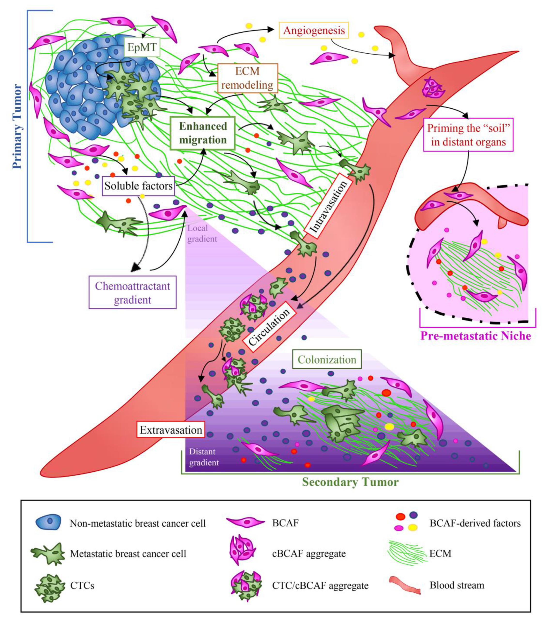
5.1. Primary, Circulating, and Metastatic BCAFs: The Onset of the Metastatic Voyage

Recent data have demonstrated that BCAFs can spread beyond the primary tumor and move through the bloodstream as circulating BCAFs (cBCAFs) or BCAF aggregates, with or without cancer cells (Figure 3). Clusters of cBCAFs alone and of clusters of circulating tumor cells (CTCs) with cBCAFs have been detected in the peripheral blood of metastatic BC patients. In addition to providing clinical evidence of the association of cBCAFs with metastatic disease, these findings also suggest the diagnostic potential of cBCAFs and CTCs as biomarkers for BC metastasis [25]. Given the presence of CAFs in the pre-metastatic niche before the appearance of cancer cells [201], it is possible to speculate that, at the end of their metastatic voyage through the blood circulation, cBCAFs colonize a new body site and prime the ”soil” for future tumor cell colonization. Furthermore, cBCAFs can also prime the “soil” by establishing and maintaining a gradient of specific chemoattractive factors, which could trigger the directional migration of BC cells and promote dissemination and colonization of the metastatic niche (Figure 3). For example, Farmaki et al. showed that CCL8 production is enhanced in fibroblasts at the margins of breast tumors. Thus, by establishing a CCL8-gradient, CAFs attract cancer cells towards the tumor margins, precisely where the highest amounts of CCL8 are detected. In response to elevated levels of CCL8, BC cells seed the peripheral “soil”, and enter the circulation by establishing CTC populations. In turn, by following the CCL8 gradient in the blood circulation, CTCs establish secondary tumors in the lungs and brain, as evidenced by the presence of elevated levels of CCL8 in both organs [202].
Additionally, in vitro and in vivo studies have shown that BCAFs promote metastasis by inducing the formation of tumor cell clusters. Specifically, BCAF-derived SDF-1 and TGF-β, via Src activation, induce the formation of multicellular clusters composed of two distinct tumor cell populations: one is composed of tumor cells in a highly epithelial (Ehi) state, as exemplified by the upregulation of the epithelial marker E-cadherin and the oncogenic cell–cell adhesion molecules, CAM5 and CAM6. The other, instead, consists of cancer cells in a hybrid epithelial/mesenchymal (E/M) state, as exemplified by the upregulation of the mesenchymal marker ZEB1. Both populations are fundamental for BC metastasis; in fact, the suppression of either the epithelial markers or ZEB1 reduces metastasis formation. In particular, Ehi cancer cells allow the formation of multicellular clusters and metastatic seeding by promoting cell–cell adhesion, aggregation, anti-apoptosis, and cell proliferation. On the other hand, E/M cancer cells act as leader cells that, by maintaining cell-cell interaction with the follower Ehi cancer cells, drive their collective cell invasion and metastatic colonization [203]. The potential clinical role of BCAFs in cancer cell cluster formation and metastasis progression is also supported by findings indicating the presence of BC inflammatory emboli characterized by increased cell–cell adhesion with high E-cadherin expression and the hybrid E/M state [203,204]. Additionally, Chung et al. demonstrated that brain metastatic BCAF aggregates attract BC cells in vitro much more strongly than BCAF aggregates from the primary tumor (pBCAFs) or normal fibroblast aggregates. Brain metastatic BCAF aggregates express higher levels of CXCL12 (also known as SDF-1) and CXCL16 than normal breast fibroblast or pBCAF aggregates. Therefore, blocking the interaction between CXCR6-CXCL16 and/or CXCR4-CXCL12 significantly inhibits BC cell migration to brain metastatic BCAF aggregates. These data further support the important role of CXCL16 and CXCL12 in eliciting migration of CTCs to the brain metastatic niche [205].
Hemalatha et al. speculate that BC cells harboring mutations in genes involved in cell motility, such as BRCA1, may convert BCAFs into the so-called metastasis associated fibroblasts (MAFs), i.e., CAFs with enhanced capability to promote cancer cell migration, invasion and metastasis. They demonstrated that BCAFs treated with the conditioned medium of BRCA1-mutated BC cells acquire an altered phenotype, characterized by increased expression of metastatic and EpMT markers, including Ezrin, CCL5, and N-Cadherin. Interestingly, inhibition of Ezrin and CCL5 in MAFs reduces their capability to promote cancer cell migration and invasion in vitro. Therefore, Ezrin and CCL5 could represent promising targets to prevent the differentiation of BCAFs into a more metastatic phenotype, especially in BRCA1 mutated tumors [206]. A recent study demonstrated that pBCAFs and metastatic BCAFs (mBCAFs) isolated from skin, lung, liver, and bone metastases exhibit distinct gene expression profiles. In particular, mBCAFs show greater upregulation of genes related to the interferon (IFN) signaling pathway, ECM organization, hypoxia, and MAPK signaling pathway than pBCAFs do. The enhanced pro-tumorigenic and pro-metastatic properties of mBCAFs are partially due to increased secretion of IGF2. In fact, inhibition of the IGF signaling pathway not only dramatically decreases the mBCAF pro-tumorigenic phenotype [207] but also significantly inhibits lung metastasis in a TNBC xenograft model [208].
A recent study shows that the gene expression profile of fibroblasts co-evolves with the metastatic disease. In particular, each stage of the metastatic process can be defined by a specific gene signature that, in turn, correlates with a specific functional profile of fibroblasts. Therefore, the transcriptional rewiring of MAFs during the metastatic progression is fundamental for the generation of a permissive and suitable metastatic niche for cancer cell colonization and growth [209].

5.2. Role of BCAFs in BC Metastasis through ECM Remodeling

In some tumors, including BC, cancer cells start spreading long before the detection and the surgical removal of the primary tumor [210,211]. Interestingly, some tumors may be smaller, owing to the enhanced capability acquired by cancer cells to migrate away from the primary tumor mass and enter the circulation. This enhanced cell dissemination can be facilitated by the abnormal ECM found in tumors [210]. In this scenario, BCAFs regulate ECM composition and organization, and thus provide a favorable environment for BC dissemination and metastasis. Intriguingly, in vitro and in vivo studies show that BCAFs deposit an abundant and well aligned ECM, whereas normal fibroblasts from disease-free reduction mammoplasty tissues deposit an ECM with a random mesh-like organization. When non-malignant epithelial cells interact with BCAF-derived ECM, they acquire a mesenchymal phenotype, which is a necessary step for cell dissemination. In particular, these epithelial cells—which appear very elongated and regularly arranged in a parallel pattern—enhance the expression of the mesenchymal marker Thy-1, and upregulate the TGF-β-dependent Smad, Erk1/2, and Jun signaling pathways. Both non-malignant and malignant cells use the parallel organization of the ECM fiber bundles, generated by BCAFs, as tracks to disseminate in the blood circulation (Figure 3). Conversely, most of the time, normal fibroblasts not only fail to trigger the mesenchymal phenotype in epithelial cells but also suppress the tumorigenic and metastatic potential of malignant cells in vivo. Moreover, although the acquisition of the mesenchymal phenotype is necessary, it is insufficient to promote metastasis. Indeed, even when fibroblasts from disease-free reduction induce the mesenchymal phenotype in epithelial cells, they nonetheless fail to promote metastasis successfully. Therefore, only BCAFs can induce additional alterations in tumor cells, which consequently acquire a metastatic potential [210].
A major player in ECM remodeling is the tumor suppressor PTEN. PTEN downregulation and deletion characterize the activated stroma in solid tumors and about 50% of invasive BC, respectively [212]. The loss of PTEN dramatically triggers stromal microenvironment remodeling, which is associated with increased collagen deposition around mammary ducts, MMP-9 expression and activity, and macrophage infiltration [212]. PTEN-null fibroblasts in normal bearing mice promote collagen alignment parallel to that of the mammary duct [212]. This collagen organization resembles that of the so-called tumor-associated collagen signature 2 (TACS-2), which is characterized by a parallel linearization and organization of collagen at the tumor edge [212,213]. These results suggest that loss of PTEN in fibroblasts may prime gland mammary microenvironment for tumor development [212]. In addition, PTEN-null fibroblasts in tumor bearing mice increase the number of fibers oriented perpendicularly to the tumor edge. This perpendicular collagen orientation closely resembles that of the TACS-3, which creates tracks for cancer cells to spread beyond the primary tumor [212,213]. This is consistent with in vitro studies showing that murine mammary adenocarcinoma cells cultured on the ECM derived from PTEN-null fibroblasts are characterized by enhanced contractility and migratory capability [212]. Interestingly, inhibition of fibroblast contractility abolishes the increased collagen alignment normally observed with PTEN loss. Therefore, the signaling pathways involved in fibroblast contractility, such as the Rho/ROCK pathway, may represent potential targets to reduce BC metastasis caused by CAF-mediated ECM remodeling [214]. For instance, the ROCK inhibitor fasudil, approved by the Food and Drug Administration for the treatment of cerebral vasospasms, dramatically reduces BC metastasis in a BC mouse model [215]. Furthermore, it has been reported that TACS-3 is associated with significantly decreased overall survival in BC patients [216], suggesting that changes in collagen organization observed with loss of stromal PTEN may evolve with tumor progression [212].
Another player in ECM remodeling is the IGF/IGF-R1 signaling pathway. Indeed, BC cells, via IGF secretion, activate the IGF/IGF-R1signaling pathways in CAFs. In turn, the IGF/IGF-R1 axis promotes fibroblast motility and upregulates the promigratory protease urokinase (uPA) and its receptor uPAR. The synergistic occurrence of these events drives ECM remodeling, thus enhancing BC cell migration and invasion [217]. Noticeably, ongoing clinical trials targeting the IGF/IGF-1R pathway in BC patients are exploiting anti-IGF-1R monoclonal antibodies (such as dalotuzumab, figitumumab, cixutumumab, ganitumab, R1507, AVE1642), anti-IGF-I and IGF-II monoclonal antibodies (MEDI-573 and BI 836845), and a small IGF-IR-tyrosine kinase inhibitor (OSI-906) as a strategy to slow down, and, in the best case scenario, to block metastatic spread [218].
Adding to the complexity of ECM remodeling and BC metastasis is the fact that distinct subtypes of BCAFs produce different types of ECM, which, in turn, can differently modulate BC progression and invasion. For instance, CD146 BCAFs promote a higher number of metastases than do CD146+ BCAFs. Furthermore, clinical data have shown that CD146 BCAFs in BC predict an increased risk of developing lymph node metastasis even in patients with small primary tumors (<5 cm). This enhanced capability to develop metastasis is partly due to the ability of CD146 BCAFs to secrete an ECM enriched for pro-metastatic proteins. In particular, CD146 BCAF-derived matrix contains numerous ECM regulators, such as LOX, and structural ECM proteins, such as FN1 and TNC, all of which are overexpressed in very aggressive tumors [219]. For example, when TNC activates the EGFR signaling pathway, it induces an invasive phenotype in BC cells. This may explain why TNC is often upregulated at the invasive tumor front of aggressive and metastatic BCs [145,220]. Conversely, the basement membrane proteins, which act as an important barrier to cancer cell invasion, are significantly decreased in CD146 BCAF-derived matrix [219]. These results are consistent with other data showing a stronger overexpression of FN, biglycan, and LOX enzyme in BCAFs with higher metastasis capability [144,210].
In addition, compelling evidence has demonstrated that chronic inflammation plays a fundamental role in cancer development and progression. Tumor infiltrating immune cells, especially neutrophils and eosinophils, contribute to the generation and maintenance of the tumor inflammatory milieu by producing massively myeloperoxidase (MPO) and eosinophil peroxidase (EPO), respectively. Indeed, MPO and EPO are strongly upregulated in the stroma of human BCs [221] and in mouse model of breast tumors [222]. Interestingly, these inflammatory enzymes contribute to BC invasion and metastasis by modulating fibroblast behaviors. Particularly, MPO and EPO strongly promote human mammary fibroblast migration, as well as increasing the fibroblast production of collagen I and VI, MMP-1 and MMP-3, and COX-2 [223], all of which act as important drivers of BC invasion and metastasis [224,225,226,227,228]. Additionally, MPO and EPO promote ECM structural remodeling by inducing collagen fiber alignment, which significantly increases cancer cell adhesion and invasion [223].
MMPs have been identified as markers of poor prognosis in BC patients, since they modify the TME. In particular, MMPs drive cancer progression and metastasis by directly degrading the ECM to promote tumor cell migration, and by destroying the basement membrane of blood or lymphatic vessels to promote tumor cell intravasation and metastatic dissemination [229,230]. Several in vitro and in vivo studies have reported that CAFs induce BC cell migration, invasion, and metastasis by producing various MMPs, including MMP-1, -2, -3, -7, -9, -13, and -14 [223,227,230,231,232,233]. Despite the prior disappointing clinical trials implementing the use of broad spectrum, small molecule catalytic site inhibitors, the identification of the mechanisms, by which these specific MMPs drive BC development and progression, offers promising targets to prevent and/or reduce BC progression [229].
COX-2 is still another important inflammatory mediator involved in tumor angiogenesis, invasion, and metastasis [234]. It is massively produced by both the stromal and the cancer cell compartments [223,234]. High COX-2 levels are associated with worse prognosis in BC patients and can predict larger tumor size and multiple lymph node metastases [235]. Furthermore, COX-2 triggers CAF activation in the tumor mass, as well as shape ECM structure and function. Krishnamachary et al. demonstrated that COX-2 overexpression significantly increases the number of activated CAFs and the amount of collagen 1 fiber density and volume [234]. In the tumor mass, collagen 1 fibers are mainly synthesized by CAFs [234] and facilitate BC metastasis by promoting the ameboid movement of tumor cells [236]. These data are consistent with studies showing a correlation between high density of collagen 1 fibers and the risk of developing metastasis [237,238]. Conversely, COX-2 downregulation significantly decreases the number of CAFs in the primary tumor and favors the formation of fewer and smaller metastatic lung nodules [234]. It is notable that fibroblasts increase COX-2 expression in BC cells by inducing the transcriptional activity of the nuclear factor kappa-light-chain-enhancer of activated B cells (NF-κB). NF-κB-mediated transcription of COX-2 is also associated with enhanced MMP-14 expression and MMP-9 protease activity.
In addition to these COX-2-driven molecular events that facilitate BC progression and invasion [239], COX-2-overexpressing BC cells can also promote indoleamine 2,3-dioxygenase (IDO) expression in fibroblasts. More specifically, IDO-expressing fibroblasts secrete large amounts of kynurenine, which induces E-cadherin ubiquitination and degradation, thus leading to BC cell migration [240]. Collectively, these data support the theory that disrupting the paracrine crosstalk between BC cells and CAFs constitutes a novel therapeutic strategy for preventing BC growth and metastatic dissemination. Moreover, COX-2 and IDO could represent promising targets for BC therapy. In fact, Chen et al. demonstrated that COX-2 and IDO inhibitors exert a strong anti-tumor effect on COX-2-overexpressing BCs. Moreover, they also demonstrated that simultaneous targeting of COX-2 and IDO markedly inhibits BC growth, despite not resulting in an additive effect [240]. Furthermore, the use of NSAIDs or selective COX-2 inhibitors has been intensively investigated for the treatment and prevention of several cancer types [241,242]. Using an animal model of BC, Harris et al. demonstrated that the use of celecoxib, a specific COX-2 inhibitor, decreases the incidence and size of BC significantly. These authors also showed that the commonly used NSAID ibuprofen induces potent anti-tumor effects in vivo, albeit to a lesser degree than celecoxib [243]. Consistently, several human epidemiologic investigations have confirmed the efficacy of NSAIDs not only in eliciting anti-tumor effects, but also in significantly decreasing the risk of BC [244,245,246]. In line with these findings, other compelling studies have reported that suppression of COX-2 expression in primary cutaneous myofibroblasts induces an in vitro anti-tumor phenotype [247,248].
Another crucial protein responsible for BC initiation, progression, and metastasis is Cav-1. Loss of Cav-1 in breast fibroblasts promotes BC progression and metastasis by generating the so-called “lethal” TME, which is associated with poor clinical outcome. In particular, Trimmer et al. demonstrated that Cav-1-deficient fibroblasts increase the production of proteins associated with collagen synthesis and processing, especially collagen VI [249], which is upregulated in BC patients prone to develop lymph node metastasis [250].
Taken together, these data suggest that the stromal collagenous ECM and BCAFs can represent promising targets for BC treatment. Maintaining a normal ECM environment and normalizing a matrix-rich desmoplastic stroma by targeting ECM proteins may offer plausible novel therapeutic strategies, able to block cancer growth and dissemination and enhance the delivery and efficacy of chemotherapy. For instance, Weaver et al., by reverting the malignant BC cell phenotype to the normal phenotype through ECM modification, demonstrated that the ECM is dominant in determining the cell phenotype over the cellular genotype. In particular, they showed that tumor phenotypes can be reverted by culturing BC cells onto a 3-D basement membrane in the presence of integrin β1 blocking antibodies. The “normalization” of the tumor cell phenotype in vitro is also maintained in vivo, where the malignant potential of tumor cells treated with β1-integrin blocking antibodies is reduced or totally lost [251]. Strikingly, pharmacological inhibition of αv integrin with GLPG0187 sharply reduces bone metastasis in BC mouse models [252]. A case in point is zoledronate. This drug, approved for the clinical treatment of patients with multiple myeloma and bone metastases [253], also inhibits BC growth, migration and invasion by targeting various ECM proteins, including integrins ανβ3, ανβ5, and α5β1, and Sdc-1, and by suppressing the expression of MMP-2 and MMP-9 [254]. Furthermore, targeting Sdc-1 with radioimmunotherapy has proved to be a successful therapeutic strategy for the treatment of mice with TNBC [255].
FN, highly expressed by BCAFs, represents another promising target for cancer therapy. Interestingly, the use of inhibitors of FN polymerization can be very beneficial for cancer therapy, since FN is not actively produced in healthy tissues, except for wounded tissues and during development. Therefore, the advantage of using FN inhibitors is that they have minimal off-target effects [256]. Hielscher et al. showed that the treatment of co-cultures of fibroblasts and BC cells with pUR4B, a small FN inhibitor, decreases not only the deposition and organization of FN in the ECM, but also the deposition of other ECM proteins. Furthermore, inhibition of FN also affects the vascular morphogenesis of endothelial cells cultured on a FN-null matrix. Overall, knowing that FN deposition participates in ECM assembly and supports angiogenesis is instrumental for the development of novel anti-tumor agents able to disrupt cancer and vascular cell interactions with an FN-rich matrix [257]. Indeed, Tabariès et al. demonstrated that the use of neutralizing antibodies targeting α5β1 or α2β1 complexes can efficiently inhibit the binding of claudin 2-overexpressing BC cells to FN and collagen IV, consequently reducing the ability of BC cells to metastasize to the liver [258]. Furthermore, Takai et al. demonstrated that pirfenidone (PFD), which is an anti-fibrotic agent and a TGF-β antagonist, inhibits tumor growth of TNBC in vitro by targeting CAFs and by significantly reducing the density of collagen 1 fibers. Conversely, in vivo PFD monotherapy inhibits tumor fibrosis and TGF-β signaling without affecting cancer growth and lung metastasis. Moreover, PFD in combination with doxorubicin synergistically thwarts BC growth and lung metastasis. On the other hand, doxorubicin alone has been shown to inhibit primary tumor growth, but not lung metastasis in in vivo experiments [259]. Losartan, a potent anti-fibrotic compound, inhibits collagen I synthesis by BCAFs both in vitro and in vivo. Interestingly, in vivo studies demonstrated that losartan enhances tumor penetration and distribution of drugs and, in particular, increases the anti-tumor effect of i.v. injected pegylated liposomal doxorubicin [260]. Notably, Hu et al. encapsulated losartan into hydrogels, in order to allow a sustainable release of the encapsulated drug after intra-tumoral injection. When they used it in combination with doxorubicin-loaded liposomes (Dox-L), the losartan-loaded hydrogels significantly improved the intra-tumoral accumulation and penetration of Dox-L. Remarkably, this combination therapy was able to delay primary tumor growth by 64% and lung metastasis by 80% compared to Dox-L monotherapy [261]. Therefore, losartan efficiently destroys the dense collagen network that tumors efficiently exploit to reduce the penetration and efficacy of chemotherapeutic drugs [260]. Another promising compound that inhibits BC tumor progression is the selective tyrosine kinase inhibitor imatinib mesylate. This compound has been shown to inhibit not only the proliferation and invasiveness of human epithelial BC cells, but also to act as a potent anti-fibrotic agent by inhibiting both fibroblast proliferation and fibroblast-derived collagen accumulation. In particular, imatinib mesylate exerts a potent anti-tumor effect in the stromal compartment by downregulating the synthesis of collagen I and III—the main collagen types in BCM [262].
Lastly, the implementation of nanomedicine in the fight against a variety of different tumors, not least BC, is gaining increasing momentum. For instance, it has been demonstrated that, by functionalizing the surface of nanoparticles, with LOX inhibiting antibodies, it is possible to suppress mammary cancer cell growth and invasion more effectively than soluble anti-LOX antibodies both in vitro and in vivo. These results, besides substantiating the theory that ECM normalization is a useful strategy for BC treatment, they also provide a proof of concept for treating cancer with higher efficacy and decreased side effects by using nanomedicine [263].

5.3. Role of BCAFs in BC Metastasis via Induction of Angiogenesis

Angiogenesis and hypoxia, by supporting each other, play an essential role in the formation of BC metastasis [264,265]. In the rapidly growing tumor mass, the morphological and functional abnormalities of the vasculature often result in heterogeneously distributed hypoxic areas [266] whose low levels of oxygen, in turn, promote and sustain angiogenesis [267]. Reduction in the oxygen supply concerns and influences both cancer and stromal cell compartments [268]. Ren et al. demonstrated that hypoxia not only induces BCAF activation, but also indirectly provokes BCAFs to promote angiogenesis and cell motility via HIF-1α and its target gene G protein estrogen receptor (GPER). In particular, BCAF-derived GPER and HIF-1α cooperatively promote the transcription of VEGF and connective tissue growth factor (CTGF), which are involved in angiogenesis and BC invasion, respectively [266]. Furthermore, BCAFs promote angiogenesis by secreting high levels of SDF-1, a protein that mobilizes and recruits endothelial progenitor cells (EPCs) from the bone marrow into the tumor mass. In the tumor, EPCs differentiate into the tumor-associated vascular endothelial cells [269] lining tumor blood vessels, which represent escape routes for metastatic cancer cells. Additionally, BCAFs induce endothelial changes by targeting cells via soluble factors. In particular, BCAF-derived IL-1β enhances the expression of several adhesion molecules, especially Selp (Selectin P), and the vascular cell adhesion molecule 1 (VCAM1) in endothelial cells. These adhesion proteins facilitate intravasation and extravasation of cancer cells by increasing tumor cell adhesion and transmigration through the endothelium. In addition, BCAF-derived IL-1 β also sustains and enhances invasion of BC cells by upregulating MMP-1, -3 and -5 [270]. Notably, BCAF-derived SDF-1 enhances cancer cell migration by binding to the receptor CXCR4 expressed on cancer cells. Consequently, SDF-1/CXCR4 binding induces actin polymerization and pseudopodia formation in cancer cells. These events drive the metastatic spread of cancer cells beyond the borders of the primary tumor toward organs that constitutively express SDF-1, such as lungs, bones, lymph nodes, and the brain. This explains why SDF-1-rich organs represent the most common metastatic sites of BC [271]. Therefore, since SDF-1 is a key factor involved in the growth, angiogenesis, invasion, and metastasis of BC, pharmacologically targeting SDF-1 and its downstream signaling pathway could have a relevant therapeutic potential for BC. Indeed, Gründker et al. have demonstrated that inhibition of the SDF-1/CXCR4 signal axis by kisspeptin-10 markedly reduces BC cell invasion [272]. Remarkably, several in vitro and in vivo studies have indeed identified several peptides or small molecule inhibitors of CXCR4 able to inhibit BC growth and progression dramatically [273,274,275,276,277,278,279].
Recently, a very interesting study investigated whether podoplanin, a glycoprotein with a key role in lymphatic vasculature formation and function, could contribute to spurring the metastatic properties of BC cells. In particular, this protein is highly expressed by fibroblasts present in the peripheral stroma at the margin of BCs, and in close proximity to tumor blood vessels. As expected, the study indicated that podoplanin expression favors fibroblast migration in the tumor stroma. This finding explains why podoplanin-rich BCAFs are preferentially detected in the peripheral tumor zone. Furthermore, fibroblasts expressing high levels of podoplanin have been found to interact efficiently with endothelial cells, as well as affect the tumor vascular network by promoting pseudo-tube formation. In addition, the results of clinical studies showing the presence of a higher number of podoplanin-rich BCAFs in IDC than in ductal carcinoma in situ (DCIS), further corroborate the pivotal role of podoplanin-positive BCAFs in BC progression and dissemination [280]. Niemiec et al. hypothesized that podoplanin-positive BCAFs could travel with metastatic cancer cells from the primary BC toward lymph node metastasis. In fact, they found that whereas podoplanin-positive BCAFs were comparably present in podoplanin-positive primary BCs and corresponding lymph nodes, they were absent in the lymph node metastases deriving from podoplanin-negative primary BCs [281].
Furthermore, preclinical and clinical studies showing the involvement of Sdc-1 in promoting and enhancing BC angiogenesis have been equally revealing [142]. In particular, it has been hypothesized that Sdc-1, via its heparan sulfate (HS) chains, activates the proangiogenic factors FGF2 and VEGF, thereby extensively contributing to tumor angiogenesis [142,143]. This theory is also strongly supported by several other studies showing that HS chains require many growth factors to spark the tumor signaling pathways [282,283]. Consistently, heparanase, by enhancing MMP-9 expression, induces expression and shedding of Sdc-1 in myeloma and BC. Indeed, by cleaving the core protein of Sdc-1, which remains biologically active [284], MMP 9 promotes BC progression by stimulating both angiogenesis [142] and cell dissemination [144]. Therefore, targeting Sdc-1 and/or inhibiting MMP-9 or heparanase could represent yet another potential therapeutic strategy for BC [285].

6. Conclusions

In both normal and neoplastic mammary glands, the interactions of stromal cells with either normal epithelial cells or cancer cells represent principal regulators of mammary gland homeostasis and cancer development, respectively. During BC growth, BCAFs exploit many altered morphogenetic signaling pathways, which are instead tightly regulated during physiological mammary gland branching morphogenesis. The comparison of mammary fibroblast and BCAF interactions with normal and cancer epithelial cells, which affect mammary gland branching morphogenesis and tumor development, could facilitate the understanding of alterations driving BCAF differentiation, BCM remodeling and cancer dissemination. Furthermore, a better understanding of the mechanisms underlying the formation of the pre-metastatic niche by BCAFs can be of fundamental importance in BC research. It has been well established that fibroblast populations from early-stage IDC are represented by BCAFs deriving from the tumor zone, myofibroblasts from the interface zone, and normal fibroblasts from the non-tumorigenic zone [137]. Therefore, we believe that the analysis of the highly complex interactions between cancer cells and fibroblast populations before CAF differentiation offers an important opportunity to better understand the role of normal fibroblasts in solid tumor development and dissemination.

Author Contributions

Conceptualization, A.A. (Alessandro Arcucci); writing—original draft preparation, A.A. (Alessandro Arcucci), A.A. (Angelica Avagliano); writing—review and editing A.A. (Alessandro Arcucci), A.A. (Angelica Avagliano), G.F., M.R.R., N.M., E.V., L.I., D.R., A.A. (Antonello Accurso), S.M. (Stefania Masone), S.M. (Stefania Montagnani). All authors have read and agreed to the published version of the manuscript.

Funding

This study was funded by the Italian Ministry of Education, Universities and Research through the grant: “Integrated mechanobiology approaches for a precise medicine in cancer treatment” (award number: PRIN-20177TTP3S) and by the Regione Campania “SATIN” grant 2018–2020.

Acknowledgments

We thank Paola Merolla for editing the review article.

Conflicts of Interest

The authors declare no conflict of interest.

References

  1. Wolfson, B.; Zhang, Y.; Gernapudi, R.; Duru, N.; Yao, Y.; Lo, P.-K.; Zhou, Q. A High-Fat Diet Promotes Mammary Gland Myofibroblast Differentiation through MicroRNA 140 Downregulation. Mol. Cell. Biol. 2017, 37. [Google Scholar] [CrossRef] [Green Version]
  2. Koledova, Z. 3D coculture of mammary organoids with fibrospheres: A model for studying epithelial–stromal interactions during mammary branching morphogenesis. In Methods in Molecular Biology; Humana Press Inc.: Totowa, NJ, USA, 2017; Volume 1612, pp. 107–124. [Google Scholar]
  3. Polyak, K.; Kalluri, R. The role of the microenvironment in mammary gland development and cancer. Cold Spring Harb. Perspect. Biol. 2010, 2, a003244. [Google Scholar] [CrossRef] [PubMed] [Green Version]
  4. Jena, M.K.; Jaswal, S.; Kumar, S.; Mohanty, A.K. Molecular mechanism of mammary gland involution: An update. Dev. Biol. 2019, 445, 145–155. [Google Scholar] [CrossRef] [PubMed]
  5. Jena, M.K.; Mohanty, A.K. New insights of mammary gland during different stages of development. Asian J. Pharm. Clin. Res. 2017, 10, 35–40. [Google Scholar] [CrossRef] [Green Version]
  6. Monkkonen, T.; Lewis, M.T. New paradigms for the Hedgehog signaling network in mammary gland development and breast Cancer. Biochim. Biophys. Acta Rev. Cancer 2017, 1868, 315–332. [Google Scholar] [CrossRef] [PubMed]
  7. Zuo, Y.; Oh, W.; Ulu, A.; Frost, J.A. Minireview: Mouse models of Rho GTPase function in mammary gland development, tumorigenesis, and metastasis. Mol. Endocrinol. 2016, 30, 278–289. [Google Scholar] [CrossRef] [PubMed]
  8. Macias, H.; Hinck, L. Mammary gland development. Wiley Interdiscip. Rev. Dev. Biol. 2012, 1, 533–557. [Google Scholar] [CrossRef] [PubMed] [Green Version]
  9. Koledova, Z.; Zhang, X.; Streuli, C.; Clarke, R.B.; Klein, O.D.; Werb, Z.; Lu, P. SPRY1 regulates mammary epithelial morphogenesis by modulating EGFR-dependent stromal paracrine signaling and ECM remodeling. Proc. Natl. Acad. Sci. USA 2016, 113, E5731–E5740. [Google Scholar] [CrossRef] [Green Version]
  10. Inman, J.L.; Robertson, C.; Mott, J.D.; Bissell, M.J. Mammary gland development: Cell fate specification, stem cells and the microenvironment. Development 2015, 142, 1028–1042. [Google Scholar] [CrossRef] [Green Version]
  11. Lühr, I.; Friedl, A.; Overath, T.; Tholey, A.; Kunze, T.; Hilpert, F.; Sebens, S.; Arnold, N.; Rösel, F.; Oberg, H.H.; et al. Mammary fibroblasts regulate morphogenesis of normal and tumorigenic breast epithelial cells by mechanical and paracrine signals. Cancer Lett. 2012, 325, 175–188. [Google Scholar] [CrossRef] [Green Version]
  12. Ruocco, M.R.; Avagliano, A.; Granato, G.; Imparato, V.; Masone, S.; Masullo, M.; Nasso, R.; Montagnani, S.; Arcucci, A. Involvement of Breast Cancer-Associated Fibroblasts in Tumor Development, Therapy Resistance and Evaluation of Potential Therapeutic Strategies. Curr. Med. Chem. 2018, 25, 3414–3434. [Google Scholar] [CrossRef] [PubMed]
  13. Deshmukh, S.K.; Srivastava, S.K.; Poosarla, T.; Dyess, D.L.; Holliday, N.P.; Singh, A.P.; Singh, S. Inflammation, immunosuppressive microenvironment and breast cancer: Opportunities for cancer prevention and therapy. Ann. Transl. Med. 2019, 7, 593. [Google Scholar] [CrossRef] [PubMed]
  14. Avagliano, A.; Ruocco, M.R.; Aliotta, F.; Belviso, I.; Accurso, A.; Masone, S.; Montagnani, S.; Arcucci, A. Mitochondrial Flexibility of Breast Cancers: A Growth Advantage and a Therapeutic Opportunity. Cells 2019, 8, 401. [Google Scholar] [CrossRef] [PubMed] [Green Version]
  15. Hill, B.S.; Sarnella, A.; D’Avino, G.; Zannetti, A. Recruitment of stromal cells into tumour microenvironment promote the metastatic spread of breast cancer. Semin. Cancer Biol. 2020, 60, 202–213. [Google Scholar] [CrossRef] [PubMed]
  16. Wu, Q.; Li, J.; Zhu, S.; Wu, J.; Chen, C.; Liu, Q.; Wei, W.; Zhang, Y.; Sun, S. Breast cancer subtypes predict the preferential site of distant metastases: A SEER based study. Oncotarget 2017, 8, 27990–27996. [Google Scholar] [CrossRef] [PubMed] [Green Version]
  17. Makki, J. Diversity of breast carcinoma: Histological subtypes and clinical relevance. Clin. Med. Insights Pathol. 2015, 8, 23–31. [Google Scholar] [CrossRef] [PubMed] [Green Version]
  18. Avagliano, A.; Granato, G.; Ruocco, M.R.; Romano, V.; Belviso, I.; Carfora, A.; Montagnani, S.; Arcucci, A. Metabolic Reprogramming of Cancer Associated Fibroblasts: The Slavery of Stromal Fibroblasts. Biomed. Res. Int. 2018, 2018, 6075403. [Google Scholar] [CrossRef] [Green Version]
  19. Da Cunha, B.R.; Domingos, C.; Buzzo Stefanini, A.C.; Henrique, T.; Polachini, G.M.; Castelo-Branco, P.; Tajara, E.H. Cellular interactions in the tumor microenvironment: The role of secretome. J. Cancer 2019, 10, 4574–4587. [Google Scholar] [CrossRef] [PubMed] [Green Version]
  20. Cacho-Díaz, B.; García-Botello, D.R.; Wegman-Ostrosky, T.; Reyes-Soto, G.; Ortiz-Sánchez, E.; Herrera-Montalvo, L.A. Tumor microenvironment differences between primary tumor and brain metastases. J. Transl. Med. 2020, 18, 1. [Google Scholar] [CrossRef] [Green Version]
  21. Arcucci, A.; Ruocco, M.R.; Granato, G.; Sacco, A.M.; Montagnani, S. Cancer: An Oxidative Crosstalk Between Solid Tumor Cells and Cancer Associated Fibroblasts. Biomed. Res. Int. 2016; 201. [Google Scholar]
  22. Tashireva, L.A.; Denisov, E.V.; Gerashchenko, T.S.; Pautova, D.N.; Buldakov, M.A.; Zavyalova, M.V.; Kzhyshkowska, J.; Cherdyntseva, N.V.; Perelmuter, V.M. Intratumoral heterogeneity of macrophages and fibroblasts in breast cancer is associated with the morphological diversity of tumor cells and contributes to lymph node metastasis. Immunobiology 2017, 222, 631–640. [Google Scholar] [CrossRef]
  23. Tower, H.; Ruppert, M.; Britt, K. The immune microenvironment of breast cancer progression. Cancers 2019, 11, 1375. [Google Scholar] [CrossRef] [PubMed] [Green Version]
  24. Li, J.; Hao, Q.; Cao, W.; Vadgama, J.V.; Wu, Y. Celecoxib in breast cancer prevention and therapy. Cancer Manag. Res. 2018, 10, 4653–4667. [Google Scholar] [CrossRef] [PubMed] [Green Version]
  25. Ao, Z.; Shah, S.H.; Machlin, L.M.; Parajuli, R.; Miller, P.C.; Rawal, S.; Williams, A.J.; Cote, R.J.; Lippman, M.E.; Datar, R.H.; et al. Identification of cancer-associated fibroblasts in circulating blood from patients with metastatic breast cancer. Cancer Res. 2015, 75, 4681–4687. [Google Scholar] [CrossRef] [PubMed] [Green Version]
  26. Weigelt, B.; Peterse, J.L.; Van’t Veer, L.J. Breast cancer metastasis: Markers and models. Nat. Rev. Cancer 2005, 5, 591–602. [Google Scholar] [CrossRef] [PubMed]
  27. Pellacani, D.; Tan, S.; Lefort, S.; Eaves, C.J. Transcriptional regulation of normal human mammary cell heterogeneity and its perturbation in breast cancer. EMBO J. 2019, 38, e100330. [Google Scholar] [CrossRef] [PubMed]
  28. Conklin, M.W.; Keely, P.J. Why the stroma matters in breast cancer: Insights into breast cancer patient outcomes through the examination of stromal biomarkers. Cell Adhes. Migr. 2012, 6, 249–260. [Google Scholar] [CrossRef] [Green Version]
  29. Kaushik, S.; Pickup, M.W.; Weaver, V.M. From transformation to metastasis: Deconstructing the extracellular matrix in breast cancer. Cancer Metastasis Rev. 2016, 35, 655–667. [Google Scholar] [CrossRef]
  30. Brownfield, D.G.; Venugopalan, G.; Lo, A.; Mori, H.; Tanner, K.; Fletcher, D.A.; Bissell, M.J. Patterned collagen fibers orient branching mammary epithelium through distinct signaling modules. Curr. Biol. 2013, 23, 703–709. [Google Scholar] [CrossRef] [Green Version]
  31. Ingman, W.V.; Wyckoff, J.; Gouon-Evans, V.; Condeelis, J.; Pollard, J.W. Macrophages promote collagen fibrillogenesis around terminal end buds of the developing mammary gland. Dev. Dyn. 2006, 235, 3222–3229. [Google Scholar] [CrossRef]
  32. Peuhu, E.; Kaukonen, R.; Lerche, M.; Saari, M.; Guzmán, C.; Rantakari, P.; De Franceschi, N.; Wärri, A.; Georgiadou, M.; Jacquemet, G.; et al. SHARPIN regulates collagen architecture and ductal outgrowth in the developing mouse mammary gland. EMBO J. 2017, 36, 165–182. [Google Scholar] [CrossRef]
  33. Cichon, M.A.; Degnim, A.C.; Visscher, D.W.; Radisky, D.C. Microenvironmental influences that drive progression from benign breast disease to invasive breast cancer. J. Mammary Gland Biol. Neoplasia 2010, 15, 389–397. [Google Scholar] [CrossRef] [PubMed] [Green Version]
  34. Ewald, A.J.; Brenot, A.; Duong, M.; Chan, B.S.; Werb, Z. Collective Epithelial Migration and Cell Rearrangements Drive Mammary Branching Morphogenesis. Dev. Cell 2008, 14, 570–581. [Google Scholar] [CrossRef] [PubMed] [Green Version]
  35. Paine, I.S.; Lewis, M.T. The Terminal End Bud: The Little Engine that Could. J. Mammary Gland Biol. Neoplasia 2017, 22, 93–108. [Google Scholar] [CrossRef] [PubMed] [Green Version]
  36. McCave, E.J.; Cass, C.A.P.; Burg, K.J.L.; Booth, B.W. The normal microenvironment directs mammary gland development. J. Mammary Gland Biol. Neoplasia 2010, 15, 291–299. [Google Scholar] [CrossRef]
  37. Mallepell, S.; Krust, A.; Chambon, P.; Brisken, C. Paracrine signaling through the epithelial estrogen receptor α is required for proliferation and morphogenesis in the mammary gland. Proc. Natl. Acad. Sci. USA 2006, 103, 2196–2201. [Google Scholar] [CrossRef] [Green Version]
  38. Brisken, C.; Park, S.; Vass, T.; Lydon, J.P.; O’Malley, B.W.; Weinberg, R.A. A paracrine role for the epithelial progesterone receptor in mammary gland development. Proc. Natl. Acad. Sci. USA 1998, 95, 5076–5081. [Google Scholar] [CrossRef] [Green Version]
  39. Russo, J.; Russo, I.H. Development of the human breast. Maturitas 2004, 49, 2–15. [Google Scholar] [CrossRef]
  40. Morsing, M.; Klitgaard, M.C.; Jafari, A.; Villadsen, R.; Kassem, M.; Petersen, O.W.; Rønnov-Jessen, L. Evidence of two distinct functionally specialized fibroblast lineages in breast stroma. Breast Cancer Res. 2016, 18, 108. [Google Scholar] [CrossRef] [Green Version]
  41. Hutson, S.W.; Cowen, P.N.; Bird, C.C. Morphometric studies of age related changes in normal human breast and their significance for evolution of mammary cancer. J. Clin. Pathol. 1985, 38, 281–287. [Google Scholar] [CrossRef] [Green Version]
  42. Henson, D.E.; Tarone, R.E. On the Possible Role of Involution in the Natural History of Breast Cancer. Cancer 1993, 7, 2154–2156. [Google Scholar] [CrossRef]
  43. Figueroa, J.D.; Pfeiffer, R.M.; Patel, D.A.; Linville, L.; Brinton, L.A.; Gierach, G.L.; Yang, X.R.; Papathomas, D.; Visscher, D.; Mies, C.; et al. Terminal Duct Lobular Unit Involution of the Normal Breast: Implications for Breast Cancer Etiology. J. Natl. Cancer Inst. 2014, 106, dju286. [Google Scholar] [CrossRef] [PubMed] [Green Version]
  44. Milanese, T.R.; Hartmann, L.C.; Sellers, T.A.; Frost, M.H.; Vierkant, R.A.; Maloney, S.D.; Pankratz, V.S.; Degnim, A.C.; Vachon, C.M.; Reynolds, C.A.; et al. Age-related lobular involution and risk of breast cancer. J. Natl. Cancer Inst. 2006, 98, 1600–1607. [Google Scholar] [CrossRef] [PubMed] [Green Version]
  45. Watson, C.J.; Khaled, W.T. Mammary development in the embryo and adult: A journey of morphogenesis and commitment. Development 2008, 135, 995–1003. [Google Scholar] [CrossRef] [PubMed] [Green Version]
  46. Rybinski, B.; Franco-Barraza, J.; Cukierman, E. The wound healing, chronic fibrosis, and cancer progression triad. Physiol. Genom. 2014, 46, 223–244. [Google Scholar] [CrossRef] [PubMed]
  47. DeClerck, Y.A. Desmoplasia: A response or a niche? Cancer Discov. 2012, 2, 772–774. [Google Scholar] [CrossRef] [Green Version]
  48. Seo, B.R.; Bhardwaj, P.; Choi, S.; Gonzalez, J.; Eguiluz, R.C.A.; Wang, K.; Mohanan, S.; Morris, P.G.; Du, B.; Zhou, X.K.; et al. Obesity-dependent changes in interstitial ECM mechanics promote breast tumorigenesis. Sci. Transl. Med. 2015, 7. [Google Scholar] [CrossRef] [Green Version]
  49. Park, J.; Scherer, P.E. Adipocyte-derived endotrophin promotes malignant tumor progression. J. Clin. Invest. 2012, 122, 4243–4256. [Google Scholar] [CrossRef] [Green Version]
  50. Kojima, Y.; Acar, A.; Eaton, E.N.; Mellody, K.T.; Scheel, C.; Ben-Porath, I.; Onder, T.T.; Wang, Z.C.; Richardson, A.L.; Weinberg, R.A.; et al. Autocrine TGF-β and stromal cell-derived factor-1 (SDF-1) signaling drives the evolution of tumor-promoting mammary stromal myofibroblasts. Proc. Natl. Acad. Sci. USA 2010, 107, 20009–20014. [Google Scholar] [CrossRef] [Green Version]
  51. Unsworth, A.; Anderson, R.; Britt, K. Stromal fibroblasts and the immune microenvironment: Partners in mammary gland biology and pathology? J. Mammary Gland Biol. Neoplasia 2014, 19, 169–182. [Google Scholar] [CrossRef]
  52. Jackson, D.; Bresnick, J.; Rosewell, I.; Crafton, T.; Poulsom, R.; Stamp, G.; Dickson, C. Fibroblast growth factor receptor signalling has a role in lobuloalveolar development of the mammary gland. J. Cell Sci. 1997, 110, 1261–1268. [Google Scholar]
  53. Schedin, P.; Mitrenga, T.; McDaniel, S.; Kaeck, M. Mammary ECM Composition and function are altered by reproductive state. Mol. Carcinog. 2004, 41, 207–220. [Google Scholar] [CrossRef]
  54. Talhouk, R.S.; Bissell, M.J.; Werb, Z. Coordinated expression of extracellular matrix-degrading proteinases and their inhibitors regulates mammary epithelial function during involution. J. Cell Biol. 1992, 118, 1271–1282. [Google Scholar] [CrossRef] [PubMed] [Green Version]
  55. Kim, E.J.; Jung, H.S.; Lu, P. Pleiotropic functions of fibroblast growth factor signaling in embryonic mammary gland development. J. Mammary Gland Biol. Neoplasia 2013, 18, 139–142. [Google Scholar] [CrossRef] [PubMed]
  56. Soriano, J.V.; Pepper, M.S.; Orci, L.; Montesano, R. Roles of Hepatocyte Growth Factor/Scatter Factor and Transforming Growth Factor-β1 in Mammary Gland Ductal Morphogenesis. J. Mammary Gland Biol. Neoplasia 1998, 3, 133–150. [Google Scholar] [CrossRef]
  57. Pond, A.C.; Bin, X.; Batts, T.; Roarty, K.; Hilsenbeck, S.; Rosen, J.M. Fibroblast growth factor receptor signaling is essential for normal mammary gland development and stem cell function. Stem Cells 2013, 31, 178–189. [Google Scholar] [CrossRef] [PubMed] [Green Version]
  58. Lu, P.; Ewald, A.J.; Martin, G.R.; Werb, Z. Genetic mosaic analysis reveals FGF receptor 2 function in terminal end buds during mammary gland branching morphogenesis. Dev. Biol. 2008, 321, 77–87. [Google Scholar] [CrossRef] [Green Version]
  59. Xian, W.; Schwertfeger, K.L.; Vargo-Gogola, T.; Rosen, J.M. Pleiotropic effects of FGFR1 on cell proliferation, survival, and migration in a 3D mammary epithelial cell model. J. Cell Biol. 2005, 171, 663–673. [Google Scholar] [CrossRef]
  60. Sumbal, J.; Koledova, Z. FGF signaling in mammary gland fibroblasts regulates multiple fibroblast functions and mammary epithelial morphogenesis. Development 2019, 146. [Google Scholar] [CrossRef]
  61. Sternlicht, M.D.; Kouros-Mehr, H.; Lu, P.; Werb, Z. Hormonal and local control of mammary branching morphogenesis. Differentiation 2006, 74, 365–381. [Google Scholar] [CrossRef] [Green Version]
  62. Kawazoe, T.; Taniguchi, K. The Sprouty/Spred family as tumor suppressors: Coming of age. Cancer Sci. 2019, 110, 1525–1535. [Google Scholar] [CrossRef] [Green Version]
  63. Zhang, X.; Qiao, G.; Lu, P. Modulation of fibroblast growth factor signaling is essential for mammary epithelial morphogenesis. PLoS ONE 2014, 9. [Google Scholar] [CrossRef] [PubMed] [Green Version]
  64. Shi, T.; Yao, L.; Han, Y.; Hao, P.; Lu, P. Quantitative phosphoproteomics reveals system-wide phosphorylation network altered by spry in mouse mammary stromal fibroblasts. Int. J. Mol. Sci. 2019, 20, 5400. [Google Scholar] [CrossRef] [PubMed]
  65. Maennling, A.E.; Tur, M.K.; Niebert, M.; Klockenbring, T.; Zeppernick, F.; Gattenlöhner, S.; Meinhold-Heerlein, I.; Hussain, A.F. Molecular targeting therapy against egfr family in breast cancer: Progress and future potentials. Cancers 2019, 11, 5400. [Google Scholar] [CrossRef] [Green Version]
  66. Andrae, J.; Gallini, R.; Betsholtz, C. Role of platelet-derived growth factors in physiology and medicine. Genes Dev. 2008, 22, 1276–1312. [Google Scholar] [CrossRef] [PubMed] [Green Version]
  67. Hammer, A.M.; Sizemore, G.M.; Shukla, V.C.; Avendano, A.; Sizemore, S.T.; Chang, J.J.; Kladney, R.D.; Cuitiño, M.C.; Thies, K.A.; Verfurth, Q.; et al. Stromal PDGFR-α Activation Enhances Matrix Stiffness, Impedes Mammary Ductal Development, and Accelerates Tumor Growth. Neoplasia 2017, 19, 496–508. [Google Scholar] [CrossRef] [PubMed]
  68. Guo, Q.; Minnier, J.; Burchard, J.; Chiotti, K.; Spellman, P.; Schedin, P. Physiologically activated mammary fibroblasts promote postpartum mammary cancer. JCI Insight 2017, 2, e89206. [Google Scholar] [CrossRef]
  69. Tian, Z.; Tang, J.; Yang, Q.; Li, X.; Zhu, J.; Wu, G. Atypical ubiquitin-binding protein SHARPIN promotes breast cancer progression. Biomed. Pharmacother. 2019, 119, 109414. [Google Scholar] [CrossRef]
  70. Kasirer-Friede, A.; Tjahjono, W.; Eto, K.; Shattil, S.J. SHARPIN at the nexus of integrin, immune, and inflammatory signaling in human platelets. Proc. Natl. Acad. Sci. USA 2019, 116, 4983–4988. [Google Scholar] [CrossRef] [Green Version]
  71. Pouwels, J.; DeFranceschi, N.; Rantakari, P.; Auvinen, K.; Karikoski, M.; Mattila, E.; Potter, C.; Sundberg, J.P.; Hogg, N.; Gahmberg, C.G.; et al. SHARPIN regulates uropod detachment in migrating lymphocytes. Cell Rep. 2013, 5, 619–628. [Google Scholar] [CrossRef]
  72. Rantala, J.K.; Pouwels, J.; Pellinen, T.; Veltel, S.; Laasola, P.; Mattila, E.; Potter, C.S.; Duffy, T.; Sundberg, J.P.; Kallioniemi, O.; et al. SHARPIN is an endogenous inhibitor of β1-integrin activation. Nat. Cell Biol. 2011, 13, 1315–1324. [Google Scholar] [CrossRef] [Green Version]
  73. Peuhu, E.; Salomaa, S.I.; De Franceschi, N.; Potter, C.S.; Sundberg, J.P.; Pouwels, J. Integrin beta 1 inhibition alleviates the chronic hyperproliferative dermatitis phenotype of SHARPIN-deficient mice. PLoS ONE 2017, 12. [Google Scholar] [CrossRef] [PubMed] [Green Version]
  74. Lerche, M.; Elosegui-Artola, A.; Kechagia, J.Z.; Guzmán, C.; Georgiadou, M.; Andreu, I.; Gullberg, D.; Roca-Cusachs, P.; Peuhu, E.; Ivaska, J. Integrin Binding Dynamics Modulate Ligand-Specific Mechanosensing in Mammary Gland Fibroblasts. iScience 2020, 23, 100907. [Google Scholar] [CrossRef] [PubMed] [Green Version]
  75. Deome, K.B.; Faulkin, L.J., Jr.; Bern, H.A.; Blair, P.B. Development of Mammary Tumors from Hyperplastic Alveolar Nodules Transplanted Into Gland-Free Mammary Fat Pads of Female C3H Mice. Cancer Res. 1959, 19, 515–520. [Google Scholar]
  76. Kuperwasser, C.; Chavarria, T.; Wu, M.; Magrane, G.; Gray, J.W.; Carey, L.; Richardson, A.; Weinberg, R.A. Reconstruction of functionally normal and malignant human breast tissues in mice. Proc. Natl. Acad. Sci. USA 2004, 101, 4966–4971. [Google Scholar] [CrossRef] [PubMed] [Green Version]
  77. Lu, P.; Weaver, V.M.; Werb, Z. The extracellular matrix: A dynamic niche in cancer progression. J. Cell Biol. 2012, 196, 395–406. [Google Scholar] [CrossRef] [PubMed]
  78. Egeblad, M.; Rasch, M.G.; Weaver, V.M. Dynamic interplay between the collagen scaffold and tumor evolution. Curr. Opin. Cell Biol. 2010, 22, 697–706. [Google Scholar] [CrossRef] [Green Version]
  79. Coleman, W.B. Breast Ductal Carcinoma in Situ: Precursor to Invasive Breast Cancer. Am. J. Pathol. 2019, 189, 942–945. [Google Scholar] [CrossRef] [Green Version]
  80. Eiro, N.; Gonzalez, L.O.; Fraile, M.; Cid, S.; Schneider, J.; Vizoso, F.J. Breast cancer tumor stroma: Cellular components, phenotypic heterogeneity, intercellular communication, prognostic implications and therapeutic opportunities. Cancers 2019, 11, 664. [Google Scholar] [CrossRef] [Green Version]
  81. Sappino, A.P.; Skalli, O.; Jackson, B.; Schürch, W.; Gabbiani, G. Smooth-muscle differentiation in stromal cells of malignant and non-malignant breast tissues. Int. J. Cancer 1988, 41, 707–712. [Google Scholar] [CrossRef]
  82. Rønnov-Jessen, L.; Petersen, O.W.; Koteliansky, V.E.; Bissell, M.J. The origin of the myofibroblasts in breast cancer: Recapitulation of tumor environment in culture unravels diversity and implicates converted fibroblasts and recruited smooth muscle cells. J. Clin. Invest. 1995, 95, 859–873. [Google Scholar] [CrossRef] [Green Version]
  83. Vaughan, M.B.; Howard, E.W.; Tomasek, J.J. Transforming growth factor-β1 promotes the morphological and functional differentiation of the myofibroblast. Exp. Cell Res. 2000, 257, 180–189. [Google Scholar] [CrossRef] [PubMed] [Green Version]
  84. Stuelten, C.H.; Busch, J.I.; Tang, B.; Flanders, K.C.; Oshima, A.; Sutton, E.; Karpova, T.S.; Roberts, A.B.; Wakefield, L.M.; Niederhuber, J.E. Transient tumor-fibroblast interactions increase tumor cell malignancy by a TGF-b Mediated Mechanism in a Mouse Xenograft Model of Breast Cancer. PLoS ONE 2010, 5, e9832. [Google Scholar] [CrossRef] [PubMed]
  85. Arcucci, A.; Ruocco, M.R.; Albano, F.; Granato, G.; Romano, V.; Corso, G.; Bancone, C.; De Vendittis, E.; Della Corte, A.; Montagnani, S. Analysis of extracellular superoxide dismutase and Akt in ascending aortic aneurysm with tricuspid or bicuspid aortic valve. Eur. J. Histochem. 2014, 58, 200–206. [Google Scholar] [CrossRef] [PubMed] [Green Version]
  86. Arcucci, A.; Ruocco, M.R.; Amatruda, N.; Riccio, A.; Tarantino, G.; Albano, F.; Mele, V.; Montagnani, S. Analysis of Extracellular Superoxide Dismutase in Fibroblasts from Patients With Systemic Sclerosis. J. Biol. Regul. Homeost. Agents 2011, 25, 647–654. [Google Scholar]
  87. Denicola, G.M.; Karreth, F.A.; Humpton, T.J.; Gopinathan, A.; Wei, C.; Frese, K.; Mangal, D.; Yu, K.H.; Yeo, C.J.; Calhoun, E.S.; et al. Oncogene-induced Nrf2 transcription promotes ROS detoxification and tumorigenesis. Nature 2011, 475, 106–110. [Google Scholar] [CrossRef]
  88. Wilmanski, T.; Zhou, X.; Zheng, W.; Shinde, A.; Donkin, S.S.; Wendt, M.; Burgess, J.R.; Teegarden, D. Inhibition of pyruvate carboxylase by 1α,25-dihydroxyvitamin D promotes oxidative stress in early breast cancer progression. Cancer Lett. 2017, 411, 171–181. [Google Scholar] [CrossRef] [Green Version]
  89. Biernacka, A.; Dobaczewski, M.; Frangogiannis, N.G. TGF-β signaling in fibrosis. Growth Factors 2011, 29, 196–202. [Google Scholar] [CrossRef] [Green Version]
  90. Casey, T.M.; Eneman, J.; Crocker, A.; White, J.; Tessitore, J.; Stanley, M.; Harlow, S.; Bunn, J.Y.; Weaver, D.; Muss, H.; et al. Cancer associated fibroblasts stimulated by transforming growth factor beta1 (TGF-β1) increase invasion rate of tumor cells: A population study. Breast Cancer Res. Treat. 2008, 110, 39–49. [Google Scholar] [CrossRef]
  91. Shangguan, L.; Ti, X.; Krause, U.; Hai, B.; Zhao, Y.; Yang, Z.; Liu, F. Inhibition of TGF-β/Smad signaling by BAMBI blocks differentiation of human mesenchymal stem cells to carcinoma-associated fibroblasts and abolishes their protumor effects. Stem Cells 2012, 30, 2810–2819. [Google Scholar] [CrossRef]
  92. Goulet, C.R.; Bernard, G.; Tremblay, S.; Chabaud, S.; Bolduc, S.; Pouliot, F. Exosomes induce fibroblast differentiation into cancer-associated fibroblasts through TGFb signaling. Mol. Cancer Res. 2018, 16, 1196–1204. [Google Scholar] [CrossRef] [Green Version]
  93. Hinz, B.; Celetta, G.; Tomasek, J.J.; Gabbiani, G.; Chaponnier, C. Alpha-smooth muscle actin expression upregulates fibroblast contractile activity. Mol. Biol. Cell 2001, 12, 2730–2741. [Google Scholar] [CrossRef] [PubMed] [Green Version]
  94. Qiao, A.; Gu, F.; Guo, X.; Zhang, X.; Fu, L. Breast cancer-associated fibroblasts: Their roles in tumor initiation, progression and clinical applications. Front. Med. 2016, 10, 33–40. [Google Scholar] [CrossRef] [PubMed]
  95. Buchsbaum, R.J.; Oh, S.Y. Breast cancer-associated fibroblasts: Where we are and where we need to go. Cancers 2016, 8, 19. [Google Scholar] [CrossRef] [Green Version]
  96. Welch, D.R.; Hurst, D.R. Defining the Hallmarks of Metastasis. Cancer Res. 2019, 79, 3011–3027. [Google Scholar] [CrossRef]
  97. Pantel, K.; Brakenhoff, R.H. Dissecting the metastatic cascade. Nat. Rev. Cancer 2004, 4, 448–456. [Google Scholar] [CrossRef]
  98. Kalluri, R. EMT: When epithelial cells decide to become mesenchymal-like cells. J. Clin. Invest. 2009, 119, 1417–1419. [Google Scholar] [CrossRef] [PubMed] [Green Version]
  99. Yilmaz, M.; Christofori, G. EMT, the cytoskeleton, and cancer cell invasion. Cancer Metastasis Rev. 2009, 28, 215–233. [Google Scholar] [CrossRef] [Green Version]
  100. Melzer, C.; von der Ohe, J.; Hass, R. Breast Carcinoma: From Initial Tumor Cell Detachment to Settlement at Secondary Sites. Biomed. Res. Int. 2017, 2017, 8534371. [Google Scholar] [CrossRef]
  101. Jain, R.K.; Carmeliet, P. SnapShot: Tumor angiogenesis. Cell 2012, 149. [Google Scholar] [CrossRef] [Green Version]
  102. Raica, M.; Ribatti, D. Targeting Tumor Lymphangiogenesis: An Update. Curr. Med. Chem. 2010, 17, 698–708. [Google Scholar] [CrossRef]
  103. Alitalo, A.; Detmar, M. Interaction of tumor cells and lymphatic vessels in cancer progression. Oncogene 2012, 31, 4499–4508. [Google Scholar] [CrossRef] [PubMed] [Green Version]
  104. Ginter, P.S.; Karagiannis, G.S.; Entenberg, D.; Lin, Y.; Condeelis, J.; Jones, J.G.; Oktay, M.H. Tumor microenvironment of metastasis (TMEM) doorways are restricted to the blood vessel endothelium in both primary breast cancers and their lymph node metastases. Cancers 2019, 11, 1507. [Google Scholar] [CrossRef] [PubMed] [Green Version]
  105. Karagiannis, G.S.; Condeelis, J.S.; Oktay, M.H. Chemotherapy-induced metastasis: Mechanisms and translational opportunities. Clin. Exp. Metastasis 2018, 35, 269–284. [Google Scholar] [CrossRef] [PubMed]
  106. Karagiannis, G.S.; Goswami, S.; Jones, J.G.; Oktay, M.H.; Condeelis, J.S. Signatures of breast cancer metastasis at a glance. J. Cell Sci. 2016, 129, 1751–1758. [Google Scholar] [CrossRef] [PubMed] [Green Version]
  107. Oktay, M.H.; Jones, J.G. TMEM: A novel breast cancer dissemination marker for the assessment of metastatic risk. Biomark. Med. 2015, 9, 81–84. [Google Scholar] [CrossRef] [PubMed]
  108. Gertler, F.; Condeelis, J. Metastasis: Tumor cells becoming MENAcing. Trends Cell Biol. 2011, 21, 81–90. [Google Scholar] [CrossRef] [PubMed] [Green Version]
  109. Harney, A.S.; Arwert, E.N.; Entenberg, D.; Wang, Y.; Guo, P.; Qian, B.Z.; Oktay, M.H.; Pollard, J.W.; Jones, J.G.; Condeelis, J.S. Real-time imaging reveals local, transient vascular permeability, and tumor cell intravasation stimulated by TIE2hi macrophage–derived VEGFA. Cancer Discov. 2015, 5, 932–943. [Google Scholar] [CrossRef] [Green Version]
  110. Sparano, J.A.; Gray, R.; Oktay, M.H.; Entenberg, D.; Rohan, T.; Xue, X.; Donovan, M.; Peterson, M.; Shuber, A.; Hamilton, D.A.; et al. A metastasis biomarker (MetaSite BreastTM Score) is associated with distant recurrence in hormone receptor-positive, HER2-negative early-stage breast cancer. NPJ Breast Cancer 2017, 3. [Google Scholar] [CrossRef] [Green Version]
  111. Walker, C.; Mojares, E.; Del Río Hernández, A. Role of extracellular matrix in development and cancer progression. Int. J. Mol. Sci. 2018, 19, 3028. [Google Scholar] [CrossRef] [Green Version]
  112. Tamimi, S.O.; Ahmed, A. Stromal changes in invasive breast carcinoma: An ultrastructural study. J. Pathol. 1987, 153, 163–170. [Google Scholar] [CrossRef]
  113. Insua-Rodríguez, J.; Oskarsson, T. The extracellular matrix in breast cancer. Adv. Drug Deliv. Rev. 2016, 97, 41–55. [Google Scholar] [CrossRef] [PubMed]
  114. Lee, S.K.; Huang, S.; Lu, W.; Lev, D.C.; Price, J.E. Vascular endothelial growth factor expression promotes the growth of breast cancer brain metastases in nude mice. Clin. Exp. Metastasis 2004, 21, 107–118. [Google Scholar] [CrossRef]
  115. Nduom, E.K.; Yang, C.; Merrill, M.J.; Zhuang, Z.; Lonser, R.R. Characterization of the blood-brain barrier of metastatic and primary malignant neoplasms. J. Neurosurg. 2013, 119, 427–433. [Google Scholar] [CrossRef] [PubMed]
  116. Uzunalli, G.; Dieterly, A.M.; Kemet, C.M.; Weng, H.Y.; Soepriatna, A.H.; Goergen, C.J.; Shinde, A.B.; Wendt, M.K.; Lyle, L.T. Dynamic transition of the blood-brain barrier in the development of non-small cell lung cancer brain metastases. Oncotarget 2019, 10, 6334–6348. [Google Scholar] [CrossRef] [Green Version]
  117. Levental, K.R.; Yu, H.; Kass, L.; Lakins, J.N.; Egeblad, M.; Erler, J.T.; Fong, S.F.T.; Csiszar, K.; Giaccia, A.; Weninger, W.; et al. Matrix Crosslinking Forces Tumor Progression by Enhancing Integrin Signaling. Cell 2009, 139, 891–906. [Google Scholar] [CrossRef] [Green Version]
  118. Ramaswamy, S.; Ross, K.N.; Lander, E.S.; Golub, T.R. A molecular signature of metastasis in primary solid tumors. Nat. Genet. 2003, 33, 49–54. [Google Scholar] [CrossRef]
  119. Acerbi, I.; Cassereau, L.; Dean, I.; Shi, Q.; Au, A.; Park, C.; Chen, Y.Y.; Liphardt, J.; Hwang, E.S.; Weaver, V.M. Human breast cancer invasion and aggression correlates with ECM stiffening and immune cell infiltration. Integr. Biol. 2015, 7, 1120–1134. [Google Scholar] [CrossRef] [Green Version]
  120. Vallet, S.D.; Ricard-Blum, S. Lysyl oxidases: From enzyme activity to extracellular matrix cross-links. Essays Biochem. 2019, 63, 349–364. [Google Scholar]
  121. Erler, J.T.; Giaccia, A.J. Lysyl oxidase mediates hypoxic control of metastasis. Cancer Res. 2006, 66, 10238–10241. [Google Scholar] [CrossRef] [Green Version]
  122. Mäki, J.M.; Sormunen, R.; Lippo, S.; Kaarteenaho-Wiik, R.; Soininen, R.; Myllyharju, J. Lysyl oxidase is essential for normal development and function of the respiratory system and for the integrity of elastic and collagen fibers in various tissues. Am. J. Pathol. 2005, 167, 927–936. [Google Scholar] [CrossRef] [Green Version]
  123. Tang, X.; Hou, Y.; Yang, G.; Wang, X.; Tang, S.; Du, Y.E.; Yang, L.; Yu, T.; Zhang, H.; Zhou, M.; et al. Stromal miR-200s contribute to breast cancer cell invasion through CAF activation and ECM remodeling. Cell Death Differ. 2016, 23, 132–145. [Google Scholar] [CrossRef] [PubMed] [Green Version]
  124. Cox, T.R.; Gartland, A.; Erler, J.T. Lysyl oxidase, a targetable secreted molecule involved in cancer metastasis. Cancer Res. 2016, 76, 188–192. [Google Scholar] [CrossRef] [PubMed] [Green Version]
  125. Schito, L.; Rey, S. Hypoxic pathobiology of breast cancer metastasis. Biochim. Biophys. Acta Rev. Cancer 2017, 1868, 239–245. [Google Scholar] [CrossRef] [PubMed]
  126. Rundqvist, H.; Johnson, R.S. Tumour oxygenation: Implications for breast cancer prognosis. J. Intern. Med. 2013, 274, 105–112. [Google Scholar] [CrossRef]
  127. Schito, L.; Semenza, G.L. Hypoxia-Inducible Factors: Master Regulators of Cancer Progression. Trends Cancer 2016, 2, 758–770. [Google Scholar] [CrossRef] [Green Version]
  128. Dales, J.P.; Garcia, S.; Meunier-Carpentier, S.; Andrac-Meyer, L.; Haddad, O.; Lavaut, M.N.; Allasia, C.; Bonnier, P.; Charpin, C. Overexpression of hypoxia-inducible factor HIF-1α predicts early relapse in breast cancer: Retrospective study in a series of 745 patients. Int. J. Cancer 2005, 116, 734–739. [Google Scholar] [CrossRef]
  129. Winter, S.C.; Buffa, F.M.; Silva, P.; Miller, C.; Valentine, H.R.; Turley, H.; Shah, K.A.; Cox, G.J.; Corbridge, R.J.; Homer, J.J.; et al. Relation of a hypoxia metagene derived from head and neck cancer to prognosis of multiple cancers. Cancer Res. 2007, 67, 3441–3449. [Google Scholar] [CrossRef] [Green Version]
  130. Cox, T.R.; Rumney, R.M.H.; Schoof, E.M.; Perryman, L.; Høye, A.M.; Agrawal, A.; Bird, D.; Latif, N.A.; Forrest, H.; Evans, H.R.; et al. The hypoxic cancer secretome induces pre-metastatic bone lesions through lysyl oxidase. Nature 2015, 522, 106–110. [Google Scholar] [CrossRef] [Green Version]
  131. Psaila, B.; Lyden, D. The metastatic niche: Adapting the foreign soil. Nat. Rev. Cancer 2009, 9, 285–293. [Google Scholar] [CrossRef]
  132. Smithen, D.A.; Leung, L.M.H.; Challinor, M.; Lawrence, R.; Tang, H.; Niculescu-Duvaz, D.; Pearce, S.P.; McLeary, R.; Lopes, F.; Aljarah, M.; et al. 2-Aminomethylene-5-sulfonylthiazole Inhibitors of Lysyl Oxidase (LOX) and LOXL2 Show Significant Efficacy in Delaying Tumor Growth. J. Med. Chem. 2020, 63, 2308–2324. [Google Scholar] [CrossRef]
  133. Chang, J.; Lucas, M.C.; Leonte, L.E.; Garcia-Montolio, M.; Singh, L.B.; Findlay, A.D.; Deodhar, M.; Foot, J.S.; Jarolimek, W.; Timpson, P.; et al. Pre-clinical evaluation of small molecule LOXL2 inhibitors in breast cancer. Oncotarget 2017, 8, 26066–26078. [Google Scholar] [CrossRef] [PubMed] [Green Version]
  134. Liu, J.; Liao, S.; Diop-Frimpong, B.; Chen, W.; Goel, S.; Naxerova, K.; Ancukiewicz, M.; Boucher, Y.; Jain, R.K.; Xu, L. TGF-β blockade improves the distribution and efficacy of therapeutics in breast carcinoma by normalizing the tumor stroma. Proc. Natl. Acad. Sci. USA 2012, 109, 16618–16623. [Google Scholar] [CrossRef] [PubMed] [Green Version]
  135. Li, J.; Zormpas-Petridis, K.; Boult, J.K.R.; Reeves, E.L.; Heindl, A.; Vinci, M.; Lopes, F.; Cummings, C.; Springer, C.J.; Chesler, L.; et al. Investigating the contribution of collagen to the tumor biomechanical phenotype with noninvasive magnetic resonance elastography. Cancer Res. 2019, 79, 5874–5883. [Google Scholar] [CrossRef] [Green Version]
  136. Sasaki, T.; Fässler, R.; Hohenester, E. Laminin: The crux of basement membrane assembly. J. Cell Biol. 2004, 164, 959–963. [Google Scholar] [CrossRef] [Green Version]
  137. Kim, B.G.; Gao, M.Q.; Choi, Y.P.; Kang, S.; Park, H.R.; Kang, K.S.; Cho, N.H. Invasive breast cancer induces laminin-332 upregulation and integrin β4 neoexpression in myofibroblasts to confer an anoikis-resistant phenotype during tissue remodeling. Breast Cancer Res. 2012, 14. [Google Scholar] [CrossRef] [PubMed] [Green Version]
  138. Lin, T.-C.; Yang, C.-H.; Cheng, L.-H.; Chang, W.-T.; Lin, Y.-R.; Cheng, H.-C. Fibronectin in Cancer: Friend or Foe. Cells 2019, 9, 27. [Google Scholar] [CrossRef] [Green Version]
  139. Fernandez-Garcia, B.; Eiró, N.; Marín, L.; González-Reyes, S.; González, L.O.; Lamelas, M.L.; Vizoso, F.J. Expression and prognostic significance of fibronectin and matrix metalloproteases in breast cancer metastasis. Histopathology 2014, 64, 512–522. [Google Scholar] [CrossRef]
  140. Balanis, N.; Wendt, M.K.; Schiemann, B.J.; Wang, Z.; Schiemann, W.P.; Carlin, C.R. Epithelial to Mesenchymal Transition Promotes Breast Cancer Progression via a Fibronectin-dependent STAT3 Signaling Pathway. J. Biol. Chem. 2013, 288, 17954–17967. [Google Scholar] [CrossRef] [Green Version]
  141. Wang, K.; Wu, F.; Seo, B.R.; Fischbach, C.; Chen, W.; Hsu, L.; Gourdon, D. Breast cancer cells alter the dynamics of stromal fibronectin-collagen interactions. Matrix Biol. 2017, 60–61, 86–95. [Google Scholar] [CrossRef]
  142. Maeda, T.; Desouky, J.; Friedl, A. Syndecan-1 expression by stromal fibroblasts promotes breast carcinoma growth in vivo and stimulates tumor angiogenesis. Oncogene 2006, 25, 1408–1412. [Google Scholar] [CrossRef] [Green Version]
  143. Stanley, M.J.; Stanley, M.W.; Sanderson, R.D.; Zera, R. Syndecan-1 Expression Is Induced in the Stroma of Infiltrating Breast Carcinoma. Am. J. Clin. Pathol. 1999, 112. [Google Scholar] [CrossRef] [PubMed]
  144. Yang, N.; Mosher, R.; Seo, S.; Beebe, D.; Friedl, A. Syndecan-1 in breast cancer stroma fibroblasts regulates extracellular matrix fiber organization and carcinoma cell motility. Am. J. Pathol. 2011, 178, 325–335. [Google Scholar] [CrossRef] [PubMed]
  145. Yang, Z.; Ni, W.; Cui, C.; Fang, L.; Xuan, Y. Tenascin C is a prognostic determinant and potential cancer-associated fibroblasts marker for breast ductal carcinoma. Exp. Mol. Pathol. 2017, 102, 262–267. [Google Scholar] [CrossRef] [PubMed]
  146. Degen, M.; Brellier, F.; Schenk, S.; Driscoll, R.; Zaman, K.; Stupp, R.; Tornillo, L.; Terracciano, L.; Chiquet-Ehrismann, R.; Rüegg, C.; et al. Tenascin-W, a new marker of cancer stroma, is elevated in sera of colon and breast cancer patients. Int. J. Cancer 2008, 122, 2454–2461. [Google Scholar] [CrossRef]
  147. Degen, M.; Brellier, F.; Kain, R.; Ruiz, C.; Terracciano, L.; Orend, G.; Chiquet-Ehrismann, R. Tenascin-W is a novel marker for activated tumor stroma in low-grade human breast cancer and influences cell behavior. Cancer Res. 2007, 67, 9169–9179. [Google Scholar] [CrossRef] [Green Version]
  148. Le Bras, G.F.; Taubenslag, K.J.; Andl, C.D. The regulation of cell-cell adhesion during epithelial-mesenchymal transition, motility and tumor progression. Cell Adhes. Migr. 2012, 6, 365–373. [Google Scholar] [CrossRef] [Green Version]
  149. Moreno-Bueno, G.; Portillo, F.; Cano, A. Transcriptional regulation of cell polarity in EMT and cancer. Oncogene 2008, 27, 6958–6969. [Google Scholar] [CrossRef] [Green Version]
  150. Aiello, N.M.; Maddipati, R.; Norgard, R.J.; Balli, D.; Li, J.; Yuan, S.; Yamazoe, T.; Black, T.; Sahmoud, A.; Furth, E.E.; et al. EMT Subtype Influences Epithelial Plasticity and Mode of Cell Migration. Dev. Cell 2018, 45, 681–695. [Google Scholar] [CrossRef] [Green Version]
  151. Tsai, J.H.; Yang, J. Epithelial-mesenchymal plasticity in carcinoma metastasis. Genes Dev. 2013, 27, 2192–2206. [Google Scholar] [CrossRef] [Green Version]
  152. Albano, F.; Chiurazzi, F.; Mimmi, S.; Vecchio, E.; Pastore, A.; Cimmino, C.; Frieri, C.; Iaccino, E.; Pisano, A.; Golino, G.; et al. The expression of inhibitor of bruton’s tyrosine kinase gene is progressively up regulated in the clinical course of chronic lymphocytic leukaemia conferring resistance to apoptosis. Cell Death Dis. 2018, 9, 13. [Google Scholar] [CrossRef] [Green Version]
  153. Pastushenko, I.; Blanpain, C. EMT Transition States during Tumor Progression and Metastasis. Trends Cell Biol. 2019, 29, 212–226. [Google Scholar] [CrossRef] [PubMed] [Green Version]
  154. Shinde, A.; Libring, S.; Alpsoy, A.; Abdullah, A.; Schaber, J.A.; Solorio, L.; Wendt, M.K. Autocrine fibronectin inhibits breast cancer metastasis. Mol. Cancer Res. 2018, 16, 1579–1589. [Google Scholar] [CrossRef] [PubMed] [Green Version]
  155. Shinde, A.; Paez, J.S.; Libring, S.; Hopkins, K.; Solorio, L.; Wendt, M.K. Transglutaminase-2 facilitates extracellular vesicle-mediated establishment of the metastatic niche. Oncogenesis 2020, 9. [Google Scholar] [CrossRef] [PubMed] [Green Version]
  156. Luo, M.; Brooks, M.; Wicha, M. Epithelial-Mesenchymal Plasticity of Breast Cancer Stem Cells: Implications for Metastasis and Therapeutic Resistance. Curr. Pharm. Des. 2015, 21, 1301–1310. [Google Scholar] [CrossRef] [Green Version]
  157. Libring, S.; Shinde, A.; Chanda, M.K.; Nuru, M.; George, H.; Saleh, A.M.; Abdullah, A.; Kinzer-Ursem, T.L.; Calve, S.; Wendt, M.K.; et al. The dynamic relationship of breast cancer cells and fibroblasts in fibronectin accumulation at primary and metastatic tumor sites. Cancers 2020, 12, 1270. [Google Scholar] [CrossRef] [PubMed]
  158. Sleeman, J.P. The metastatic niche and stromal progression. Cancer Metastasis Rev. 2012, 31, 429–440. [Google Scholar] [CrossRef] [PubMed] [Green Version]
  159. Galluzzi, L.; Pietrocola, F.; Bravo-San Pedro, J.M.; Amaravadi, R.K.; Baehrecke, E.H.; Cecconi, F.; Codogno, P.; Debnath, J.; Gewirtz, D.A.; Karantza, V.; et al. Autophagy in malignant transformation and cancer progression. EMBO J. 2015, 34, 856–880. [Google Scholar] [CrossRef]
  160. Pisano, A.; Albano, F.; Vecchio, E.; Renna, M.; Scala, G.; Quinto, I.; Fiume, G. Revisiting bacterial ubiquitin ligase effectors: Weapons for host exploitation. Int. J. Mol. Sci. 2018, 19, 3576. [Google Scholar] [CrossRef] [Green Version]
  161. Clarke, A.J.; Simon, A.K. Autophagy in the renewal, differentiation and homeostasis of immune cells. Nat. Rev. Immunol. 2019, 19, 170–183. [Google Scholar] [CrossRef]
  162. Münz, C. Autophagy proteins in antigen processing for presentation on MHC molecules. Immunol. Rev. 2016, 272, 17–27. [Google Scholar] [CrossRef]
  163. Molina, M.L.; García-Bernal, D.; Martinez, S.; Valdor, R. Autophagy in the immunosuppressive perivascular microenvironment of glioblastoma. Cancers 2020, 12, 102. [Google Scholar] [CrossRef] [PubMed] [Green Version]
  164. White, E.; Mehnert, J.M.; Chan, C.S. Autophagy, Metabolism, and Cancer. Clin. Cancer Res. 2015, 21, 5037–5046. [Google Scholar] [CrossRef] [PubMed] [Green Version]
  165. Yun, H.R.; Jo, Y.H.; Kim, J.; Shin, Y.; Kim, S.S.; Choi, T.G. Roles of autophagy in oxidative stress. Int. J. Mol. Sci. 2020, 21, 3289. [Google Scholar] [CrossRef] [PubMed]
  166. Yun, C.W.; Lee, S.H. The roles of autophagy in cancer. Int. J. Mol. Sci. 2018, 19, 3466. [Google Scholar] [CrossRef] [PubMed] [Green Version]
  167. Pisano, A.; Ceglia, S.; Palmieri, C.; Vecchio, E.; Fiume, G.; De Laurentiis, A.; Mimmi, S.; Falcone, C.; Iaccino, E.; Scialdone, A.; et al. CRL3IBTK regulates the tumor suppressor Pdcd4 through ubiquitylation coupled to proteasomal degradation. J. Biol. Chem. 2015, 290, 13958–13971. [Google Scholar] [CrossRef] [PubMed] [Green Version]
  168. Liang, X.H.; Jackson, S.; Seaman, M.; Brown, K.; Kempkes, B.; Hibshoosh, H.; Levine, B. Induction of autophagy and inhibition of tumorigenesis by beclin 1. Nature 1999, 402, 672–676. [Google Scholar] [CrossRef]
  169. Takamura, A.; Komatsu, M.; Hara, T.; Sakamoto, A.; Kishi, C.; Waguri, S.; Eishi, Y.; Hino, O.; Tanaka, K.; Mizushima, N. Autophagy-deficient mice develop multiple liver tumors. Genes Dev. 2011, 25, 795–800. [Google Scholar] [CrossRef] [Green Version]
  170. Peng, Y.F.; Shi, Y.H.; Ding, Z.B.; Ke, A.W.; Gu, C.Y.; Hui, B.; Zhou, J.; Qiu, S.J.; Dai, Z.; Fan, J. Autophagy inhibition suppresses pulmonary metastasis of HCC in mice via impairing anoikis resistance and colonization of HCC cells. Autophagy 2013, 9, 2056–2068. [Google Scholar] [CrossRef] [Green Version]
  171. Akalay, I.; Janji, B.; Hasmim, M.; Noman, M.Z.; André, F.; De Cremoux, P.; Bertheau, P.; Badoual, C.; Vielh, P.; Larsen, A.K.; et al. Epithelial-to-mesenchymal transition and autophagy induction in breast carcinoma promote escape from t-cell-mediated lysis. Cancer Res. 2013, 73, 2418–2427. [Google Scholar] [CrossRef] [Green Version]
  172. You, L.; Wang, Z.; Li, H.; Shou, J.; Jing, Z.; Xie, J.; Sui, X.; Pan, H.; Han, W. The role of STAT3 in autophagy. Autophagy 2015, 11, 729–739. [Google Scholar] [CrossRef] [Green Version]
  173. Shen, S.; Niso-Santano, M.; Adjemian, S.; Takehara, T.; Malik, S.A.; Minoux, H.; Souquere, S.; Mariño, G.; Lachkar, S.; Senovilla, L.; et al. Cytoplasmic STAT3 Represses Autophagy by Inhibiting PKR Activity. Mol. Cell 2012, 48, 667–680. [Google Scholar] [CrossRef] [PubMed] [Green Version]
  174. Oh, H.M.; Yu, C.R.; Dambuza, I.; Marrero, B.; Egwuagu, C.E. STAT3 protein interacts with class O forkhead transcription factors in the cytoplasm and regulates nuclear/cytoplasmic localization of FoxO1 and FoxO3a proteins in CD4+ T cells. J. Biol. Chem. 2012, 287, 30436–30443. [Google Scholar] [CrossRef] [PubMed] [Green Version]
  175. Pires, B.R.B.; Mencalha, A.L.; Ferreira, G.M.; De Souza, W.F.; Morgado-Díaz, J.A.; Maia, A.M.; Corrêa, S.; Abdelhay, E.S.F.W. NF-kappaB is involved in the regulation of EMT genes in breast cancer cells. PLoS ONE 2017, 12. [Google Scholar] [CrossRef] [PubMed] [Green Version]
  176. Fiume, G.; Rossi, A.; de Laurentiis, A.; Falcone, C.; Pisano, A.; Vecchio, E.; Pontoriero, M.; Scala, I.; Scialdone, A.; Masci, F.F.; et al. Eukaryotic Initiation Factor 4H Is under Transcriptional Control of p65/NF-κB. PLoS ONE 2013, 8. [Google Scholar] [CrossRef] [PubMed] [Green Version]
  177. Wang, Y.; Xiong, H.; Liu, D.; Hill, C.; Ertay, A.; Li, J.; Zou, Y.; Miller, P.; White, E.; Downward, J.; et al. Autophagy inhibition specifically promotes epithelial-mesenchymal transition and invasion in RAS-mutated cancer cells. Autophagy 2019, 15, 886–899. [Google Scholar] [CrossRef] [PubMed]
  178. Gugnoni, M.; Sancisi, V.; Manzotti, G.; Gandolfi, G.; Ciarrocchi, A. Autophagy and epithelial–mesenchymal transition: An intricate interplay in cancer. Cell Death Dis. 2016, 7, e2520. [Google Scholar] [CrossRef] [PubMed]
  179. Hardy, S.D.; Shinde, A.; Wang, W.H.; Wendt, M.K.; Geahlen, R.L. Regulation of epithelial-mesenchymal transition and metastasis by TGF-β, P-bodies, and autophagy. Oncotarget 2017, 8, 103302–103314. [Google Scholar] [CrossRef] [Green Version]
  180. Shinde, A.; Hardy, S.D.; Kim, D.; Akhand, S.S.; Jolly, M.K.; Wang, W.H.; Anderson, J.C.; Khodadadi, R.B.; Brown, W.S.; George, J.T.; et al. Spleen tyrosine kinase–mediated autophagy is required for epithelial–mesenchymal plasticity and metastasis in breast cancer. Cancer Res. 2019, 79, 1831–1843. [Google Scholar] [CrossRef] [Green Version]
  181. Zeng, K.; He, B.; Yang, B.B.; Xu, T.; Chen, X.; Xu, M.; Liu, X.; Sun, H.; Pan, Y.; Wang, S. The pro-metastasis effect of circANKS1B in breast cancer 11 Medical and Health Sciences 1112 Oncology and Carcinogenesis. Mol. Cancer 2018, 17. [Google Scholar] [CrossRef]
  182. Lebret, S.C.; Newgreen, D.F.; Thompson, E.W.; Ackland, M.L. Induction of epithelial to mesenchymal transition in PMC42-LA human breast carcinoma cells by carcinoma-associated fibroblast secreted factors. Breast Cancer Res. 2007, 9. [Google Scholar] [CrossRef] [Green Version]
  183. Yu, Y.; Xiao, C.H.; Tan, L.D.; Wang, Q.S.; Li, X.Q.; Feng, Y.M. Cancer-associated fibroblasts induce epithelial-mesenchymal transition of breast cancer cells through paracrine TGF-β signalling. Br. J. Cancer 2014, 110, 724–732. [Google Scholar] [CrossRef] [PubMed] [Green Version]
  184. Giulianelli, S.; Cerliani, J.P.; Lamb, C.A.; Fabris, V.T.; Bottino, M.C.; Gorostiaga, M.A.; Novaro, V.; Góngora, A.; Baldi, A.; Molinolo, A.; et al. Carcinoma-associated fibroblasts activate progesterone receptors and induce hormone independent mammary tumor growth: A role for the FGF-2/FGFR-2 axis. Int. J. Cancer 2008, 123, 2518–2531. [Google Scholar] [CrossRef] [PubMed]
  185. Jedeszko, C.; Victor, B.C.; Podgorski, I.; Sloane, B.F. Fibroblast hepatocyte growth factor promotes invasion of human mammary ductal carcinoma in situ. Cancer Res. 2009, 69, 9148–9155. [Google Scholar] [CrossRef] [PubMed] [Green Version]
  186. Cao, X.C.; Zhang, W.R.; Cao, W.F.; Liu, B.W.; Zhang, F.; Zhao, H.M.; Meng, R.; Zhang, L.; Niu, R.F.; Hao, X.S.; et al. Aquaporin3 Is Required for FGF-2-Induced Migration of Human Breast Cancers. PLoS ONE 2013, 8, e56735. [Google Scholar] [CrossRef]
  187. Liu, Y.; Geng, Y.H.; Yang, H.; Yang, H.; Zhou, Y.T.; Zhang, H.Q.; Tian, X.X.; Fang, W.G. Extracellular ATP drives breast cancer cell migration and metastasis via S100A4 production by cancer cells and fibroblasts. Cancer Lett. 2018, 430, 1–10. [Google Scholar] [CrossRef]
  188. Sjöberg, E.; Meyrath, M.; Milde, L.; Herrera, M.; Lövrot, J.; Hägerstrand, D.; Frings, O.; Bartish, M.; Rolny, C.; Sonnhammer, E.; et al. A novel ACKR2-Dependent role of fibroblast-derived CXCL14 in epithelial-to-mesenchymal transition and metastasis of breast cancer. Clin. Cancer Res. 2019, 25, 3702–3717. [Google Scholar] [CrossRef] [Green Version]
  189. Liu, Y.; Zhang, J.; Sun, X.; Su, Q.; You, C. Down-regulation of miR-29b in carcinoma associated fibroblasts promotes cell growth and metastasis of breast cancer. Oncotarget 2017, 8, 39559–39570. [Google Scholar] [CrossRef] [Green Version]
  190. Tsuyada, A.; Chow, A.; Wu, J.; Somlo, G.; Chu, P.; Loera, S.; Luu, T.; Li, A.X.; Wu, X.; Ye, W.; et al. CCL2 mediates cross-talk between cancer cells and stromal fibroblasts that regulates breast cancer stem cells. Cancer Res. 2012, 72, 2768–2779. [Google Scholar] [CrossRef] [Green Version]
  191. Osuala, K.O.; Sameni, M.; Shah, S.; Aggarwal, N.; Simonait, M.L.; Franco, O.E.; Hong, Y.; Hayward, S.W.; Behbod, F.; Mattingly, R.R.; et al. Il-6 signaling between ductal carcinoma in situ cells and carcinoma-associated fibroblasts mediates tumor cell growth and migration. BMC Cancer 2015, 15, 584. [Google Scholar] [CrossRef] [Green Version]
  192. Xu, K.; Tian, X.; Oh, S.Y.; Movassaghi, M.; Naber, S.P.; Kuperwasser, C.; Buchsbaum, R.J. The fibroblast Tiam1-osteopontin pathway modulates breast cancer invasion and metastasis. Breast Cancer Res. 2016, 18, 14. [Google Scholar] [CrossRef] [Green Version]
  193. Shinde, A.; Wilmanski, T.; Chen, H.; Teegarden, D.; Wendt, M.K. Pyruvate carboxylase supports the pulmonary tropism of metastatic breast cancer. Breast Cancer Res. 2018, 20. [Google Scholar] [CrossRef] [PubMed] [Green Version]
  194. Wen, S.; Hou, Y.; Fu, L.; Xi, L.; Yang, D.; Zhao, M.; Qin, Y.; Sun, K.; Teng, Y.; Liu, M. Cancer-associated fibroblast (CAF)-derived IL32 promotes breast cancer cell invasion and metastasis via integrin β3–p38 MAPK signalling. Cancer Lett. 2019, 442, 320–332. [Google Scholar] [CrossRef] [PubMed]
  195. Hugo, H.J.; Lebret, S.; Tomaskovic-Crook, E.; Ahmed, N.; Blick, T.; Newgreen, D.F.; Thompson, E.W.; Ackland, M.L. Contribution of Fibroblast and Mast Cell (Afferent) and Tumor (Efferent) IL-6 Effects within the Tumor Microenvironment. Cancer Microenviron. 2012, 5, 83–93. [Google Scholar] [CrossRef] [PubMed] [Green Version]
  196. Studebaker, A.W.; Storci, G.; Werbeck, J.L.; Sansone, P.; Sasser, A.K.; Tavolari, S.; Huang, T.; Chan, M.W.Y.; Marini, F.C.; Rosol, T.J.; et al. Fibroblasts Isolated from Common Sites of Breast Cancer Metastasis Enhance Cancer Cell Growth Rates and Invasiveness in an Interleukin-6–Dependent Manner. Cancer Res. 2008, 68, 9087–9095. [Google Scholar] [CrossRef] [PubMed] [Green Version]
  197. Jayatilaka, H.; Tyle, P.; Chen, J.J.; Kwak, M.; Ju, J.; Kim, H.J.; Lee, J.S.H.; Wu, P.-H.; Gilkes, D.M.; Fan, R.; et al. Synergistic IL-6 and IL-8 paracrine signalling pathway infers a strategy to inhibit tumour cell migration. Nat. Commun. 2017, 8, 15584. [Google Scholar] [CrossRef] [PubMed] [Green Version]
  198. Tan, W.; Zhang, W.; Strasner, A.; Grivennikov, S.; Cheng, J.Q.; Hoffman, R.M.; Karin, M. Tumour-infiltrating regulatory T cells stimulate mammary cancer metastasis through RANKL–RANK signalling. Nature 2011, 470, 548–553. [Google Scholar] [CrossRef] [PubMed] [Green Version]
  199. Chen, Y.; Zeng, C.; Zhan, Y.; Wang, H.; Jiang, X.; Li, W. Aberrant low expression of p85α in stromal fibroblasts promotes breast cancer cell metastasis through exosome-mediated paracrine Wnt10b. Oncogene 2017, 36, 4692–4705. [Google Scholar] [CrossRef] [Green Version]
  200. Luga, V.; Zhang, L.; Viloria-Petit, A.M.; Ogunjimi, A.A.; Inanlou, M.R.; Chiu, E.; Buchanan, M.; Hosein, A.N.; Basik, M.; Wrana, J.L. Exosomes mediate stromal mobilization of autocrine Wnt-PCP signaling in breast cancer cell migration. Cell 2012, 151, 1542–1556. [Google Scholar] [CrossRef] [Green Version]
  201. McCarthy, J.B.; El-Ashry, D.; Turley, E.A. Hyaluronan, cancer-associated fibroblasts and the tumor microenvironment in malignant progression. Front. Cell Dev. Biol. 2018, 6. [Google Scholar] [CrossRef]
  202. Farmaki, E.; Chatzistamou, I.; Kaza, V.; Kiaris, H. A CCL8 gradient drives breast cancer cell dissemination. Oncogene 2016, 35, 6309–6318. [Google Scholar] [CrossRef]
  203. Matsumura, Y.; Ito, Y.; Mezawa, Y.; Sulidan, K.; Daigo, Y.; Hiraga, T.; Mogushi, K.; Wali, N.; Suzuki, H.; Itoh, T.; et al. Stromal fibroblasts induce metastatic tumor cell clusters via epithelial–mesenchymal plasticity. Life Sci. Alliance 2019, 2, e201900425. [Google Scholar] [CrossRef] [PubMed]
  204. Jolly, M.K.; Boareto, M.; Debeb, B.G.; Aceto, N.; Farach-Carson, M.C.; Woodward, W.A.; Levine, H. Inflammatory breast cancer: A model for investigating cluster-based dissemination. NPJ Breast Cancer 2017, 3, 21. [Google Scholar] [CrossRef] [PubMed] [Green Version]
  205. Chung, B.; Esmaeili, A.A.; Gopalakrishna-Pillai, S.; Murad, J.P.; Andersen, E.S.; Reddy, N.K.; Srinivasan, G.; Armstrong, B.; Chu, C.; Kim, Y.; et al. Human brain metastatic stroma attracts breast cancer cells via chemokines CXCL16 and CXCL12. NPJ Breast Cancer 2017, 3, 6. [Google Scholar] [CrossRef] [Green Version]
  206. Hemalatha, S.K.; Sengodan, S.K.; Nadhan, R.; Dev, J.; Sushama, R.R.; Somasundaram, V.; Thankappan, R.; Rajan, A.; Latha, N.R.; Varghese, G.R.; et al. Brcal Defective Breast Cancer Cells Induce in vitro Transformation of Cancer Associated Fibroblasts (CAFs) to Metastasis Associated Fibroblasts (MAF). Sci. Rep. 2018, 8, 13903. [Google Scholar] [CrossRef] [PubMed]
  207. Gui, Y.; Aguilar-Mahecha, A.; Krzemien, U.; Hosein, A.; Buchanan, M.; Lafleur, J.; Pollak, M.; Ferrario, C.; Basik, M. Metastatic breast carcinoma–associated fibroblasts have enhanced protumorigenic properties related to increased IGF2 expression. Clin. Cancer Res. 2019, 25, 7229–7242. [Google Scholar] [CrossRef] [PubMed] [Green Version]
  208. Daubriac, J.; Han, S.; Grahovac, J.; Smith, E.; Hosein, A.; Buchanan, M.; Basik, M.; Boucher, Y. The crosstalk between breast carcinoma-associated fibroblasts and cancer cells promotes RhoA-dependent invasion via IGF-1 and PAI-1. Oncotarget 2018, 9, 10375–10387. [Google Scholar] [CrossRef] [Green Version]
  209. Shani, O.; Raz, Y.; Megides, O.; Shacham, H.; Cohen, N.; Silverbush, D.; Monteran, L.; Sharan, R.; Tsarfaty, I.; Erez, N. Evolution of metastases-associated fibroblasts in the lung microenvironment is driven by stage-specific transcriptional plasticity. bioRxiv 2019, 778936. [Google Scholar] [CrossRef] [Green Version]
  210. Dumont, N.; Liu, B.; DeFilippis, R.A.; Chang, H.; Rabban, J.T.; Karnezis, A.N.; Tjoe, J.A.; Marx, J.; Parvin, B.; Tlsty, T.D. Breast Fibroblasts Modulate Early Dissemination, Tumorigenesis, and Metastasis through Alteration of Extracellular Matrix Characteristics. Neoplasia 2013, 15, 249–262. [Google Scholar] [CrossRef] [Green Version]
  211. Hüsemann, Y.; Geigl, J.B.; Schubert, F.; Musiani, P.; Meyer, M.; Burghart, E.; Forni, G.; Eils, R.; Fehm, T.; Riethmüller, G.; et al. Systemic Spread Is an Early Step in Breast Cancer. Cancer Cell 2008, 13, 58–68. [Google Scholar] [CrossRef] [Green Version]
  212. Trimboli, A.J.; Cantemir-Stone, C.Z.; Li, F.; Wallace, J.A.; Merchant, A.; Creasap, N.; Thompson, J.C.; Caserta, E.; Wang, H.; Chong, J.L.; et al. Pten in stromal fibroblasts suppresses mammary epithelial tumours. Nature 2009, 461, 1084–1091. [Google Scholar] [CrossRef] [Green Version]
  213. Provenzano, P.P.; Eliceiri, K.W.; Campbell, J.M.; Inman, D.R.; White, J.G.; Keely, P.J. Collagen reorganization at the tumor-stromal interface facilitates local invasion. BMC Med. 2006, 4, 38. [Google Scholar] [CrossRef] [PubMed] [Green Version]
  214. Jones, C.E.; Hammer, A.M.; Cho, Y.; Sizemore, G.M.; Cukierman, E.; Yee, L.D.; Ghadiali, S.N.; Ostrowski, M.C.; Leight, J.L. Stromal PTEN Regulates Extracellular Matrix Organization in the Mammary Gland. Neoplasia 2019, 21, 132–145. [Google Scholar] [CrossRef] [PubMed]
  215. Ying, H. The Rho kinase inhibitor fasudil inhibits tumor progression in human and rat tumor models. Mol. Cancer Ther. 2006, 5, 2158–2164. [Google Scholar] [CrossRef] [PubMed] [Green Version]
  216. Conklin, M.W.; Eickhoff, J.C.; Riching, K.M.; Pehlke, C.A.; Eliceiri, K.W.; Provenzano, P.P.; Friedl, A.; Keely, P.J. Aligned Collagen Is a Prognostic Signature for Survival in Human Breast Carcinoma. Am. J. Pathol. 2011, 178, 1221–1232. [Google Scholar] [CrossRef]
  217. De Vincenzo, A.; Belli, S.; Franco, P.; Telesca, M.; Iaccarino, I.; Botti, G.; Carriero, M.V.; Ranson, M.; Stoppelli, M.P. Paracrine recruitment and activation of fibroblasts by c-Myc expressing breast epithelial cells through the IGFs/IGF-1R axis. Int. J. Cancer 2019, 145, 2827–2839. [Google Scholar] [CrossRef]
  218. Iams, W.T.; Lovly, C.M. Molecular Pathways: Clinical Applications and Future Direction of Insulin-like Growth Factor-1 Receptor Pathway Blockade. Clin. Cancer Res. 2015, 21, 4270–4277. [Google Scholar] [CrossRef] [Green Version]
  219. Brechbuhl, H.M.; Barrett, A.S.; Kopin, E.; Hagen, J.C.; Han, A.L.; Gillen, A.E.; Finlay-Schultz, J.; Cittelly, D.M.; Owens, P.; Horwitz, K.B.; et al. Fibroblast subtypes define a metastatic matrisome in breast cancer. JCI Insight 2020, 5, e130751. [Google Scholar] [CrossRef] [Green Version]
  220. Hancox, R.A.; Allen, M.D.; Holliday, D.L.; Edwards, D.R.; Pennington, C.J.; Guttery, D.S.; Shaw, J.A.; Walker, R.A.; Pringle, J.H.; Jones, J.L. Tumour-associated tenascin-C isoforms promote breast cancer cell invasion and growth by matrix metalloproteinase-dependent and independent mechanisms. Breast Cancer Res. 2009, 11, R24. [Google Scholar] [CrossRef] [Green Version]
  221. Samoszuk, M.K.; Nguyen, V.; Gluzman, I.; Pham, J.H. Occult Deposition of Eosinophil Peroxidase in a Subset of Human Breast Carcinomas. Am. J. Pathol. 1996, 148, 701–706. [Google Scholar]
  222. Zhang, N.; Francis, K.P.; Prakash, A.; Ansaldi, D. Enhanced detection of myeloperoxidase activity in deep tissues through luminescent excitation of near-infrared nanoparticles. Nat. Med. 2013, 19, 500–505. [Google Scholar] [CrossRef]
  223. Panagopoulos, V.; Leach, D.A.; Zinonos, I.; Ponomarev, V.; Licari, G.; Liapis, V.; Ingman, W.V.; Anderson, P.; DeNichilo, M.O.; Evdokiou, A. Inflammatory peroxidases promote breast cancer progression in mice via regulation of the tumour microenvironment. Int. J. Oncol. 2017, 50, 1191–1200. [Google Scholar] [CrossRef] [PubMed] [Green Version]
  224. Van’t Veer, L.J.; Dai, H.; van de Vijver, M.J.; He, Y.D.; Hart, A.A.M.; Mao, M.; Peterse, H.L.; van der Kooy, K.; Marton, M.J.; Witteveen, A.T.; et al. Gene expression profiling predicts clinical outcome of breast cancer. Nature 2002, 415, 530–536. [Google Scholar] [CrossRef] [PubMed] [Green Version]
  225. Iyengar, P.; Espina, V.; Williams, T.W.; Lin, Y.; Berry, D.; Jelicks, L.A.; Lee, H.; Temple, K.; Graves, R.; Pollard, J.; et al. Adipocyte-derived collagen VI affects early mammary tumor progression in vivo, demonstrating a critical interaction in the tumor/stroma microenvironment. J. Clin. Invest. 2005, 115, 1163–1176. [Google Scholar] [CrossRef] [PubMed] [Green Version]
  226. Gupta, G.P.; Nguyen, D.X.; Chiang, A.C.; Bos, P.D.; Kim, J.Y.; Nadal, C.; Gomis, R.R.; Manova-Todorova, K.; Massagué, J. Mediators of vascular remodelling co-opted for sequential steps in lung metastasis. Nature 2007, 446, 765–770. [Google Scholar] [CrossRef]
  227. Eck, S.M.; Côté, A.L.; Winkelman, W.D.; Brinckerhoff, C.E. CXCR4 and matrix metalloproteinase-1 are elevated in breast carcinoma-associated fibroblasts and in normal mammary fibroblasts exposed to factors secreted by breast cancer cells. Mol. Cancer Res. 2009, 7, 1033–1044. [Google Scholar] [CrossRef] [Green Version]
  228. Holliday, D.L.; Hughes, S.; Shaw, J.A.; Walker, R.A.; Jones, J.L. Intrinsic genetic characteristics determine tumor-modifying capacity of fibroblasts: Matrix metalloproteinase-3 5A/5A genotype enhances breast cancer cell invasion. Breast Cancer Res. 2007, 9, R67. [Google Scholar] [CrossRef] [Green Version]
  229. Radisky, E.S.; Radisky, D.C. Matrix metalloproteinases as breast cancer drivers and therapeutic targets. Front. Biosci. Landmark 2015, 20, 1144–1163. [Google Scholar] [CrossRef]
  230. Nabeshima, K.; Inoue, T.; Shimao, Y.; Sameshima, T. Matrix metalloproteinases in tumor invasion: Role for cell migration. Pathol. Int. 2002, 52, 255–264. [Google Scholar] [CrossRef]
  231. Kessenbrock, K.; Plaks, V.; Werb, Z. Matrix Metalloproteinases: Regulators of the Tumor Microenvironment. Cell 2010, 141, 52–67. [Google Scholar] [CrossRef] [Green Version]
  232. Del Casar, J.M.; González, L.O.; Alvarez, E.; Junquera, S.; Marín, L.; González, L.; Bongera, M.; Vázquez, J.; Vizoso, F.J. Comparative analysis and clinical value of the expression of metalloproteases and their inhibitors by intratumor stromal fibroblasts and those at the invasive front of breast carcinomas. Breast Cancer Res. Treat. 2009, 116, 39–52. [Google Scholar] [CrossRef] [Green Version]
  233. Wang, T.N.; Albo, D.; Tuszynski, G.P. Fibroblasts Promote Breast Cancer Cell Invasion by Upregulating Tumor Matrix metalloproteinase-9 Production. Surgery 2002, 132. [Google Scholar] [CrossRef] [PubMed]
  234. Krishnamachary, B.; Stasinopoulos, I.; Kakkad, S.; Penet, M.-F.; Jacob, D.; Wildes, F.; Mironchik, Y.; Pathak, A.P.; Solaiyappan, M.; Bhujwalla, Z.M. Breast cancer cell cyclooxygenase-2 expression alters extracellular matrix structure and function and numbers of cancer associated fibroblasts. Oncotarget 2017, 8, 17981–17994. [Google Scholar] [CrossRef] [PubMed] [Green Version]
  235. Xu, F.; Li, M.; Zhang, C.; Cui, J.; Liu, J.; Li, J.; Jiang, H. Clinicopathological and prognostic significance of COX-2 immunohistochemical expression in breast cancer: A metaanalysis. Oncotarget 2017, 8, 6003–6012. [Google Scholar] [CrossRef] [PubMed] [Green Version]
  236. Wolf, K.; Mazo, I.; Leung, H.; Engelke, K.; Von Andrian, U.H.; Deryugina, E.I.; Strongin, A.Y.; Bröcker, E.B.; Friedl, P. Compensation mechanism in tumor cell migration: Mesenchymal-amoeboid transition after blocking of pericellular proteolysis. J. Cell Biol. 2003, 160, 267–277. [Google Scholar] [CrossRef] [Green Version]
  237. Provenzano, P.P.; Inman, D.R.; Eliceiri, K.W.; Knittel, J.G.; Yan, L.; Rueden, C.T.; White, J.G.; Keely, P.J. Collagen density promotes mammary tumor initiation and progression. BMC Med. 2008, 6, 11. [Google Scholar] [CrossRef] [Green Version]
  238. Kakkad, S.M.; Solaiyappan, M.; Argani, P.; Sukumar, S.; Jacobs, L.K.; Leibfritz, D.; Bhujwalla, Z.M.; Glunde, K. Collagen I fiber density increases in lymph node positive breast cancers: Pilot study. J. Biomed. Opt. 2012, 17, 116017. [Google Scholar] [CrossRef] [Green Version]
  239. Hu, M.; Peluffo, G.; Chen, H.; Gelman, R.; Schnitt, S.; Polyak, K. Role of COX-2 in epithelial-stromal cell interactions and progression of ductal carcinoma in situ of the breast. Proc. Natl. Acad. Sci. USA 2009, 106, 3372–3377. [Google Scholar] [CrossRef] [Green Version]
  240. Chen, J.Y.; Li, C.F.; Kuo, C.C.; Tsai, K.K.; Hou, M.F.; Hung, W.C. Cancer/stroma interplay via cyclooxygenase-2 and indoleamine 2,3-dioxygenase promotes breast cancer progression. Breast Cancer Res. 2014, 16, 410. [Google Scholar] [CrossRef] [Green Version]
  241. Pereg, D.; Lishner, M. Non-steroidal anti-inflammatory drugs for the prevention and treatment of cancer. J. Intern. Med. 2005, 258, 115–123. [Google Scholar] [CrossRef]
  242. Albano, F.; Arcucci, A.; Granato, G.; Romano, S.; Montagnani, S.; De Vendittis, E.; Ruocco, M.R. Markers of mitochondrial dysfunction during the diclofenac-induced apoptosis in melanoma cell lines. Biochimie 2013, 95, 934–945. [Google Scholar] [CrossRef]
  243. Harris, R.E.; Alshafie, G.A.; Abou-Issa, H.; Seibert, K. Chemoprevention of Breast Cancer in Rats by Celecoxib, a Cyclooxygenase 2 Inhibitor. Cancer Res. 2000, 60, 2101–2103. [Google Scholar]
  244. Ulrich, C.M.; Bigler, J.; Potter, J.D. Non-steroidal anti-inflammatory drugs for cancer prevention: Promise, perils and pharmacogenetics. Nat. Rev. Cancer 2006, 6, 130–140. [Google Scholar] [CrossRef]
  245. Egan, K.M.; Stampfer, M.J.; Giovannucci, E.; Rosner, B.A.; Colditz, G.A. Prospective Study of Regular Aspirin Use and the Risk of Breast Cancer. J. Natl. Cancer Inst. 1996, 88, 988–993. [Google Scholar] [CrossRef] [Green Version]
  246. Arun, B.; Goss, P. The role of COX-2 inhibition in breast cancer treatment and prevention. Semin. Oncol. 2004, 31, 22–29. [Google Scholar] [CrossRef]
  247. Granato, G.; Ruocco, M.R.; Iaccarino, A.; Masone, S.; Calì, G.; Avagliano, A.; Russo, V.; Bellevicine, C.; Di Spigna, G.; Fiume, G.; et al. Generation and analysis of spheroids from human primary skin myofibroblasts: An experimental system to study myofibroblasts deactivation. Cell Death Discov. 2017, 3, 17038. [Google Scholar] [CrossRef] [Green Version]
  248. Avagliano, A.; Ruocco, M.R.; Nasso, R.; Aliotta, F.; Sanità, G.; Iaccarino, A.; Bellevicine, C.; Calì, G.; Fiume, G.; Masone, S.; et al. Development of a Stromal Microenvironment Experimental Model Containing Proto-Myofibroblast Like Cells and Analysis of Its Crosstalk with Melanoma Cells: A New Tool to Potentiate and Stabilize Tumor Suppressor Phenotype of Dermal Myofibroblasts. Cells 2019, 8, 1435. [Google Scholar] [CrossRef] [Green Version]
  249. Trimmer, C.; Sotgia, F.; Whitaker-Menezes, D.; Balliet, R.M.; Eaton, G.; Martinez-Outschoorn, U.E.; Pavlides, S.; Howell, A.; Iozzo, R.V.; Pestell, R.G.; et al. Caveolin-1 and mitochondrial SOD2 (MnSOD) function as tumor suppressors in the stromal microenvironment: A new genetically tractable model for human cancer associated fibroblasts. Cancer Biol. Ther. 2011, 11, 383–394. [Google Scholar] [CrossRef]
  250. Pavlides, S.; Tsirigos, A.; Vera, I.; Flomenberg, N.; Frank, P.G.; Casimiro, M.C.; Wang, C.; Pestell, R.G.; Martinez-Outschoorn, U.E.; Howell, A.; et al. Transcriptional evidence for the “Reverse Warburg Effect” in human breast cancer tumor stroma and metastasis: Similarities with oxidative stress, inflammation, Alzheimer’s disease, and “Neuron-Glia Metabolic Coupling”. Aging 2010, 2, 185–199. [Google Scholar] [CrossRef] [Green Version]
  251. Weaver, V.M.; Petersen, O.W.; Wang, F.; Larabell, C.A.; Briand, P.; Damsky, C.; Bissell, M.J. Reversion of the Malignant Phenotype of Human Breast Cells in Three-Dimensional Culture and In Vivo by Integrin Blocking Antibodies. J. Cell Biol. 1997, 137, 231–245. [Google Scholar] [CrossRef] [Green Version]
  252. Li, Y.; Drabsch, Y.; Pujuguet, P.; Ren, J.; van Laar, T.; Zhang, L.; van Dam, H.; Clément-Lacroix, P.; ten Dijke, P. Genetic depletion and pharmacological targeting of αv integrin in breast cancer cells impairs metastasis in zebrafish and mouse xenograft models. Breast Cancer Res. 2015, 17, 28. [Google Scholar] [CrossRef]
  253. Sayyad, M.R.; Puchalapalli, M.; Vergara, N.G.; Wangensteen, S.M.; Moore, M.; Mu, L.; Edwards, C.; Anderson, A.; Kall, S.; Sullivan, M.; et al. Syndecan-1 facilitates breast cancer metastasis to the brain. Breast Cancer Res. Treat. 2019, 178, 35–49. [Google Scholar] [CrossRef]
  254. Dedes, P.G.; Gialeli, C.; Tsonis, A.I.; Kanakis, I.; Theocharis, A.D.; Kletsas, D.; Tzanakakis, G.N.; Karamanos, N.K. Expression of Matrix Macromolecules and Functional Properties of Breast Cancer Cells Are Modulated by the Bisphosphonate Zoledronic Acid. Biochim. Biophys. Acta 2012, 1820, 1926–1939. [Google Scholar] [CrossRef]
  255. Rousseau, C.; Ruellan, A.L.; Bernardeau, K.; Kraeber-Bodéré, F.; Gouard, S.; Loussouarn, D.; Saï-Maurel, C.; Faivre-Chauvet, A.; Wijdenes, J.; Barbet, J.; et al. Syndecan-1 antigen, a promising new target for triple-negative breast cancer immuno-PET and radioimmunotherapy. A preclinical study on MDA-MB-468 xenograft tumors. EJNMMI Res. 2011, 1, 1–11. [Google Scholar] [CrossRef] [Green Version]
  256. Wang, J.P.; Hielscher, A. Fibronectin: How its aberrant expression in tumors may improve therapeutic targeting. J. Cancer 2017, 8, 674–682. [Google Scholar] [CrossRef] [Green Version]
  257. Hielscher, A.; Ellis, K.; Qiu, C.; Porterfield, J.; Gerecht, S. Fibronectin Deposition Participates in Extracellular Matrix Assembly and Vascular Morphogenesis. PLoS ONE 2016, 11, e0147600. [Google Scholar] [CrossRef] [Green Version]
  258. Tabariès, S.; Dong, Z.; Annis, M.G.; Omeroglu, A.; Pepin, F.; Ouellet, V.; Russo, C.; Hassanain, M.; Metrakos, P.; Diaz, Z.; et al. Claudin-2 is selectively enriched in and promotes the formation of breast cancer liver metastases through engagement of integrin complexes. Oncogene 2011, 30, 1318–1328. [Google Scholar] [CrossRef] [Green Version]
  259. Takai, K.; Le, A.; Weaver, V.M.; Werb, Z. Targeting the cancer-associated fibroblasts as a treatment in triple-negative breast cancer. Oncotarget 2016, 7, 82889–82901. [Google Scholar] [CrossRef] [Green Version]
  260. Diop-Frimpong, B.; Chauhan, V.P.; Krane, S.; Boucher, Y.; Jain, R.K. Losartan inhibits collagen I synthesis and improves the distribution and efficacy of nanotherapeutics in tumors. Proc. Natl. Acad. Sci. USA 2011, 108, 2909–2914. [Google Scholar] [CrossRef] [Green Version]
  261. Hu, C.; Liu, X.; Ran, W.; Meng, J.; Zhai, Y.; Zhang, P.; Yin, Q.; Yu, H.; Zhang, Z.; Li, Y. Regulating Cancer Associated Fibroblasts With Losartan-Loaded Injectable Peptide Hydrogel to Potentiate Chemotherapy in Inhibiting Growth and Lung Metastasis of Triple Negative Breast Cancer. Biomaterials 2017, 144, 60–72. [Google Scholar] [CrossRef]
  262. Gioni, V.; Karampinas, T.; Voutsinas, G.; Roussidis, A.E.; Papadopoulos, S.; Karamanos, N.K.; Kletsas, D. Imatinib Mesylate Inhibits Proliferation and Exerts an Antifibrotic Effect in Human Breast Stroma Fibroblasts. Mol. Cancer Res. 2008, 6, 706–714. [Google Scholar] [CrossRef] [Green Version]
  263. Kanapathipillai, M.; Mammoto, A.; Mammoto, T.; Kang, J.H.; Jiang, E.; Ghosh, K.; Korin, N.; Gibbs, A.; Mannix, R.; Ingber, D.E. Inhibition of mammary tumor growth using lysyl oxidase-targeting nanoparticles to modify extracellular matrix. Nano Lett. 2012, 12, 3213–3217. [Google Scholar] [CrossRef]
  264. Van Den Eynden, G.G.; Van Der Auwera, I.; Van Laere, S.J.; Colpaert, C.G.; Turley, H.; Harris, A.L.; Van Dam, P.; Dirix, L.Y.; Vermeulen, P.B.; Van Marck, E.A. Angiogenesis and hypoxia in lymph node metastases is predicted by the angiogenesis and hypoxia in the primary tumour in patients with breast cancer. Br. J. Cancer 2005, 93, 1128–1136. [Google Scholar] [CrossRef] [Green Version]
  265. Longatto Filho, A.; Lopes, J.M.; Schmitt, F.C. Angiogenesis and breast cancer. J. Oncol. 2010, 2010, 576384. [Google Scholar] [CrossRef]
  266. Ren, J.; Guo, H.; Wu, H.; Tian, T.; Dong, D.; Zhang, Y.; Sui, Y.; Zhang, Y.; Zhao, D.; Wang, S.; et al. GPER in CAFs regulates hypoxia-driven breast cancer invasion in a CTGF-dependent manner. Oncol. Rep. 2015, 33, 1929–1937. [Google Scholar] [CrossRef] [Green Version]
  267. De Francesco, E.M.; Lappano, R.; Santolla, M.F.; Marsico, S.; Caruso, A.; Maggiolini, M. HIF-1α/GPER signaling mediates the expression of VEGF induced by hypoxia in breast cancer associated fibroblasts (CAFs). Breast Cancer Res. 2013, 15. [Google Scholar] [CrossRef] [Green Version]
  268. Petrova, V.; Annicchiarico-Petruzzelli, M.; Melino, G.; Amelio, I. The hypoxic tumour microenvironment. Oncogenesis 2018, 7, 10. [Google Scholar] [CrossRef]
  269. Orimo, A.; Gupta, P.B.; Sgroi, D.C.; Arenzana-Seisdedos, F.; Delaunay, T.; Naeem, R.; Carey, V.J.; Richardson, A.L.; Weinberg, R.A. Stromal fibroblasts present in invasive human breast carcinomas promote tumor growth and angiogenesis through elevated SDF-1/CXCL12 secretion. Cell 2005, 121, 335–348. [Google Scholar] [CrossRef]
  270. Ershaid, N.; Sharon, Y.; Doron, H.; Raz, Y.; Shani, O.; Cohen, N.; Monteran, L.; Leider-Trejo, L.; Ben-Shmuel, A.; Yassin, M.; et al. NLRP3 inflammasome in fibroblasts links tissue damage with inflammation in breast cancer progression and metastasis. Nat. Commun. 2019, 10. [Google Scholar] [CrossRef] [Green Version]
  271. Mukherjee, D.; Zhao, J. The Role of Chemokine Receptor CXCR4 in Breast Cancer Metastasis. Am. J. Cancer Res. 2013, 3, 46–57. [Google Scholar]
  272. Gründker, C.; Bauerschmitz, G.; Knapp, J.; Schmidt, E.; Olbrich, T.; Emons, G. Inhibition of SDF-1/CXCR4-induced epithelial–mesenchymal transition by kisspeptin-10. Breast Cancer Res. Treat. 2015, 152, 41–50. [Google Scholar] [CrossRef]
  273. Tamamura, H.; Hori, A.; Kanzaki, N.; Hiramatsu, K.; Mizumoto, M.; Nakashima, H.; Yamamoto, N.; Otaka, A.; Fujii, N. T140 analogs as CXCR4 antagonists identified as anti-metastatic agents in the treatment of breast cancer. FEBS Lett. 2003, 550, 79–83. [Google Scholar] [CrossRef]
  274. Liang, Z.; Wu, T.; Lou, H.; Yu, X.; Taichman, R.S.; Lau, S.K.; Nie, S.; Umbreit, J.; Shim, H. Inhibition of breast cancer metastasis by selective synthetic polypeptide against CXCR4. Cancer Res. 2004, 64, 4302–4308. [Google Scholar] [CrossRef] [Green Version]
  275. Smith, M.C.P.; Luker, K.E.; Garbow, J.R.; Prior, J.L.; Jackson, E.; Piwnica-Worms, D.; Luker, G.D. CXCR4 regulates growth of both primary and metastatic breast cancer. Cancer Res. 2004, 64, 8604–8612. [Google Scholar] [CrossRef] [Green Version]
  276. Kim, B.; Park, B. Baohuoside I suppresses invasion of cervical and breast cancer cells through the downregulation of CXCR4 chemokine receptor expression. Biochemistry 2014, 53, 7562–7569. [Google Scholar] [CrossRef]
  277. Chua, A.W.L.; Hay, H.S.; Rajendran, P.; Shanmugam, M.K.; Li, F.; Bist, P.; Koay, E.S.C.; Lim, L.H.K.; Kumar, A.P.; Sethi, G. Butein downregulates chemokine receptor CXCR4 expression and function through suppression of NF-κB activation in breast and pancreatic tumor cells. Biochem. Pharmacol. 2010, 80, 1553–1562. [Google Scholar] [CrossRef]
  278. Zhan, W.; Liang, Z.; Zhu, A.; Kurtkaya, S.; Shim, H.; Snyder, J.P.; Liotta, D.C. Discovery of small molecule CXCR4 antagonists. J. Med. Chem. 2007, 50, 5655–5664. [Google Scholar] [CrossRef]
  279. Yang, Q.; Zhang, F.; Ding, Y.; Huang, J.; Chen, S.; Wu, Q.; Wang, Z.; Chen, C. Antitumour activity of the recombination polypeptide GST-NT21MP is mediated by inhibition of CXCR4 pathway in breast cancer. Br. J. Cancer 2014, 110, 1288–1297. [Google Scholar] [CrossRef] [Green Version]
  280. Suchanski, J.; Tejchman, A.; Zacharski, M.; Piotrowska, A.; Grzegrzolka, J.; Chodaczek, G.; Nowinska, K.; Rys, J.; Dziegiel, P.; Kieda, C.; et al. Podoplanin increases the migration of human fibroblasts and affects the endothelial cell network formation: A possible role for cancer-associated fibroblasts in breast cancer progression. PLoS ONE 2017, 12, e0184970. [Google Scholar] [CrossRef] [Green Version]
  281. Niemiec, J.; Adamczyk, A.; Harazin-Lechowska, A.; Ambicka, A.; Grela-Wojewoda, A.; Majchrzyk, K.; Kruczak, A.; Sas-Korczyńska, B.; Ryś, J. Podoplanin-positive Cancer-associated stromal fibroblasts in primary tumor and synchronous lymph node metastases of HER2-overexpressing breast carcinomas. Anticancer Res. 2018, 38, 1957–1965. [Google Scholar]
  282. Bernfield, M.; Götte, M.; Park, P.W.; Reizes, O.; Fitzgerald, M.L.; Lincecum, J.; Zako, M. Functions of Cell Surface Heparan Sulfate Proteoglycans. Annu. Rev. Biochem. 1999, 68, 729–777. [Google Scholar] [CrossRef]
  283. Derksen, P.W.B.; Keehnen, R.M.J.; Evers, L.M.; Van Oers, M.H.J.; Spaargaren, M.; Pals, S.T. Cell surface proteoglycan syndecan-1 mediates hepatocyte growth factor binding and promotes Met signaling in multiple myeloma. Blood 2002, 99, 1405–1410. [Google Scholar] [CrossRef] [PubMed] [Green Version]
  284. Purushothaman, A.; Uyama, T.; Kobayashi, F.; Yamada, S.; Sugahara, K.; Rapraeger, A.C.; Sanderson, R.D. Heparanase-enhanced shedding of syndecan-1 by myeloma cells promotes endothelial invasion and angiogenesis. Blood 2010, 115, 2449–2457. [Google Scholar] [CrossRef] [PubMed] [Green Version]
  285. Szatmári, T.; Ötvös, R.; Hjerpe, A.; Dobra, K. Syndecan-1 in Cancer: Implications for Cell Signaling, Differentiation, and Prognostication. Dis. Markers 2015, 2015, 796052. [Google Scholar] [CrossRef] [PubMed] [Green Version]
Figure 1. (A,B) Haematoxylin and eosin staining. (A) Normal structure of human terminal duct lobular units (TDLUs), composed of an inner layer of luminal epithelial cells (red arrow), and an outer layer of myoepithelial cell (yellow arrow), separated from the stroma by a basement membrane (blue arrow). (B) Early cancerization of a human mammary duct with initial stromal invasion. Magnification X 20. Whole Slide Imaging were digitized with an Aperio AT2 scanner with 40x optics. (C,D) Schematic representations of (C) terminal end bud (TEB) during mammary branching morphogenesis and (D) ductal and alveolar structure during breast cancer (BC). (C) The figure shows the structure of TEB which consists of inner layers of epithelial precursors, also known as body cells, surrounded by an outer layer of myoepithelial precursors, also known as cap cells. A basement membrane, lining the neck of the TEB and the subtending duct, separates the epithelium and the stroma. In front of the expanding TEB, the basement membrane is transiently disrupted, to allow epithelial growth and ductal expansion. Fibroblasts, together with adipocytes (not shown), represent the major cellular component of the mammary stroma, and play a fundamental role in mammary gland development. Indeed, they sustain mammary gland formation by promoting epithelial cell expansion, normal duct elongation, and invasion into the fat pad. (D) BC cells break the normal architecture of myoepithelial and epithelial cells in the ducts and alveoli of mammary gland. Epithelial cells can undergo malignant transformation. Myoepithelial cells surrounding the neoplastic lesion disappear. Fibroblasts become constitutively activated in breast cancer-associated fibroblasts (BCAFs), and the basement membrane gradually thins out. BCAFs sustain BC growth and progression by enhancing epithelial tumor cell proliferation, migration, and invasion by paracrine interactions. These events lead to BC expansion inside and outside the mammary gland.
Cancers 12 01697 g001
Figure 2. Role of fibroblasts in mammary gland branching and BC growth. Mammary branching morphogenesis and BC growth share many cellular processes, including epithelial cell proliferation, migration, invasion, and extracellular matrix (ECM) remodeling. These similarities explain why many morphogenic processes induced by fibroblasts are exploited intensively also in BC. (A) In normal conditions, fibroblasts guide mammary epithelial branching through paracrine signals that are mediated by growth factors, such as insulin-like growth factor (IGF), hepatocyte growth factor (HGF), fibroblast growth factor (FGF)2, and FGF9, low concentrations of transforming growth factor (TGF)-β and tightly regulated activation of the epithelial growth factor receptor (EGFR) signaling pathway. The expression of Sprouty/Spred family members in stromal cells plays a very important role during mammary branching. These morphogenic processes activated by fibroblasts are tightly controlled to ensure normal mammary gland development. (B) However, they become dysregulated and subverted during BC. In fact, BCAFs support BC growth by increasing extensively the same morphogenic pathways required for normal gland development. BCAFs massively upregulate the platelet derived growth factor receptor (PDGFR)α and EGFR signaling pathways, and produce higher levels of FGFs, HGF, and TGF-β than normal fibroblasts. The loss of Spry genes promotes, in fibroblasts, a cancer-associated fibroblast (CAF)-like phenotype and thus leads to BC growth.
Cancers 12 01697 g002
Figure 3. Role of BCAFs in BC metastasis. BCAFs can create favorable physical conditions for BC metastasis. Through ECM remodeling, enhanced and aberrant angiogenesis, soluble factor release and establishment of local and distant chemoattractant gradients, BCAFs sustain and enhance BC cell migration, intravasation in blood circulation (as circulating tumor cells (CTCs), CTC aggregate and/or CTC/BCAF aggregate), extravasation, and, ultimately, the homing and colonization of secondary sites. Additionally, through paracrine interactions, BCAFs promote epithelial-mesenchymal transition (EpMT), and the activation of several signaling pathways in BC cells. All of these processes enhance the capability of BC cells to migrate, produce proteases, enter the blood circulation and finally colonize a secondary tumor site. Notably, BCAFs can disseminate through the blood circulation as cBCAFs and/or aggregates without cancer cells, colonize and modify the “soil” in distant organs, thereby creating a receptive microenvironment (the pre-metastatic niche) for distant tumor growth. The color gradient (from lighter to darker violet), starting in the primary tumor, proceeding in the blood stream, and ending in the secondary tumor, indicates the gradual increase of BCAF-derived chemoattractive factors. Lighter colors depict lower concentrations. Darker colors depict higher concentrations.
Cancers 12 01697 g003

