Article

Ultrasonication-Assisted Synthesis of ZnxCd1−xS for Enhanced Visible-Light Photocatalytic Activity

1
School of Chemistry and Materials Science, Huaibei Normal University, Huaibei 235000, China
2
Department of Materials and Chemical Engineering, Bengbu University, Bengbu 233030, China
3
Key Laboratory of Pesticide & Chemical Biology of Ministry of Education, Institute of Environmental & Applied Chemistry, College of Chemistry, Central China Normal University, Wuhan 430079, China
*
Authors to whom correspondence should be addressed.
Submission received: 6 February 2020 / Revised: 19 February 2020 / Accepted: 25 February 2020 / Published: 1 March 2020
(This article belongs to the Special Issue Photocatalytic Degradation of Organic Wastes in Water)

Abstract

:
ZnxCd1−xS as a solid solution photocatalyst has attracted widespread attention for its unique adjustable band gap structure and good and stable performance. A novel synthesis approach for ZnxCd1−xS is still required to further improve its performance. In this study, we synthesized a series of ZnxCd1−xS (x = 0−1) solid solutions via an ultrasonication-assisted hydrothermal route. In comparison with conventional methods of preparation, the sample prepared by our innovative method showed enhanced photocatalytic activity for the degradation of a methyl orange (MO) solution under visible light due to its high crystallinity and small crystallite size. Furthermore, the composition and bandgap of ZnxCd1−xS can be tuned by adjusting the mole ratio of Zn2+/Cd2+. Zn0.3Cd0.7S shows the highest level of activity and stability for the degradation of MO with k = 0.85 h−1, which is 2.2 times higher than that of CdS. The balance between band gap structure-directed redox capacity and light absorption of Zn0.3Cd0.7S accounts for its high photocatalytic performance, both of which are determined by the composition of the solid solution. Also, a degradation mechanism of MO over the sample is tentatively proposed. This study demonstrates a new strategy to synthesize highly efficient sulfide photocatalysts.

Graphical Abstract

1. Introduction

Organic dyes discharged from the synthetic textile industry and other industrial processes are one of the largest groups of pollutants released into aquatic environments [1,2]. The removal of these dyes is vital not only for the protection and purification of water, but also for the maintenance of human and ecological health. Sunlight-driven heterogeneous photocatalysis is an ideal technique for the degradation of organic dyes in wastewater [3]. Almost 200 visible-light photocatalysts have been developed for the abatement of environmental pollutants and the extraction of H2 from water [4,5,6,7,8,9]. Among them, CdS is regarded as a good candidate due to its reasonably narrow band gap (2.4 eV) for visible light absorption and high efficiency for the separation of charge carriers [9,10]. However, CdS instability due to self-photocorrosion by photogenerated holes (h+) (CdS + 2h+ → Cd2+ + S) has restricted its use [9,11]. Thus, improving the stability of CdS emerges as a key issue for the application of CdS-based photocatalysis.
Some reasonable strategies, including modification with a cocatalyst, the construction of heterojunctions, and incorporation with other sulfides to form solid solutions, have been developed to improve the stability of CdS [12,13,14,15,16,17,18,19]. Among them, forming solid solutions offers a more straightforward and effective approach to manipulate the composition of CdS, and consequently, gives rise to the possibility of improving the photostability of the samples. Furthermore, the continuous adjustment of the band gap structure, i.e., not only the band gap energy but also the band edge position, can be achieved through the formation of a solid solution [11,20]. For example, the band structure of CdS and its photocatalytic performance can be tuned by the introduction of Zn2+ in the CdS lattice to form a ZnxCd1−xS solid solution (ZCS). Compared to pristine CdS and ZnS, the activity and stability of ZCS can be optimized by changing the mole ratio of Zn2+/Cd2+ [21,22,23]. The conduction band (CB) and valence band (VB) of the ZCS shift to more negative and positive positions, respectively, with the incorporation of Zn2+ into CdS [24,25]. It is well known that more negative conduction-band potential (Ecb) and more positive valence-band potential (Evb) favor the formation of O2 and OH, respectively [9,26,27,28,29]. These active oxygen species (O2 and OH) play important roles in the photocatalytic degradation of dyes [28].
Various methods, such as hard-template [30,31], microwave [28], coprecipitation [23], solvothermal [20,32], and hydrothermal [29,33,34], have been developed for the preparation of ZCS. In general, the hydrothermal route is a green and cost-effective technique to prepare highly crystallized samples with fewer defects; however, it has drawbacks, e.g., it is difficult to regulate the particle size of the product, and it requires a long reaction time [9,29,33,35]. Thus for ZCS preparation, an optimized hydrothermal procedure to decrease particle size and treatment time is highly desirable.
An ultrasonication technique was employed to prepare various nanoparticles. This method not only promotes the dispersion of reactants and products, but also increases the rate of the reactions [12,36,37,38]. Some studies [12,36,38] have indicated that photocatalysts prepared via the ultrasonication-assisted technique showed much higher photocatalytic activities. With these considerations in mind, a combination of ultrasonication and hydrothermal treatment was used to prepare a series of ZCS solid solutions with a short reaction time. The photocatalytic performance of the samples was then assessed by the degradation of MO, a typical pollutant obtained in dye wastewater [9]. This study will describe a new strategy to synthesize highly efficient CdS-based photocatalysts in a short time.

2. Results and Discussion

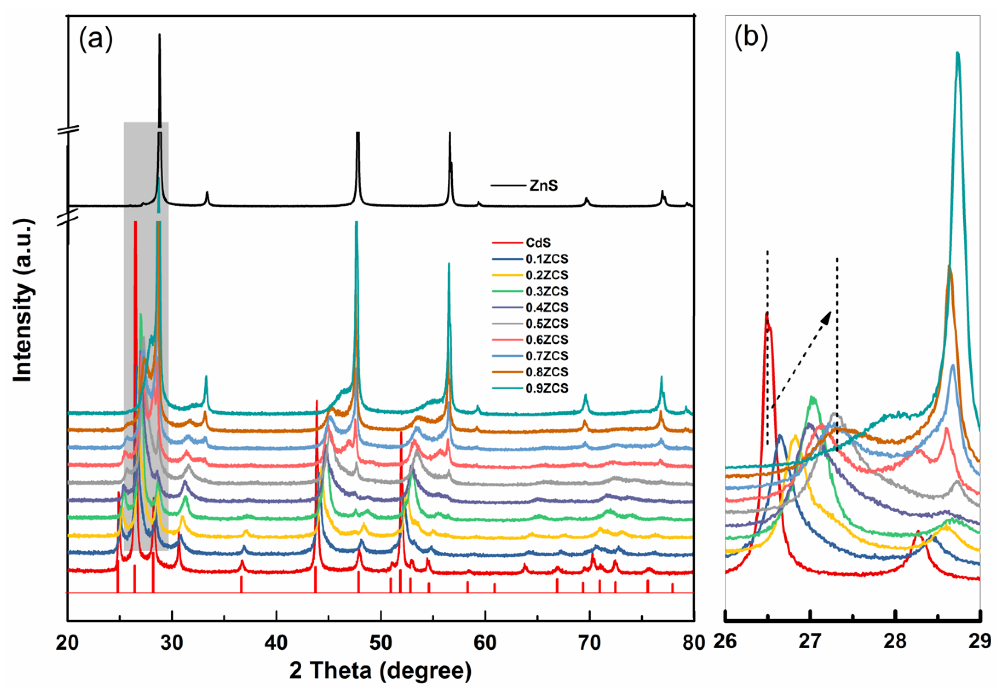
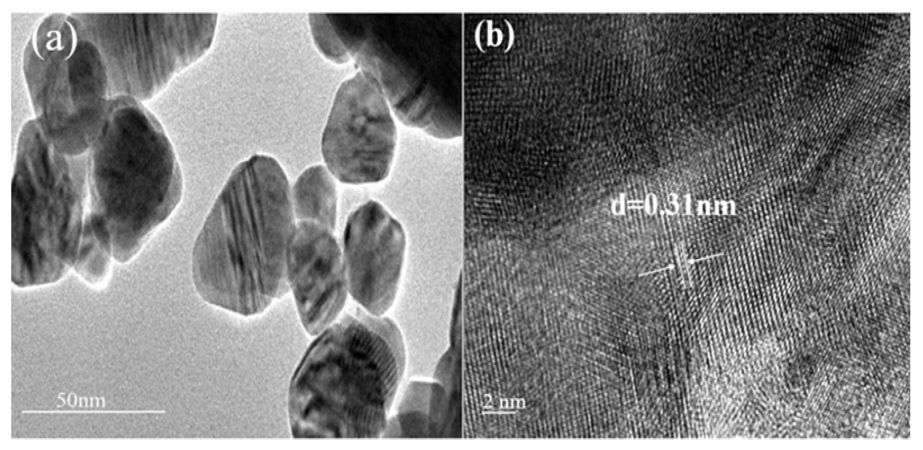
2.1. Phase Structures and Morphology

The crystal structure and the average crystallite size of the prepared xZCS samples were analyzed by X-ray diffraction (XRD). As shown in Figure 1a, the diffraction peaks of CdS located at 24.8°, 26.4°, 28.2°, 43.7°, and 51.0° can be attributed to the (100), (002), (101), (110), and (112) planes of hexagonal CdS (JCPDF Card No. 06-0314), whereas the peaks of ZnS at 28.5°, 47.5°, and 56.2° can be ascribed to the (111), (220), and (311) of cubic ZnS (JCPDF Card No.05-0566). xZCS changes gradually from the hexagonal to the cubic phase with the gradual increase of Zn content. The incorporation of Zn atoms into the lattice of CdS influences the positions of Cd atoms, and subsequently changes the lattice structure of the ZCS. The gradual phase transition is thermodynamically favored by the fact that the zinc blende structure (cubic phase ZnS) is more stable than wurtzite, whereas the wurtzite phase is more stable for CdS [20]. Furthermore, the diffraction intensity of xZCS gradually decreases with an increase of Zn content, and the peaks shift toward a high diffraction angle (Figure 1b, i.e., the strongest peaks), corresponding to a gradual decrease of d-spacing value (Table 1). The changes indicated the formation of the xZCS [22,39,40]. A similar observation has been reported in Huang’s work [41]. The differences in the cation radii of Zn2+ (0.74 Å) and Cd2+ (0.97 Å) are responsible for the successive peak shifts.
The crystallite size of the xZCS was calculated by the Scherrer equation. As shown in Table 1, the crystallite size first decreases from 28.45 to 12.93 nm, and then increases from 12.93 to 37.7 nm with the increase of Zn content. The obtained results are consistent with results of other studies [20,35].
The morphology and microstructure of the ZCS was further examined by transmission electron microscopy (TEM). As shown in Figure 2a, 0.3ZCS presents a nanosheet structure. The nanosheets are cumulative and the diameter of the aggregates is about 10–30 nm. The high-resolution transmission electron microscopy (HRTEM) image (Figure 2b) shows the interplanar spacing of 0.31 nm that corresponds to the (002) plane of hexagonal 0.3ZCS. The result is in agreement with that of XRD, as shown in Table 1.

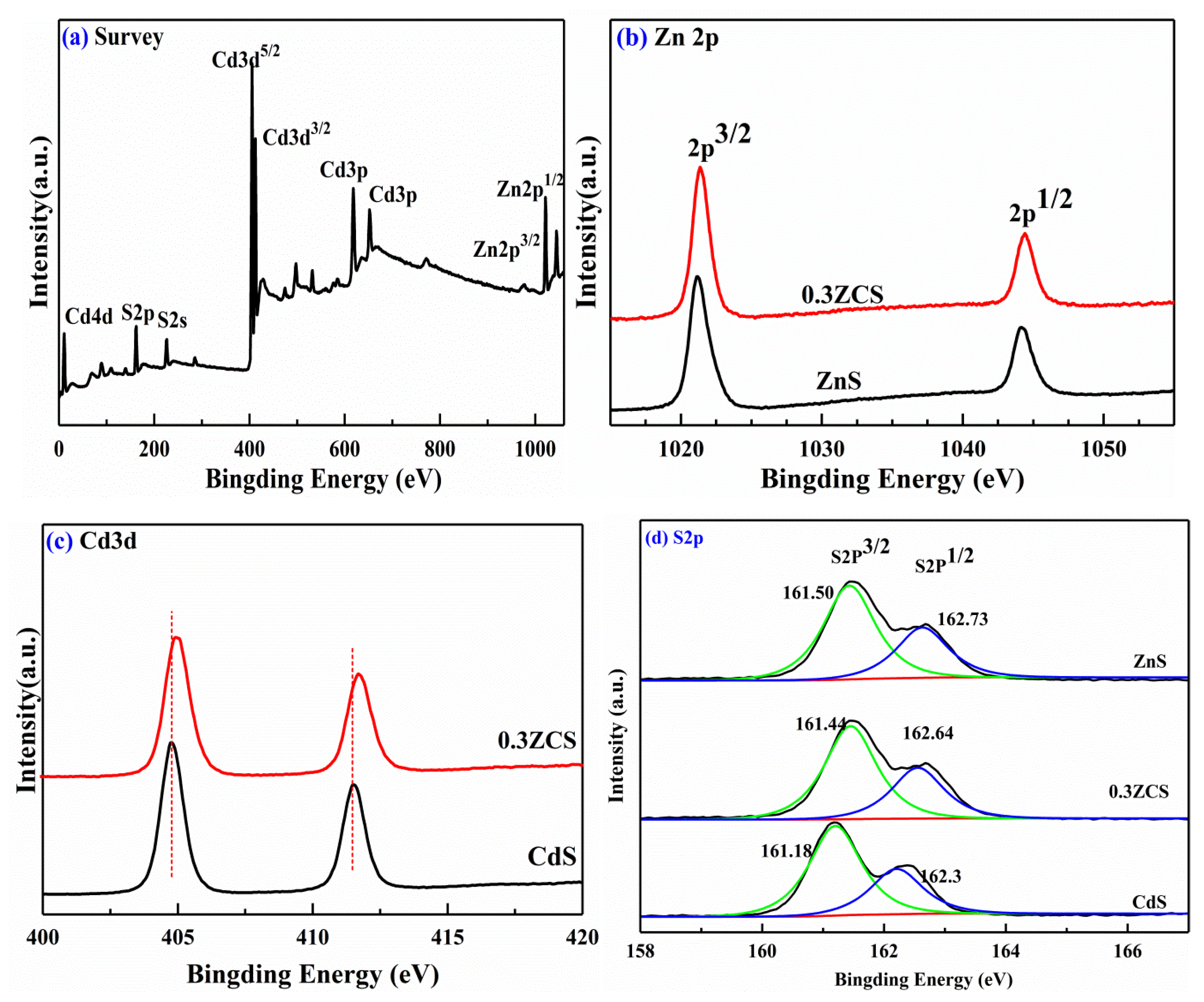
2.2. X-ray Photoelectron Spectroscopy (XPS) Analysis

An XPS analysis was performed to identify the chemical composition of the 0.3ZCS. Due to its high photocatalytic degradation efficiency of methyl orange (MO) among all the samples, 0.3ZCS was chosen for XPS detection. The survey spectrum (Figure 3a) shows that the sample is composed of Zn, Cd, and S. High-resolution XPS spectra of Zn 2p (Figure 3b ) and Cd 3d (Figure 3c ) exhibit a pair of symmetrical peaks with the binding energy of 1021.4 eV (Zn 2p3/2), 1044.5 eV (Zn 2p1/2), 404.9 eV (Cd 3d5/2), and 411.7 eV (Cd 3d3/2), respectively, indicating that the valence states of Zn and Cd elements are +2 [32,42,43]. As seen in Figure 3d, S 2p peaks located at 161.44 eV (S 2p3/2) and 162.64 (S 2p1/2) confirm the presence of S2 in 0.3ZCS [26,29,31]. In addition, the binding energy of Zn 2p and Cd 3d in 0.3ZCS is slightly higher than that of ZnS and CdS, respectively (Figure 3b,c), as also reported by other studies [39,44]. This can be attributed to the changed chemical states of the Zn and Cd atoms in 0.3ZCS [35]. The binding energy of S 2p in 0.3ZCS is slightly higher than that of CdS, but is close to to that of ZnS (Figure 3d). The shifts of the binding energies indicate the formation of a solid solution [29,39].

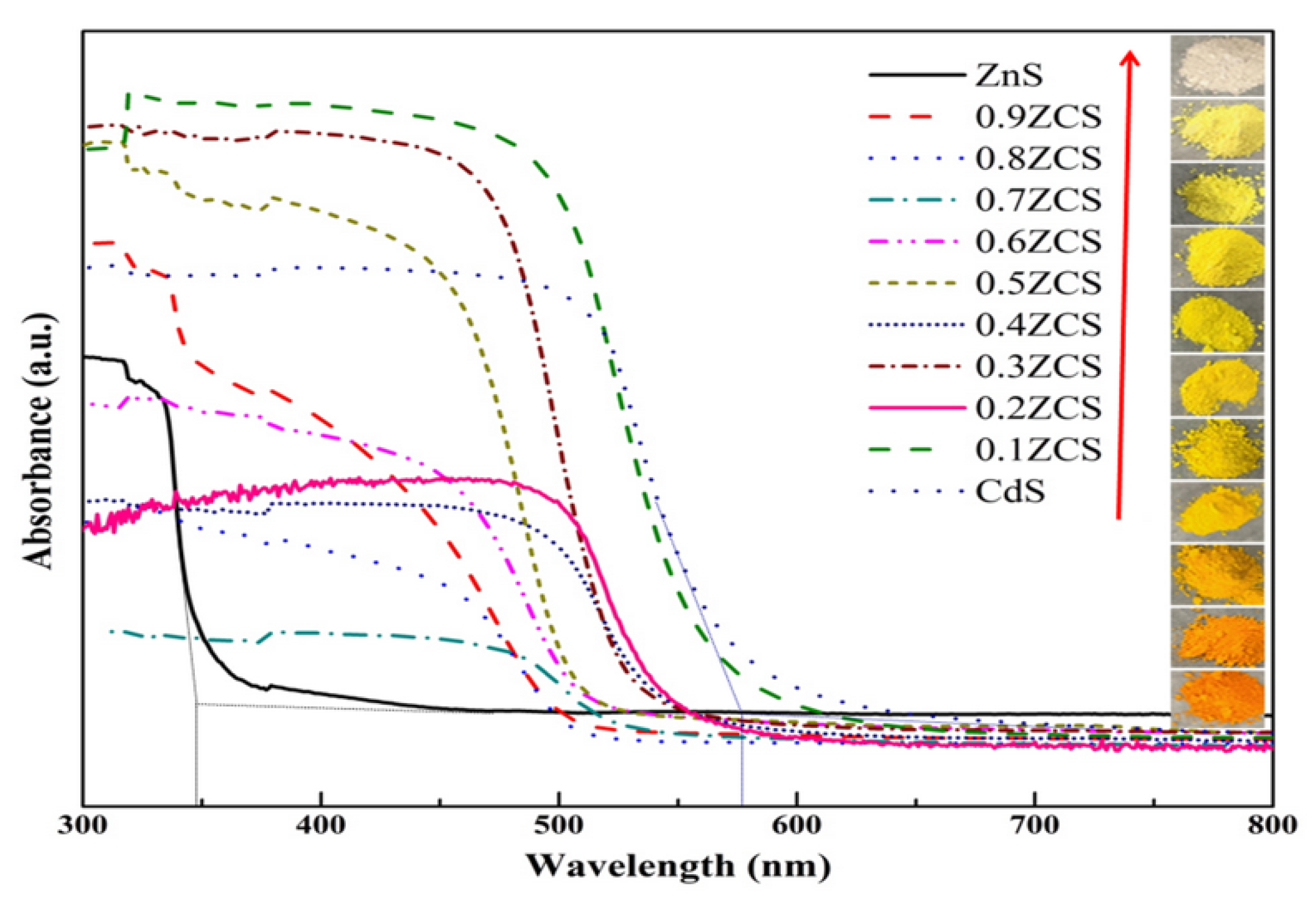
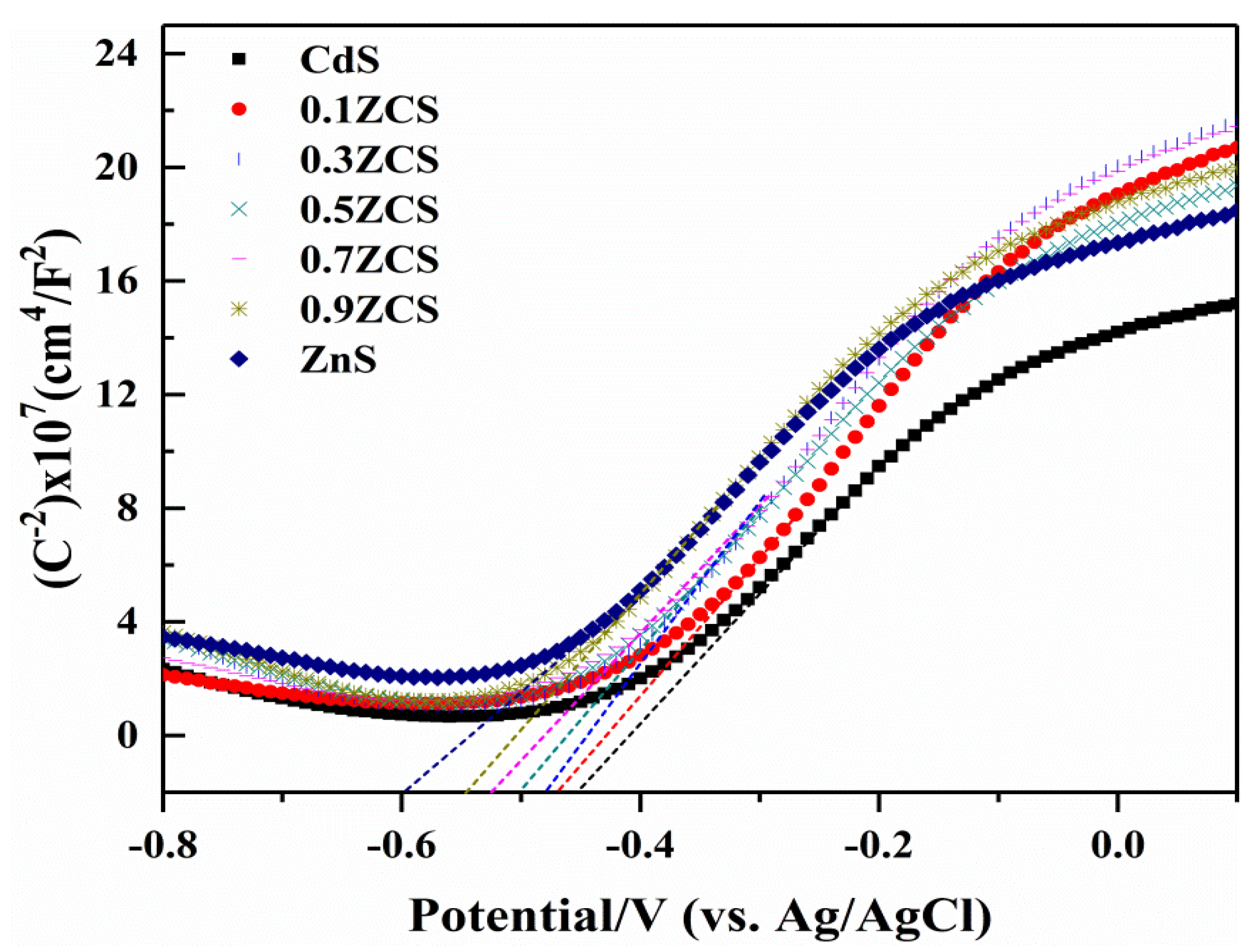
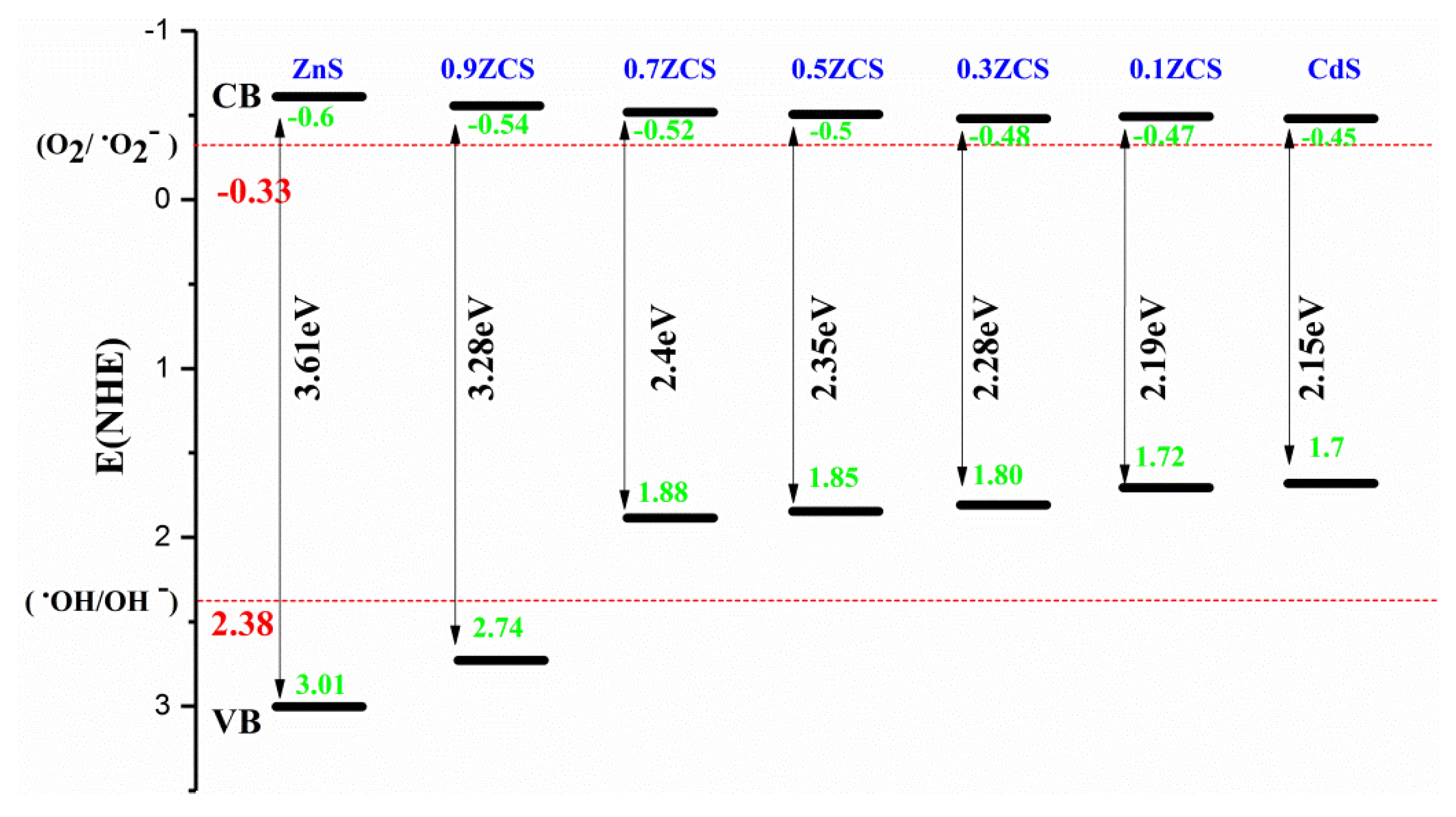
2.3. Optical Property and Band Structure Analysis

The UV-Vis diffuse reflectance spectroscopy (UV-Vis DRS) and the corresponding colors of the prepared xZCS samples are shown in Figure 4. With an increasing Zn content, the absorption edge of the prepared solid solution exhibits an obvious blue-shift from 576 to 343 nm, and the color of the sample changes gradually from dark orange to gray-white (inset of Figure 4).
The band gap energy (Eg) values of the prepared ZCS samples are estimated by Equation (1), represented as follows:
Eg = 1240/λ
where λ is the absorption threshold. The results are summarized in Table 1. The Eg of the xZCS samples increases gradually from 2.15 to 3.61 eV with the increasing Zn content. This demonstrates that the intrinsic Eg of the ZCS can be regulated by changing the ratio of Zn:Cd [24,31,35].
A Mott–Schottky (M-S) measurement was further performed to determine the band structure of the ZCS samples (Figure 5).
As shown in Figure 5, the positive slopes indicate that the xZCS samples are n-type semiconductors [32]. The flat-band potentials (Efb) of the xZCS samples can be measured by extrapolating the linear portion of the M-S curves to the X axis; the results are −0.45, −0.47, −0.48, −0.5, −0.52, −0.54, and −0.6 V (vs. Ag/AgCl) for CdS, 0.1ZCS, 0.3ZCS, 0.5ZCS, 0.7ZCS, 0.9ZCS, and ZnS, respectively. Generally, the Ecb of n-type semiconductor is more negative, i.e., about −0.1 or −0.2 V, compared to Efb [32,45]. Thus, the Ecb of CdS, 0.1ZCS, 0.3ZCS, 0.5ZCS, 0.7ZCS, 0.9ZCS, and ZnS are −0.65, −0.67, −0.68, −0.7, −0.72, −0.74, and −0.8 V, respectively.
Based on the measured Eg and Ecb values, the Evb of the prepared ZCS samples can be calculated by Evb = Ecb + Eg. Then, the resulting Ecb and Evb of the prepared ZCS samples are referred to as Normal Hydrogen Electrode Potential (ENHE = EAg/AgCl + 0.197 V) [45]; the results are schematically illustrated in Figure 6.
As shown in Figure 6, the Ecb of the ZCS samples are more negative than the standard redox potential of O2/O2 (−0.33 V vs. NHE) [28], while the Evb are less than the standard redox potential of OH/OH (2.38 V vs. NHE) [28]. A more negative Ecb enables the photogenerated electrons to reduce O2 into O2 radicals, which can further induce the production of HO2 or OH radicals [26,27]. These active oxygen species (O2, OH, and HO2) can conduct photocatalytic degradation of the dyes [28]. On the other hand, although the Evb of xZCS (excluding x = 0.9 and 1) is less than the standard redox potential of OH/OH and cannot oxidize OH to form OH radicals, the photogenerated holes can directly oxidize the dyes [26,28,29].

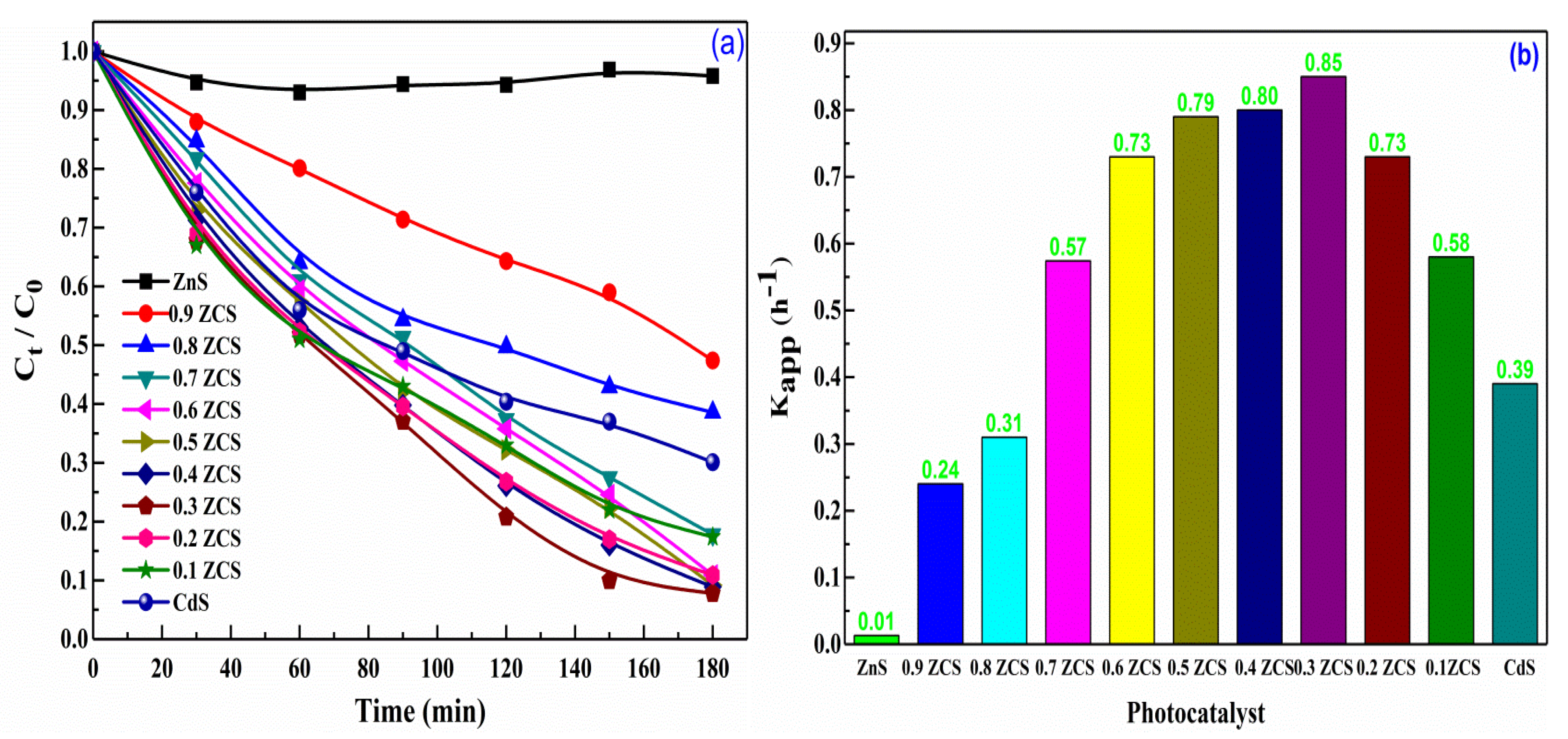
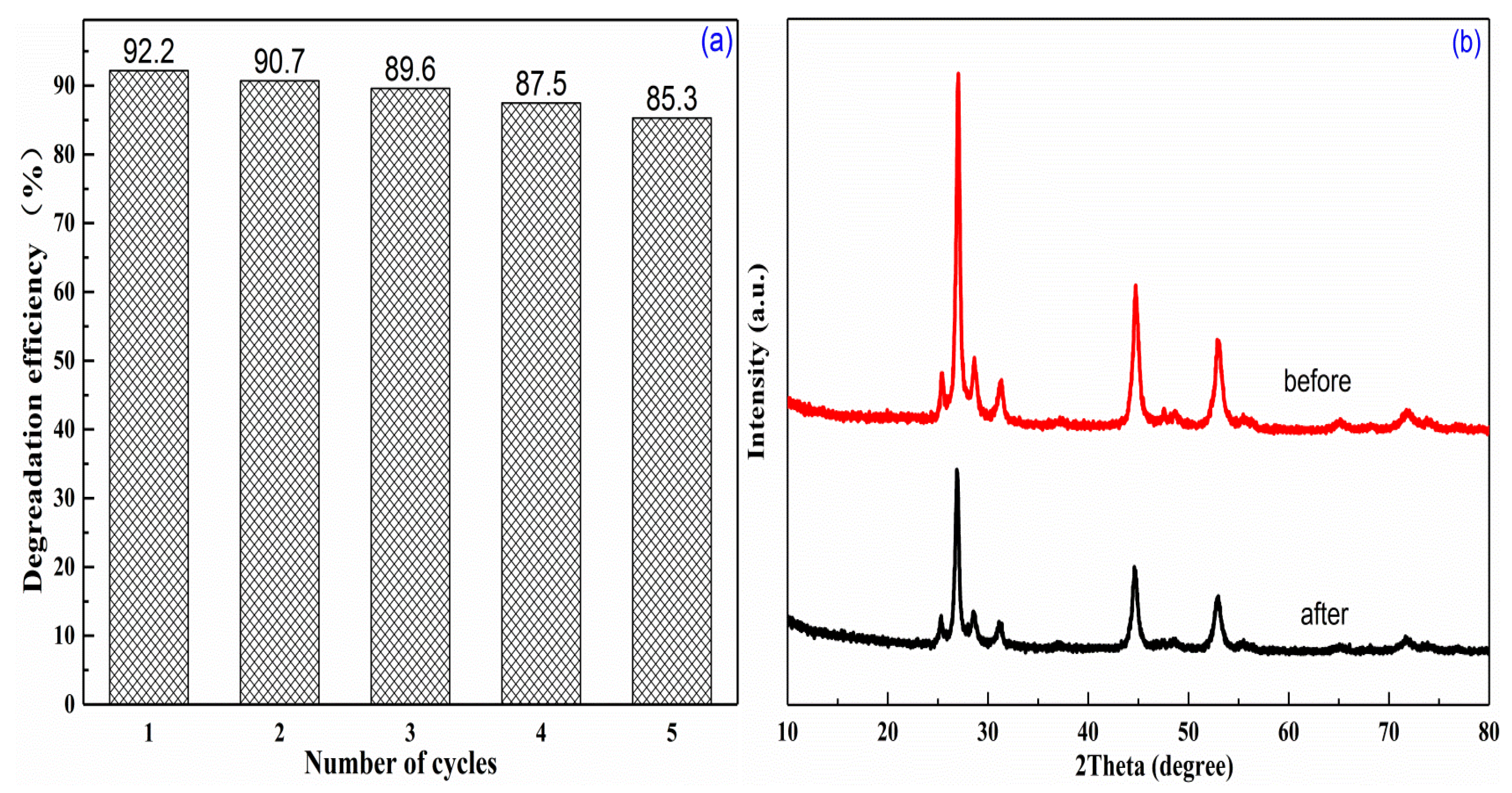
2.4. Photocatalytic Activity and Stability

The photocatalytic activity of the prepared xZCS was evaluated by the degradation of MO solution under visible light (λ > 420 nm). The variations of MO concentration with irradiation time are illustrated in Figure 7a. This indicated that the wide band gap ZnS shows no apparent activity for the decolorization of MO under visible light irradiation, whereas the ZCS samples show a monotonic decrease in the concentration of MO with irradiation time. Compared to pristine CdS, the introduction of Zn (x = from 0 to 0.3) into CdS crystal lattice forming ZCS solid solutions can substantially improve the photocatalytic activity. It was shown that 0.3ZCS yielded the best performance, that is, about 92% of MO was decolorized after irradiation for 3 h. Increasing the Zn content further from x = 0.3 to 0.9 leads to a decline of the activity.
The degradation kinetic of MO over the prepared samples was further analyzed by a pseudo first-order kinetic Equation (2):
lnC0/Ct = kapp × t
A model extensively used for the degradation of dyes was studied previously [6,46]. The resulting degradation rate constants (kapp) are summarized and compared in Figure 7b. Apparently, 0.3ZCS shows the highest degradation rate (0.85 h−1), i.e., almost 2.2 times higher than that of CdS (0.39 h−1). The superior photocatalytic performance of the 0.3ZCS sample for the degradation of the dyes was also reported in other studies [28,30,33]. In comparison, when using the same amount of the 0.3ZCS sample to degrade a MO solution of the same volume and concentration under the same test conditions, over 90% of the MO was decolorized after visible light irradiation for 3 h by the sample synthesized through the ultrasonic-assisted hydrothermal process, and after irradiation for no less than 5 h by the sample synthesized via traditional hydrothermal [33], microwave, and solvothermal routes [28], respectively. The best photocatalytic performance of 0.3ZCS prepared by our process may be attributed to its fabrication method as well as its proper composition, which offers a suitable band structure to strike a good balance between the light absorption and the redox capability of photoinduced charge carriers. The photocatalytic degradation mechanism is further discussed in the next paragraphs.
To investigate the stability of the prepared ZCS, five consecutive tests were carried out on the prepared 0.3ZCS sample.
As shown in Figure 8a, the high efficiency of 0.3ZCS still can be largely preserved after five consecutive tests. The slight drop of the performance was caused by the loss of the photocatalyst which occurred during the determination of MO concentration. The XRD results (Figure 8b) indicated that the spent 0.3ZCS after the five cycling tests shows an identical XRD pattern to the fresh one, further confirming the stability of the sample.

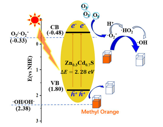
2.5. Mechanism on Enhancement in Photocatalytic Activity

The absorption of light and the oxidation capability of the photoinduced charge carriers are key factors to determine the visible light photocatalytic degradation of dyes [33]. Generally, Cd-rich ZCS samples show better activity than Zn-rich samples because the former has a narrow band gap, and consequently, can generate more photoinduced charge carriers. However, a smaller band gap is not always better, because the redox capability of photo-generated charge carriers will decrease as the band gap becomes narrow. Therefore, to pursue a high photocatalytic performance of a solid solution photocatalyst, its composition needs to be optimized not only to maximize its visible light absorption range, but also to avoid a significant sacrifice of the redox capability of charge carriers. Our results indicate that x = 0.3−0.5 is the appropriate composition of ZCS, and 0.3ZCS shows the highest activity, which agrees well with the findings in previously reported works [28,30,33].
In addition, the fabrication method may also affect the photocatalytic activity of the photocatalysts. As mentioned above, compared with conventional hydrothermal, microwave, and solvothermal methods, a 0.3ZCS solid solution prepared by the ultrasonication-assisted hydrothermal process in this study has best photocatalytic performance. This is due to: (1) the ultrasonication treatment making the precursors mix well and react completely to form high-crystallinity crystals [35], and (2) once thioacetamide as a sulfur source combines with water to form H2S gas, Zn2+ and Cd2+ quickly bind to the S2− to form ZnxCd1−xS nanoparticles, thus yielding improved photocatalytic activity [47].

3. Materials and Methods

3.1. Preparation of ZnxCd1−xS

All reagents were purchased from Sinopharm Chemical Reagent Co., Ltd (Shanghai, China). ZnxCd1−xS was prepared by the following procedure. First, the reaction materials of Zn(Ac)2·2H2O, Cd(Ac)2·2H2O (with different mole ratios as given in Table 1), and 0.94 g CH3CSNH2 were mixed in 40 mL of deionized water under ultrasonic vibration. Then, 10 mL of NaOH aqueous solution (4 mol·L−1) was added dropwise into the above solution. After ultrasonic treatment for 0.5 h, the reaction mixture was transferred into a 100-mL, Teflon-lined, stainless-steel autoclave and then hydrothermally treated for 24 h at 180 °C. Next, the products were washed several times with deionized water and absolute ethanol, respectively, and dried in a vacuum oven for 8 h at 60 °C to obtain the final ZnxCd1−xS samples. Finally, according to the value of x in ZnxCd1−xS (x = 0–1), the as-synthesized products were marked as xZCS, as exemplified in Table 1.

3.2. Photocatalytic Tests

The visible-light-responsive photocatalytic activities of the xZCS were evaluated by measuring the degradation rate of MO under visible light irradiation. For this, 0.04 g photocatalyst was dispersed in 80 mL of MO aqueous solution with an initial concentration of 20 mg·L−1 under magnetic stirring; then, the suspension was further stirred for 0.5 h in the dark to establish an adsorption–desorption equilibrium between the MO and the photocatalyst. Next, the suspension was exposed to visible-light irradiation from a 300 W xenon lamp (Beijing Zhongjiao Jinyuan Technology Co., Ltd, Beijing, China) equipped with a 420 nm cut-off filter for 3 h. At a given time interval, 5 mL suspension was taken out from the reaction solution and centrifuged to obtain a clear solution. The absorbance of MO was measured spectrophotometrically (722N visible spectrophotometer, Shanghai Precision & Scientific Instrument Co. Ltd. Shanghai, China) at its maximum absorption wavelength (463 nm). Finally, the photocatalytic degradation efficiency was estimated according to the time profiles of Ct/C0; here, C0 refers to the initial concentration of MO, and Ct corresponds to the concentration after irradiation for t min. The stability of the catalysts was evaluated by five cycles of photocatalytic degradation of MO.

3.3. Characterization

The crystal structure of the sample was determined by a Bruker D8 advance X-ray diffractometer (Bruker AXS, Karlsruhe, Germany) employing Cu Kα radiation (λ = 1.5406 Å) operated at 40 kV and 40 mA in air. The morphology and microstructure were analyzed by TEM and HRTEM using a JEOL JEM-2010 electron microscope (JEOL Ltd, Tokyo, Japan) with an acceleration voltage of 200 kV. The XPS measurements were conducted on an X-ray photoelectron spectrometer (ESCA LAB 250 Xi, Thermo Fisher Scientific, Waltham, MA, USA). The UV-Vis absorption spectra were recorded by UV-vis DRS (UV-3600, Shimadzu, Japan) with a wavelength range of 200–800 nm.

4. Conclusions

A series of ZCSs with nanosheet structures were successfully synthesized by an ultrasonication-assisted hydrothermal method. All the solid solutions (except for x = 0.8−1.0) showed enhanced efficiency for the photocatalytic degradation of MO solution under visible light irradiation in comparison with CdS; the highest degradation efficiency of 92% was achieved for Zn0.3Cd0.7S. The balance between the band gap structure-directed redox capacity and light absorption may open doors in the exploration of efficient photocatalysts with high stability.

Author Contributions

Conceptualization was done by L.Y., L.L., and S.C.; methodology was done by L.Y.; software analysis was performed by M.L. and X.F.; validation was done by M.Z. and Y.F.; formal analysis was done by H.B.; investigation was carried out by L.Y.; writing—original draft preparation was done by L.Y. and M.Z.; writing—review and editing was executed by L.L. and S.C.; visualization was done by M.Z. and Y.F.; supervision was done under L.L.; project administration was carried out by L.L. and S.C.; funding acquisition was done by L.L. and S.C. All authors have read and agreed to the published version of the manuscript.

Funding

This research was funded by the Natural Science Foundation of Anhui Province for Distinguished Young Scholars (grant number 1808085J24), the Natural Science Foundation of Anhui Province (grant number 1808085MB45) and the Natural Science Foundation of Anhui Education Bureau (grant number KJ2018B03).

Conflicts of Interest

The authors declare no conflict of interest.

References

  1. Han, F.; Kambala, V.S.R.; Srinivasan, M.; Rajarathnam, D.; Naidu, R. Tailored titanium dioxide photocatalysts for the degradation of organic dyes in wastewater treatment: A review. Appl. Catal. A Gen. 2009, 359, 25–40. [Google Scholar] [CrossRef]
  2. Lam, S.M.; Sin, J.C.; Abdullah, A.Z.; Mohamed, A.R. Degradation of wastewaters containing organic dyes photocatalysed by zinc oxide: A review. Desalin. Water Treat. 2012, 41, 131–169. [Google Scholar] [CrossRef]
  3. Chong, M.N.; Jin, B.; Chow, C.W.; Saint, C. Recent developments in photocatalytic water treatment technology: A review. Water Res. 2010, 44, 2997–3027. [Google Scholar] [CrossRef] [PubMed]
  4. Abdulkarem, A.M.; Aref, A.A.; Abdulhabeeb, A.; Li, Y.F.; Yu, Y. Synthesis of Bi2O3/Cu2O nanoflowers by hydrothermal method and its photocatalytic activity enhancement under simulated sunlight. J. Alloys Compd. 2013, 560, 132–141. [Google Scholar] [CrossRef]
  5. Yan, S.C.; Li, Z.S.; Zou, Z.G. Photodegradation performance of g-C3N4 fabricated by directly heating melamine. Langmuir 2009, 25, 10397–10401. [Google Scholar] [CrossRef] [PubMed]
  6. Zhang, M.L.; Yang, L.; Wang, Y.J.; Li, L.F.; Chen, S.F. High yield synthesis of homogeneous boron doping C3N4 nanocrystals with enhanced photocatalytic property. Appl. Surf. Sci. 2019, 489, 631–638. [Google Scholar] [CrossRef]
  7. Li, L.F.; Zhang, W.X.; Feng, C.; Luan, X.W.; Jiang, J.; Zhang, M.L. Preparation of nanocrystalline Cu2O by a modified solid-state reaction method and its photocatalytic activity. Mater. Lett. 2013, 107, 123–125. [Google Scholar] [CrossRef]
  8. Zhang, Y.X.; Ye, Y.J.; Zhou, X.B.; Liu, Z.L.; Ma, D.; Li, B.; Liu, Q.H.; Zhu, G.P.; Chen, S.; Li, X. Facile preparation of a monodispersed CuO yolk-shelled structure with enhanced photochemical performance. Cryst. Eng. Commun. 2016, 18, 7994–8003. [Google Scholar] [CrossRef]
  9. Cheng, L.; Xiang, Q.J.; Liao, Y.L.; Zhang, H.W. CdS-Based photocatalysts. Environ. Sci. Technol. 2018, 11, 1362–1391. [Google Scholar] [CrossRef]
  10. Mort, J.; Spear, W.E. Hole drift mobility and lifetime in CdS crystals. Phys. Rev. Lett. 1962, 8, 314–315. [Google Scholar] [CrossRef]
  11. Deng, Q.; Miao, T.; Wang, Z.; Xu, Y.; Fu, X. Compositional regulation and modification of the host CdS for efficient photocatalytic hydrogen production: Case study on MoS2 decorated Co0.2Cd0.8S nanorods. Chem. Eng. J. 2019, 378, 122139. [Google Scholar] [CrossRef]
  12. Xie, Y.; Ali, G.; Yoo, S.H.; Cho, S.O. Sonication-assisted synthesis of CdS quantum-dot-sensitized TiO2 nanotube arrays with enhanced photoelectrochemical and photocatalytic activity. ACS Appl. Mater. Inter. 2010, 2, 2910–2914. [Google Scholar] [CrossRef]
  13. Fu, J.; Chang, B.B.; Tian, Y.L.; Xi, F.N.; Dong, X.P. Novel C3N4-CdS composite photocatalysts with organic-inorganic heterojunctions: In situ synthesis, exceptional activity, high stability and photocatalytic mechanism. J. Mater. Chem. A 2013, 1, 3083–3090. [Google Scholar] [CrossRef]
  14. Lin, Y.F.; Hsu, Y.J. Interfacial charge carrier dynamics of type-II semiconductor nanoheterostructures. Appl. Catal. B Environ. 2013, 130, 93–98. [Google Scholar] [CrossRef]
  15. Ren, Z.; Zhang, J.; Xiao, F.X.; Xiao, G. Revisiting the construction of graphene–CdS nanocomposites as efficient visible-light-driven photocatalysts for selective organic transformation. J. Mater. Chem. A 2014, 2, 5330–5339. [Google Scholar] [CrossRef]
  16. Fan, Q.J.; Huang, Y.N.; Zhang, C.; Liu, J.J.; Piao, L.Y.; Yu, Y.C.; Zuo, S.L.; Li, B.S. Superior nanoporous graphitic carbon nitride photocatalyst coupled with CdS quantum dots for photodegradation of RhB. Catal. Today 2016, 264, 250–256. [Google Scholar] [CrossRef]
  17. Tian, Q.; Wu, W.; Liu, J.; Wu, Z.; Yao, W.; Ding, J.; Jiang, C. Dimensional heterostructures of 1D CdS/2D ZnIn2S4 composited with 2D graphene: Designed synthesis and superior photocatalytic performance. Dalton Trans. 2017, 46, 2770–2777. [Google Scholar] [CrossRef]
  18. Lai, J.; Qin, Y.M.; Lan, Y.; Zhang, C. GSH-assisted hydrothermal synthesis of MnxCd1xS solid solution hollow spheres and their application in photocatalytic degradation. Mat. Sci. Semicon. Proc. 2016, 52, 82–90. [Google Scholar] [CrossRef]
  19. Swafford, L.A.; Weigand, L.A.; Bowers, M.J.; McBride, J.R.; Rapaport, J.L.; Watt, T.L.; Dixit, S.K.; Feldman, L.C.; Rosenthal, S.J. Homogeneously alloyed CdSxSe1-x nanocrystals: Synthesis, characterization, and composition/size-dependent band gap. J. Am. Chem. Soc. 2006, 128, 12299–12306. [Google Scholar] [CrossRef]
  20. Kaur, M.; Nagaraja, C.M. Template-free synthesis of Zn1−xCdxS nanocrystals with tunable band structure for efficient water splitting and reduction of nitroaromatics in water. ACS Sustain. Chem. Eng. 2017, 5, 4293–4303. [Google Scholar] [CrossRef]
  21. Zhao, X.; Luo, Z.; Hei, T.; Jiang, Y. One-pot synthesis of ZnxCd1−xS nanoparticles with nano-twin structure. J. Photochem. Photobiol. A 2019, 382, 11919. [Google Scholar] [CrossRef]
  22. Chen, J.; Lv, S.; Shen, Z.; Tian, P.; Chen, J.; Li, Y. Novel ZnCdS quantum dots engineering for enhanced visible-light-driven hydrogen evolution. ACS Sustain. Chem. Eng. 2019, 7, 13805–13814. [Google Scholar] [CrossRef]
  23. Tang, L.; Kuai, L.; Li, Y.; Li, H.; Zhou, Y.; Zou, Z. ZnxCd1−xS tunable band structure-directing photocatalytic activity and selectivity of visible-light reduction of CO2 into liquid solar fuels. Nanotechnology 2018, 29, 064003. [Google Scholar] [CrossRef] [PubMed]
  24. Lou, S.; Wang, W.; Jia, X.; Wang, Y.; Zhou, S. A unique nanoporous graphene-ZnxCd1xS hybrid nanocomposite for enhanced photocatalytic degradation of water pollutants. Ceram. Int. 2016, 42, 16775–16781. [Google Scholar] [CrossRef]
  25. Yang, M.; Wang, Y.B.; Ren, Y.K.; Liu, E.Z.; Fan, J.; Hu, X.Y. Zn/Cd ratio-dependent synthetic conditions in ternary ZnCdS quantum dots. J. Alloys Compd. 2018, 752, 260–266. [Google Scholar] [CrossRef]
  26. Wang, X.; Tian, H.; Cui, X.; Zheng, W.; Liu, Y. One-pot hydrothermal synthesis of mesoporous ZnxCd1−xS/reduced graphene oxide hybrid material and its enhanced photocatalytic activity. Dalton Trans. 2014, 43, 12894–12903. [Google Scholar] [CrossRef]
  27. Li, W.; Li, D.; Xian, J.; Chen, W.; Hu, Y.; Shao, Y.; Fu, X. Specific analyses of the active species on Zn0.28Cd0.72S and TiO2 photocatalysts in the degradation of methyl orange. J. Phys. Chem. C 2010, 114, 21482–21492. [Google Scholar] [CrossRef]
  28. Li, W.J.; Li, D.Z.; Zhang, W.J.; Hu, Y.; He, Y.H.; Fu, X.Z. Microwave synthesis of ZnxCd1−xS nanorods and their photocatalytic activity under visible light. J. Phys. Chem. C 2010, 114, 2154–2159. [Google Scholar] [CrossRef]
  29. Abideen, Z.U.; Teng, F. Effect of alkaline treatment on photochemical activity and stability of Zn0.3Cd0.7S. Appl. Surf. Sci. 2019, 465, 459–469. [Google Scholar] [CrossRef]
  30. Lee, Y.Y.; Moon, J.H.; Choi, Y.S.; Park, G.O.; Jin, M.; Jin, L.Y.; Li, D.; Lee, J.Y.; Son, S.U.; Kim, J.M. Visible-light driven photocatalytic degradation of organic dyes over ordered mesoporous CdxZn1–xS materials. J. Phys. Chem. C 2017, 121, 5137–5144. [Google Scholar] [CrossRef]
  31. Zhao, X.; Feng, J.; Liu, J.; Shi, W.; Yang, G.; Wang, G.C.; Cheng, P. An efficient, visible-light-driven, hydrogen evolution catalyst NiS/ZnxCd1−xS nanocrystal derived from a metal-organic framework. Angew. Chem. Int. 2018, 57, 9790–9794. [Google Scholar] [CrossRef] [PubMed]
  32. Liu, Y.; Wang, G.; Li, Y.; Jin, Z. 2D/1D Zn0.7Cd0.3S p-n heterogeneous junction enhanced with NiWO4 for efficient photocatalytic hydrogen evolution. J. Colloid Interface Sci. 2019, 554, 113–124. [Google Scholar] [CrossRef] [PubMed]
  33. Li, W.; Li, D.; Chen, Z.; Huang, H.; Sun, M.; He, Y.; Fu, X. High-efficient degradation of dyes by ZnxCd1xS solid solutions under visible light irradiation. J. Phys. Chem. C 2008, 112, 14943–14947. [Google Scholar] [CrossRef]
  34. Yu, J.; Yang, B.; Cheng, B. Noble-metal-free carbon nanotube-Cd0.1Zn0.9S composites for high visible-light photocatalytic H2-production performance. Nanoscale 2012, 4, 2670–2677. [Google Scholar] [CrossRef] [PubMed]
  35. Li, Q.; Meng, H.; Zhou, P.; Zheng, Y.; Wang, J.; Yu, J.; Gong, J. Zn1−xCdxS solid solutions with controlled bandgap and enhanced visible-light photocatalytic H2-production activity. ACS Catal. 2013, 3, 882–889. [Google Scholar] [CrossRef]
  36. Pugazhenthiran, N.; Sathishkumar, P.; Murugesan, S.; Anandan, S. Effective degradation of acid orange 10 by catalytic ozonation in the presence of Au-Bi2O3 nanoparticles. Chem. Eng. J. 2011, 168, 1227–1233. [Google Scholar] [CrossRef]
  37. Liao, K.H.; Lin, Y.S.; Macosko, C.W.; Haynes, C.L. Cytotoxicity of graphene oxide and graphene in human erythrocytes and skin fibroblasts. ACS Appl. Mater. Inter. 2011, 3, 2607–2615. [Google Scholar] [CrossRef]
  38. Ghows, N.; Entezari, M.H. Exceptional catalytic efficiency in mineralization of the reactive textile azo dye (RB5) by a combination of ultrasound and core-shell nanoparticles (CdS/TiO2). J. Hazard. Mater. 2011, 195, 132–138. [Google Scholar] [CrossRef]
  39. Zhong, J.; Zhang, Y.; Hu, C.; Hou, R.; Yin, H.; Li, H.; Huo, Y. Supercritical solvothermal preparation of a ZnxCd1xS visible photocatalyst with enhanced activity. J. Mater. Chem. A 2014, 2, 19641–19647. [Google Scholar] [CrossRef]
  40. Zhong, X.; Feng, Y.; Knoll, W.; Han, M. Alloyed ZnxCd1−xS nanocrystals with highly narrow luminescence spectral width. J. Am. Chem. Soc. 2003, 125, 13559–13563. [Google Scholar] [CrossRef]
  41. Huang, M.; Yu, J.; Deng, C.; Huang, Y.; Fan, M.; Li, B.; Tong, Z.; Zhang, F.; Dong, L. 3D nanospherical CdxZn1-xS/reduced graphene oxide composites with superior photocatalytic activity and photocorrosion resistance. Appl. Surf. Sci. 2016, 365, 227–239. [Google Scholar] [CrossRef]
  42. Wu, Y.J.; Yue, Z.K.; Liu, A.J.; Yang, P.; Zhu, M.S. P-type Cu-doped Zn0.3Cd0.7S/Graphene photocathode for efficient water splitting in a photoelectrochemical tandem cell. ACS Sustain. Chem. Eng. 2016, 4, 2569–2577. [Google Scholar] [CrossRef]
  43. Liu, Y.; Wang, G.R.; Ma, Y.L.; Jin, Z.L. Noble-metal-free visible light driven hetero-structural Ni/ZnxCd1−xS photocatalyst for efficient hydrogen production. Catal. Lett. 2019, 149, 1788–1799. [Google Scholar] [CrossRef]
  44. Zhang, J.; Yu, J.; Jaroniec, M.; Gong, J.R. Noble metal-free reduced graphene oxide-ZnxCd1−xS nanocomposite with enhanced solar photocatalytic H2-production performance. Nano Lett. 2012, 12, 4584–4589. [Google Scholar] [CrossRef]
  45. Yi, S.S.; Yan, J.M.; Wulan, B.R.; Li, S.J.; Liu, K.H.; Jiang, Q. Noble-metal-free cobalt phosphide modified carbon nitride: An efficient photocatalyst for hydrogen generation. Appl. Catal. B Environ. 2017, 200, 477–483. [Google Scholar] [CrossRef]
  46. Wu, C.H.; Chang, H.W.; Chern, J.M. Basic dye decomposition kinetics in a photocatalytic slurry reactor. J. Hazard. Mater. 2006, 137, 336–343. [Google Scholar] [CrossRef]
  47. Wang, W.; Shen, H.; He, X.; Li, J. Effects of sulfur sources on properties of Cu2ZnSnS4 nanoparticles. J. Nanopart. Res. 2014, 16, 1–8. [Google Scholar] [CrossRef]
Figure 1. (a) XRD patterns of the prepared CdS, ZnS, and xZCS samples, (b) local enlarged patterns.
Catalysts 10 00276 g001
Figure 2. (a) TEM and (b) HRTEM images of the 0.3ZCS sample.
Catalysts 10 00276 g002
Figure 3. (a) Survey and high resolution (b) Zn 2p; (c) Cd 3d; (d) S 2p XPS spectra of the prepared 0.3ZCS sample.
Catalysts 10 00276 g003
Figure 4. The UV-Vis DRS spectra of the ZnxCd1−xS (x = 0–1.0) samples.
Catalysts 10 00276 g004
Figure 5. Mott–Schottky plots of the prepared ZCS samples as electrode materials.
Catalysts 10 00276 g005
Figure 6. The conduction and valence band potentials of the prepared ZCS samples.
Catalysts 10 00276 g006
Figure 7. (a) Photocatalytic degradation of MO on the prepared xZCS samples; (b) Kapp values of the prepared xZCS samples of the photocatalytic degradation of MO under visible light (> 420 nm) irradiation.
Catalysts 10 00276 g007
Figure 8. (a) Recycling tests for the photocatalytic degradation of MO over 0.3ZCS sample under visible light irradiation; (b) Comparison of XRD patterns of 0.3ZCS sample before and after five cycles tests.
Catalysts 10 00276 g008
Table 1. Elemental composition and characterization results of the prepared samples.
SampleCd/Zn/S Molar RatioSize (nm)Eg (eV)d Value (Å)
CdS
0.1ZCS
0.2 ZCS
0.3 ZCS
0.4 ZCS
0.5 ZCS
0.6ZCS
0.7 ZCS
0.8 ZCS
0.9ZCS
ZnS
Zn0Cd1.0S
Zn0.1Cd0.9S
Zn0.2Cd0.8S
Zn0.3Cd0.7S
Zn0.4Cd0.6S
Zn0.5Cd0.5S
Zn0.6Cd0.4S
Zn0.7Cd0.3S
Zn0.8Cd0.2S
Zn0.9Cd0.1S
Zn1.0Cd0S
28.45
21.70
20.00
14.50
13.60
13.10
12.93
21.00
28.64
28.96
37.7
2.15
2.19
2.25
2.28
2.31
2.35
2.37
2.40
2.45
3.28
3.61
3.36
3.34
3.32
3.30
3.29
3.28
3.26
3.12
3.11
3.10
3.09

