Article

The Efficacy of Silver Nitrate (AgNO3) as a Coating Agent to Protect Paper against High Deteriorating Microbes

by
Amr Fouda
1,*,
Gomaa Abdel-Maksoud
2,
Hosam A. Saad
3,
Adil A. Gobouri
3,
Zuhair M. Mohammedsaleh
4 and
Mohamad Abdel-Haleem El-Sadany
5
1
Botany and Microbiology Department, Faculty of Science, Al-Azhar University, Nasr City, Cairo 11884, Egypt
2
Conservation Department, Faculty of Archaeology, Cairo University, Giza 12613, Egypt
3
Department of Chemistry, College of Science, Taif University, P.O. Box 11099, Taif 21944, Saudi Arabia
4
Department of Medical Laboratory Technology, Faculty of Applied Medical Sciences, University of Tabuk, Tabuk 71491, Saudi Arabia
5
Adminstration of Conservation of Islamic Monuments in Mansoura, Dakahlia Governorate, Ministry of Antiquities, Cairo 26854, Egypt
*
Author to whom correspondence should be addressed.
Submission received: 10 February 2021 / Revised: 18 February 2021 / Accepted: 20 February 2021 / Published: 26 February 2021
(This article belongs to the Special Issue Gold, Silver and Copper Catalysis)

Abstract

:
This study focuses on the efficacy of silver nitrate (AgNO3) as a coating agent used to preserve papers against microbial deterioration. To this end, the in vitro cytotoxicity of AgNO3 was assessed against two normal cell lines, WI-38 and HFB-4, to detect a safe dose that can be used as a coating agent, which was 80 µg mL−1. Bacillus subtilis B3 and Penicillium chrysogenum F9 were selected as high deteriorating microbes, previously isolated from a historical manuscript dating back to 1677 A.-D. The microbial growth inhibition, color change, mechanical properties, and cellulosic fibers of untreated/treated papers were evaluated. The data showed the efficacy of AgNO3 to inhibit the growth of B. subtilis with a percentage of 100% after 7 days, while it inhibits the growth of P. chrysogenum with a percentage of 85.9 ± 1.1% after 21 days. The color and mechanical properties of treated paper in the presence/absence of microbial inoculation were slightly changed, although they changed greatly due to microbial growth in the absence of AgNO3. The EDX analysis confirmed the successful adsorption of Ag-ion on papers, with a weight percentage of 1.9%. The cellulosic fibers of untreated paper in the presence of microbial growth were highly deteriorated as compared with treated and standard filter paper (shown by FT-IR and SEM).

1. Introduction

History is registered on paper, historical or archeological manuscripts, and documents, which makes the preservation of these materials very important. Like other materials, papers can undergo chemical, physical, and biological degradation through the ages [1]. Physical and chemical agents such as UV, pollutants, heat, humidity, light exposure, pH, poor ventilation, and metal ions contribute to aspects of deterioration [2]. Biological degradation consists of the ability of microbes to degrade the main components of paper, especially cellulose, lignin, and hemicellulose, through their efficacy in the secretion of hydrolytic enzymes [3]. Microbes, especially fungi, can degrade other materials involved in paper manufactures, such as fillers and sizing materials, because they have high sugar and protein content [1]. Different microbes including bacteria, fungi, and actinomycetes such as Bacillus spp., Sphingomonas sp. Pseudomonas, Serratia, Acinetobacter, Staphylococcus, Alternaria, Cladosporium, Penicillium, Chaetomium, Aspergillus, Cladosporium, Streptomyces, and Nocardia sp. are characterized by their ability to degrade cellulosic fibers [4,5,6,7,8]. These organisms possess the ability to secrete different cellulase enzymes such as endo-glucanase (EC:3.2.1.4), exo-glucanase (EC:3.2.1.19), and β-glucosidase (EC:3.2.1.21) enzymes, which synergistically contribute to cellulosic fiber degradation [9]. In addition to their potential to deteriorate cellulosic fibers, these organisms can produce toxic substances such as mycotoxins and allergenic compounds, which have negative impacts on the health of visitors, readers, and library workers [10]. Therefore, it is urgent to develop a new approach that does not affect human health and is effective in preserving historical or archeological papers from degrading microorganisms.
Different strategies, including chemical and physical methods, are used to preserve documents and archeological manuscripts from deterioration. Among chemical methods, alcohols, azole antifungals, phenol derivatives, acids and salts, essential oils, and photocatalysts can be used in the preservation processes [11,12,13]. Physical methods such as dehydration, γ-radiation, freezing, UV-radiation, high temperature, and high-frequency currents are utilized in preservation [14,15,16]. Nanosized materials such as zinc oxide, magnesium, calcium hydroxide, and silver can be used to inhibit the growth of deteriorating microbes, as well as the deacidification of paper [17,18,19].
Recently, metal compounds, particularly silver nitrate (AgNO3), have been intensely used in different biotechnological applications [20,21]. Silver nitrate has the potential to inhibit the growth of a wide range of microbes such as bacteria, fungi, actinomycetes, and viruses [22,23]. Silver has been defined as an “oligodynamic or biocidal agent” because of its ability to inhibit microbial growth at low concentrations [24]. Recently, silver ions have been used as anti-inflammatory, antifungal, and antibacterial agents at low concentrations. Besides, it can be used to make dental resin, bone cement, coating for medical textiles, and medical devices [25,26]. Moreover, the activity of AgNO3 against different cancer and tumor cells has been investigated [21,27]. The researchers reported that although AgNO3 has high activity against different pathogenic microbes, it has low toxicity on normal or healthy cells [28,29]. Hence silver salts, particularly in the form of AgNO3, have been used for biotechnological and biomedical applications [30]. To date, AgNO3 has not been used as a coating agent to increase paper quality and preserve it against deteriorating microbes.
Therefore, this study aims to investigate the efficacy of AgNO3 as a coating agent for Whatman filter paper as a standard for the experimental study. To this end, the in vitro cytotoxic efficacy of AgNO3 was assessed on two normal cell lines to detect a safe dose that can be used for a coating agent. The antimicrobial activity of the selected dose was investigated against high deteriorating bacterial and fungal strains previously isolated from a deteriorating historical manuscript. The color changes, mechanical properties including tensile strength and elongation, chemical changes using FT-IR, and colonization of bacterial and fungal strains to treated/untreated filter paper were investigated at different interval times using a scanning electron microscope (SEM).

2. Results and Discussion

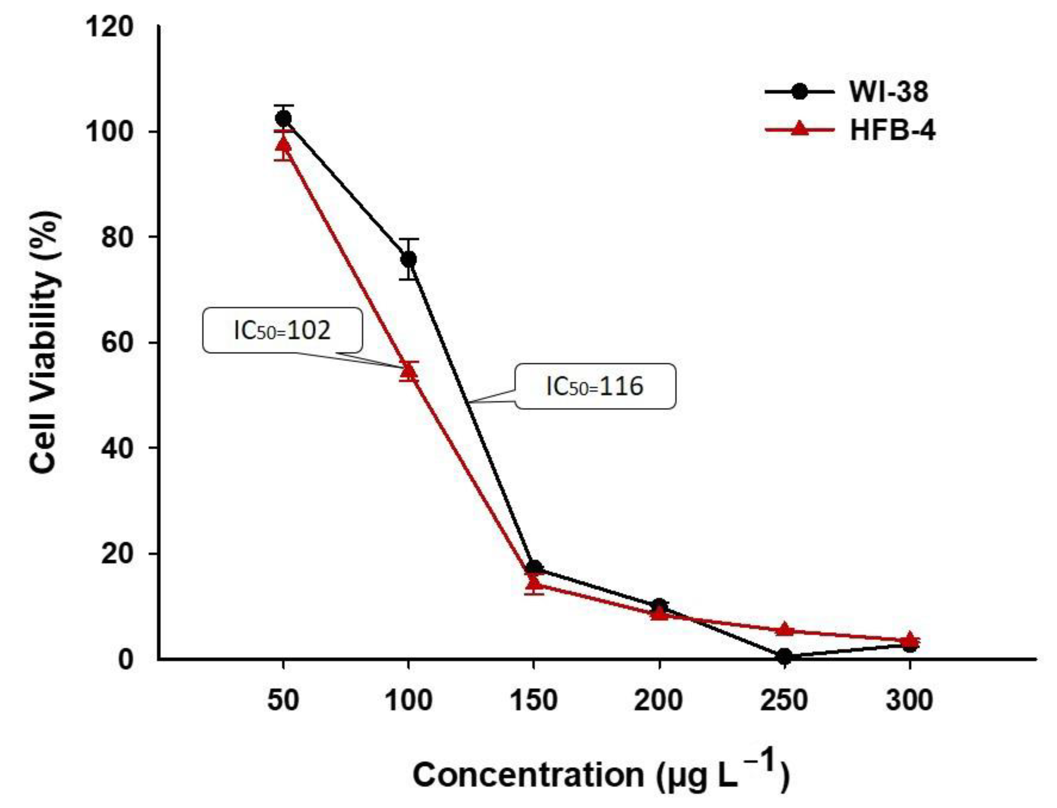
2.1. In Vitro Cytotoxic Efficacy of Filter Paper Loaded with AgNO3 on the Normal Cell Line

Silver ions are among the most commonly used chemical compounds in different medical and biotechnological applications. The toxicity of AgNO3 and its inhibitory effect on differentiation and proliferation of different cell lines such as Caco-2, Vero, PC-12, HEP-G2, A-549, and NIH-3T3 cell lines have been investigated [31,32,33]. In this study, the efficacy of Ag+ from AgNO3 loaded on Whatman filter paper toward two normal cell lines, WI 38 and HFB-4, was assessed to detect the safe concentration can be used as a coating agent for a historical manuscript. Approximately, 0.7 cm of filter paper loaded with different AgNO3 concentrations was incubated with a microplate containing normal cell lines. Data analysis showed that the viability of normal cell lines was dose-dependent, as detected by the MTT assay method (Figure 1). The IC50 values (the concentration of AgNO3 that decrease the absorbance to 50% compared to control) were 116 and 102 µg mL−1 for WI 38 and HFB 4, respectively.
The toxicity of AgNO3 and hence IC50 values differ according to the type of cell line. Silver nitrate exhibits in vitro cytotoxic efficacy against ovarian (OVCAR-3), breast (MB157), and cervical (HeLa) cell lines with IC50 35.0, 5.0, and 50 µM after exposure to 72 h [33]. Kaba and Egorova [34] reported a moderate cytotoxic effect on U937 (human leukemia monocyte cell line) because of treatment by different concentrations of AgNO3. Frazer [35] reported that silver ions do not exert any harmful effects on mammalian cells if used in the correct manner and at an appropriate dose. Therefore, in this study, we used a safe dose (80 µg mL−1) as a coating material to protect the historical manuscript against deteriorating microbes.

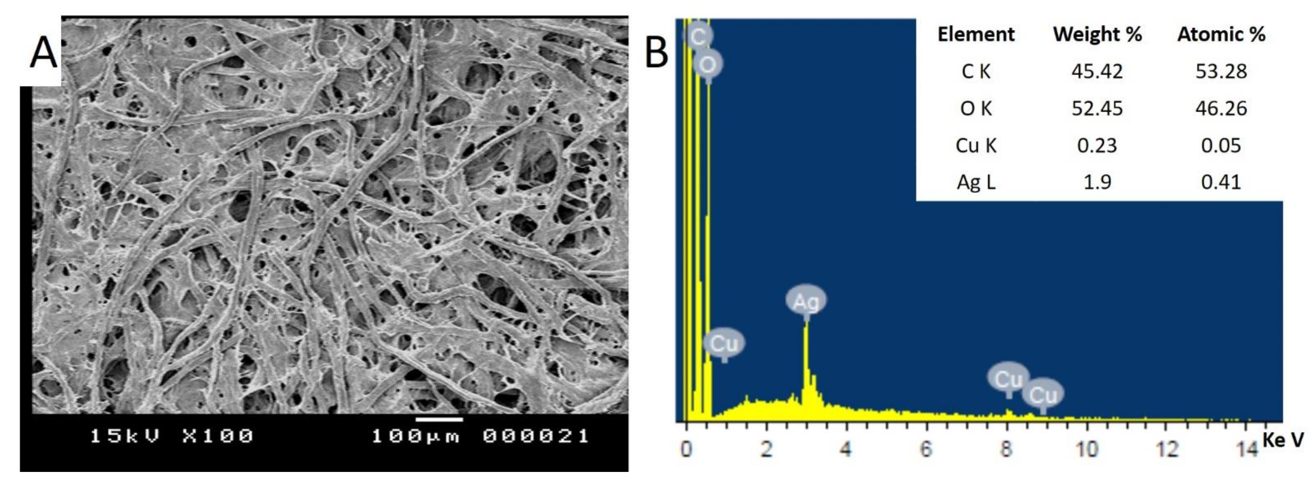
2.2. Assessment of Successful AgNO3 Loading on the Surface of Whatman Filter Paper

The surface characteristics of cellulosic fibers of filter paper treated with 80 µg mL−1 of AgNO3 and their metal contents were investigated using a scanning electron microscope, connected with energy dispersive spectroscopy (SEM-EDX). The data showed that the cellulosic fibers of the treated filter paper without microbial inoculation appeared as normal (Figure 2A). The weight and atomic percentages of deposited AgNO3 on the surface of filter paper were assessed by EDX analysis. The data showed that the peak of Ag ions was nearly 3 Ke V, which is characteristic of silver ions as reported previously [36,37]. Moreover, the EDX spectrum showed that the Ag ion occupied about 1.9% of the total elements besides C and O ions, which occupied weight percentages 45.42% and 52.45%, respectively (Figure 2B). Similarly, the weight and atomic percentages of Ag loaded on a fabric surface have been previously detected by EDX analysis [38]. The obtained data confirm the successful loading of Ag ions on the surface of filter paper without any negative impacts on cellulosic fibers.

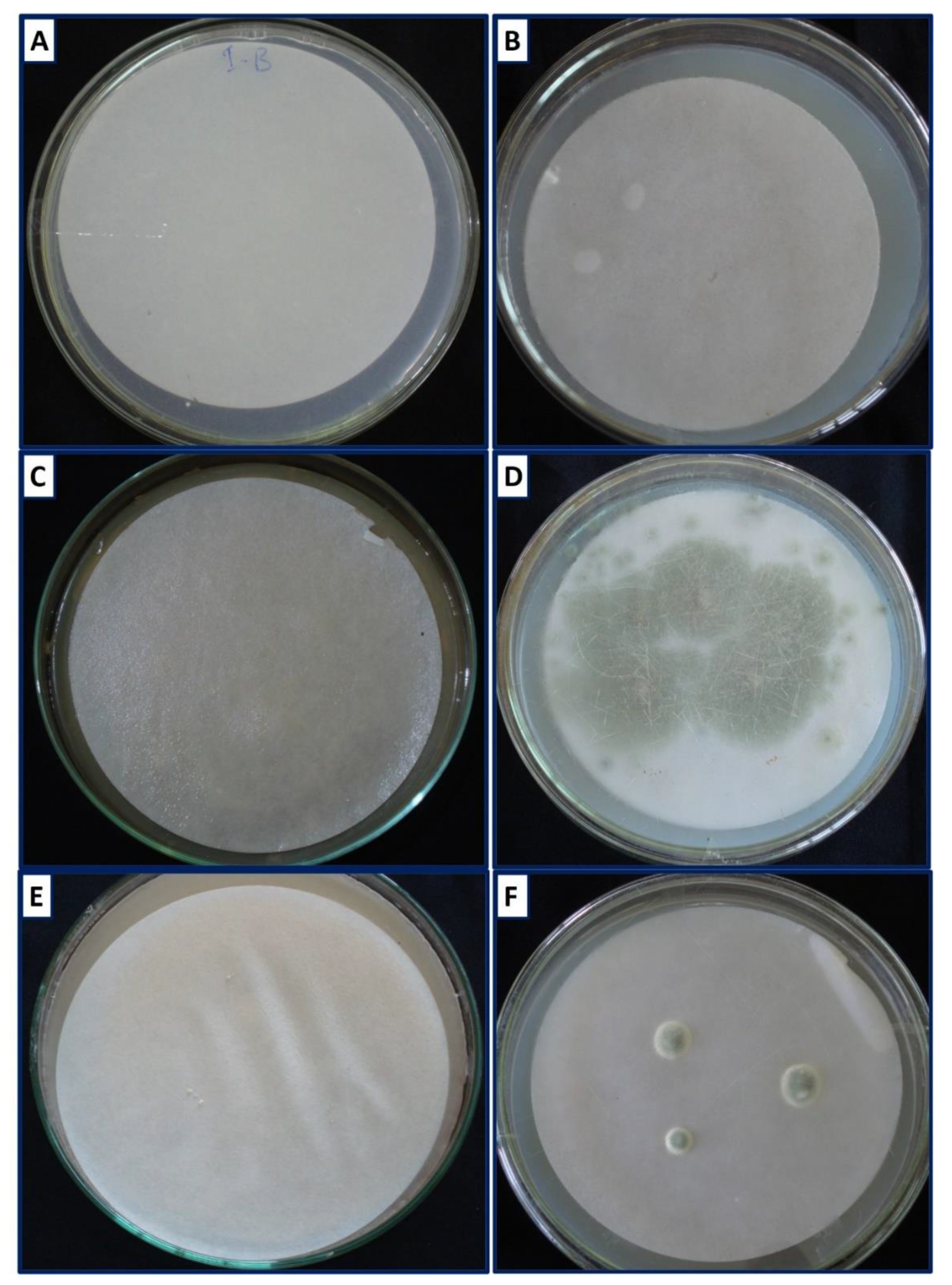
2.3. Assessment of Microbial Growth

The bacterial and fungal strains B. subtilis and P. chrysogenum were selected based on their efficacy in cellulose degradation as the main components of papers. These strains exhibit high cellulase enzyme activities represented by a clear zone (30.3 ± 0.33 mm for B. subtilis and 41.7 ± 0.33 mm for P. chrysogenum) as previously mentioned [4]. The selected strains were cultivated on Whatman filter paper treated with 80 µg mL−1 of AgNO3 as a safe dose based on an in vitro cytotoxic experiment and the results were recorded as growth inhibitions after 7, 14, and 21 days. Because we could not perform this experiment on the historical manuscript, Whatman filter paper was used as a model because it was free of additives and contained a high content of pure cellulose (98%) [39].
The bacterial strain Bacillus subtilis was completely grown on untreated filter paper after seven days, forming a mucous layer that deteriorated the quality of the fibers (Figure 3C). Interestingly, the dose of AgNO3 used was able to inhibit the bacterial growth completely after 7, 14, and 21 days (Table 1 and Figure 3E). The obtained data are consistent with those recorded in our recently published study in which Ag nanoparticles (NPs) were able to inhibit the growth of B. subtilis at a concentration of 1.0 mM after being inoculated onto Ag-NPs treated paper [4].
On the other hand, the dose of AgNO3 can inhibit fungal growth to a varying degree. As shown in Table 1, the growth of P. chrysogenum was inhibited with percentages 43.8 ± 3.4%, 68.8 ± 1.5%, and 85.9 ± 1.1% after 7, 14, and 21 days of incubation. At the dose used, the fungal growth showed a growth rate of 56.7% after 7 incubation days and decreased to 14.1% after 21 days of aging. This growth rate can be neglected in nature as a result of the absence of minerals required for fungal growth. The growth of these fungal strains was previously inhibited when grown on filter paper treated with a low Ag-NPs concentration (1 mM) with percentages of 29.0 ± 3.7%, 56.2 ± 0.9%, and 59.9 ± 0.9% after 7, 14, and 21 days, respectively, whereas its growth was completely inhibited with 2 mM Ag-NPs [4]. The growth of Aspergillus niger strain A2 isolated from the archeological manuscript was able to be inhibited with percentages of 48.9 ± 4.69%, 53.5 ± 0.0%, and 53.5 ± 5.50% after 7, 14, and 21 incubation days, respectively, as a result of treatment with a low concentration of Ag-NPs (1 mM). The high Ag-NPs concentration (2 mM) showed inhibition percentages of 100.0 ± 0.0%, 97.7 ± 2.30%, and 97.1 ± 1.50% after 7, 14, and 21 days, respectively [19].
Silver nitrate is characterized by its efficacy as an antimicrobial agent [40]. Lansdown Alan [26] reported that silver ions have an inhibitory effect on different bacterial and fungal species while exhibiting low toxicity on human cells. The inhibitory effect of silver nitrate could be attributed to its efficacy in binding with proteins’ thiol groups and hence denaturing them. Furthermore, silver ions can react with proteins in the cell wall and disrupt the permeability function. Moreover, silver ions can react with DNA or RNA once entering the microbial cell and convert them from a natural to a condensed form and hence inhibit their replication. The inhibitory action could also be related to enhancing the production of reactive oxygen species (ROS) because of the liberation of Ag+ into the cell. ROS can destroy the proteins and enzymes particularly involved in cellular respiration, thus causing cell death [41,42,43].

2.4. Color Change Measurement

The aspects of deterioration due to treatment and/or microbial inoculations were investigated based on paper color change compared with references [44]. The color changes due to different treatments were investigated through the CIE L*a*b* color space system as represented in Table 2.
The lightness values (L*) for untreated filter paper without microbial inoculation but deposited on the surface of mineral salt agar (MSA) media (negative control) were decreased with aging, recording values of 89.4 ± 1.2, 89.1 ± 0.7, and 87.7 ± 0.9 after 7, 14, and 21 days as compared with 90.6 for reference paper (filter paper without treatment, without microbial inoculation, and without adding to the surface of MSA media). These slight changes could be attributed to the action of wetting [45]. The lightness values of filter paper inoculated with fungal strain without AgNO3 treatment (positive control) were highly decreased as compared with the positive control of bacterial strain and negative control. The L* for the fungal positive control showed values 15.2 ± 1.1, 9.9 ± 0.3, and 10.3 ± 0.7 after aging time 7, 14, and 21 days, respectively, compared with the bacterial positive control, which showed L* values 82.2 ± 1.4, 82.02 ± 1.5, and 80.6 ± 1.2 after the same aging times. This high reduction in L* values can be attributed to different pigments synthesized by the fungal strain P. chrysogenum as compared with those synthesized by bacterial strains [46]. The treated filter paper inoculated with the B. subtilis strain displayed brightness values of 87.1 ± 0.7 after 21 aging days, with a reduction percentage of 0.6% as compared with the negative control. Although the growth of P. chrysogenum was inhibited with a percentage of 85%, the lightness values were highly decreased, with a value of 65.9 ± 1.9 after 21 aging days as compared with the negative control (87.7 ± 0.9).
The color change from red to green due to AgNO3 treatment in the presence/absence of microbial inoculation was represented by a* values. As shown in Table 2, there was a slight color change between the negative control and filter paper treated with AgNO3 without microbial inoculation, with values of −0.17 ± 0.01 and −0.14 ± 0.0, respectively, as compared with the reference (−0.24 ± 0.07) after 21 aging days. Untreated filter paper inoculated with B. subtilis and P. chrysogenum tended to be a red color, recording the maximum change with values of 1.3 ± 0.1 and −0.5 ± 0.05 after 21 days. These changes returned to normal as a result of silver nitrate coating and hence complete inhibition of bacterial growth, showing a* values of −0.23 ± 0.02 as compared with the negative control (−0.17 ± 0.01) and the reference sample (−0.24 ± 0.07). This phenomenon could be attributed to reactions of AgNO3 with fibers and inhibiting bacterial and fungal growth [47].
Moreover, the color change to yellow (b* value) is another way to investigate the effect of AgNO3 in the presence/absence of microbial inoculation on filter paper. Data represented in Table 2 showed that the highest change (yellow color) was achieved as a result of B. subtilis growth without silver nitrate treatment, which displayed b* values of 20.1 ± 0.9, 21.03 ± 1.3, and 21.7 ± 1.7 after aging time 7, 14, and 21 days, respectively. These changes were reduced to 3.5 ± 0.4, 3.79 ± 0.1, and 3.7 ± 0.1 after 7, 14, and 21 days respectively, because of AgNO3 coating. In addition, a yellowish color appeared due to the growth of P. chrysogenum on untreated filter paper, showing a b* value of 4.7 ± 0.6 after 21 aging days. The highest yellowish color in the bacterial or fungal positive control may be attributed to a mucoid layer formed due to B. subtilis growth and their efficacy in producing a yellowish carotenoid pigment [48], as well as the ability of P. chrysogenum to produce a yellowish chrysogine pigment [46]. The color change to a yellowish color was reduced because the coating of the filter paper with 80 µg mL−1 AgNO3 inhibits bacterial and fungal growth. Data showed that the b* value of the B. subtilis positive control was reduced from 21.7 ± 1.7 after 21 aging days to 3.7 ± 0.1 because of silver nitrate treatment, which inhibited the bacterial growth. Moreover, the b* value of the positive control of P. chrysogenum was reduced from 4.7 ± 0.6 to 2.4 ± 0.3 after 21 days. The yellowish-brown color which appeared on the negative control and the treated filter paper without microbial inoculations may be related to the production of yellowish chromophores as a result of the oxidation process, imparting its color to the paper [49].
According to the L*a*b* results, the highest color change (ΔE) was recorded for untreated filter paper inoculated with P. chrysogenum, which recorded an ΔE value equal to 81.2 ± 1.2 after 21 aging days, and this value was reduced to 23.4 ± 1.5 for inoculated treated filter paper. Moreover, the total color difference (ΔE) for treated filter paper inoculated with B. subtilis was 5.5 ± 0.4 as compared with untreated inoculated filter paper (15.0 ± 0.7).
Δ E = ( Δ L ) 2   + ( Δ a ) 2 + ( Δ b ) 2

2.5. Tensile Strength and Elongation

The mechanical properties including tensile strength and elongation of filter paper in the presence/absence of AgNO3 and microbial inoculations were investigated after aging times of 7, 14, and 21 days (Table 3). Tensile strength indicates fiber strength through the measurement of some parameters such as length, strength, and bonding of fibers [50]. In this study, the tensile strength of the negative control after 7 days was slightly increased (23.54 ± 0.2 N) as compared with the reference filter paper sample, which showed a tensile strength of 22.54 ± 0.4 N after 7 days. By increasing aging time, the tensile strength decreased as compared with the reference sample. This phenomenon could be attributed to the increasing aging time of the filter paper on the surface of semi-solid MSA media. On the other hand, the tensile strength of treated filter paper without microbial inoculation increased as compared with the reference and negative control (Table 3); this is due to the deposit and interaction of AgNO3 with cellulosic fibers. The incorporation of Ag ions into cellulosic fibers could be attributed to electrostatic interactions [51]. The obtained data were compatible with those obtained by Ariafar et al. [52], who reported that the tensile strength of nanoparticle treated filter paper was increased as compared with untreated paper. Data analysis showed that the tensile strength of untreated filter papers and those inoculated with B. subtilis B3 and P. chrysogenum F9 (positive control) were highly decreased, showing 11.27 ± 1.2 and 13.2 ± 0.7 after 21 aging days for bacterial and fungal species, respectively. This could be attributed to the high cellulase enzymatic activities secreted by these microbial strains, as previously mentioned in our study [4]. Interestingly, the tensile strength of treated filter paper inoculated with microbial strains is increased due to the inhibitory action of AgNO3 on microbial growth. In some treatment especially in the presence of AgNO3, the tensile strength values were reduced at 14 days, and returns increased again at 21 days. This phenomenon could be attributed to some dehydration occurring in the sample after 14 days of incubation, which reduced the tensile strength, while at the end of the incubation period, the silver ions had enough time to form a metal complex with the hydroxyl group of fibers, which act as crosslinking that help to increase tensile strength.
Elongation is considered one of the most mechanical properties of paper and is considered one of the main components of paper-based materials, helping with formability without damage [53]. The elongation test depends on three factors: single fiber properties, bonds between interfiber, and the fiber network formed during the manufacturing process [54,55]. Therefore, any external stress that affects the quality and bonding between fibers directly affects the elongation test. In this study, the elongation of coated filter paper without microbial inoculation significantly increased, with values of 3.51% as compared with the reference sample (1.34 ± 0.02%) and negative control (1.19 ± 0.08%) after 21 aging days. Moreover, the elongation percentages were highly decreased for untreated paper inoculated with the microbial strain, recording percentages of 0.32 ± 0.02% and 0.79 ± 0.04% for B. subtilis B3 and P. chrysogenum F9 (positive control) after 21 incubation days. These values returned to normal after silver nitrate treatment due to the inhibition of bacterial and fungal growth. After 21 aging days, the elongation percentages were 2.27 ± 0.2% and 2.42 ± 0.4% for treated filter paper inoculated with B. subtilis and P. chrysogenum, respectively. The simultaneous increase in tensile strength and elongation of filter paper treated with AgNO3 could be attributed to the hydrogen bond formed between coating material (AgNO3) and the substrate (filter paper), as mentioned previously by Sherazy et al. [56]. The obtained data reveal the efficacy of AgNO3 as a coating agent to reduce the hydrolytic efficacy of deteriorating microbes and hence improve the mechanical properties of papers.

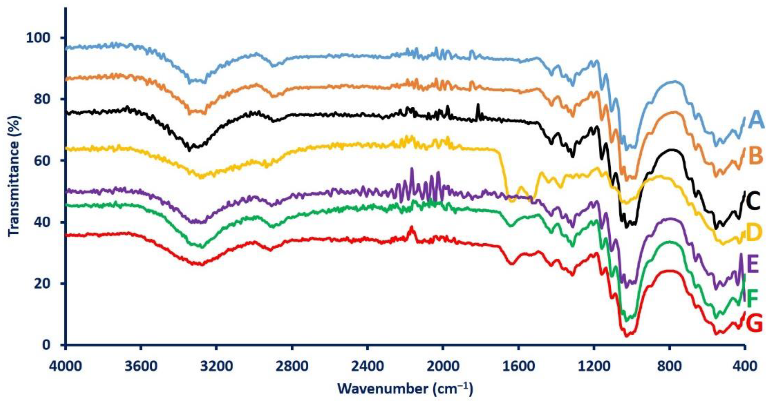
2.6. Attenuated Total Reflection Fourier Transform Infrared (ATR-FTIR) Spectroscopy

The chemical structures of filter paper before and after AgNO3 treatment and in the presence/absence of microbial inoculation were investigated after 21 aging days. As shown, the ATR-FTIR spectra for the reference sample (Figure 4A) and the negative control (Figure 4B) are identical. The peaks at 3343 cm−1 and 3257 cm−1 indicate N–H stretching for aliphatic primary amines [57]. Moreover, peaks at 2900 cm−1 and 1315 cm−1 signify C–H stretching and O–H bending for alkene and phenol, respectively. The peak at 1423 cm−1 corresponds to O–H for carboxylic acid, whereas the peak at 1030 cm−1 may correspond to the S=O sulfoxide stretching group. The peaks that appear at a wavelength between 1060 cm−1 and 1180 cm−1 signify C–N stretching amines. The peaks at 3343 cm−1 and 3257 cm−1 completely disappeared or were decreased in other treatments (Figure 4C–G), which signifies the absence of hydroxyl functional groups [58]. The peak observed at 1635 cm−1 corresponds to the carboxylic (C=O) group, which indicates the oxidative degradation of cellulosic fibers due to bacterial and fungal cellulase enzymes, or the presence of this peak may indicate protein materials secreted because of the growth of microorganisms (Figure 4D,F,G). This peak disappeared in the reference, negative control, treated filter paper without microbial inoculations, and treated filter paper inoculated with B. subtilis. The disappearance of this peak could be attributed to the absence of microbial growth as well as the efficacy of AgNO3 to inhibit bacterial growth [59,60]. The ATR-FTIR spectra of treated filter paper inoculated with bacterial strains exhibit the same bands as shown in the reference sample and negative control. However, those treated with AgNO3 and inoculated with fungal strains were slightly different compared with the reference and negative control due to fungal growth, as shown in Table 1. These data confirm the efficacy of a safe dose of AgNO3 in the treatment of deteriorating microbes without causing a significant change in paper quality.

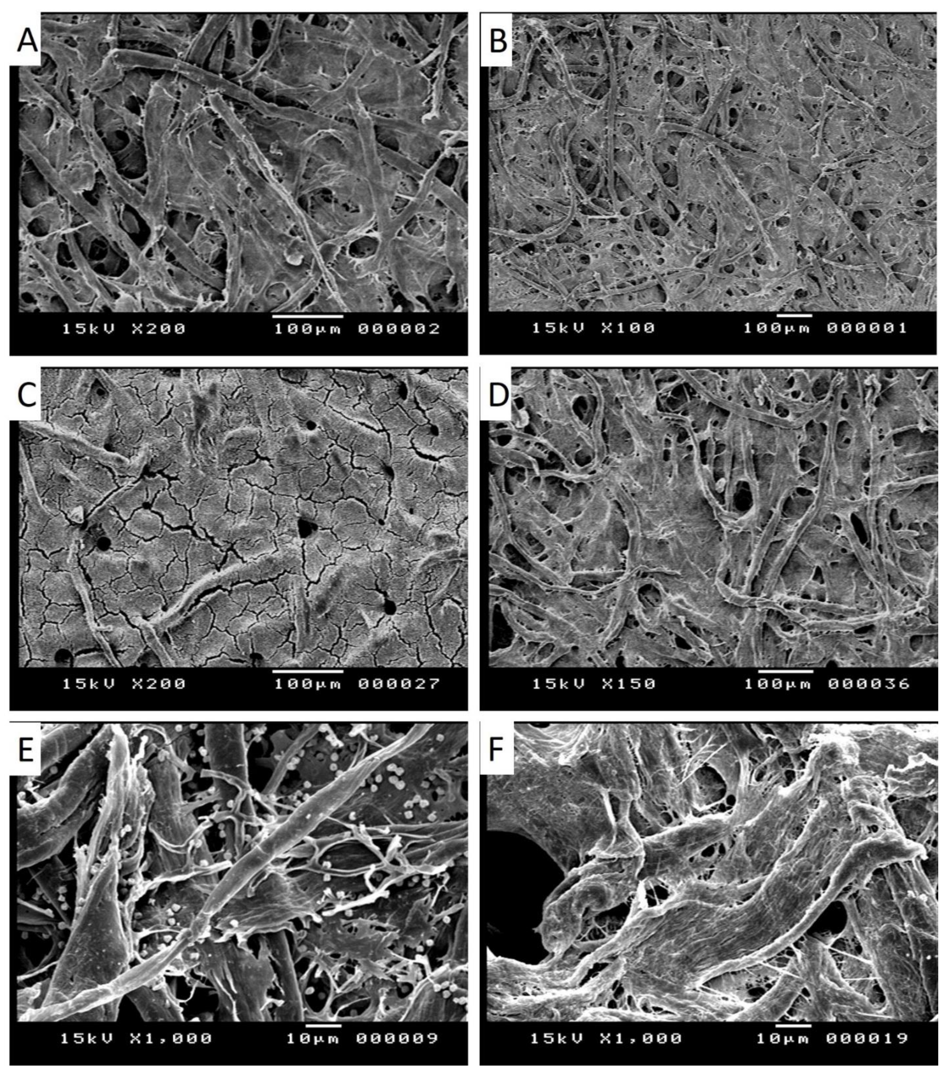
2.7. Assessment of the Cellulosic Fibers of Treated/Untreated Filter Paper in the Presence/Absence of Microbial Inoculations Using SEM Analysis

The morphological fiber changes of treated/untreated filter paper in the presence/absence of bacterial and fungal growth were investigated after 21 aging days using scanning electron microscopy (SEM). The cellulosic fibers of the reference filter paper and the negative control (untreated filter paper added to MSA media without microbial inoculation) exhibited normal results without any indicators of deterioration (Figure 5A,B).
The network cellulosic fibers of untreated filter paper were completely deteriorated because of the high growth of B. subtilis B3 and P. chrysogenum F9. As a result of a mucoid layer formed due to the growth of B. subtilis (bacterial positive control) and colonization of P. chrysogenum conidia on the untreated filter paper (fungal positive control), the normal network cellulosic fibers were completely distorted (Figure 5C,E, respectively). Consistent with our results, Lech [61] studied the efficacy of B. subtilis to deteriorate parchment dating to the thirteenth century using SEM analysis. Naraian and Gautam [62] also reported that Penicillium spp. has a high potential to colonize and deteriorate cellulosic fiber through the synthesis of different lytic enzymes.
The silver nitrate dose used in this study has the efficacy to inhibit the growth of B. subtilis B3 and P. chrysogenum F9, as shown in Table 1 and Figure 3. Therefore, the cellulosic fibers of treated filter paper inoculated with bacterial and fungal strains were not affected (Figure 5D,F) compared with those untreated inoculated filter paper. According to the obtained results especially of FT-IR and SEM analyses, it can be concluded that Ag ions show the ability to protect papers against deteriorating microbes without any effects on paper quality.

3. Materials and Methods

3.1. Materials

The silver nitrate (AgNO3) and medium components used in the current study were of analytical grade and obtained from Sigma Aldrich (Cairo, Egypt). The filter paper used as a model to study the biodegradable activity of bacterial and fungal strains was Whatman filter paper No. 1. According to El Bergadi et al. [39], the used filter paper is characterized by high cellulose purity (98%) and free of other additives. All biological syntheses in the current study were achieved using distilled water (dis. H2O).

3.2. Cytotoxic Efficacy of Whatman Filter Paper Loaded with Different Concentrations of AgNO3 on Normal Cells

The cell viability of filter paper due to treatment with different concentrations of AgNO3 was assessed using two normal cell lines namely WI 38 (diploid human lung fibroblast) and HFB-4 (human normal melanocytes), which were obtained from ATCC (American Type Culture Collection, Manassas, VA, USA). The MTT assay method (3-(4,5-dimethylthiazol-2-yl)-2,5-diphenyl tetrazolium bromide) was used to measure the viability of the treated cell lines as follows: each normal cell (at concentration 1 × 10⁵ cell mL−1) was grown in a 12-well culture plate and incubated with 0.7 cm of filter paper loaded with different AgNO3 concentrations (300, 250, 200, 150, 100, and 50 µg mL−1) at 37 °C for 48 h. After that, the MTT reagent (5 mg mL−1 in phosphate buffer) was mixed with treated cells and incubated at the following conditions: 37 °C; 5% CO2 for 5 h. At the end of the incubation period, each well was mixed with 150 µL of DMSO (dimethyl sulfoxide), wrapped the plate in foil, and placed on an orbital shaker for 20 min to completely dissolve the MTT formazan. The formed color was measured at wavelength 560 nm using an ELIZA reader (Thermo Fisher Scentific, Inc., Waltham, MA, USA) [63]. The cell viability percentages were calculated based on the following equation:
Cell   viability   ( % ) =   Absorbance   of   treatment Absorbance   of   control   × 100

3.3. Evaluate the Efficacy of a Safe Dose of AgNO3 on Paper Quality

3.3.1. Confirm Successful Loaded of AgNO3 on Sterilized Filter Paper

Before the experiment, the Whatman filter paper was sterilized at 121 °C for 15 min. After that, the sterilized filter paper was immersed on safe AgNO3 concentration (selected based on an in vitro cytotoxicity test) for one minute under aseptic conditions and removed after complete adsorption to stay dry for one hour. To confirm the successful adsorption of AgNO3 on the surface of filter paper, Scanning Electron Microscope connected with Energy Dispersive Spectroscopy (SEM-EDX, JEOL JSM-6360LA) was achieved.

3.3.2. The Bacterial and Fungal Strains Used in This Study

The bacterial Bacillus subtilis strain B3 and the fungal Penicillium chrysogenum strain F9 were isolated previously from a historical manuscript “The biography of prophet Mohamed, Peace be upon him”, and selected according to their high efficacy to cellulose hydrolysis [4]. The bacterial and fungal isolates were identified based on 16S rRNA (for bacteria) and ITS (for fungi) sequence analysis and the obtained sequences were deposited in GeneBank under accession number MK447110 and MK452266, respectively.

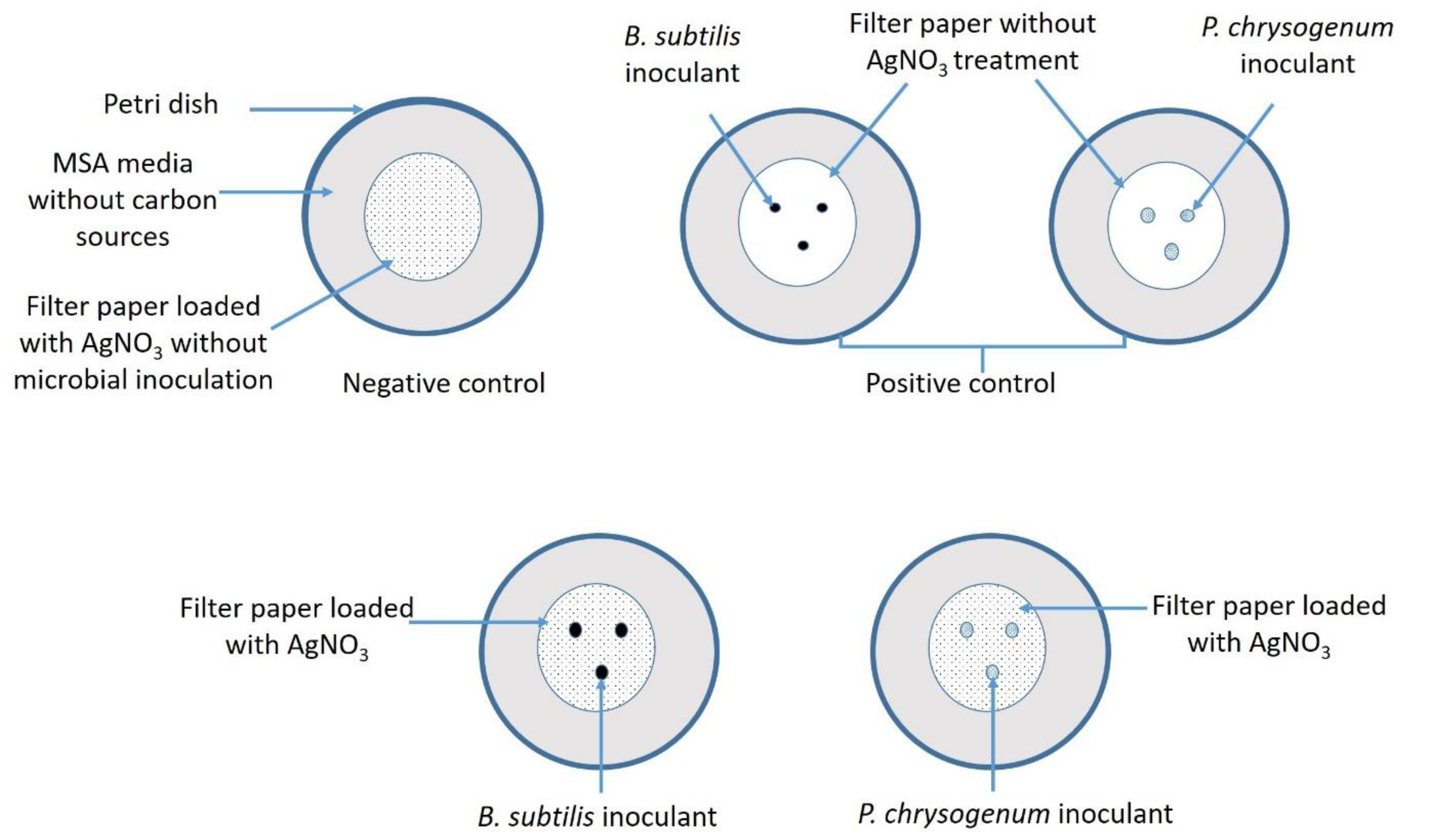
3.3.3. Design of the Experiment

The sterilized loaded filter papers were placed on the surface of Petri dishes (15 cm in diameter) containing sterilized mineral salt agar (MSA) media (containing g L−1: NaNO3, 6; KCl, 5; KH2PO4, 1.5; MgSO4·7H2O, 0.5; ZnSO4, 0.01; FeSO4, 0.01 and agar, 15) without a carbon source. The sterilized loaded filter paper served as a sole carbon source for microbial growth. The following treatments were conducted: (A) filter paper treated with AgNO3 without microbial inoculation (negative control), (B) untreated sterilized filter paper inoculated by B. subtilis (positive control), (C) untreated sterilized filter paper inoculated by P. chrysogenum (positive control), (D) filter paper treated with AgNO3 and inoculated with B. subtilis, (E) filter paper treated with AgNO3 and inoculated with P. chrysogenum. The paper properties based on the previous treatment were compared with a reference sample (filter paper without microbial inoculations and any treatment). The microbial inoculations were achieved as follows: three inoculant spots for each filter paper, each spot containing either 20 µL of B. subtilis culture (adjusted O.D.600 nm at 1.0 by taking samples from the bacterial culture at interval times and measuring their O.D.) or a heavy growth disk (6 mm in diameter) of P. chrysogenum (Figure 6). The experiment was achieved in triplicates.

3.3.4. Assessment of Bacterial and Fungal Growth

Measurement of the colony diameter was used to assess the microbial growth after 7, 14, and 21 days of incubation. The inhibition percentages (%) of the microbial growth due to AgNO3 treatment were calculated according to the following equation [64]:
Inhibition   percentages   ( % ) =   D c   D t D c   × 100
where Dc is the average of microbial growth in control (mm), Dt is the average of microbial growth in treatment (mm).

3.3.5. Color Change Measurement

The color changes due to silver nitrate and/or microbial growth were investigated after 7, 14, and 21 days of incubation according to CIELAB color space using ultra-scan PRO, Hunter lab, USA, UV spectrophotometer [65]. The CIE system contains one channel for detect lightness (L*) and two channels, one of them to measure color change from red to green (a*) and another to measure color change from yellow to blue (b*). The total color difference (ΔE) was measured according to the following equation [66]:
Δ E =   ( Δ L ) 2 + ( Δ a ) 2 +   ( Δ b ) 2
where ΔL, Δa, and Δb were calculated as the difference between the values of L*, a*, and b* for the control and treated samples.

3.3.6. Tensile Strength and Elongation

The tensile strength and elongations for different treatments were measured after 7, 14, and 21 days using hums filed test (LTD); S series; H 10K-SUTM according to TAPPI standard T 494 om-01 [67]. Briefly, Whatman filter paper after different treatments were cut into strips (width ≈ 1.5 cm) and subjected to standard test conditions (50% relative humidity (RH) at 25 °C for 24 h) followed by measuring tensile strength and elongation [45].

3.3.7. Attenuated Total Reflection Fourier Transform Infrared (ATR-FTIR) Spectroscopy

The change in the functional groups of filter papers due to AgNO3 treatment and/or microbial growth was analyzed using ATR-FTIR analysis and compared to control. The ATR-FTIR analysis was achieved for samples after 21 days of incubation using Cary 630 FT-IR spectrometer (Agilent Technologies Inc., Santa Clara, CA, USA) in the range between 4000–400 cm−1.

3.3.8. Scanning Electron Microscopy (SEM)

The efficacy of bacterial and fungal isolates to adhere to the cellulosic fiber of treated Whatman filter paper was confirmed using a Scanning Electron Microscope (JSM-5400, Jeol, Tokyo, Japan) after 21 days of incubation. A strip of filter paper was coated with gold thin film before being mounted on the specimen’s holder.

3.4. Statistical Analysis

All results presented in this study are the means of three independent replicates. Data were subjected to analysis of variance (ANOVA) by a statistical package SPSS v17. The mean difference comparison between the treatments was analyzed by the Tukey HSD test at p < 0.05.

4. Conclusions

In this study, previously identified B. subtilis B3 and P. chrysogenum F9 isolated from a historical manuscript were selected as the most potent cellulolytic microbes to investigate the efficacy of AgNO3 as a coating agent to protect papers against deteriorating microbes. The data showed that a safe dose of AgNO3 used as a coating agent was 80 µg mL−1 based on in vitro cytotoxicity against two normal cell lines, namely Wi-38 and FHB-4. This concentration was loaded on filter paper and showed the ability to inhibit the growth of B. subtilis B3 and P. chrysogenum F9 with percentages of 100% and 85.9 ± 1.1%, respectively, when inoculated on treated filter paper. Moreover, the color, tensile strength, and elongation percentages of treated filter paper in the absence/presence of microbial inoculation were slightly changed as compared with the reference sample. Untreated inoculated filter paper exhibited a large color change and decreased tensile strength and elongation values as compared with treated inoculated filter paper. In addition, the chemical structure of loaded filter paper and that of paper inoculated/uninoculated with microbial strains did not exhibit any change compared with the reference sample. Moreover, the cellulosic network fibers of treated filter paper in the presence/absence of microbial strains appeared healthy and were not deteriorating as compared with untreated filter paper in the presence of microbial strains. The data obtained here may contribute to the introduction of a successful strategy to protect historical papers from highly deteriorating bacterial and fungal strains.

Author Contributions

Conceptualization, A.F.; G.A.-M., and M.A.-H.E.-S.; methodology, A.F.; G.A.-M. and M.A.-H.E.-S.; software, A.F.; G.A.-M., Z.M.M., and M.A.-H.E.-S.; validation, A.F.; G.A.-M., H.A.S., A.A.G., and M.A.-H.E.-S.; formal analysis, A.F.; Z.M.M., G.A.-M., and M.A.-H.E.-S.; investigation, A.F.; G.A.-M., H.A.S., A.A.G., and M.A.-H.E.-S.; resources, A.F.; G.A.-M., Z.M.M., H.A.S., A.A.G., and M.A.-H.E.-S.; data curation, A.F.; G.A.-M., and M.A.-H.E.-S.; writing—original draft preparation, A.F.; G.A.-M., and M.A.-H.E.-S.; writing—review and editing, A.F.; G.A.-M., H.A.S., Z.M.M., A.A.G., and M.A.-H.E.-S.; visualization, A.F.; G.A.-M., H.A.S., A.A.G., and M.A.-H.E.-S.; supervision, A.F., and G.A.-M.; project administration, A.F., and G.A.-M.; funding acquisition, H.A.S. and A.A.G. All authors have read and agreed to the published version of the manuscript.

Funding

This research received no external funding.

Data Availability Statement

The data presented in this study are available on request from the corresponding author.

Acknowledgments

We thank the researchers at Taif University, Taif, Saudi Arabia (supporting project number: TURSP-2020/07).

Conflicts of Interest

The authors declare no conflict of interest.

References

  1. Sequeira, S.; Cabrita, E.J.; Macedo, M.F. Antifungals on paper conservation: An overview. Int. Biodeterior. Biodegrad. 2012, 74, 67–86. [Google Scholar] [CrossRef]
  2. Strlič, M.; Kolar, J. Ageing and Stabilisation of Paper; National and University Library Ljubljana: Ljubljana, Slovenia, 2005. [Google Scholar]
  3. Thapa, S.; Mishra, J.; Arora, N.; Mishra, P.; Li, H.; O′Hair, J.; Bhatti, S.; Zhou, S. Microbial cellulolytic enzymes: Diversity and biotechnology with reference to lignocellulosic biomass degradation. Rev. Environ. Sci. Biol. Technol. 2020, 19, 621–648. [Google Scholar]
  4. Fouda, A.; Abdel-Maksoud, G.; Abdel-Rahman, M.A.; Salem, S.S.; Hassan, S.E.-D.; El-Sadany, M.A.-H. Eco-friendly approach utilizing green synthesized nanoparticles for paper conservation against microbes involved in biodeterioration of archaeological manuscript. Int. Biodeterior. Biodegrad. 2019, 142, 160–169. [Google Scholar] [CrossRef]
  5. Fouda, A.H.; Hassan, S.E.-D.; Eid, A.M.; Ewais, E.E.-D. Biotechnological applications of fungal endophytes associated with medicinal plant Asclepias sinaica (Bioss.). Ann. Agric. Sci. 2015, 60, 95–104. [Google Scholar] [CrossRef] [Green Version]
  6. Alkahtani, M.; Fouda, A.; Attia, K.A.; Alotaibi, F.E.; Eid, A.M.; Ewais, E.E.; Hijri, M.; St-Arnaud, M.; Hassan, S.E.; Khan, N.; et al. Isolation and Characterization of Plant Growth Promoting Endophytic Bacteria from Desert Plants and Their Application as Bioinoculants for Sustainable Agriculture. Agronomy 2020, 10, 1325. [Google Scholar] [CrossRef]
  7. Khalil, A.M.A.; Hassan, S.E.-D.; Alsharif, S.M.; Eid, A.M.; Ewais, E.E.-D.; Azab, E.; Gobouri, A.A.; Elkelish, A.; Fouda, A. Isolation and Characterization of Fungal Endophytes Isolated from Medicinal Plant Ephedra pachyclada as Plant Growth-Promoting. Biomolecules 2021, 11, 140. [Google Scholar] [CrossRef]
  8. Saini, A.; Aggarwal, N.K.; Sharma, A.; Yadav, A. Actinomycetes: A Source of Lignocellulolytic Enzymes. Enzym. Res. 2015, 2015, 279381. [Google Scholar] [CrossRef] [Green Version]
  9. Zhang, Y.H.; Lynd, L.R. Toward an aggregated understanding of enzymatic hydrolysis of cellulose: Noncomplexed cellulase systems. Biotechnol. Bioeng. 2004, 88, 797–824. [Google Scholar] [CrossRef]
  10. Di Bella, M.; Randazzo, D.; Di Carlo, E.; Barresi, G.; Palla, F. Monitoring Biological Damage on Paper-based Documents in the Historical Archive of the Palermo Astronomical Observatory. Conserv. Sci. Cult. Herit. 2017, 15, 85–94. [Google Scholar]
  11. Paulus, W. Directory of Microbicides for the Protection of Materials: A Handbook; Springer Science & Business Media: Berlin/Heidelberg, Germany, 2005. [Google Scholar]
  12. Gupta, P.; Aggarwal, M. Chapter 55—Toxicity of Fungicides, Veterinary Toxicology; Academic Press: Boston, MA, USA, 2012. [Google Scholar]
  13. Zervos, S.; Alexopoulou, I. Paper conservation methods: A literature review. Cellulose 2015, 22, 2859–2897. [Google Scholar] [CrossRef]
  14. Garside, P.; Knight, B. The behaviour of books in changing environmental conditions and the implications for collection storage. In Proceedings of the ICOM Committee for Conservation, Preprints of the 16th Triennial Conference, Lisbon, Portugal, 19–23 September 2011. [Google Scholar]
  15. Belloni, F.; Nassisi, V.; Alifano, P.; Monaco, C.; Panzanaro, S. The Effects of UV Laser Radiation as Sterilizer for Cultural Heritage. Macromol. Symp. 2006, 238, 52–56. [Google Scholar] [CrossRef]
  16. Alexopoulou, I.; Zervos, S. Paper conservation methods: An international survey. J. Cult. Herit. 2016, 21, 922–930. [Google Scholar] [CrossRef]
  17. El-Belely, E.F.; Farag, M.M.S.; Said, H.A.; Amin, A.S.; Azab, E.; Gobouri, A.A.; Fouda, A. Green Synthesis of Zinc Oxide Nanoparticles (ZnO-NPs) Using Arthrospira platensis (Class: Cyanophyceae) and Evaluation of their Biomedical Activities. Nanomaterials 2021, 11, 95. [Google Scholar] [CrossRef] [PubMed]
  18. Wójciak, A. Washing, Spraying and Brushing. A Comparison of Paper Deacidification by Magnesium Hydroxide Nanoparticles. Restaur. Int. J. Preserv. Libr. Arch. Mater. 2015, 36, 3–23. [Google Scholar] [CrossRef]
  19. Fouda, A.; Abdel-Maksoud, G.; Abdel-Rahman, M.A.; Eid, A.M.; Barghoth, M.G.; El-Sadany, M.A.-H. Monitoring the effect of biosynthesized nanoparticles against biodeterioration of cellulose-based materials by Aspergillus niger. Cellulose 2019, 26, 6583–6597. [Google Scholar] [CrossRef]
  20. Salem, S.S.; El-Belely, E.F.; Niedbała, G.; Alnoman, M.M.; Hassan, S.E.-D.; Eid, A.M.; Shaheen, T.I.; Elkelish, A.; Fouda, A. Bactericidal and In-Vitro Cytotoxic Efficacy of Silver Nanoparticles (Ag-NPs) Fabricated by Endophytic Actinomycetes and Their Use as Coating for the Textile Fabrics. Nanomaterials 2020, 10, 2082. [Google Scholar] [CrossRef]
  21. Kaplan, A.; Kutlu, H.M. Investigation of Silver Nitrate on Cytotoxicity and Apoptosis in MCF7 Human Breast Carcinoma Cells. Asian Pac. J. Cancer Biol. 2020, 5, 49–56. [Google Scholar] [CrossRef]
  22. Cuin, A.; Massabni, A.C.; Pereira, G.A.; Leite, C.Q.; Pavan, F.R.; Sesti-Costa, R.; Heinrich, T.A.; Costa-Neto, C.M. 6-Mercaptopurine complexes with silver and gold ions: Anti-tuberculosis and anti-cancer activities. Biomed. Pharmacother. Biomed. Pharmacother. 2011, 65, 334–338. [Google Scholar] [CrossRef]
  23. Radko, L.; Stypuła-Trębas, S.; Posyniak, A.; Żyro, D.; Ochocki, J. Silver(I) Complexes of the Pharmaceutical Agents Metronidazole and 4-Hydroxymethylpyridine: Comparison of Cytotoxic Profile for Potential Clinical Application. Molecules 2019, 24, 1949. [Google Scholar] [CrossRef] [Green Version]
  24. Raffi, M.; Hussain, F.; Bhatti, T.; Akhter, J.; Hameed, A.; Hasan, M. Antibacterial characterization of silver nanoparticles against E. coli ATCC-15224. J. Mater. Sci. Technol. 2008, 24, 192–196. [Google Scholar]
  25. Soliman, A.M.; Abdel-Latif, W.; Shehata, I.H.; Fouda, A.; Abdo, A.M.; Ahmed, Y.M. Green Approach to Overcome the Resistance Pattern of Candida spp. Using Biosynthesized Silver Nanoparticles Fabricated by Penicillium chrysogenum F9. Biol. Trace Elem. Res. 2021, 199, 800–811. [Google Scholar] [CrossRef]
  26. Lansdown, A.B. Silver in health care: Antimicrobial effects and safety in use. Curr. Probl. Dermatol. 2006, 33, 17–34. [Google Scholar] [CrossRef] [Green Version]
  27. Mohamed, A.; Fouda, A.; Elgamal, M.S.; EL-Din Hassan, S.; Shaheen, T.I.; Salem, S.S. Enhancing of Cotton Fabric Antibacterial Properties by Silver Nanoparticles Synthesized by New Egyptian Strain Fusarium keratoplasticum A1-3. Egypt J. Chem. 2017, 60, 63–71. [Google Scholar] [CrossRef] [Green Version]
  28. Kaplan, A.; Akalin Ciftci, G.; Kutlu, H.M. Cytotoxic, anti-proliferative and apoptotic effects of silver nitrate against H-ras transformed 5RP7. Cytotechnology 2016, 68, 1727–1735. [Google Scholar] [CrossRef] [Green Version]
  29. Kolesarova, A.; Capcarova, M.; Sirotkin, A.V.; Medvedova, M.; Kovacik, J. In vitro assessment of silver effect on porcine ovarian granulosa cells. J. Trace Elem. Med. Biol. 2011, 25, 166–170. [Google Scholar] [CrossRef] [PubMed]
  30. Alexander, J.W. History of the medical use of silver. Surg. Infect. 2009, 10, 289–292. [Google Scholar] [CrossRef] [PubMed] [Green Version]
  31. Alsharif, S.M.; Salem, S.S.; Abdel-Rahman, M.A.; Fouda, A.; Eid, A.M.; El-Din Hassan, S.; Awad, M.A.; Mohamed, A.A. Multifunctional properties of spherical silver nanoparticles fabricated by different microbial taxa. Heliyon 2020, 6, e03943. [Google Scholar] [CrossRef]
  32. Sambale, F.; Wagner, S.; Stahl, F.; Khaydarov, R.R.; Scheper, T.; Bahnemann, D. Investigations of the Toxic Effect of Silver Nanoparticles on Mammalian Cell Lines. J. Nanomater. 2015, 2015, 136765. [Google Scholar] [CrossRef]
  33. Medvetz, D.A.; Hindi, K.M.; Panzner, M.J.; Ditto, A.J.; Yun, Y.H.; Youngs, W.J. Anticancer Activity of Ag(I) N-Heterocyclic Carbene Complexes Derived from 4,5-Dichloro-1H-Imidazole. Metal-Based Drugs 2008, 2008, 384010. [Google Scholar] [CrossRef] [PubMed]
  34. Kaba, S.I.; Egorova, E.M. In vitro studies of the toxic effects of silver nanoparticles on HeLa and U937 cells. Nanotechnol. Sci. Appl. 2015, 8, 19–29. [Google Scholar] [CrossRef] [Green Version]
  35. Frazer, R. Use of Silver Nanoparticles in HIV Treatment Protocols: A Research Proposal. J. Nanomed. Nanotechnol. 2011, 3. [Google Scholar] [CrossRef]
  36. Magudapathy, P.; Gangopadhyay, P.; Panigrahi, B.; Nair, K.; Dhara, S. Electrical transport studies of Ag nanoclusters embedded in glass matrix. Phys. B Condens. Matter 2001, 299, 142–146. [Google Scholar] [CrossRef]
  37. Fouda, A.; Hassan, S.E.; Abdo, A.M.; El-Gamal, M.S. Antimicrobial, Antioxidant and Larvicidal Activities of Spherical Silver Nanoparticles Synthesized by Endophytic Streptomyces spp. Biol. Trace Elem. Res. 2020, 195, 707–724. [Google Scholar] [CrossRef]
  38. Fouda, A.; Saad, E.; Salem, S.S.; Shaheen, T.I. In-Vitro cytotoxicity, antibacterial, and UV protection properties of the biosynthesized Zinc oxide nanoparticles for medical textile applications. Microb. Pathog. 2018, 125, 252–261. [Google Scholar] [CrossRef] [PubMed]
  39. El Bergadi, F.; Laachari, F.; Elabed, S.; Mohammed, I.H.; Ibnsouda, S.K. Cellulolytic potential and filter paper activity of fungi isolated from ancients manuscripts from the Medina of Fez. Ann. Microbiol. 2014, 64, 815–822. [Google Scholar] [CrossRef]
  40. Human, Z.; Munyaneza, A.; Omondi, B.; Sanabria, N.M.; Meijboom, R.; Cronjé, M.J. The induction of cell death by phosphine silver(I) thiocyanate complexes in SNO-esophageal cancer cells. Biometals Int. J. Role Metal Ions Biol. Biochem. Med. 2015, 28, 219–228. [Google Scholar] [CrossRef]
  41. Salem, S.S.; Fouda, A. Green Synthesis of Metallic Nanoparticles and Their Prospective Biotechnological Applications: An Overview. Biol. Trace Elem. Res. 2021, 199, 344–370. [Google Scholar] [CrossRef]
  42. Eid, A.M.; Fouda, A.; Niedbała, G.; Hassan, S.E.; Salem, S.S.; Abdo, A.M.; Hetta, H.F.; Shaheen, T.I. Endophytic Streptomyces laurentii Mediated Green Synthesis of Ag-NPs with Antibacterial and Anticancer Properties for Developing Functional Textile Fabric Properties. Antibiotics 2020, 9, 641. [Google Scholar] [CrossRef]
  43. Kanwal, Z.; Raza, M.A.; Riaz, S.; Manzoor, S.; Tayyeb, A.; Sajid, I.; Naseem, S. Synthesis and characterization of silver nanoparticle-decorated cobalt nanocomposites (Co@AgNPs) and their density-dependent antibacterial activity. R. Soc. Open Sci. 2019, 6, 182135. [Google Scholar] [CrossRef] [Green Version]
  44. Zervos, S.; Moropoulou, A. Methodology and Criteria for the Evaluation of Paper Conservation Interventions: A Literature Review. Restaur. Int. J. Preserv. Libr. Arch. Mater. Restaur. 2006, 27, 219–274. [Google Scholar] [CrossRef]
  45. Rushdy, A.; Noshy, W.; Youssef, A.; Kamel, S. Influence of Bleaching Materials on Mechanical and Morphological Properties for Paper Conservation. Egypt. J. Chem. 2017, 60, 893–903. [Google Scholar]
  46. Viggiano, A.; Salo, O.; Ali, H.; Szymanski, W.; Lankhorst, P.P.; Nygård, Y.; Bovenberg, R.A.L.; Driessen, A.J.M. Pathway for the Biosynthesis of the Pigment Chrysogine by Penicillium chrysogenum. Appl. Environ. Microbiol. 2018, 84, e02246-17. [Google Scholar] [CrossRef] [Green Version]
  47. Amornkitbamrung, L.; Mohan, T.; Hribernik, S.; Reichel, V.; Faivre, D.; Gregorova, A.; Engel, P.; Kargl, R.; Ribitsch, V. Polysaccharide stabilized nanoparticles for deacidification and strengthening of paper. RSC Adv. 2015, 5, 32950–32961. [Google Scholar] [CrossRef] [Green Version]
  48. Khaneja, R.; Perez-Fons, L.; Fakhry, S.; Baccigalupi, L.; Steiger, S.; To, E.; Sandmann, G.; Dong, T.; Ricca, E.; Fraser, P. Carotenoids found in Bacillus. J. Appl. Microbiol. 2010, 108, 1889–1902. [Google Scholar] [PubMed]
  49. Rosenau, T.; Potthast, A.; Krainz, K.; Hettegger, H.; Henniges, U.; Yoneda, Y.; Rohrer, C.; French, A.D. Chromophores in cellulosics, XI: Isolation and identification of residual chromophores from bacterial cellulose. Cellulose 2014, 21, 2271–2283. [Google Scholar] [CrossRef]
  50. Bajpai, P. Chapter 2—Paper and Its Properties. In Biermann’s Handbook of Pulp and Paper, 3rd ed.; Bajpai, P., Ed.; Elsevier: Amsterdam, The Netherlands, 2018; pp. 35–63. [Google Scholar]
  51. Tang, B.; Li, J.; Hou, X.; Afrin, T.; Sun, L.; Wang, X. Colorful and Antibacterial Silk Fiber from Anisotropic Silver Nanoparticles. Ind. Eng. Chem. Res. 2013, 52, 4556–4563. [Google Scholar] [CrossRef]
  52. Ariafar, A.A.; Afsharpour, M.; Samanian, K. Use of TiO2/chitosan nanoparticles for enhancing the preservative effects of carboxymethyl cellulose in paper-art-works against biodeterioration. Int. Biodeterior. Biodegrad. 2018, 131, 67–77. [Google Scholar] [CrossRef]
  53. Vishtal, A.; Retulainen, E. Deep-drawing of paper and paperboard: The role of material properties. BioResources 2012, 7, 4424–4450. [Google Scholar]
  54. Seth, R.S. Optimizing reinforcement pulps by fracture toughness. Tappi J. 1996, 79, 170–178. [Google Scholar]
  55. Zeng, X.; Vishtal, A.; Retulainen, E.; Sivonen, E.; Fu, S. The Elongation Potential of Paper—How Should Fibres be Deformed to Make Paper Extensible? Bioresources 2013, 8. [Google Scholar] [CrossRef] [Green Version]
  56. Sherazy, E.; Saad, M.; Kobesy, O.; Almetwally, A.A.; Aly, N.M. Characterization of the Tensile Strength Properties of Hybrid Sandwich Composites. Int. Des. J. 2015, 5, 1285–1292. [Google Scholar]
  57. Hamza, M.F.; Abdel-Rahman, A.A.-H. Extraction studies of some hazardous metal ions using magnetic peptide resins. J. Dispers. Sci. Technol. 2015, 36, 411–422. [Google Scholar] [CrossRef]
  58. Hamza, M.F.; Abdel-Rahman, A.A.-H.; Guibal, E. Magnetic glutamine-grafted polymer for the sorption of U(VI), Nd(III) and Dy(III). J. Chem. Technol. Biotechnol. 2018, 93, 1790–1806. [Google Scholar] [CrossRef]
  59. Böhm, A.; Trosien, S.; Avrutina, O.; Kolmar, H.; Biesalski, M. Covalent attachment of enzymes to paper fibers for paper-based analytical devices. Front. Chem. 2018, 6, 214. [Google Scholar] [CrossRef] [PubMed]
  60. Kruer-Zerhusen, N.; Cantero-Tubilla, B.; Wilson, D.B. Characterization of cellulose crystallinity after enzymatic treatment using Fourier transform infrared spectroscopy (FTIR). Cellulose 2018, 25, 37–48. [Google Scholar] [CrossRef]
  61. Lech, T. Evaluation of a parchment document, the 13th century incorporation charter for the city of Krakow, Poland, for microbial hazards. Appl. Environ. Microbiol. 2016, 82, 2620–2631. [Google Scholar] [CrossRef] [Green Version]
  62. Naraian, R.; Gautam, R.L. Penicillium enzymes for the saccharification of lignocellulosic feedstocks. In New and Future Developments in Microbial Biotechnology and Bioengineering; Elsevier: Amsterdam, The Netherlands, 2018; pp. 121–136. [Google Scholar]
  63. Salem, S.S.; Fouda, M.M.G.; Fouda, A.; Awad, M.A.; Al-Olayan, E.M.; Allam, A.A.; Shaheen, T.I. Antibacterial, Cytotoxicity and Larvicidal Activity of Green Synthesized Selenium Nanoparticles Using Penicillium corylophilum. J. Clust. Sci. 2021, 32, 351–361. [Google Scholar] [CrossRef]
  64. Ansari, N.A.; Khan, M.W.; Muheet, A. Evaluation of some fungicides for seed treatment and foliar application in management of damping-off of seedlings and blight of rapeseed caused by Alternaria brassicae. Mycopathologia 1990, 110, 163–167. [Google Scholar] [CrossRef]
  65. Abdel-Maksoud, G.; Marcinkowska, E. Changes in some properties of aged and historical parchment. Restaur. Int. J. Preserv. Libr. Arch. Mater. 2000, 21, 138–157. [Google Scholar] [CrossRef]
  66. Reis-Menezes, A.A.; Gambale, W.; Giudice, M.C.; Shirakawa, M.A. Accelerated testing of mold growth on traditional and recycled book paper. Int. Biodeterior. Biodegrad. 2011, 65, 423–428. [Google Scholar] [CrossRef]
  67. Tappi, T. Tensile properties of paper and paperboard (using constant rate of elongation apparatus). Tech. Assoc. Pulp Pap. Ind. Atlanta 2001. [Google Scholar]
Figure 1. In-vitro cytotoxic efficacy of filter paper loaded with AgNO3 on normal cell line Wi-38 and HFB-4.
Catalysts 11 00310 g001
Figure 2. (A) SEM image of treated Whatman filter paper without microbial inoculation; (B) EDX profile exhibiting the chemical composition of treated filter paper.
Catalysts 11 00310 g002
Figure 3. Photographic images for Whatman filter paper without any treatment (A), treated with 80 µg mL−1 without microbial inoculation (B), untreated filter paper inoculated with B. subtilis B3 and P. chrysogenum F9 (C,D respectively), and treated filter paper inoculated with B. subtilis B3 and P. chrysogenum F9 after 21 days of aging (E,F, respectively).
Catalysts 11 00310 g003
Figure 4. ATR-FTIR of treated/untreated filter paper in presence/absence of microbial inoculations after 21 aging days. A is reference sample; B is negative control; C is treated filter paper without microbial inoculation; D is untreated filter paper inoculated with B. subtilis; E is treated filter paper inoculated with B. subtilis; F is untreated filter paper inoculated with P. chrysogenum, and G is the treated filter paper inoculated with P. chrysogenum.
Catalysts 11 00310 g004
Figure 5. Scanning electron microscope (SEM) analysis of treated/untreated filter paper in the presence/absence of B. subtilis B3 and P. chrysogenum F9 after 21 aging days. (A) is the reference sample; (B) is negative control; (C) is untreated filter paper inoculated with B. subtilis B3; (D) is treated filter paper inoculated with B. subtilis B3; (E) is untreated filter paper inoculated with P. chrysogenum F9, and (F) is the treated filter paper inoculated with P. chrysogenum F9.
Catalysts 11 00310 g005
Figure 6. Outline showed the design of the experiment to evaluate the efficacy of AgNO3 on microbial growth and paper quality.
Catalysts 11 00310 g006
Table 1. The inhibitory effect of AgNO3 toward Bacillus subtilis B3 and Penicillium chrysogenum F9 inoculated onto filter paper treated with 80 µg mL−1 AgNO3.
Filter Paper Treated withInoculated byGrowth Inhibition (%):
7 Days14 Days21 Days
AgNO3 (80 µg mL−1)Bacillus subtilis B3100 ± 0.0 a100 ± 0.0 a100 ± 0.0 a
AgNO3 (80 µg mL−1)Penicillium chrysogenum F943.8 ± 3.4 c68.8 ± 1.5 b85.9 ± 1.1 d
For each treatment, values within the same row with different letters are significantly different (p ≤ 0.05) based on Tukey’s test, values are means ± SE (n = 3).
Table 2. Color changes measured according to CIE L*a*b* system using Ultra-scan PRO, UV spectrophotometer of filter papers treated with 80 µg mL−1 AgNO3.
TreatmentInoculated with7 Days14 Days21 Days
L*a*b*ΔEL*a*b*ΔEL*a*b*ΔE
Reference-91.6 ± 1.8 a−0.24 ± 0.07 c0 ± 0.00 ± 0.091.6 ± 1.8 a−0.24 ± 0.01 c0 ± 0.00 ± 0.091.6 ± 1.8 a−0.24 ± 0.07 c0 ± 0.00 ± 0.0
Negative control-89.4 ± 1.2 ab−0.16 ± 0.07 c1.8 ± 0.2 c2.6 ± 0.3 e89.1 ± 0.7 b−0.17 ± 0.01 c1.76 ± 0.1 c1.9 ± 0.2 d87.7 ± 0.9 a−0.17 ± 0.01 c2.3 ± 0.3 c2.5 ± 0.4 e
FB + 80 µg mL−1 AgNO3-87.7 ± 1.5 b−0.06 ± 0.0 b2.4 ± 0.2 bc3.3 ± 0.4 de88.1 ± 1.3 b−0.14 ± 0.06 c2.87 ± 0.1 bc3.9 ± 0.5 c87.7 ± 0.9 a−0.14 ± 0.0 c2.9 ± 0.2 c3.7 ± 0.2 de
Positive controlBacillus subtilis B3 82.2 ± 1.4 c0.86 ± 1.1 a20.1 ± 0.9 a15.4 ± 1.1 c82.02 ± 1.5 c0.69 ± 0.08 a21.03 ± 1.3 a21.2 ± 1.7 b80.6 ± 1.2 b1.3 ± 0.1 a21.7 ± 1.7 a15.0 ± 0.7 c
FB + 80 µg mL−1 Ag NO3Bacillus subtilis B3 87.8 ± 0.6 b−0.22 ± 0.02 c3.5 ± 0.4 b4.8 ± 0.2 d85.9 ± 0.4 c−0.25 ± 0.01 c3.79 ± 0.1 b5.3 ± 0.7 c87.1 ± 0.7 a−0.23 ± 0.02 c3.7 ± 0.1 bc5.5 ± 0.4 d
Positive controlP. chrysogenum F915.2 ± 1.1 e−0.13 ± 0.007 c3.39 ± 0.4 b75.2 ± 1.5 a9.9 ± 0.3 e−0.32 ± 0.02 b3.99 ± 0.1 b80.3 ± 1.3 a10.3 ± 0.7 d−0.5 ± 0.05 b4.7 ± 0.6 b81.2 ± 1.2 a
FB + 80 µg mL−1 Ag NO3P. chrysogenum F954.7 ± 0.7 d−0.32 ± 0.02 c2.13 ± 0.3 bc35.1 ± 0.9 b66.8 ± 1.3 d−0.42 ± 0.06 b2.41 ± 0.3 bc24.1 ± 0.5 b65.9 ± 1.9 c−0.5 ± 0.03 b2.4 ± 0.3 c23.4 ± 1.5 b
FB, Filter paper; -, non-inoculated; L* value measure darkness to brightness, a* value measures red color to green, and b* value measure yellow color to blue. The total color difference (ΔE) was calculated as (Equation (1)) where ΔL, Δa, and Δb are the difference between the value of L*, a*, and b* for the negative control sample and treated sample, Reference meaning filter paper without AgNO3 treatment, without microbial inoculation, and without adding on the surface of mineral salt agar (MSA) media; negative control meaning filter paper without AgNO3 treatment, without microbial inoculation, but added to MSA media. The values within the same column with different letters are significantly different (p ≤ 0.05) by Tukey’s test, values are means ± SE (n = 3).
Table 3. Tensile strength (N/15 mm) and elongation (%) measurement of filter paper with/without 80 µg mL−1 AgNO3 in the presence/absence of microbial inoculations.
TreatmentInoculated byAfter 7 DaysAfter 14 DaysAfter 21 Days
Tensile Strength (N)Elongation %Tensile StrengthElongation %Tensile StrengthElongation %
Reference-22.54 ± 0.4 b1.37 ± 0.1 ab22.54 ± 0.4 a1.36 ± 0.1 b22.54 ± 0.4 b1.34 ± 0.02 b
Negative control-23.54 ± 0.2 b2.24 ± 0.2 ab22.19 ± 0.7 a1.29 ± 0.2 b21.85 ± 0.5 b1.19 ± 0.08 b
FB + 80 µg mL−1 AgNO3-24.91 ± 0.4 ab2.53 ± 0.1 a24.21 ± 0.6 a2.59 ± 0.3 a24.57 ± 0.9 b3.51 ± 0.4 a
Positive controlBacillus subtilis B315.41 ± 0.6 c0.81 ± 0.02 b12.02 ± 1.5 b0.53 ± 0.03 c11.27 ± 1.2 c0.32 ± 0.02 c
FB + 80 µg mL−1 AgNO3Bacillus subtilis B326.16 ± 1.2 a2.76 ± 0.1 a23.38 ± 1.7 a2.3 ± 0.3 a29.39 ± 7.01 a2.27 ± 0.2 ab
Positive controlP. chrysogenum F915.62 ± 1.1 c0.98 ± 0.03 b11.78 ± 1.3 b0.72 ± 0.02 c13.2 ± 0.7 c0.79 ± 0.04 c
FB + 80 µg mL−1 AgNO3P. chrysogenum F925.97 ± 1.7 ab2.71 ± 0.2 a23.26 ± 1.4 a2.27 ± 0.2 a24.01 ± 1.6 b2.42 ± 0.4 ab
FB, Filter paper; -, non-inoculated; Reference meaning filter paper without AgNO3 treatment, without microbial inoculation, and without adding on the surface of mineral salt agar (MSA) media; negative control meaning filter paper without AgNO3 treatment, without microbial inoculation, but added to MSA media surface; positive control meaning filter paper without AgNO3 treatment but inoculated with microbial stain. The values within the same column with different letters (a, b, and c) are significantly different (p ≤ 0.05) by Tukey’s test, values means ± SE (n = 3).
Publisher’s Note: MDPI stays neutral with regard to jurisdictional claims in published maps and institutional affiliations.

