
Negative Regulation of FGFR (Fibroblast Growth Factor Receptor) Signaling
by
 , and
, and
Patrycja Szybowska
1,2, 
Michal Kostas
1,2,
Jørgen Wesche
1,2,
Ellen Margrethe Haugsten
1,2,* and
Antoni Wiedlocha
2,3,4,* 1
Department of Tumor Biology, Institute for Cancer Research, The Norwegian Radium Hospital, Oslo University Hospital, Montebello, 0379 Oslo, Norway
2
Centre for Cancer Cell Reprogramming, Institute of Clinical Medicine, Faculty of Medicine, University of Oslo, Montebello, 0379 Oslo, Norway
3
Department of Molecular Cell Biology, Institute for Cancer Research, The Norwegian Radium Hospital, Oslo University Hospital, Montebello, 0379 Oslo, Norway
4
Military Institute of Hygiene and Epidemiology, 01-163 Warsaw, Poland
*
Authors to whom correspondence should be addressed.
Cells 2021, 10(6), 1342; https://0-doi-org.brum.beds.ac.uk/10.3390/cells10061342
Submission received: 29 April 2021
/
Revised: 25 May 2021
/
Accepted: 25 May 2021
/
Published: 28 May 2021
(This article belongs to the Collection Fibroblast Growth Factor Receptor (FGFR) Signaling Pathway in Cancer and Inflammation)
Abstract
:FGFR (fibroblast growth factor receptor) signaling controls fundamental processes in embryonic, fetal and adult human life. The magnitude, duration, and location of FGFR signaling must be strictly controlled in order to induce the correct biological response. Uncontrolled receptor signaling has been shown to lead to a variety of diseases, such as skeletal disorders and cancer. Here we review the numerous cellular mechanisms that regulate and turn off FGFR signaling, once the receptor is activated. These mechanisms include endocytosis and endocytic sorting, phosphatase activity, negative regulatory proteins and negative feedback phosphorylation events. The mechanisms act together simultaneously or sequentially, controlling the same or different steps in FGFR signaling. Although more work is needed to fully understand the regulation of FGFR signaling, it is clear that the cells in our body have evolved an extensive repertoire of mechanisms that together keep FGFR signaling tightly controlled and prevent excess FGFR signaling.
1. Introduction
The pleiotropic biological actions of FGFs (fibroblast growth factors) are exerted through binding to and activation of four conserved, transmembrane cell-surface FGFRs (fibroblast growth factor receptors) with tyrosine kinase activity, named FGFR1-4. In humans, 22 structurally related FGFs have been identified based on sequence homology [1]. All FGFs are recognized by a highly conserved core of around 140 amino acids that exhibits a beta-trefoil structure [2,3]. The beta-trefoil protein fold belongs to the oldest evolutionary protein folds [4]. FGFs probably originated from a beta-trefoil FGF-like domain present in a choanoflagellate metazoan ancestor [5,6]. Eighteen of these FGFs function as high affinity ligands for the four FGFRs and multiple splice variants of three of them (FGFR1-3). FGFRs consist of two or three extracellular immunoglobulin (Ig)-like domains, a transmembrane domain and an intracellular split tyrosine kinase domain. Of particular importance is the alternative splicing of FGFR1-3 at the third Ig-like domain giving rise to FGFR1-3 b or c forms with altered affinity for different FGFs and alternative expression patterns [7]. While the FGFRb forms are generally expressed in epithelial cells, the FGFRc forms are generally found in mesenchymal cells [8].
The FGFs act as tissue growth factors (canonical FGFs) or metabolic hormones (endocrine FGFs) [9] and form together with the different FGFRs a regulatory system that is operative in vertebrates as well as in invertebrates, controlling fundamental processes in embryonic, fetal and adult life [1,10]. FGF/FGFR signaling is an ancient metazoan cell communication mechanism predating the cnidarian-bilaterian divergence to even before the Cambrian explosion events, over half a billion years ago and is expressed by all extant taxa surveyed up to now [5,11]. The FGF/FGFR families have evolved through gene amplification and differentiation as a regulatory signaling system crucially important for a sophisticated organization of tissues in multi-cellular organisms [1,12,13]. It appears that FGF-induced signaling is highly adaptive and promotes/facilitates the possibilities of great variety in life forms [1,14].
In vertebrates, FGFs/FGFRs form one of the largest protein signaling families. Already from this point of view, it is not surprising that aberrations in the FGF/FGFR axis signaling are often found in developmental and metabolic disorders as well as in highly malignant diseases like breast, lung and prostate cancers [15,16,17]. Deregulation in the control of FGFR signaling also leads to skeletal disorders. Mutations in FGFR3 cause achondroplasia, a frequent form of dwarfism [18]. Additionally, mutations in FGFR1-3 can result in development of chondrodysplasia, craniosynostosis and skeletal overgrowth syndromes [19]. Knowledge of the regulatory mechanisms that govern FGF signaling is important in order to understand many aspects of FGF/FGFR biology and disease.
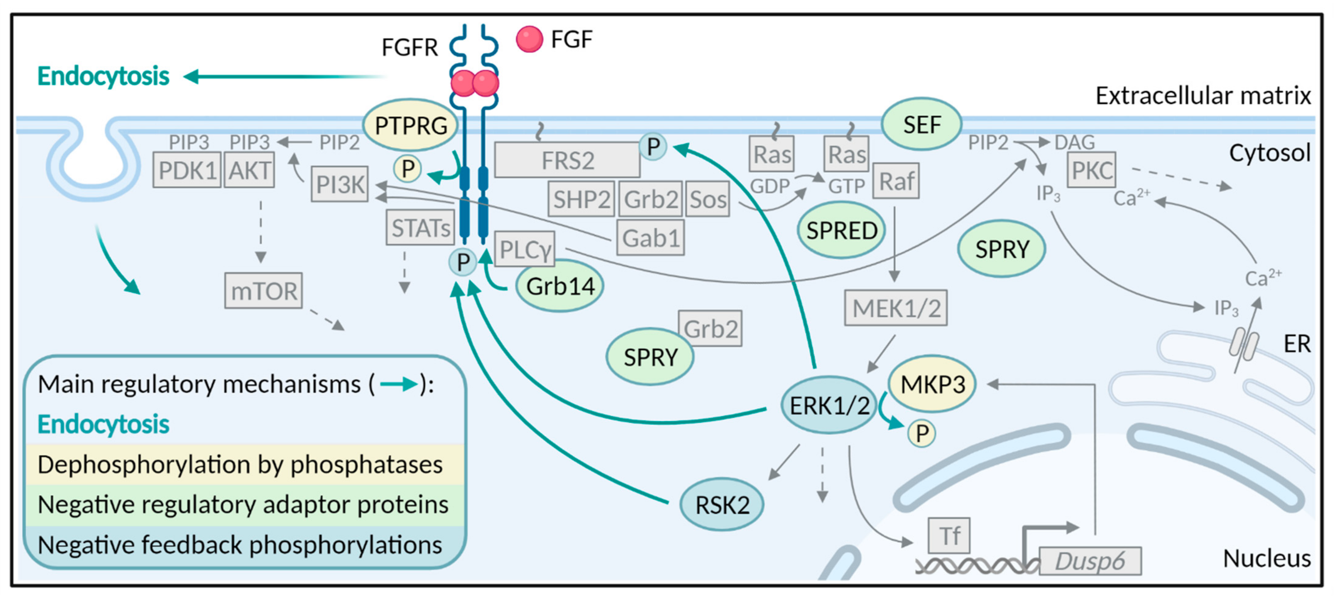
The binding of FGF ligands to their specific receptors results in receptor dimerization and conformational changes in the cytoplasmic part of the receptor. The conformational changes lead to trans-autophosphorylation of the tyrosine kinase domains of the receptors and subsequently the induction of several downstream signaling pathways. There are four major signaling pathways activated by FGF/FGFRs; the Ras-MAPK (mitogen-activated protein kinase) pathway, the PI3K (phosphatidylinositol-4,5-bisphosphate 3-kinase)-AKT pathway, the PLCγ (phospholipase Cγ)/PKC (protein kinase C) pathway and the STAT (signal transducer and activator of transcription) pathway [9] (Figure 1). There are several excellent reviews on FGFR signaling and therefore, this will not be discussed in further detail here [9,16,20]. We will focus here on how FGFR signaling is inhibited and turned off, once the receptor is activated.
Accurate signaling from receptor tyrosine kinases can be maintained through tight regulation by several mechanisms [21]. However, in contrast to the well-studied mechanism of FGFR activation and signaling, the mechanisms ensuring receptor deactivation are less understood. Downregulation of FGFR signaling has been reported to occur through mechanisms involving phosphatases, negative regulator proteins, and negative feedback phosphorylations (Figure 1). However, the main mechanism that regulates the duration and strength of receptor tyrosine signaling is endocytosis (Figure 1 and Figure 2).
2. Endocytosis
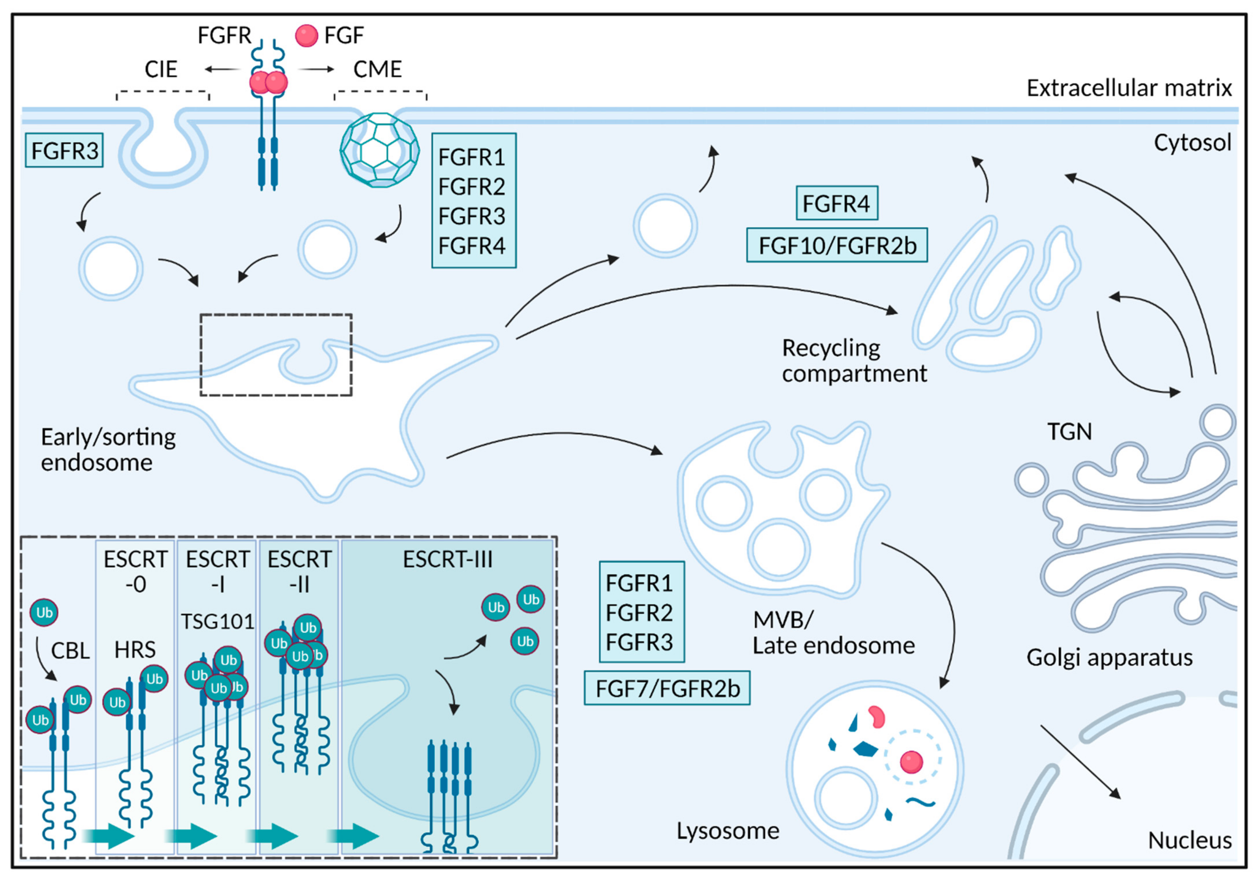
Endocytosis is a cellular process involving membrane invagination and uptake of different cargos such as receptor-ligand complexes, antigens, viruses and protein toxins into intracellular vesicles [22]. Vesicles and tubular structures with endocytic cargo are fused with early/sorting endosomes from which the cargo is sorted to different intracellular destinations (Figure 2). Endocytosed receptors might be transported into intraluminal vesicles in multivesicular bodies/late endosomes/lysosomes for degradation resulting in attenuation of signaling while some receptors are recycled back to the plasma membrane, leading to sustained signaling [23]. In the case of FGFR, endocytosis regulates signaling in several ways and depending on the receptor type, ligand type and possibly also cell type, the receptors are sorted differently resulting in altered signaling. We will here go through the mechanisms of FGFR endocytosis and how endocytosis influences FGFR signaling.
The first step in receptor-mediated endocytosis is the recruitment of active receptor-ligand complexes into growing invaginations at the plasma membrane that eventually buds off to form intracellular vesicles. Receptor internalization is often divided into clathrin-mediated endocytosis (CME) and clathrin-independent endocytosis (CIE). CIE pathways include caveolae-mediated, FEME (fast endophilin-mediated endocytosis) and CLIC/GEEC (clathrin-independent carriers/GPI-enriched early endosomal compartments) [24]. CME is the main route for many cell surface receptors and their ligands [25]. CME is characterized by the formation of clathrin coated pits at the plasma membrane which pinch off from the cell surface by the large GTPase dynamin. In the case of FGFRs, FGFR1, FGFR2 and FGFR4 are mainly internalized via CME, while FGFR3 internalization involves both CME and CIE processes [26,27,28,29,30,31,32] (Figure 2). In the case of FGFR3, it is not clear which mechanisms are involved in CIE, but dynamin seems not to be required [27,32]. It is worth mentioning that FGFRs have been observed in caveolae but it is not clear if they are internalized via caveolin-mediated endocytosis [33,34]. Additionally, FGF1 stimulation seems to induce the formation of an endophilin positive structure at the plasma membrane, indicating that FEME pathway might be involved in FGF1 uptake in some cells [35]. It is, however, not clear which FGFRs are expressed in these cells.
In the case of another RTK (receptor tyrosine kinase), namely EGFR (epidermal growth factor receptor), the receptor is taken up via both CME and CIE depending on low (<2 ng/mL) or high (>2 ng/mL) ligand concentration, respectively [36]. At even higher ligand concentrations (>50 ng/mL), EGFR is internalized via FEME [24,37]. It is not clear how this applies to other RTKs and FGFRs. In the case of FGFR1 (CME endocytosis) and FGFR3 (partial CME and CIE), ligand concentrations did not alter their dependency on clathrin for endocytosis [27].
It is uncertain what might be the signal for recruitment of FGFRs to invaginations at the plasma membrane leading to their endocytosis. Most receptors internalized via CME are recruited to the clathrin-coated membrane invaginations via tyrosine-based sequences such as YXXФ (where Ф can be any hydrophobic residue) or [FY]XNPX[FY] motives [38]. These sorting signals bind directly to the µ2 subunit of AP2, a major adaptor protein in CME, or other adaptor proteins involved in CME that contain phosphotyrosine binding (PTB) domains. Although some putative signal sequences of both types are present in FGFR1-4 (our unpublished data), none of these has yet been experimentally demonstrated to function as endocytic signals. However, a few factors seem to be crucial for FGFR endocytosis. Inhibition of receptor kinase activity results in reduced internalization, indicating that ligand binding, dimerization and trans-autophosphorylation seem to be required for proper endocytosis. An inactive, kinase dead FGFR1 (K514R) as well as treatment with FGFR inhibitors drastically reduced the rate of endocytosis [26,31,33,39,40]. In addition, the mutation of tyrosine 766 in FGFR1, which is the binding site for PLC-γ in activated FGFR1, also reduced receptor internalization [40]. However, in the case of FGFR2, tyrosine 769 (corresponding to tyrosine 766 in FGFR1) is not required for endocytosis [41]. It has also been demonstrated that phosphorylation of a specific serine (S789) in FGFR1 by RSK2 (ribosomal s6 kinase 2) is important for internalization [42] and functions as a negative feedback loop since RSK2 is activated by the Ras-MAPK pathway downstream of FGFR1 (see chapter below).
Recruitment of E3 ubiquitin ligases such as NEDD4-1 (neural precursor cell expressed developmentally down-regulated protein 4) and c-CBL (Casitas B-lineage lymphoma) either directly to the receptor or via FRS2 (FGFR substrate 2)/Grb2 (growth factor receptor-bound 2), respectively, and subsequent ubiquitination of the receptor has also been implicated in FGFR endocytosis [43,44,45]. However, a lysine mutant of FGFR1 lacking most of the potential ubiquitination sites, was endocytosed to a similar degree as wild type FGFR1 indicating that ubiquitination is not crucial for internalization [26]. Several other proteins have been implicated in FGFR endocytosis. For instance, proper kinetics of FGFR2 internalization seems to depend on the recruitment and activation of Src and Eps8 (epidermal growth factor receptor kinase substrate 8) but not Eps15 (epidermal growth factor receptor kinase substrate 15) [31,46]. In the case of endocytosis of FGFR1, interaction between FGFR1 and Esyt2 (extended synaptotagmin-2) appears to be required for proper CME [47]. Another protein implicated in FGFR endocytosis is the tumor suppressor, VHL (von Hippel-Lindau protein). Loss of VHL led to accumulation of FGFR1 at the cell surface and enhanced signaling due to impaired internalization [48,49]. Additionally cell adhesion molecules such as N-cadherin and E-cadherin seem to play a role in FGFR endocytosis. Binding of FGFR1 to N-cadherin or overexpression of E-cadherin, can delay FGFR1 endocytosis [50,51]. Syndecan-4, a heparan sulfate proteoglycan at the cell surface, is involved in macropinocytosis of FGFR1 [52]. Although recruitment of several proteins to active FGFRs seems to play a role in FGFR endocytosis, it has also been suggested that FGFR1 dimerization rather than activation is required for CME [53]. To this end, it is not completely clear how the different findings regarding recruitment of FGFRs to the endocytic machinery, choice of machinery and proteins involved add together. More research, preferentially under similar experimental settings, is needed in order to fully understand these processes and to elucidate receptor or ligand determined preferences.
Once internalized, the number of receptors available for binding of ligands at the cell surface is reduced and the cells are less responsive to ligand. The receptor can continue to signal from endosomes and it has been suggested that different intracellular locations might give rise to different signaling simply because the subsets of downstream signaling molecules might vary from compartment to compartment [54]. For example, in cells depleted for Rab11, a master regulator of recycling, FGFR4 accumulates intracellularly leading to sustained PLC-γ signaling, but reduced AKT signaling [55]. It seems that recycling is needed for FGFR4 to interact with the AKT signaling machinery to maintain AKT signaling. On the other hand, PLC-γ that binds directly to the receptor is localized with FGFR4 and signaling is sustained even if the receptor is trapped in endosomes [55]. When endocytosis of FGFR1 is reduced, due to depletion of cells for clathrin, not only prolonged MAPK signaling was observed but also a delay in activation [27]. This indicates that endocytosis of FGFR1 is needed for full activation of MAPK signaling as well as for downregulation of MAPK signaling [27]. Delayed endocytosis of FGFR1 due to binding to N-cadherin, also led to sustained MAPK signaling [50]. On the other hand, sustained MAPK signaling was observed upon FGFR1 macropinocytosis by removal of syndecan-4 from cells [52]. Additionally, trapping of FGFR1 at the cell surface upon E-cadherin overexpression, reduced MAPK signaling [51,52]. As is evident from these studies, it is not entirely clear how endocytosis influences FGFR-induced MAPK signaling. The effect of endocytosis and different intracellular localization of FGFRs on signaling might be cell type dependent or even dependent on the experimental conditions. It is however clear, that FGFRs can signal not only from the plasma membrane but also from endosomes and that their signaling properties might vary with their localization (for instance plasma membrane versus endosomal location). It is also worth mentioning that FGFs and FGFRs can traffic to the nucleus (reviewed in [56,57]). Although their function in the nucleus is not clear, nuclear localization seems to be required for promigratory effect of FGF stimulation in breast and pancreatic cancer cells, as well as HIF (hypoxia-inducible factor)-mediated hypoxic responses in prostate cancer cells [58,59,60].
Following endocytosis, receptors are transported to the lysosomes for degradation resulting in termination of signaling or for recycling back to the cell surface allowing additional rounds of signaling [23,61] (Figure 2). The newly formed endocytic carrier vesicles originating from the plasma membrane undergo fusion events to form early endosomes also called sorting endosomes and it is here, at the early/sorting endosome, the fate of the internalized receptors are decided. The receptors at the early/sorting endosomes are either sorted for lysosomal degradation or they are retrieved into tubule-vesicular transport carriers for recycling back to the plasma membrane [23,61]. Recycling back to the plasma membrane can occur via the endosomal recycling compartment in a Rab11 dependent manner (slow recycling) or directly from early/sorting endosomes back to the plasma membrane in a Rab4 dependent manner (fast recycling) [23]. Receptors destined for degradation are sorted into intraluminal vesicles originating at the early/sorting endosomal membrane. This occurs as the early endosomes mature into a late endosome [23]. Late endosomes are characterized by the appearance of multiple intraluminal vesicles and are also referred to as multivesicular bodies. Late endosomes usually fuse to lysosomes resulting in degradation of their content [23].
The pathway that FGFRs follow after internalization depends on both the receptor-type but also the bound ligand [62] (Figure 2). Upon stimulation with FGF1, FGFR1-3 are mainly sorted to lysosomes, but with lower efficiency in the case of FGFR2 and FGFR3 while FGFR4 is mainly recycled leading to sustained signaling [62]. Stimulation of FGFR2b with FGF7 also resulted in lysosomal sorting and degradation while stimulation with FGF10 led to recycling and increased signaling [29,63]. Moreover, FGFR1 bound to NCAM (neural cell adhesion molecule), an unconventional ligand for FGFR1, led to FGFR1 recycling [64]. Similarly, NEGR1 (neuronal growth regulator 1) seems to be important for FGFR2 recycling [65]. The recycling pathway might also involve transport via the TGN (trans Golgi network) as internalized FGF1/FGFR4 complexes were found to localize partially with TGN structures [28]. FGF/FGFRs can also be transported to the nucleus [59,66,67].
While the retrieval of receptors for recycling is poorly understood, sorting of receptors into intraluminal vesicles and the degradative pathway is well characterized. The key step for sorting of a receptor into intraluminal vesicles and subsequent degradation, is the attachment of ubiquitin to lysine residues in the intracellular part of the receptor by E3 ubiquitin ligases such as CBL and NEDD4 [23,68]. The ESCRT (endosomal sorting complex required for transport) complexes are recruited to the endosomal membrane and concentrate ubiquitinated proteins in degradative subdomains that eventually bud off to form intraluminal vesicles (Figure 2). The ESCRT complexes, ESCRT-0, -I, -II and -III operate in a sequential manner [69]. First, ESCRT-0 consisting of HRS (hepatocyte growth factor-regulated tyrosine kinase substrate) and STAM 1 (signal transducing adaptor molecule 1), each binding with low affinity to ubiquitin, cluster the ubiquitinated cargo. This leads to recruitment of ESCRT-I and ESCRT-II. Accumulation of ESCRT-II at the degradative subdomains leads to recruitment of ESCRT-III. ESCRT-III together with VPS4 (vacuolar protein sorting-associated protein 4) generate inwardly budding of the membrane followed by scission to generate an intraluminal receptor-containing vesicle. Before scission, the receptors are deubiquitinated and the ESCRT-0-II is dissociated [23,69,70].
To understand the different sorting of FGFR1 and FGFR4, a comparison of their intracellular sequences was performed and revealed that FGFR1 contains a higher number of lysines that are potential ubiquitination sites, than FGFR4 [62]. Indeed, it was demonstrated that FGFR1 was more efficiently ubiquitinated than FGFR4. Moreover, removing potential ubiquitination sites in FGFR1 by substituting lysines for arginines, led to reduced ubiquitination of FGFR1 and forced FGFR1 to recycle [26]. It has also been demonstrated that the binding of N-cadherin to FGFR1 reduces ubiquitination of FGFR1 resulting in increased FGFR1 stability [71]. The sorting of FGFR2b to degradation upon binding to FGF7 seems to depend on HRS (a component of ESCRT-0), TSG101 (tumor susceptibility gene 101 protein) (a component of ESCRT-I), and ubiquitination [29,72]. FGF7 induced FGFR2b ubiquitination to a greater extent than FGF10 possibly explaining why FGF7 induces lysosomal routing while FGF10 induces receptor recycling. The reduced ubiquitination of FGFR2b upon FGF10 stimulation might result from less efficient phosphorylation of FRS2 leading to reduced recruitment of the ubiquitin ligase c-CBL (discussed below) [29]. It was also reported that stimulation with FGF10 in contrast to FGF7 led to the phosphorylation of Y734 in FGFR2b and subsequent recruitment of PI3K and SH3BP4 (SH3-binding protein 4) [63]. Both phosphorylation of Y734 and the presence of SH3BP4 were required for FGF10 induced FGFR2b recycling. Interestingly, FGFR3 harboring mutations associated with skeletal disorders was found to escape lysosomal targeting due to reduced levels of ubiquitination [73]. On the other hand, decreased degradation but excessive ubiquitination of FGFR3 harboring these mutations associated with skeletal disorders, has also been reported [74,75] indicating that there might exist alternative mechanisms which independently of ubiquitination levels allow the receptors to escape into the recycling pathway. Although other factors might be involved, it seems that different levels of FGFR ubiquitination, as for many RTKs, might be the main mechanism deciding their fate at the early/sorting endosome.
It is not completely clear which ubiquitin ligases are involved in FGFR ubiquitination but many reports have implicated c-CBL [29,43,73,76,77]. It has been shown that Grb2 interacts with CBL and is recruited to phosphorylated FRS2α in an FGF-dependent manner [29,43]. In this way, CBL recruitment functions as a negative feedback loop in FGFR signaling. Interestingly, in the case of FGFR2b, FGF7 stimulation (leading to receptor degradation) also resulted in a more efficient recruitment of c-CBL than FGF10 stimulation (leading to receptor recycling) [29]. Ubiquitination of FGFR has been shown to increase with overexpression of CBL [43]. It is not clear which receptor was examined in these experiments but similar effects have been observed in the case of FGFR3 [73]. Likewise, overexpression of a dominant negative form of CBL reduced FGFR2 ubiquitination [78]. Additionally, lysosomal transport of FGF8 was delayed upon interfering with the function of CBL [79]. On the other hand, it has been reported that overexpression of wild-type c-CBL or a dominant negative c-CBL variant did not significantly alter FGFR3 ubiquitination [74]. Interestingly, FGFR3 has also been suggested to be stabilized by binding to Hsp90 (heat shock protein 90) [80,81] and it was shown that FGFR3 was ubiquitinated by CHIP (carboxyl terminus of HSP70-interacting protein) ubiquitin ligase probably leading to degradation through a proteasomal pathway rather than the endolysosomal pathway [80]. In addition, NEDD4-1 has been shown to directly bind to FGFR1 leading to ligand induced ubiquitination [44,45]. Under these experimental conditions, the knockdown of CBL did not alter the stability of active FGFR1 [44]. Taken together, several ubiquitin ligases might be involved in FGFR ubiquitination leading to receptor degradation.
Clearly, endocytosis and intracellular sorting influence receptor signaling in many ways. Receptor trafficking is not only altering the duration of signaling, but also the signaling pathways activated might vary with subcellular localization. This might give rise to a completely different signaling output. For instance, altered intracellular sorting of FGFR2b upon stimulation with FGF10 (recycling) reduced signaling duration and led to decreased breast cancer cell migration and inhibition of epithelial branching compared to stimulation with FGF7 (degradation) [63]. Similarly, in contrast to FGF2-induced degradation of FGFR1, NCAM promoted recycling of the receptor, leading to a recycling-dependent increase in cell migration [64]. Expression of ubiquitination-deficient FGFR1 mutants in adult dorsal root ganglia cells led to enhanced recycling and increased axon elongation without stimulating axon branching compared to FGFR1 wild-type [82]. By contrast, the inhibition of FGFR1 endocytosis reduced axon elongation and enhanced axonal branching [83]. Trapping FGFR1 at the cell surface as well as forcing FGFR1 to recycle (ubiquitination-deficient FGFR1 mutants), led to increased signaling but had opposite effects on axon growth. In this case, receptor localization/routing rather than the duration decided the signaling output.
Most studies on FGFR endocytosis and signaling are performed in cell lines. The understanding of the mechanisms and consequences of FGFR endocytosis in vivo is limited. It has been shown that expression of a mutant form of FGFR1, which is unable to bind to NEDD-4 leading to reduced endocytosis and sustained signaling, results in disrupted anterior neuronal patterning (head development) in zebrafish [44]. Another example is the role of endocytosis in FGF8 morphogen gradient formation [79,84]. FGF8 acts as a diffusible morphogen during vertebrate development and has key roles in a variety of developmental processes, including limb and brain development. In the nascent neuroectoderm of living zebrafish embryos, the spread of FGF8 through the tissue is controlled by endocytosis and subsequent degradation in lysosomes [84]. Upon inhibition of endocytosis, FGF8 accumulated extracellularly and spread over a greater distance in tissue while enhanced FGF8 internalization reduced the signaling range. The effects seemed to be dependent on CBL-mediated lysosomal transport of FGF8 [79]. Yet another example showing the importance of FGFR regulation by endocytosis in vivo is the regulation of FGFR2 by the cell adhesion protein, NEGR1. The knockout of NEGR1 or FGFR2 affected neuronal migration and spine density during mouse cortical development and resulted in impaired core behaviors related to autism spectrum disorders [65]. In cell lines, removal of NEGR1 led to increased degradation of FGFR2 and less signaling. In mice, overexpression of FGFR2 could rescue the effect of NEGR1 knockout indicating that the effect on mouse cortical development was due to decreased FGFR2 protein levels and signaling [65]. Taken together, endocytosis and intracellular transport regulate FGFR signaling giving rise to alternative biological outputs.
3. Phosphatases
Protein tyrosine phosphatases (PTPs) remove the phosphate group from tyrosine residues of phosphorylated proteins [85] and as such are primary suspects as negative regulators of FGFR activity. However, to date only PTPRG (protein tyrosine receptor-type G) has been found to inhibit the activity of FGFR1 by direct dephosphorylation of activated FGFR1 [86]. The phosphatase also downregulates FGFR2-4 phosphorylation. It was estimated that PTPRG accounts for ~80% of all phosphatases activity in the early stage of FGFR1 activation [86]. Cancer cells depleted of PTPRG display increased FGFR activity and are hypersensitive to stimulation by FGF1. Moreover, PTPRG depletion elevated cell growth and negatively affected the efficacy of FGFR kinase inhibitors [86]. Due to the efficient dephosphorylation of FGFRs by PTPRG (~80% for FGFR1), other PTPs involved in direct dephosphorylation and negative regulation of FGFRs may be difficult to find due to the functional redundancy and a small share in the process. However, FGFR3 is subject to context-dependent regulation by the phosphatases, PTPN1 and PTPN2 (protein tyrosine phosphatase non-receptor type 1 and 2), and loss of either PTP resulted in ligand-independent activation of FGFR3 [87]. Interestingly, while PTPRG seems to dephosphorylate FGFRs mainly at the cell surface, PTPN1 is localized to the ER and prevents FGFR3 phosphorylation during ER-Golgi processing [86,87].
Phosphatases can also play a role in the positive regulation of FGFR signaling. The best example is the phosphatase SHP2 (Src homology region 2 domain-containing phosphatase 2, also known as PTPN11 (protein tyrosine phosphatase non-receptor type 11)), which binds to phosphorylated FRS2 upon FGF stimulation, but enhances FGFR signaling [88,89]. Once recruited to FRS2, SHP2 is tyrosine phosphorylated and recruits Grb2 leading to the initiation of downstream signaling pathways [89]. Recruited SHP2 can dephosphorylate and inactivate SPRY (Sprouty) (discussed below) thereby causing dissociation of Grb2 from SPRY and suppressing negative impact of SPRY on signaling activation [88]. Another interesting example is the inositol phosphatase, SHIP2 (phosphatidylinositol-3,4,5-trisphosphate 5-phosphatase). SHIP2 has been shown to bind activated FGFRs and nucleate recruitment of Src-family kinases, leading to prolonged activation of FGFRs [90]. Yet, this function of SHIP2 is independent of its phosphatase activity, and rather depends on scaffolding properties by bringing in Src-family kinases as positive regulators. Phosphatases targeting downstream effectors of activated FGFRs are additional players in the regulation of FGFR activity. A known example is MKP3 (MAPK phosphatase 3 also known as DUSP6 (dual-specificity phosphatase 6)), which is an ERK (extracellular signal-regulated kinase)-specific phosphatase encoded by the gene Dusp6. MKP3 acts as a key modulator and controls MAPK deactivation by dephosphorylating ERK1 and ERK2 on phosphotyrosine and phosphothreonine residues [91]. Several studies show that Dusp6 transcription is activated by FGF signaling and suggest a negative feedback role for MKP3 in FGF signaling [92,93,94]. FGFR negative feedback regulation is driven by direct binding of the ERK1/2-responsive transcription factor ETS2 to the Dusp6 promoter [95]. Downregulation of FGF-induced MAPK signaling by MKP3 seems to play critical roles in regulating developmental outcomes in vertebrates. For example, loss of MKP3 in mice embryos resulted in partially penetrant postnatal lethality, skeletal malformations, and hearing loss, phenotypes that are characteristic of activating mutations in FGFRs [91]. Overexpression of MKP3 in chick embryos reduced levels of activated MAPK in the neural plate and retarded limb bud outgrowth [93]. FGF8-induced MKP3 expression seems to be important during chick, mouse and zebrafish limb/fin development [94].
4. Negative Regulatory Proteins
A class of well-studied molecules that regulate FGFR signaling are the SPRY proteins (Figure 1). Four members of the SPRY (SPRY1-4) family were discovered as ligand-inducible antagonists of RTK signaling [21]. RTK signaling can influence SPRY proteins by increasing SPRY expression, regulating recruitment to the plasma membrane and modulating SPRY activity by transient tyrosine phosphorylation [96]. ERK1/2 activity has an impact on the expression of SPRY proteins as in several studies SPRY expression was abolished by MEK (mitogen-activated protein kinase kinase) inhibition [97]. SPRY proteins regulate FGFR signaling at various levels and in several ways. First of all, upon FGF stimulation, SPRY1 and SPRY2 translocate to the plasma membrane where they are phosphorylated on a conserved N-terminal tyrosine residue (Y53 or Y55, respectively) and interact with Grb2 [98]. The binding of SPRY1/2 to Grb2 prevents Grb2 from binding to either FRS2 or SHP2 and consequently inhibits MAPK signaling [98]. In addition, SPRY2 has also been shown to bind and inhibit the activity of Raf, a serine/threonine kinase that functions downstream of Ras [99]. There are probably at least two distinct pools of SPRY2, one that binds PP2A (protein phosphatase 2A) and another that binds c-CBL [100]. PP2A binding is required for dephosphorylation of SPRY2, while direct interaction with c-CBL possibly directs SPRY2 for degradation via ubiquitination [100]. Overexpression of SPRY1 and 2 resulted in decreased ubiquitination and increased stability of FGFR2 and FGFR3, respectively [101,102]. The authors suggested that overexpression of SPRY proteins sequesters CBL away from FGFR:FRS2:Grb2 complexes and thus prevents FGFR ubiquitination. In PC12 cells, the activity and binding of CK1 (casein kinase 1) to SPRY2 was necessary for the inhibitory function of SPRY2 in FGFR signaling by promoting binding of SPRY2 to Grb2 [103]. SPRY2 has been shown to act as a key regulator of FGFR signaling in fetal lung development as SPRY2 knockout rats had severe defects in lung morphogenesis [104]. Contrary, TESK1 (testicular protein kinase 1) and DYRK1A (dual-specificity tyrosine phosphorylation-regulated kinase 1A) can promote FGFR signaling by reversing the inhibitory effects of SPRY2 in both in vivo and in vitro models [105,106]. SPRY4, another member of the SPRY protein family, can also significantly abolish FGF2-induced ERK activation by sequestering Sos1 (Son of sevenless homolog 1) [107]. Interestingly, hetero-oligomers that formed between SPRY1 and SPRY4 more effectively suppressed the ERK activation by inhibiting the association of the Grb2-Sos1 complex with FRS2 [107]. Taken together, SPRY proteins repress FGFR signaling through multiple mechanisms, which likely depend on the cellular context, growth factor- and receptor-type and/or the experimental conditions.
SPRY share several features with the SPRED (Sprouty related with EVH1 (Ena/VASP homology 1)) protein family composed of SPRED-1, SPRED-2, and SPRED-3. Both protein families have a homologous cysteine-rich C terminus and function as inhibitors of the Ras-MAPK pathway downstream of various stimuli like growth factors and cytokines [108,109]. As for SPRY proteins, the mechanisms by which the SPRED proteins negatively regulate Ras signaling are not entirely clear. One way that SPRED proteins suppress Ras signaling is by the recruitment of a the Ras GTPase activating protein, NF-1 (neurofibromin) to the plasma membrane [110]. The recruitment of NF-1 leads to hydrolysis of GTP to GDP in Ras and inactivation of the Ras-MAPK signaling pathway. Other reports suggest that SPRED proteins downregulate Ras signaling by directly preventing Raf phosphorylation and activation [111]. In the case of FGFR signaling, increased ERK phosphorylation upon FGF stimulation was observed in SPRED2-deficient mice [112]. Simultaneously, a dwarf phenotype similar to human achondroplasia, usually caused by activating mutations in FGFR3, was also observed in the SPRED2 knockout mice. Loss of SPRED2 seems to inhibit chondrocyte proliferation due to increased FGF-induced ERK activation, resulting in reduced bone growth [112]. Moreover, SPRED2 seems to play a role in FGFR degradation through direct binding to late endosomal protein NBR1 (neighbor of BRCA1 gene 1 protein) [113]. Similarly to SPRY, SPRED2 interacts with CBL and this interplay controls protein levels of SPRED2 as ubiquitination by CBL targets SPRED2 for degradation [114]. The binding of p85, a subunit of PI3K, to SPRED2 augments the SPRED2-mediated inhibitory effect by increasing Ras binding to SPRED2 and decreasing SPRED2 ubiquitination [115].
Another molecule that acts as an antagonist of FGFR signaling is SEF (similar expression to FGF), a conserved inhibitor of the MAPK pathway [116]. However, the precise role of SEF in FGFR signaling is still not clear. SEF has been proposed to act directly in the MAPK signaling pathway as well as at the FGFR itself. In the MAPK pathway, SEF might act at several points. Reports have shown that SEF acts downstream of or at MEK level by inhibiting ERK phosphorylation [117]. On the contrary, it was also reported that SEF can act upstream of MEK possibly at the level of Ras in prostate cancer cells [118]. It has been proposed that SEF directly interacts with Ras at the plasma membrane causing inhibition of Ras activation by FGFR [119]. Results showing that SEF interacts with FGFR across different species and cell types and can inhibit FGFR phosphorylation support the observation that SEF might inhibit components early in the signaling cascade (prior to Ras activation) [119,120]. Direct binding of SEF to FGFR might influence receptor dimerization and consecutive trans-autophosphorylation or can modify receptor-ligand interaction leading to attenuation of signal transduction [120]. The finding that SEF acts on the receptor level or at early steps in FGFR signaling and inhibits various signaling pathways is additionally supported by results showing that both mouse and human SEF mediates a reduction in AKT signaling [120,121]. Overexpression of SEF in the lens of mice resulted in impaired lens and eye development and increased apoptosis due to inhibition of FGFR signaling during lens morphogenesis [122]. The intracellular, extracellular and the transmembrane domain of SEF seem to be important for SEF-mediated negative regulation of FGF signaling [123].
Another protein that might modulate FGFR signaling is FGFR5 (FGFRL1). FGFR5 is the fifth member of the FGFR family but lacks tyrosine kinase activity and thus is not an active receptor. Instead, FGFR5 has been predicted to negatively regulate FGF signaling by competing with other FGFRs for ligand binding or by forming heterodimers with other members of the FGFR family and thereby preventing trans-autophosphorylation [124]. However, experimental results showed that FGFR5 does not function as a decoy receptor, but rather promotes intracellular signaling as it increases activation of MAPK signaling due to association with the phosphatase SHP1 [125]. Flotillin-1, a multifunctional protein also involved in endocytosis, has been shown to compete with FGFR for binding to FRS2 and thereby interferes with signaling [126]. A flotillin-1 knockdown resulted in increased tyrosine phosphorylation of FRS2, as well as inhibition of ERK activity.
Grb2 has also been suggested to play a role as a regulator of FGFR2 signaling [127]. It was reported that dimeric Grb2 can bind directly to unliganded FGFR2, preventing receptor phosphorylation. Upon stimulation, the activated FGFR2 phosphorylates Grb2, leading to Grb2 dissociation and the full activation of FGFR2 downstream signaling.
The adaptor protein Grb14 has been implicated in the regulation of PLCγ signaling downstream of FGFRs [128]. Upon receptor activation, PLCγ is directly recruited to phosphorylated tyrosine 766 in FGFR1 [129]. Similarly, Grb14 was found to bind to the same phosphorylated tyrosine in FGFR1 (Y766) and is thought to compete with PLCγ for binding [130]. In addition, it was suggested that the binding of Grb14 to phosphorylated Y766 in FGFR1 led to conformational changes in Grb14 that unmasked a PLCγ binding motif in Grb14 [131]. Once the binding motif is revealed, PLCγ is trapped away from the active FGFR and cannot be phosphorylated. Thus, Grb14 seems to have a dual way of inhibiting FGFR-induced PLCγ signaling.
Clearly, multiple proteins can function at various steps as negative regulators in the FGFR signaling pathways to turn off FGFR signaling. In addition, some of these regulatory proteins might have numerous roles.
5. Negative Feedback Phosphorylations
In addition to several negative feedback mechanisms governing FGFR signaling such as FGF-induced CBL recruitment, MKP3 production and others discussed above, more direct negative feedback mechanism involving phosphorylation events also exists to prevent excess FGFR signaling (Figure 1). Nearly all components of the MAPK signaling pathways are regulated through such negative feedback phosphorylations by downstream kinases [97]. For example, upon activation, FGFRs phosphorylate FRS2α on tyrosine residues leading to recruitment of Grb2 and activation of MAPK signaling and ERK1/2 activation. Active ERK1/2 can then phosphorylate eight threonine residues in FRS2 [132]. Mutation of the FRS2α threonine phosphorylation sites led to constitutive tyrosine phosphorylation of FRS2α in unstimulated cells and enhanced FRS2α tyrosine phosphorylation in FGF-stimulated cells [132]. Similarly, a specific serine residue (S777) in the C-terminal region of FGFR1 was shown to be directly phosphorylated by active ERK1 and ERK2 upon FGF1 stimulation [133]. Mutating S777 to alanine led to prolonged FGFR1 tyrosine phosphorylation indicating a direct negative feedback mechanism attenuating FGFR1 signaling [133]. A negative feedback loop mediated by ERK1/2 pathway was also identified for FGFR2 [134]. It was shown that substituting serine 780 in FGFR2 (corresponding to S777 in FGFR1) with alanine resulted in increased FGFR2 phosphorylation and signaling. Hence, ERK1/2-mediated phosphorylation of S780 in FGFR2, similarly to S777 in FGFR1, acts as a negative feedback loop to prevent excess signaling. It is not known how phosphorylation of S777 (FGFR1) and S780 (FGFR2) regulate receptor activity. It is also worth mentioning that, a serine close to S780 in FGFR2 (S782 or S779 dependent on the numbering) is phosphorylated by PKCε, providing a docking site for the adaptor protein 14-3-3 and leading to sustained ERK activation [135]. It is not known how these negative and positive feedback phosphorylation events in FGFR2 cooperate to regulate FGFR2 signaling. Interestingly, the serine/threonine kinase RSK2, which is activated downstream of ERK1/2, can bind directly to FGFR1 and phosphorylate serine 789 (S789) in FGFR1 [42]. Phosphorylation of S789 in FGFR1 is important for proper FGFR1 endocytosis and ubiquitination [42].
6. Summary
Clearly, many mechanisms exist to control FGFR signaling. The tight regulation of signaling is necessary to maintain body homeostasis. These mechanisms can occur simultaneously or sequentially, and in different or, sometimes, the same steps of the signaling pathways. They might operate differently depending on cell type, the ligand or the receptor type. It is clear that our cells have evolved a whole menu of mechanisms reinforcing each other to control FGFR signaling. In case one of the mechanisms is not operational, other mechanisms and proteins involved in the regulation of cell signaling can take over and the cell might still be able to prevent excessive FGFR signaling. The development of all of these different mechanisms indicates the importance of keeping FGFR signaling under control. As mentioned in the introduction, several human conditions are caused by excessive FGFR signaling, among them dwarfism and cancer [15,16,17,18]. Clearly, the magnitude, duration, and location of FGFR signaling must be strictly controlled in order to induce the correct biological response and prevent development of diseases.
Author Contributions
Writing—original draft preparation, P.S., E.M.H., A.W.; writing—review and editing, P.S., M.K., J.W., E.M.H., A.W.; visualization, E.M.H.; funding acquisition, J.W., A.W. All authors have read and agreed to the published version of the manuscript.
Funding
This work was supported by the Norwegian Cancer Society (project number 198093 and 207006), the Research Council of Norway through its Centers of Excellence funding scheme (project number 262652), the Norway Grants 2014–2021 via the National Centre for Research and Development, Project Contract No. NOR/POLNOR/DUALDRUG/0058/2019-00.
Acknowledgments
We thank Birgitte Bjørnerud for a thorough review of the manuscript.
Conflicts of Interest
The authors declare no conflict of interest.
Abbreviations
| AP2 | Adaptor protein 2 |
| CBL | Casitas B-lineage lymphoma |
| CHIP | Carboxyl terminus of HSP70-interacting protein |
| CIE | Clathrin-independent endocytosis |
| CK1 | Casein kinase 1 |
| CLIC/GEEC | Clathrin-independent carriers/GPI-enriched early endosomal compartments |
| CME | Clathrin-mediated endocytosis |
| DUSP6 | Dual-specificity phosphatase 6 |
| DYRK1A | Dual-specificity tyrosine phosphorylation-regulated kinase 1A |
| Eps | Epidermal growth factor receptor kinase substrate |
| ERK 1/2 | Extracellular signal-regulated kinase 1/2 |
| ESCRT | Endosomal sorting complex required for transport |
| Esyt2 | Extended synaptotagmin-2 |
| FEME | Fast endophilin-mediated endocytosis |
| FGF | Fibroblast growth factor |
| FGFR | Fibroblast growth factor receptor |
| FRS2 | FGFR substrate 2 |
| Grb2/14 | Growth factor receptor-bound 2/14 |
| HIF | Hypoxia-inducible factor |
| HRS | Hepatocyte growth factor-regulated tyrosine kinase substrate |
| Hsp90 | Heat shock protein 90 |
| MAPK | Mitogen-activated protein kinase |
| MEK | Mitogen-activated protein kinase kinase |
| MKP3 | MAPK phosphatase 3 |
| NBR1 | Neighbor of BRCA1 gene 1 protein |
| NCAM | Neural cell adhesion molecules |
| NEDD4 | Neural precursor cell expressed developmentally down-regulated protein 4 |
| NEGR1 | Neuronal growth regulator 1NF-1 Neurofibromin |
| PI3K | Phosphatidylinositol-4,5-bisphosphate 3-kinase |
| PKC | Protein kinase C |
| PLCγ | Phospholipase Cγ |
| PP2A | Protein phosphatase 2A |
| PTB | Phosphotyrosine binding |
| PTP | Protein tyrosine phosphatase |
| PTPN1/2/11 | Protein tyrosine phosphatase non-receptor type 1/2/11 |
| PTPRG | Protein tyrosine phosphatase receptor type G |
| RasGAP | Ras GTPase activating protein |
| RSK2 | Ribosomal s6 kinase 2 |
| RTK | Receptor tyrosine kinase |
| SEF | Similar expression to Fgf |
| SH3BP4 | SH3-binding protein 4 |
| SHIP2 | Phosphatidylinositol-3,4,5-trisphosphate 5-phosphatase |
| SHP1/2 | Src homology region 2 domain-containing phosphatase 1/2 |
| Sos1 | Son of sevenless homolog 1 |
| SPRED | Sprouty related with EVH1 (Ena/VASP homology 1) |
| SPRY | Sprouty |
| STAM 1 | Signal transducing adaptor molecule 1 |
| STAT | Signal transducer and activator of transcription |
| TESK1 | Testicular protein kinase 1 |
| TGN | Trans Golgi network |
| TSG101 | Tumor susceptibility gene 101 protein |
| VHL | von Hippel-Lindau protein |
| VPS4 | Vacuolar protein sorting-associated protein 4 |
References
- Itoh, N.; Ornitz, D.M. Fibroblast growth factors: From molecular evolution to roles in development, metabolism and disease. J. Biochem. 2011, 149, 121–130. [Google Scholar] [CrossRef] [PubMed] [Green Version]
- Murzin, A.G.; Lesk, A.M.; Chothia, C. beta-Trefoil fold. Patterns of structure and sequence in the Kunitz inhibitors interleukins-1 beta and 1 alpha and fibroblast growth factors. J. Mol. Biol. 1992, 223, 531–543. [Google Scholar] [CrossRef]
- Blaber, M.; DiSalvo, J.; Thomas, K.A. X-ray crystal structure of human acidic fibroblast growth factor. Biochemistry 1996, 35, 2086–2094. [Google Scholar] [CrossRef] [PubMed] [Green Version]
- Orengo, C.A.; Jones, D.T.; Thornton, J.M. Protein superfamilies and domain superfolds. Nature 1994, 372, 631–634. [Google Scholar] [CrossRef]
- Bertrand, S.; Iwema, T.; Escriva, H. FGF signaling emerged concomitantly with the origin of Eumetazoans. Mol. Biol. Evol. 2014, 31, 310–318. [Google Scholar] [CrossRef] [Green Version]
- Philippe, H.; Derelle, R.; Lopez, P.; Pick, K.; Borchiellini, C.; Boury-Esnault, N.; Vacelet, J.; Renard, E.; Houliston, E.; Quéinnec, E.; et al. Phylogenomics revives traditional views on deep animal relationships. Curr. Biol. 2009, 19, 706–712. [Google Scholar] [CrossRef] [Green Version]
- Werner, S.; Duan, D.S.; de Vries, C.; Peters, K.G.; Johnson, D.E.; Williams, L.T. Differential splicing in the extracellular region of fibroblast growth factor receptor 1 generates receptor variants with different ligand-binding specificities. Mol. Cell. Biol. 1992, 12, 82–88. [Google Scholar] [CrossRef] [Green Version]
- Johnson, D.E.; Williams, L.T. Structural and functional diversity in the FGF receptor multigene family. Adv. Cancer Res. 1993, 60, 1–41. [Google Scholar]
- Ornitz, D.M.; Itoh, N. The Fibroblast Growth Factor signaling pathway. Wiley Interdiscip. Rev. Dev. Biol. 2015, 4, 215–266. [Google Scholar] [CrossRef] [Green Version]
- Thisse, B.; Thisse, C. Functions and regulations of fibroblast growth factor signaling during embryonic development. Dev. Biol. 2005, 287, 390–402. [Google Scholar] [CrossRef]
- Tulin, S.; Stathopoulos, A. Extending the family table: Insights from beyond vertebrates into the regulation of embryonic development by FGFs. Birth Defects Res. Part C Embryo Today Rev. 2010, 90, 214–227. [Google Scholar] [CrossRef] [Green Version]
- Itoh, N.; Ornitz, D.M. Evolution of the Fgf and Fgfr gene families. Trends Genet. 2004, 20, 563–569. [Google Scholar] [CrossRef]
- Popovici, C.; Roubin, R.; Coulier, F.; Birnbaum, D. An evolutionary history of the FGF superfamily. Bioessays 2005, 27, 849–857. [Google Scholar] [CrossRef]
- Brewer, J.R.; Mazot, P.; Soriano, P. Genetic insights into the mechanisms of Fgf signaling. Genes Dev. 2016, 30, 751–771. [Google Scholar] [CrossRef] [Green Version]
- Wesche, J.; Haglund, K.; Haugsten, E.M. Fibroblast growth factors and their receptors in cancer. Biochem. J. 2011, 437, 199–213. [Google Scholar] [CrossRef] [Green Version]
- Xie, Y.; Su, N.; Yang, J.; Tan, Q.; Huang, S.; Jin, M.; Ni, Z.; Zhang, B.; Zhang, D.; Luo, F.; et al. FGF/FGFR signaling in health and disease. Signal Transduct. Target. Ther. 2020, 5, 181. [Google Scholar] [CrossRef]
- Krook, M.A.; Reeser, J.W.; Ernst, G.; Barker, H.; Wilberding, M.; Li, G.; Chen, H.Z.; Roychowdhury, S. Fibroblast growth factor receptors in cancer: Genetic alterations, diagnostics, therapeutic targets and mechanisms of resistance. Br. J. Cancer 2021, 124, 880–892. [Google Scholar] [CrossRef]
- Shiang, R.; Thompson, L.M.; Zhu, Y.Z.; Church, D.M.; Fielder, T.J.; Bocian, M.; Winokur, S.T.; Wasmuth, J.J. Mutations in the transmembrane domain of FGFR3 cause the most common genetic form of dwarfism, achondroplasia. Cell 1994, 78, 335–342. [Google Scholar] [CrossRef]
- Ornitz, D.M.; Marie, P.J. Fibroblast growth factor signaling in skeletal development and disease. Genes Dev. 2015, 29, 1463–1486. [Google Scholar] [CrossRef] [Green Version]
- Babina, I.S.; Turner, N.C. Advances and challenges in targeting FGFR signalling in cancer. Nat. Rev. Cancer 2017, 17, 318–332. [Google Scholar] [CrossRef]
- Dikic, I.; Giordano, S. Negative receptor signalling. Curr. Opin. Cell Biol. 2003, 15, 128–135. [Google Scholar] [CrossRef]
- Jeger, J.L. Endosomes, lysosomes, and the role of endosomal and lysosomal biogenesis in cancer development. Mol. Biol. Rep. 2020, 47, 9801–9810. [Google Scholar] [CrossRef] [PubMed]
- Cullen, P.J.; Steinberg, F. To degrade or not to degrade: Mechanisms and significance of endocytic recycling. Nat. Rev. Mol. Cell Biol. 2018, 19, 679–696. [Google Scholar] [CrossRef]
- Renard, H.F.; Boucrot, E. Unconventional endocytic mechanisms. Curr. Opin. Cell Biol. 2021, 71, 120–129. [Google Scholar] [CrossRef]
- Kaksonen, M.; Roux, A. Mechanisms of clathrin-mediated endocytosis. Nat. Rev. Mol. Cell Biol. 2018, 19, 313–326. [Google Scholar] [CrossRef]
- Haugsten, E.M.; Malecki, J.; Bjorklund, S.M.; Olsnes, S.; Wesche, J. Ubiquitination of fibroblast growth factor receptor 1 is required for its intracellular sorting but not for its endocytosis. Mol. Biol. Cell 2008, 19, 3390–3403. [Google Scholar] [CrossRef] [Green Version]
- Haugsten, E.M.; Zakrzewska, M.; Brech, A.; Pust, S.; Olsnes, S.; Sandvig, K.; Wesche, J. Clathrin- and dynamin-independent endocytosis of FGFR3—Implications for signalling. PLoS ONE 2011, 6, e21708. [Google Scholar] [CrossRef] [Green Version]
- Haugsten, E.M.; Sorensen, V.; Kunova Bosakova, M.; de Souza, G.A.; Krejci, P.; Wiedlocha, A.; Wesche, J. Proximity Labeling Reveals Molecular Determinants of FGFR4 Endosomal Transport. J. Proteome Res. 2016, 15, 3841–3855. [Google Scholar] [CrossRef]
- Belleudi, F.; Leone, L.; Nobili, V.; Raffa, S.; Francescangeli, F.; Maggio, M.; Morrone, S.; Marchese, C.; Torrisi, M.R. Keratinocyte growth factor receptor ligands target the receptor to different intracellular pathways. Traffic 2007, 8, 1854–1872. [Google Scholar] [CrossRef]
- Marchese, C.; Mancini, P.; Belleudi, F.; Felici, A.; Gradini, R.; Sansolini, T.; Frati, L.; Torrisi, M.R. Receptor-mediated endocytosis of keratinocyte growth factor. J. Cell Sci. 1998, 111 Pt 23, 3517–3527. [Google Scholar] [CrossRef]
- Auciello, G.; Cunningham, D.L.; Tatar, T.; Heath, J.K.; Rappoport, J.Z. Regulation of fibroblast growth factor receptor signalling and trafficking by Src and Eps8. J. Cell Sci. 2013, 126, 613–624. [Google Scholar] [CrossRef] [PubMed] [Green Version]
- Sieber, S.; Gigout, A. Sprifermin (recombinant human FGF18) is internalized through clathrin- and dynamin-independent pathways and degraded in primary chondrocytes. Exp. Cell Res. 2020, 395, 112236. [Google Scholar] [CrossRef] [PubMed]
- Citores, L.; Khnykin, D.; Sørensen, V.; Wesche, J.; Klingenberg, O.; Wiedłocha, A.; Olsnes, S. Modulation of intracellular transport of acidic fibroblast growth factor by mutations in the cytoplasmic receptor domain. J. Cell Sci. 2001, 114, 1677–1689. [Google Scholar] [CrossRef] [PubMed]
- Feng, L.; Liao, W.X.; Luo, Q.; Zhang, H.H.; Wang, W.; Zheng, J.; Chen, D.B. Caveolin-1 orchestrates fibroblast growth factor 2 signaling control of angiogenesis in placental artery endothelial cell caveolae. J. Cell. Physiol. 2012, 227, 2480–2491. [Google Scholar] [CrossRef] [Green Version]
- Boucrot, E.; Ferreira, A.P.; Almeida-Souza, L.; Debard, S.; Vallis, Y.; Howard, G.; Bertot, L.; Sauvonnet, N.; McMahon, H.T. Endophilin marks and controls a clathrin-independent endocytic pathway. Nature 2015, 517, 460–465. [Google Scholar] [CrossRef]
- Sigismund, S.; Argenzio, E.; Tosoni, D.; Cavallaro, E.; Polo, S.; Di Fiore, P.P. Clathrin-mediated internalization is essential for sustained EGFR signaling but dispensable for degradation. Dev. Cell 2008, 15, 209–219. [Google Scholar] [CrossRef] [Green Version]
- Giangreco, G.; Malabarba, M.G.; Sigismund, S. Specialised endocytic proteins regulate diverse internalisation mechanisms and signalling outputs in physiology and cancer. Biol. Cell. 2021, 113, 165–182. [Google Scholar] [CrossRef]
- Mettlen, M.; Chen, P.H.; Srinivasan, S.; Danuser, G.; Schmid, S.L. Regulation of Clathrin-Mediated Endocytosis. Annu. Rev. Biochem. 2018, 87, 871–896. [Google Scholar] [CrossRef]
- Muñoz, R.; Klingenberg, O.; Wiedłocha, A.; Rapak, A.; Falnes, P.O.; Olsnes, S. Effect of mutation of cytoplasmic receptor domain and of genistein on transport of acidic fibroblast growth factor into cells. Oncogene 1997, 15, 525–536. [Google Scholar] [CrossRef] [Green Version]
- Sorokin, A.; Mohammadi, M.; Huang, J.; Schlessinger, J. Internalization of fibroblast growth factor receptor is inhibited by a point mutation at tyrosine 766. J. Biol. Chem. 1994, 269, 17056–17061. [Google Scholar] [CrossRef]
- Ceridono, M.; Belleudi, F.; Ceccarelli, S.; Torrisi, M.R. Tyrosine 769 of the keratinocyte growth factor receptor is required for receptor signaling but not endocytosis. Biochem. Biophys. Res. Commun. 2005, 327, 523–532. [Google Scholar] [CrossRef]
- Nadratowska-Wesolowska, B.; Haugsten, E.M.; Zakrzewska, M.; Jakimowicz, P.; Zhen, Y.; Pajdzik, D.; Wesche, J.; Wiedlocha, A. RSK2 regulates endocytosis of FGF receptor 1 by phosphorylation on serine 789. Oncogene 2014, 33, 4823–4836. [Google Scholar] [CrossRef] [Green Version]
- Wong, A.; Lamothe, B.; Lee, A.; Schlessinger, J.; Lax, I. FRS2 alpha attenuates FGF receptor signaling by Grb2-mediated recruitment of the ubiquitin ligase Cbl. Proc. Natl. Acad. Sci. USA 2002, 99, 6684–6689. [Google Scholar] [CrossRef] [Green Version]
- Persaud, A.; Alberts, P.; Hayes, M.; Guettler, S.; Clarke, I.; Sicheri, F.; Dirks, P.; Ciruna, B.; Rotin, D. Nedd4-1 binds and ubiquitylates activated FGFR1 to control its endocytosis and function. EMBO J. 2011, 30, 3259–3273. [Google Scholar] [CrossRef] [Green Version]
- Persaud, A.; Alberts, P.; Mari, S.; Tong, J.; Murchie, R.; Maspero, E.; Safi, F.; Moran, M.F.; Polo, S.; Rotin, D. Tyrosine phosphorylation of NEDD4 activates its ubiquitin ligase activity. Sci. Signal. 2014, 7, ra95. [Google Scholar] [CrossRef]
- Belleudi, F.; Visco, V.; Ceridono, M.; Leone, L.; Muraro, R.; Frati, L.; Torrisi, M.R. Ligand-induced clathrin-mediated endocytosis of the keratinocyte growth factor receptor occurs independently of either phosphorylation or recruitment of eps15. FEBS Lett. 2003, 553, 262–270. [Google Scholar] [CrossRef] [Green Version]
- Jean, S.; Mikryukov, A.; Tremblay, M.G.; Baril, J.; Guillou, F.; Bellenfant, S.; Moss, T. Extended-synaptotagmin-2 mediates FGF receptor endocytosis and ERK activation in vivo. Dev. Cell 2010, 19, 426–439. [Google Scholar] [CrossRef] [Green Version]
- Hsu, T.; Adereth, Y.; Kose, N.; Dammai, V. Endocytic function of von Hippel-Lindau tumor suppressor protein regulates surface localization of fibroblast growth factor receptor 1 and cell motility. J. Biol. Chem. 2006, 281, 12069–12080. [Google Scholar] [CrossRef] [Green Version]
- Champion, K.J.; Guinea, M.; Dammai, V.; Hsu, T. Endothelial function of von Hippel-Lindau tumor suppressor gene: Control of fibroblast growth factor receptor signaling. Cancer Res. 2008, 68, 4649–4657. [Google Scholar] [CrossRef] [Green Version]
- Suyama, K.; Shapiro, I.; Guttman, M.; Hazan, R.B. A signaling pathway leading to metastasis is controlled by N-cadherin and the FGF receptor. Cancer Cell 2002, 2, 301–314. [Google Scholar] [CrossRef] [Green Version]
- Bryant, D.M.; Wylie, F.G.; Stow, J.L. Regulation of endocytosis, nuclear translocation, and signaling of fibroblast growth factor receptor 1 by E-cadherin. Mol. Biol. Cell 2005, 16, 14–23. [Google Scholar] [CrossRef] [PubMed]
- Elfenbein, A.; Lanahan, A.; Zhou, T.X.; Yamasaki, A.; Tkachenko, E.; Matsuda, M.; Simons, M. Syndecan 4 regulates FGFR1 signaling in endothelial cells by directing macropinocytosis. Sci. Signal. 2012, 5, ra36. [Google Scholar] [CrossRef] [PubMed] [Green Version]
- Opaliński, Ł.; Sokołowska-Wędzina, A.; Szczepara, M.; Zakrzewska, M.; Otlewski, J. Antibody-induced dimerization of FGFR1 promotes receptor endocytosis independently of its kinase activity. Sci. Rep. 2017, 7, 7121. [Google Scholar] [CrossRef] [PubMed] [Green Version]
- Miaczynska, M. Effects of membrane trafficking on signaling by receptor tyrosine kinases. Cold Spring Harb. Perspect. Biol. 2013, 5, a009035. [Google Scholar] [CrossRef]
- Haugsten, E.M.; Brech, A.; Liestøl, K.; Norman, J.C.; Wesche, J. Photoactivation approaches reveal a role for Rab11 in FGFR4 recycling and signalling. Traffic 2014, 15, 665–683. [Google Scholar] [CrossRef] [Green Version]
- Porębska, N.; Latko, M.; Kucińska, M.; Zakrzewska, M.; Otlewski, J.; Opaliński, Ł. Targeting Cellular Trafficking of Fibroblast Growth Factor Receptors as a Strategy for Selective Cancer Treatment. J. Clin. Med. 2018, 8, 7. [Google Scholar] [CrossRef] [Green Version]
- Coleman, S.J.; Bruce, C.; Chioni, A.M.; Kocher, H.M.; Grose, R.P. The ins and outs of fibroblast growth factor receptor signalling. Clin. Sci. 2014, 127, 217–231. [Google Scholar] [CrossRef]
- Chioni, A.M.; Grose, R. FGFR1 cleavage and nuclear translocation regulates breast cancer cell behavior. J. Cell Biol. 2012, 197, 801–817. [Google Scholar] [CrossRef] [Green Version]
- Coleman, S.J.; Chioni, A.M.; Ghallab, M.; Anderson, R.K.; Lemoine, N.R.; Kocher, H.M.; Grose, R.P. Nuclear translocation of FGFR1 and FGF2 in pancreatic stellate cells facilitates pancreatic cancer cell invasion. EMBO Mol. Med. 2014, 6, 467–481. [Google Scholar] [CrossRef]
- Lee, J.E.; Shin, S.-H.; Shin, H.-W.; Chun, Y.-S.; Park, J.-W. Nuclear FGFR2 negatively regulates hypoxia-induced cell invasion in prostate cancer by interacting with HIF-1 and HIF-2. Sci. Rep. 2019, 9, 3480. [Google Scholar] [CrossRef] [Green Version]
- Goh, L.K.; Sorkin, A. Endocytosis of receptor tyrosine kinases. Cold Spring Harb. Perspect. Biol. 2013, 5, a017459. [Google Scholar] [CrossRef] [Green Version]
- Haugsten, E.M.; Sorensen, V.; Brech, A.; Olsnes, S.; Wesche, J. Different intracellular trafficking of FGF1 endocytosed by the four homologous FGF receptors. J. Cell Sci. 2005, 118, 3869–3881. [Google Scholar] [CrossRef] [Green Version]
- Francavilla, C.; Rigbolt, K.T.; Emdal, K.B.; Carraro, G.; Vernet, E.; Bekker-Jensen, D.B.; Streicher, W.; Wikström, M.; Sundström, M.; Bellusci, S.; et al. Functional proteomics defines the molecular switch underlying FGF receptor trafficking and cellular outputs. Mol. Cell 2013, 51, 707–722. [Google Scholar] [CrossRef] [Green Version]
- Francavilla, C.; Cattaneo, P.; Berezin, V.; Bock, E.; Ami, D.; de Marco, A.; Christofori, G.; Cavallaro, U. The binding of NCAM to FGFR1 induces a specific cellular response mediated by receptor trafficking. J. Cell Biol. 2009, 187, 1101–1116. [Google Scholar] [CrossRef]
- Szczurkowska, J.; Pischedda, F.; Pinto, B.; Managò, F.; Haas, C.A.; Summa, M.; Bertorelli, R.; Papaleo, F.; Schäfer, M.K.; Piccoli, G.; et al. NEGR1 and FGFR2 cooperatively regulate cortical development and core behaviours related to autism disorders in mice. Brain 2018, 141, 2772–2794. [Google Scholar] [CrossRef]
- Bryant, D.M.; Stow, J.L. Nuclear translocation of cell-surface receptors: Lessons from fibroblast growth factor. Traffic 2005, 6, 947–954. [Google Scholar] [CrossRef]
- Olsnes, S.; Klingenberg, O.; Wiedłocha, A. Transport of exogenous growth factors and cytokines to the cytosol and to the nucleus. Physiol. Rev. 2003, 83, 163–182. [Google Scholar] [CrossRef] [Green Version]
- Polo, S. Signaling-mediated control of ubiquitin ligases in endocytosis. BMC Biol. 2012, 10, 25. [Google Scholar] [CrossRef] [Green Version]
- McCullough, J.; Frost, A.; Sundquist, W.I. Structures, Functions, and Dynamics of ESCRT-III/Vps4 Membrane Remodeling and Fission Complexes. Annu. Rev. Cell Dev. Biol. 2018, 34, 85–109. [Google Scholar] [CrossRef]
- Vietri, M.; Radulovic, M.; Stenmark, H. The many functions of ESCRTs. Nat. Rev. Mol. Cell Biol. 2020, 21, 25–42. [Google Scholar] [CrossRef]
- Kon, E.; Calvo-Jiménez, E.; Cossard, A.; Na, Y.; Cooper, J.A.; Jossin, Y. N-cadherin-regulated FGFR ubiquitination and degradation control mammalian neocortical projection neuron migration. eLife 2019, 8. [Google Scholar] [CrossRef]
- Belleudi, F.; Leone, L.; Maggio, M.; Torrisi, M.R. Hrs regulates the endocytic sorting of the fibroblast growth factor receptor 2b. Exp. Cell Res. 2009, 315, 2181–2191. [Google Scholar] [CrossRef]
- Cho, J.Y.; Guo, C.; Torello, M.; Lunstrum, G.P.; Iwata, T.; Deng, C.; Horton, W.A. Defective lysosomal targeting of activated fibroblast growth factor receptor 3 in achondroplasia. Proc. Natl. Acad. Sci. USA 2004, 101, 609–614. [Google Scholar] [CrossRef] [Green Version]
- Bonaventure, J.; Horne, W.C.; Baron, R. The localization of FGFR3 mutations causing thanatophoric dysplasia type I differentially affects phosphorylation, processing and ubiquitylation of the receptor. FEBS J. 2007, 274, 3078–3093. [Google Scholar] [CrossRef]
- Monsonego-Ornan, E.; Adar, R.; Rom, E.; Yayon, A. FGF receptors ubiquitylation: Dependence on tyrosine kinase activity and role in downregulation. FEBS Lett. 2002, 528, 83–89. [Google Scholar] [CrossRef] [Green Version]
- Dufour, C.; Guenou, H.; Kaabeche, K.; Bouvard, D.; Sanjay, A.; Marie, P.J. FGFR2-Cbl interaction in lipid rafts triggers attenuation of PI3K/Akt signaling and osteoblast survival. Bone 2008, 42, 1032–1039. [Google Scholar] [CrossRef]
- Kaabeche, K.; Lemonnier, J.; Le Mee, S.; Caverzasio, J.; Marie, P.J. Cbl-mediated degradation of Lyn and Fyn induced by constitutive fibroblast growth factor receptor-2 activation supports osteoblast differentiation. J. Biol. Chem. 2004, 279, 36259–36267. [Google Scholar] [CrossRef] [Green Version]
- Sévère, N.; Miraoui, H.; Marie, P.J. The Casitas B lineage lymphoma (Cbl) mutant G306E enhances osteogenic differentiation in human mesenchymal stromal cells in part by decreased Cbl-mediated platelet-derived growth factor receptor alpha and fibroblast growth factor receptor 2 ubiquitination. J. Biol. Chem. 2011, 286, 24443–24450. [Google Scholar] [CrossRef] [PubMed] [Green Version]
- Nowak, M.; Machate, A.; Yu, S.R.; Gupta, M.; Brand, M. Interpretation of the FGF8 morphogen gradient is regulated by endocytic trafficking. Nat. Cell Biol. 2011, 13, 153–158. [Google Scholar] [CrossRef] [PubMed]
- Laederich, M.B.; Degnin, C.R.; Lunstrum, G.P.; Holden, P.; Horton, W.A. Fibroblast growth factor receptor 3 (FGFR3) is a strong heat shock protein 90 (Hsp90) client: Implications for therapeutic manipulation. J. Biol. Chem. 2011, 286, 19597–19604. [Google Scholar] [CrossRef] [PubMed] [Green Version]
- Degnin, C.R.; Laederich, M.B.; Horton, W.A. Ligand activation leads to regulated intramembrane proteolysis of fibroblast growth factor receptor 3. Mol. Biol. Cell 2011, 22, 3861–3873. [Google Scholar] [CrossRef] [PubMed]
- Hausott, B.; Förste, A.; Zach, F.; Mangger, S.; Haugsten, E.M.; Klimaschewski, L. Endocytosis and Transport of Growth Factor Receptors in Peripheral Axon Regeneration: Novel Lessons from Neurons Expressing Lysine-Deficient FGF Receptor Type 1 in vitro. Anat. Rec. 2019, 302, 1268–1275. [Google Scholar] [CrossRef] [PubMed] [Green Version]
- Hausott, B.; Rietzler, A.; Vallant, N.; Auer, M.; Haller, I.; Perkhofer, S.; Klimaschewski, L. Inhibition of fibroblast growth factor receptor 1 endocytosis promotes axonal branching of adult sensory neurons. Neuroscience 2011, 188, 13–22. [Google Scholar] [CrossRef] [PubMed]
- Scholpp, S.; Brand, M. Endocytosis controls spreading and effective signaling range of Fgf8 protein. Curr. Biol. 2004, 14, 1834–1841. [Google Scholar] [CrossRef] [Green Version]
- Tonks, N.K. Protein tyrosine phosphatases--from housekeeping enzymes to master regulators of signal transduction. FEBS J. 2013, 280, 346–378. [Google Scholar] [CrossRef] [PubMed] [Green Version]
- Kostas, M.; Haugsten, E.M.; Zhen, Y.; Sorensen, V.; Szybowska, P.; Fiorito, E.; Lorenz, S.; Jones, N.; de Souza, G.A.; Wiedlocha, A.; et al. Protein Tyrosine Phosphatase Receptor Type G (PTPRG) Controls Fibroblast Growth Factor Receptor (FGFR) 1 Activity and Influences Sensitivity to FGFR Kinase Inhibitors. Mol. Cell. Proteom. 2018, 17, 850–870. [Google Scholar] [CrossRef] [PubMed] [Green Version]
- St-Germain, J.R.; Taylor, P.; Zhang, W.; Li, Z.; Ketela, T.; Moffat, J.; Neel, B.G.; Trudel, S.; Moran, M.F. Differential regulation of FGFR3 by PTPN1 and PTPN2. Proteomics 2015, 15, 419–433. [Google Scholar] [CrossRef]
- Hanafusa, H.; Torii, S.; Yasunaga, T.; Matsumoto, K.; Nishida, E. Shp2, an SH2-containing protein-tyrosine phosphatase, positively regulates receptor tyrosine kinase signaling by dephosphorylating and inactivating the inhibitor Sprouty. J. Biol. Chem. 2004, 279, 22992–22995. [Google Scholar] [CrossRef] [Green Version]
- Hadari, Y.R.; Kouhara, H.; Lax, I.; Schlessinger, J. Binding of Shp2 tyrosine phosphatase to FRS2 is essential for fibroblast growth factor-induced PC12 cell differentiation. Mol. Cell. Biol. 1998, 18, 3966–3973. [Google Scholar] [CrossRef] [Green Version]
- Fafilek, B.; Balek, L.; Bosakova, M.K.; Varecha, M.; Nita, A.; Gregor, T.; Gudernova, I.; Krenova, J.; Ghosh, S.; Piskacek, M.; et al. The inositol phosphatase SHIP2 enables sustained ERK activation downstream of FGF receptors by recruiting Src kinases. Sci. Signal. 2018, 11, eaap8608. [Google Scholar] [CrossRef] [Green Version]
- Camps, M.; Nichols, A.; Gillieron, C.; Antonsson, B.; Muda, M.; Chabert, C.; Boschert, U.; Arkinstall, S. Catalytic activation of the phosphatase MKP-3 by ERK2 mitogen-activated protein kinase. Science 1998, 280, 1262–1265. [Google Scholar] [CrossRef] [Green Version]
- Li, C.; Scott, D.A.; Hatch, E.; Tian, X.; Mansour, S.L. Dusp6 (Mkp3) is a negative feedback regulator of FGF-stimulated ERK signaling during mouse development. Development 2007, 134, 167–176. [Google Scholar] [CrossRef] [Green Version]
- Eblaghie, M.C.; Lunn, J.S.; Dickinson, R.J.; Münsterberg, A.E.; Sanz-Ezquerro, J.J.; Farrell, E.R.; Mathers, J.; Keyse, S.M.; Storey, K.; Tickle, C. Negative feedback regulation of FGF signaling levels by Pyst1/MKP3 in chick embryos. Curr. Biol. 2003, 13, 1009–1018. [Google Scholar] [CrossRef]
- Kawakami, Y.; Rodríguez-León, J.; Koth, C.M.; Büscher, D.; Itoh, T.; Raya, A.; Ng, J.K.; Esteban, C.R.; Takahashi, S.; Henrique, D.; et al. MKP3 mediates the cellular response to FGF8 signalling in the vertebrate limb. Nat. Cell Biol. 2003, 5, 513–519. [Google Scholar] [CrossRef]
- Ekerot, M.; Stavridis, M.P.; Delavaine, L.; Mitchell, M.P.; Staples, C.; Owens, D.M.; Keenan, I.D.; Dickinson, R.J.; Storey, K.G.; Keyse, S.M. Negative-feedback regulation of FGF signalling by DUSP6/MKP-3 is driven by ERK1/2 and mediated by Ets factor binding to a conserved site within the DUSP6/MKP-3 gene promoter. Biochem. J. 2008, 412, 287–298. [Google Scholar] [CrossRef] [Green Version]
- Mason, J.M.; Morrison, D.J.; Bassit, B.; Dimri, M.; Band, H.; Licht, J.D.; Gross, I. Tyrosine phosphorylation of Sprouty proteins regulates their ability to inhibit growth factor signaling: A dual feedback loop. Mol. Biol. Cell 2004, 15, 2176–2188. [Google Scholar] [CrossRef] [Green Version]
- Lake, D.; Correa, S.A.; Muller, J. Negative feedback regulation of the ERK1/2 MAPK pathway. Cell. Mol. Life Sci. 2016, 73, 4397–4413. [Google Scholar] [CrossRef] [Green Version]
- Hanafusa, H.; Torii, S.; Yasunaga, T.; Nishida, E. Sprouty1 and Sprouty2 provide a control mechanism for the Ras/MAPK signalling pathway. Nat. Cell Biol. 2002, 4, 850–858. [Google Scholar] [CrossRef]
- Yusoff, P.; Lao, D.H.; Ong, S.H.; Wong, E.S.; Lim, J.; Lo, T.L.; Leong, H.F.; Fong, C.W.; Guy, G.R. Sprouty2 inhibits the Ras/MAP kinase pathway by inhibiting the activation of Raf. J. Biol. Chem. 2002, 277, 3195–3201. [Google Scholar] [CrossRef] [Green Version]
- Lao, D.H.; Yusoff, P.; Chandramouli, S.; Philp, R.J.; Fong, C.W.; Jackson, R.A.; Saw, T.Y.; Yu, C.Y.; Guy, G.R. Direct binding of PP2A to Sprouty2 and phosphorylation changes are a prerequisite for ERK inhibition downstream of fibroblast growth factor receptor stimulation. J. Biol. Chem. 2007, 282, 9117–9126. [Google Scholar] [CrossRef] [Green Version]
- Guo, C.; Degnin, C.R.; Laederich, M.B.; Lunstrum, G.P.; Holden, P.; Bihlmaier, J.; Krakow, D.; Cho, Y.J.; Horton, W.A. Sprouty 2 disturbs FGFR3 degradation in thanatophoric dysplasia type II: A severe form of human achondroplasia. Cell. Signal. 2008, 20, 1471–1477. [Google Scholar] [CrossRef] [Green Version]
- Yang, X.; Harkins, L.K.; Zubanova, O.; Harrington, A.; Kovalenko, D.; Nadeau, R.J.; Chen, P.Y.; Toher, J.L.; Lindner, V.; Liaw, L.; et al. Overexpression of Spry1 in chondrocytes causes attenuated FGFR ubiquitination and sustained ERK activation resulting in chondrodysplasia. Dev. Biol. 2008, 321, 64–76. [Google Scholar] [CrossRef] [Green Version]
- Yim, D.G.; Ghosh, S.; Guy, G.R.; Virshup, D.M. Casein kinase 1 regulates Sprouty2 in FGF-ERK signaling. Oncogene 2015, 34, 474–484. [Google Scholar] [CrossRef]
- Friedmacher, F.; Gosemann, J.H.; Fujiwara, N.; Alvarez, L.A.; Corcionivoschi, N.; Puri, P. Spatiotemporal alterations in Sprouty-2 expression and tyrosine phosphorylation in nitrofen-induced pulmonary hypoplasia. J. Pediatr. Surg. 2013, 48, 2219–2225. [Google Scholar] [CrossRef]
- Aranda, S.; Alvarez, M.; Turro, S.; Laguna, A.; de la Luna, S. Sprouty2-mediated inhibition of fibroblast growth factor signaling is modulated by the protein kinase DYRK1A. Mol. Cell. Biol. 2008, 28, 5899–5911. [Google Scholar] [CrossRef] [Green Version]
- Chandramouli, S.; Yu, C.Y.; Yusoff, P.; Lao, D.H.; Leong, H.F.; Mizuno, K.; Guy, G.R. Tesk1 interacts with Spry2 to abrogate its inhibition of ERK phosphorylation downstream of receptor tyrosine kinase signaling. J. Biol. Chem. 2008, 283, 1679–1691. [Google Scholar] [CrossRef] [Green Version]
- Ozaki, K.; Miyazaki, S.; Tanimura, S.; Kohno, M. Efficient suppression of FGF-2-induced ERK activation by the cooperative interaction among mammalian Sprouty isoforms. J. Cell Sci. 2005, 118, 5861–5871. [Google Scholar] [CrossRef] [Green Version]
- Lorenzo, C.; McCormick, F. SPRED proteins and their roles in signal transduction, development, and malignancy. Genes Dev. 2020, 34, 1410–1421. [Google Scholar] [CrossRef] [PubMed]
- McClatchey, A.I.; Cichowski, K. SPRED proteins provide a NF-ty link to Ras suppression. Genes Dev. 2012, 26, 1515–1519. [Google Scholar] [CrossRef] [PubMed] [Green Version]
- Stowe, I.B.; Mercado, E.L.; Stowe, T.R.; Bell, E.L.; Oses-Prieto, J.A.; Hernández, H.; Burlingame, A.L.; McCormick, F. A shared molecular mechanism underlies the human rasopathies Legius syndrome and Neurofibromatosis-1. Genes Dev. 2012, 26, 1421–1426. [Google Scholar] [CrossRef] [PubMed] [Green Version]
- Wakioka, T.; Sasaki, A.; Kato, R.; Shouda, T.; Matsumoto, A.; Miyoshi, K.; Tsuneoka, M.; Komiya, S.; Baron, R.; Yoshimura, A. Spred is a Sprouty-related suppressor of Ras signalling. Nature 2001, 412, 647–651. [Google Scholar] [CrossRef]
- Bundschu, K.; Knobeloch, K.P.; Ullrich, M.; Schinke, T.; Amling, M.; Engelhardt, C.M.; Renne, T.; Walter, U.; Schuh, K. Gene disruption of Spred-2 causes dwarfism. J. Biol. Chem. 2005, 280, 28572–28580. [Google Scholar] [CrossRef] [Green Version]
- Mardakheh, F.K.; Yekezare, M.; Machesky, L.M.; Heath, J.K. Spred2 interaction with the late endosomal protein NBR1 down-regulates fibroblast growth factor receptor signaling. J. Cell Biol. 2009, 187, 265–277. [Google Scholar] [CrossRef] [Green Version]
- Lock, P.; Stacey, T.T.; Straffon, A.F.; Schieb, H.; Hovens, C.M.; Stylli, S.S. Spred-2 steady-state levels are regulated by phosphorylation and Cbl-mediated ubiquitination. Biochem. Biophys. Res. Commun. 2006, 351, 1018–1023. [Google Scholar] [CrossRef]
- Meng, S.; Zhang, M.; Pan, W.; Li, Z.; Anderson, D.H.; Zhang, S.; Ge, B.; Wang, C. Tyrosines 303/343/353 within the Sprouty-related domain of Spred2 are essential for its interaction with p85 and inhibitory effect on Ras/ERK activation. Int. J. Biochem. Cell Biol. 2012, 44, 748–758. [Google Scholar] [CrossRef]
- Furthauer, M.; Lin, W.; Ang, S.L.; Thisse, B.; Thisse, C. Sef is a feedback-induced antagonist of Ras/MAPK-mediated FGF signalling. Nat. Cell Biol. 2002, 4, 170–174. [Google Scholar] [CrossRef]
- Torii, S.; Nakayama, K.; Yamamoto, T.; Nishida, E. Regulatory mechanisms and function of ERK MAP kinases. J. Biochem. 2004, 136, 557–561. [Google Scholar] [CrossRef]
- Darby, S.; Murphy, T.; Thomas, H.; Robson, C.N.; Leung, H.Y.; Mathers, M.E.; Gnanapragasam, V.J. Similar expression to FGF (Sef) inhibits fibroblast growth factor-induced tumourigenic behaviour in prostate cancer cells and is downregulated in aggressive clinical disease. Br. J. Cancer 2009, 101, 1891–1899. [Google Scholar] [CrossRef]
- Ren, Y.; Cheng, L.; Rong, Z.; Li, Z.; Li, Y.; Li, H.; Wang, Z.; Chang, Z. hSef co-localizes and interacts with Ras in the inhibition of Ras/MAPK signaling pathway. Biochem. Biophys. Res. Commun. 2006, 347, 988–993. [Google Scholar] [CrossRef]
- Kovalenko, D.; Yang, X.; Nadeau, R.J.; Harkins, L.K.; Friesel, R. Sef inhibits fibroblast growth factor signaling by inhibiting FGFR1 tyrosine phosphorylation and subsequent ERK activation. J. Biol. Chem. 2003, 278, 14087–14091. [Google Scholar] [CrossRef] [Green Version]
- Ziv, I.; Fuchs, Y.; Preger, E.; Shabtay, A.; Harduf, H.; Zilpa, T.; Dym, N.; Ron, D. The human sef-a isoform utilizes different mechanisms to regulate receptor tyrosine kinase signaling pathways and subsequent cell fate. J. Biol. Chem. 2006, 281, 39225–39235. [Google Scholar] [CrossRef] [Green Version]
- Newitt, P.; Boros, J.; Madakashira, B.P.; Robinson, M.L.; Reneker, L.W.; McAvoy, J.W.; Lovicu, F.J. Sef is a negative regulator of fiber cell differentiation in the ocular lens. Differentiation 2010, 80, 53–67. [Google Scholar] [CrossRef] [Green Version]
- Kovalenko, D.; Yang, X.; Chen, P.Y.; Nadeau, R.J.; Zubanova, O.; Pigeon, K.; Friesel, R. A role for extracellular and transmembrane domains of Sef in Sef-mediated inhibition of FGF signaling. Cell. Signal. 2006, 18, 1958–1966. [Google Scholar] [CrossRef]
- Kilkenny, D.M.; Rocheleau, J.V. Chapter Two—The FGF21 Receptor Signaling Complex: Klothoβ, FGFR1c, and Other Regulatory Interactions. Vitam. Horm. 2016, 101, 17–58. [Google Scholar]
- Silva, P.N.; Altamentova, S.M.; Kilkenny, D.M.; Rocheleau, J.V. Fibroblast growth factor receptor like-1 (FGFRL1) interacts with SHP-1 phosphatase at insulin secretory granules and induces beta-cell ERK1/2 protein activation. J. Biol. Chem. 2013, 288, 17859–17870. [Google Scholar] [CrossRef] [Green Version]
- Tomasovic, A.; Traub, S.; Tikkanen, R. Molecular networks in FGF signaling: Flotillin-1 and cbl-associated protein compete for the binding to fibroblast growth factor receptor substrate 2. PLoS ONE 2012, 7, e29739. [Google Scholar] [CrossRef] [Green Version]
- Lin, C.C.; Melo, F.A.; Ghosh, R.; Suen, K.M.; Stagg, L.J.; Kirkpatrick, J.; Arold, S.T.; Ahmed, Z.; Ladbury, J.E. Inhibition of basal FGF receptor signaling by dimeric Grb2. Cell 2012, 149, 1514–1524. [Google Scholar] [CrossRef] [Green Version]
- Reilly, J.F.; Mickey, G.; Maher, P.A. Association of fibroblast growth factor receptor 1 with the adaptor protein Grb14. Characterization of a new receptor binding partner. J. Biol. Chem. 2000, 275, 7771–7778. [Google Scholar] [CrossRef]
- Mohammadi, M.; Honegger, A.M.; Rotin, D.; Fischer, R.; Bellot, F.; Li, W.; Dionne, C.A.; Jaye, M.; Rubinstein, M.; Schlessinger, J. A tyrosine-phosphorylated carboxy-terminal peptide of the fibroblast growth factor receptor (Flg) is a binding site for the SH2 domain of phospholipase C-gamma 1. Mol. Cell. Biol. 1991, 11, 5068–5078. [Google Scholar] [CrossRef] [Green Version]
- Cailliau, K.; Perdereau, D.; Lescuyer, A.; Chen, H.; Garbay, C.; Vilain, J.P.; Burnol, A.F.; Browaeys-Poly, E. FGF receptor phosphotyrosine 766 is a target for Grb14 to inhibit MDA-MB-231 human breast cancer cell signaling. Anticancer Res. 2005, 25, 3877–3882. [Google Scholar]
- Browaeys-Poly, E.; Blanquart, C.; Perdereau, D.; Antoine, A.F.; Goenaga, D.; Luzy, J.P.; Chen, H.; Garbay, C.; Issad, T.; Cailliau, K.; et al. Grb14 inhibits FGF receptor signaling through the regulation of PLCγ recruitment and activation. FEBS Lett. 2010, 584, 4383–4388. [Google Scholar] [CrossRef] [PubMed] [Green Version]
- Lax, I.; Wong, A.; Lamothe, B.; Lee, A.; Frost, A.; Hawes, J.; Schlessinger, J. The docking protein FRS2alpha controls a MAP kinase-mediated negative feedback mechanism for signaling by FGF receptors. Mol. Cell 2002, 10, 709–719. [Google Scholar] [CrossRef]
- Zakrzewska, M.; Haugsten, E.M.; Nadratowska-Wesolowska, B.; Oppelt, A.; Hausott, B.; Jin, Y.; Otlewski, J.; Wesche, J.; Wiedlocha, A. ERK-mediated phosphorylation of fibroblast growth factor receptor 1 on Ser777 inhibits signaling. Sci. Signal. 2013, 6, ra11. [Google Scholar] [CrossRef] [PubMed]
- Szybowska, P.; Kostas, M.; Wesche, J.; Wiedlocha, A.; Haugsten, E.M. Cancer Mutations in FGFR2 Prevent a Negative Feedback Loop Mediated by the ERK1/2 Pathway. Cells 2019, 8, 518. [Google Scholar] [CrossRef] [Green Version]
- Lonic, A.; Powell, J.A.; Kong, Y.; Thomas, D.; Holien, J.K.; Truong, N.; Parker, M.W.; Guthridge, M.A. Phosphorylation of serine 779 in fibroblast growth factor receptor 1 and 2 by protein kinase C(epsilon) regulates Ras/mitogen-activated protein kinase signaling and neuronal differentiation. J. Biol. Chem. 2013, 288, 14874–14885. [Google Scholar] [CrossRef] [Green Version]
- Cha, J.Y.; Maddileti, S.; Mitin, N.; Harden, T.K.; Der, C.J. Aberrant receptor internalization and enhanced FRS2-dependent signaling contribute to the transforming activity of the fibroblast growth factor receptor 2 IIIb C3 isoform. J. Biol. Chem. 2009, 284, 6227–6240. [Google Scholar] [CrossRef] [Green Version]
- Moffa, A.B.; Tannheimer, S.L.; Ethier, S.P. Transforming potential of alternatively spliced variants of fibroblast growth factor receptor 2 in human mammary epithelial cells. Mol. Cancer Res. 2004, 2, 643–652. [Google Scholar]
Figure 1.
Turning off FGFR signaling. Endocytosis, dephosphorylation by phosphatases, inhibitory adaptor proteins competing for target and negative feedback phosphorylation loops are all mechanisms that act to turn off FGFR signaling. The four main FGFR signaling pathways, Ras-MAPK, PI3K-AKT, PLCγ-PKC and STAT pathway are indicated in gray. Green arrows indicate a negative regulatory mechanism. Tf, transcription factor, ER, endoplasmic reticulum. Created with BioRender.com, accessed on 1 May 2021.

Figure 2.
Endocytosis of FGFs/FGFRs. Once activated by ligand-binding, FGFRs can be internalized via clathrin-mediated endocytosis (CME) and clathrin-independent endocytosis (CIE). While FGFR1, 2 and 4 are mainly internalized via CME, FGFR3 seems to be internalized partly by CME and partly by CIE mechanisms. Once internalized, the ligand-receptor complexes are localized to early/sorting endosomes from which they can be sorted to recycling either directly or via the endocytic recycling compartment or to degradation in lysosomes via multivesicular bodies (MVB) and late endosomes. FGFRs destined for degradation in lysosomes are tagged by the attachment of ubiquitin (Ub). Ubiquitination of FGFRs seems to be dependent on the E3 ubiquitin ligase CBL. Ubiquitinated receptors are then recognized by the ESCRT complexes (ESCRT-0-III) and sorted into intraluminal vesicles originating at the endosomal membrane. Ub is removed before internalization. At least two components of the ESCRT machinery have proven important for FGFR sorting into the degradative pathway, namely HRS of ESCRT-0 and TSG101 of ESCRT-I. The decision to degrade or not, depends on the receptor type as well as the bound ligand. FGFR4 seems to be mainly recycled while FGFR1, 2 and 3 are sorted more efficiently to lysosomal degradation. FGFR2b bound to FGF7 is sorted for degradation while FGFR2b bound to FGF10 is recycled. In both cases the decision to degrade or not, seems to depend on the levels of receptor ubiquitination. The recycling of FGFR4 might also occur via TGN (trans Golgi network). Moreover, both FGFs and FGFRs can translocate to the nucleus. Created with BioRender.com, accessed on 1 May 2021.

Publisher’s Note: MDPI stays neutral with regard to jurisdictional claims in published maps and institutional affiliations. |
© 2021 by the authors. Licensee MDPI, Basel, Switzerland. This article is an open access article distributed under the terms and conditions of the Creative Commons Attribution (CC BY) license (https://creativecommons.org/licenses/by/4.0/).
Share and Cite
MDPI and ACS Style
Szybowska, P.; Kostas, M.; Wesche, J.; Haugsten, E.M.; Wiedlocha, A. Negative Regulation of FGFR (Fibroblast Growth Factor Receptor) Signaling. Cells 2021, 10, 1342. https://0-doi-org.brum.beds.ac.uk/10.3390/cells10061342
AMA Style
Szybowska P, Kostas M, Wesche J, Haugsten EM, Wiedlocha A. Negative Regulation of FGFR (Fibroblast Growth Factor Receptor) Signaling. Cells. 2021; 10(6):1342. https://0-doi-org.brum.beds.ac.uk/10.3390/cells10061342
Chicago/Turabian StyleSzybowska, Patrycja, Michal Kostas, Jørgen Wesche, Ellen Margrethe Haugsten, and Antoni Wiedlocha. 2021. "Negative Regulation of FGFR (Fibroblast Growth Factor Receptor) Signaling" Cells 10, no. 6: 1342. https://0-doi-org.brum.beds.ac.uk/10.3390/cells10061342
Note that from the first issue of 2016, this journal uses article numbers instead of page numbers. See further details here.