Article

Multivalent Sialyllactose-Levan-Conjugated Gold Nanoparticles for Efficient Interaction with and Colorimetric Detection of Influenza A Virus

1
BioNano Health Guard Research Center, 125 Gwahak-ro, Yuseong-gu, Daejeon 305-806, Korea
2
BioNano Research Center, Korea Research Institute of Bioscience and Biotechnology (KRIBB), 125 Gwahak-ro, Yuseong-gu, Daejeon 305-806, Korea
*
Authors to whom correspondence should be addressed.
Submission received: 17 June 2021 / Revised: 16 July 2021 / Accepted: 16 July 2021 / Published: 19 July 2021
(This article belongs to the Section (Bio)chemical Sensing)

Abstract

:
We report a colorimetric assay to detect influenza A virus using sialyllactose-levan-conjugated gold nanoparticles (AuNPs). We successfully conjugated 2, 3- and 2, 6-sialyllactose to levan and synthesized sialyllactose-levan-conjugated AuNPs. Each sialyllactose-conjugated levan specifically interacted with a recognizable lectin. Synthesized sialyllactose-conjugated levan acted as reducing and coating agents during the formation of AuNPs. Human influenza A virus specifically bound to 2, 6-sialyllactose-levan-conjugated AuNPs. Moreover, 2, 6-sialyllactose-conjugated levan AuNPs rapidly changed color from red to blue after incubation with human influenza virus. For detecting avian influenza virus, 2, 3-sialyllactose-levan-conjugated AuNPs were more effective than 2, 6-sialyllactose-levan-conjugated AuNPs. Therefore, the efficient targeting and diagnosis of influenza virus according to origin was possible. The deployment of sialyllactose-levan-conjugated particles for the detection of influenza virus is simple and quick. The limit of detection (L.O.D) of H1N1 influenza virus was 7.4 × 10³ pfu using 2, 6-siallylactose-levan-conjugated AuNPs and H5N2 influenza virus was 4.2 × 10³ pfu using 2, 3-siallylactose-levan- conjugated AuNPs.

1. Introduction

Influenza is the most common infectious disease in the world [1]. Specifically, the H1N1 virus rapidly and widely spread in 2009, and pandemic forms of influenza A virus have also appeared all around the world [2]. Influenza virus infects the body by binding to sialic acid on cells [3,4]. Influenza A viruses have a variety of different hosts, including humans, swine, and birds. The recognition receptor of hemagglutinin (HA) in influenza virus varies according to the host of the influenza virus. Human HA specifically binds to 2, 6-sialyllactose, and avian HA binds to 2, 3-sialyllactose [5]. Receptor-specific targeting is a very useful tool for disease detection and therapy without the side effects induced via the attack of normal and non-targeted cells and tissues [6,7]. Therefore, well-designed ligand-conjugated materials are attractive for biomedical applications. The specific recognition of glycopolymers via interaction with lectin and proteins has been widely used to detect diseases and pathogens. The galactose moiety binds to the asialoglycoprotein receptor (ASGP-R) on hepatocytes [8], and the mannose moiety interacts with mannose-binding protein on macrophages [9] and several pathogens. Specifically, carbohydrate glycopolymers more strongly interact with lectin via a multivalent glycocluster effect than do monosaccharide moieties [10,11]. In the influenza virus, the sialic acid monosaccharide is more weakly bound to HA (103 M−1) than the poly and multimultivalent sialic acid moieties (1013 M−1) [12]. Therefore, multivalent carbohydrate materials have an advantage in the efficient diagnosis of diseases, including influenza.
Levan is a high-molecular-weight and amphiphilic biocompatible carbohydrate polymer. Levan has been assembled in aqueous solutions and can form nanoparticles [13]. Molecules can be conjugated with levan multivalently. Moreover, it is suitable as a reducing agent for the synthesis of inorganic nanopariticles and coating particles to ensure the stability of the colloids. Because of these advantages, levan has many applications in the biomedical field [14,15,16,17].
In the detection and diagnosis of the influenza virus, polymerase chain reaction (PCR) methods are used. PCR methods have high sensitivity and specificity, but require expensive equipment, skilled personnel and they are time consuming (Table S1). Recently, point-of-care (POC) systems have become attractive for the rapid detection and treatment of diseases [18,19,20,21]. Nanoparticles (NPs) have been widely used for biomedical applications, such as drug delivery systems and disease diagnosis. Colorimetric assays based on NPs are suitable for POC systems. A colorimetric assay is very simple and does not usually require instrumentation [22,23,24]. Therefore, diseases can be quickly diagnosed, and treatment can begin promptly. NPs such as gold, silver and magnetite particles have been used for disease detection [25,26,27,28,29]. Antibody-conjugated gold NPs (AuNPs) have also been reported for the detection of t influenza virus by a color change [30,31,32]. However, an antibody-recognizing system is highly dependent on the epitope type. Therefore, antibody conjugation site and orientation are important factors for high affinity with antigens and thus offer increased specificity (Supplementary Table S2).
As mentioned previously, sialic acid-conjugated NPs have been used for the detection of influenza virus via interaction with the HA [33,34,35]. Therefore, in this study, we synthesized sialyllactose-conjugated levan for the efficient multivalent interaction with the influenza A virus. We also designed sialyllactose-levan-conjugated AuNPs for the colorimetric detection of influenza virus. In addition, we prepared 2, 6-sialyllactose-levan- and 2, 3-sialyllactose-levan-conjugated AuNPs for the detection of human H1N1 influenza virus and avian H5N2 virus using receptor-specific colorimetric assay. The conjugation of levan with sialyllactose provides multivalent sialic acid moieties for high-affinity interactions with HA and influenza virus.

2. Materials and Methods

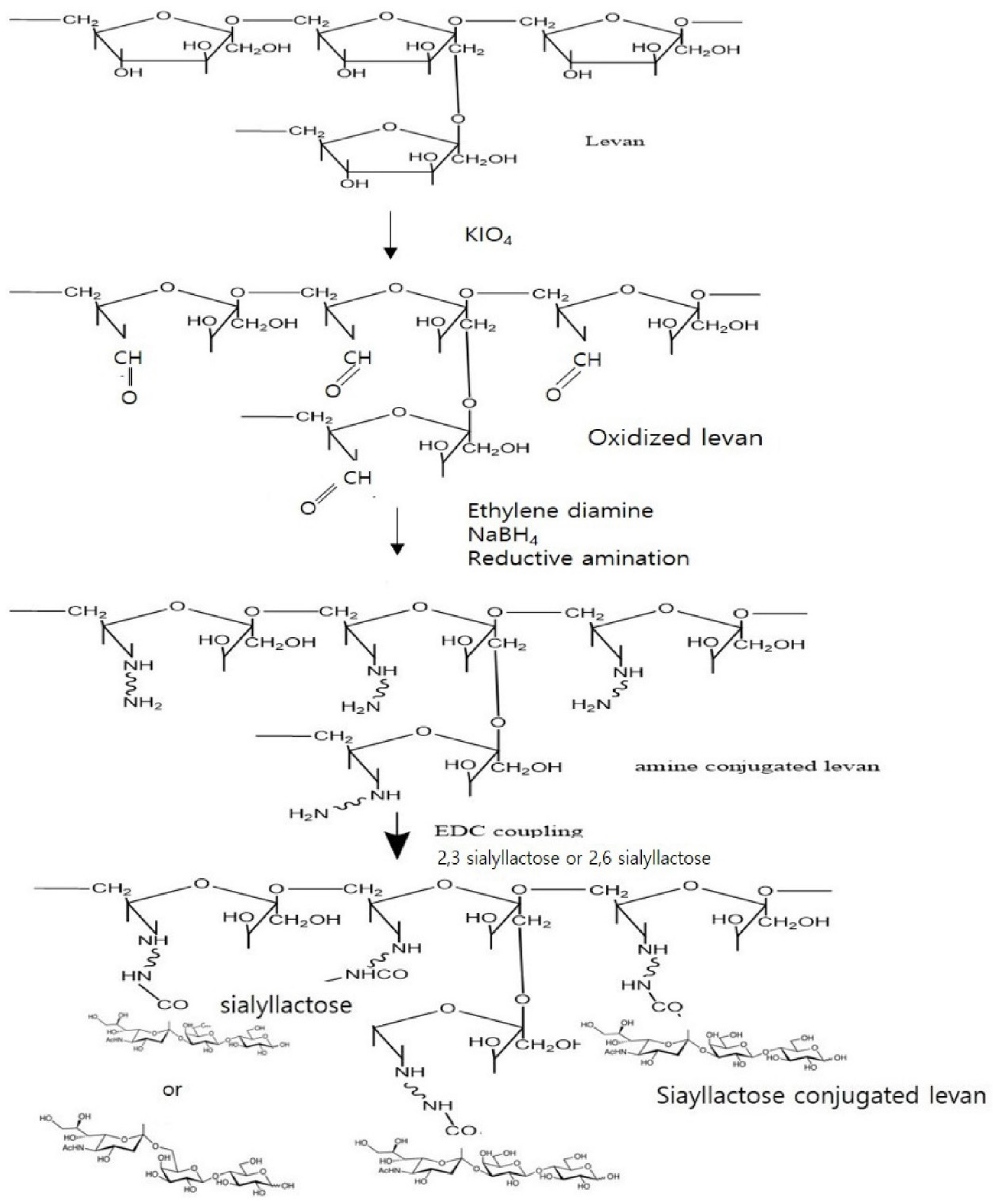
2.1. Conjugation of Levan to 2, 3- and 2, 6-Sialyllactose

To conjugate levan and sialyllactose, an amine functional group was introduced to levan (estimated MW < 2000 kDa, Realbiotech, Korea) by the reductive amination method [36,37]. First, we oxidized levan to introduce an aldehyde group. The oxidized levan was obtained by reacting levan in distilled water with potassium periodate. Oxidized levan (6.25 mmol) was dissolved in 100 mL of distilled water and added to ethylenediamine dissolved in borate buffer (0.1 M, pH 11). The solution was stirred using a magnetic stirrer at room temperature for 24 h, and NaBH4 was added to the solution. After 48 h of mixing, NaBH4 was added again to the solution, and stirring continued for 24 h. The solution was dialyzed using a dialysis membrane (M.W.C.O 12,000). The solution was lyophilized using a freeze-dryer. The determination of the amine functional group was calculated by a fluorescamine assay. The conjugation of sialyllactose (GeneChem Inc., Daejeon, Korea) and levan was performed by the EDC coupling method. An amine-functionalized levan was conjugated to a carboxylic acid group of sialyllactose (Scheme 1). Next, 25.6 mg of sialic lactose and 40 mg of EDC were dissolved in pH 5.0 acidic aqueous solution (10 mL) at room temperature for 4 h. After mixing, levan-NH2 (40 mg) was added to the mixed solution at room temperature for 24 h. The unreacted materials were removed using a dialysis membrane (MWCO 12,000). The solution was lyophilized using a freeze-dryer. The formation of products was confirmed by nuclear magnetic resonance (NMR, JEOL Ltd., JLM-AL400, Tokyo, Japan) and Fourier transform infrared (FT-IR) spectroscopy (Bruker Optics IF66, Billerica, MA, USA). NMR samples were prepared in that the products were dissolved in deuterium oxide and loaded on an NMR (400 MHz) at 25 °C to obtain the requisite 1H NMR spectra. FT-IR samples were prepared in a powder state and scanned 16 times from 4000 to 400 cm.

2.2. Interaction of Sialyllactose-Conjugated Levan with HA Protein, Lectins and Influenza Virus

To confirm that the sialyllactose-conjugated levan interacted with HA and influenza virus, we performed a modified ELISA assay. Recombinant protein, influenza virus and antibody were obtained by BioNano Health Guard Research Center. Biotin-conjugated Maackia amurensis lectin II (MALII) and biotin-conjugated Sambucus nigra lectin (SNL) were obtained from Vector Laboratories (Burlingame, CA, USA). Briefly, synthesized polymer (1 mg/mL in PBS) was incubated in a 96-well ELISA plate (Corning, Tewksbury, MA, USA) at 37 °C for 24 h. The plate was washed with PBS 3 times. To eliminate nonspecific binding, the plate was incubated with 1% bovine serum albumin (BSA) at 37 °C for 2 h. After washing, recombinant HA protein or influenza virus (A/California/07/2009) was added to the plate, which was incubated for 2 h. The plate was also washed 3 times and then incubated with mouse H1N1 antibody for 2 h. After 3 washes, incubation with horseradish peroxidase (HRP)-conjugated mouse IgG secondary antibody was performed for 2 h at room temperature. After washing, the binding affinity of polymer and influenza virus or HA protein was detected using 3, 3′, 5, 5′-tetramethylbenzidine (TMB solution) for 15 min; afterward, the stop solution was added. The absorbance (450 nm) was measured on a microplate reader. A lectin binding assay was also performed to confirm the specific binding of each sialyllactose-conjugated polymer. Briefly, synthesized polymer (100 μg/mL in PBS) and each sialyllactose (1 mg/mL in PBS) was incubated in an ELISA 96-well plate 37 °C for 24 h. The plate was washed with PBS 3 times. To eliminate nonspecific binding, the sample was incubated with 1% BSA at 37 °C for 2 h. After washing, incubation with biotin-conjugated lectin was conducted for 1 h. After washing, incubation with avidin-HRP was performed for 1 h. After washing, the binding affinity of polymers and lectin was subsequently determined using TMB for 15 min; afterwards, the stop solution was added. The absorbance (450 nm) was measured on a microplate reader.

2.3. Synthesis of Sialyllactose-Levan-Conjugated AuNPs

Five milligrams of sialyllactose-conjugated levan was dissolved in distilled water (5 mL). Two milligrams of gold chloride in distilled water (HAuCl4) was added to the solution. Sialyllactose-conjugated levan was used for the reducing and capping agent. The mixture was stirred at 70 °C until the color changed from yellow to red. The NPs were purified by centrifugation at 13,500 rpm, which was repeated 3 times. Control AuNPs were also synthesized using sodium borohydride [38]. 0.8 mg of gold chloride was dissolved in distilled water, 0.4 mg of NaBH4 was dissolved in distilled water (5 mL), and 0.8 mg of gold chloride stock solution was added to the solution slowly. The mixture was stirred at 70 °C until the color changed from yellow to red. The NPs were purified by centrifugation at 13,500 rpm. The size and morphology of the NPs were confirmed by transmission microscopy (TEM; JEM-1400 plus (JEOL, Japan)). The 10 µL of conjugate was loaded onto a carbon-coated copper grid and dried in air before being examined by TEM.

2.4. Detection of Influenza Virus Using Sialyllactose-Levan-Conjugated AuNPs

2,3-sialyllactose-levan-conjugated AuNPs, 2,6-sialyllactose-levan-conjugated AuNPs and control NPs were treated with H1N1 influenza virus (A/California/07/2009) and H5N2 influenza virus (A/aquatic bird/Korea/w351/2008) provided by BioNano Health Guard Research Center. The 100 µL of several concentration of virus were added to a 24-well plate and treated with 100 µL synthesized AuNPs and control AuNPs. After 5 min, the colorimetric change was observed by the naked eye. In addition, the absorbance of the AuNPs was measured by UV-Vis spectroscopy. NP aggregation was also observed by TEM. The samples were prepared by a method modified from previous research [39,40]. Influenza virus was purified using sucrose gradient ultracentrifugation. For the TEM analysis, purified virus (7.8 × 10⁹ pfu/mL) was mixed with each type of AuNP for 5 min and then loaded on the carbon-coated grid. The grid was washed with water, and uranyl acetate solution was added to the grid for negative staining. The specimens were observed using TEM microscopy (JEM-1400 plus (JEOL, Japan)). Several concentrations of influenza virus and 1 mg/mL of synthesized AuNPs were mixed for 5 min. After mixing, the hydrodynamic diameter of NPs measured by dynamic light scattering (DLS; Otsuka Electronics, Osaka, Japan).

2.5. Statistical Analysis

Several independent experiments were performed, and the data are expressed as the mean and standard deviation. Statistical significance was determined by Student’s t-test.

3. Results

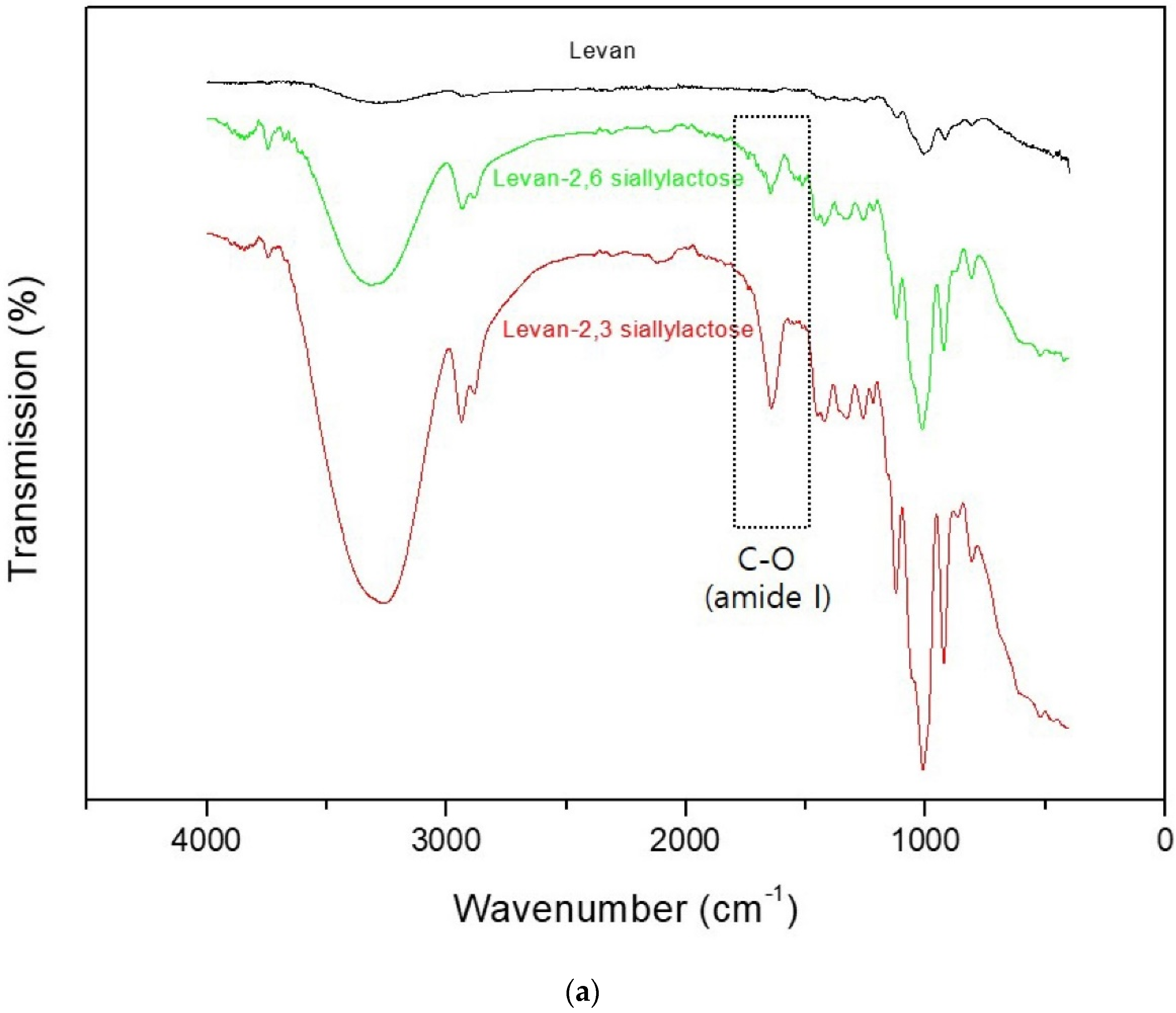
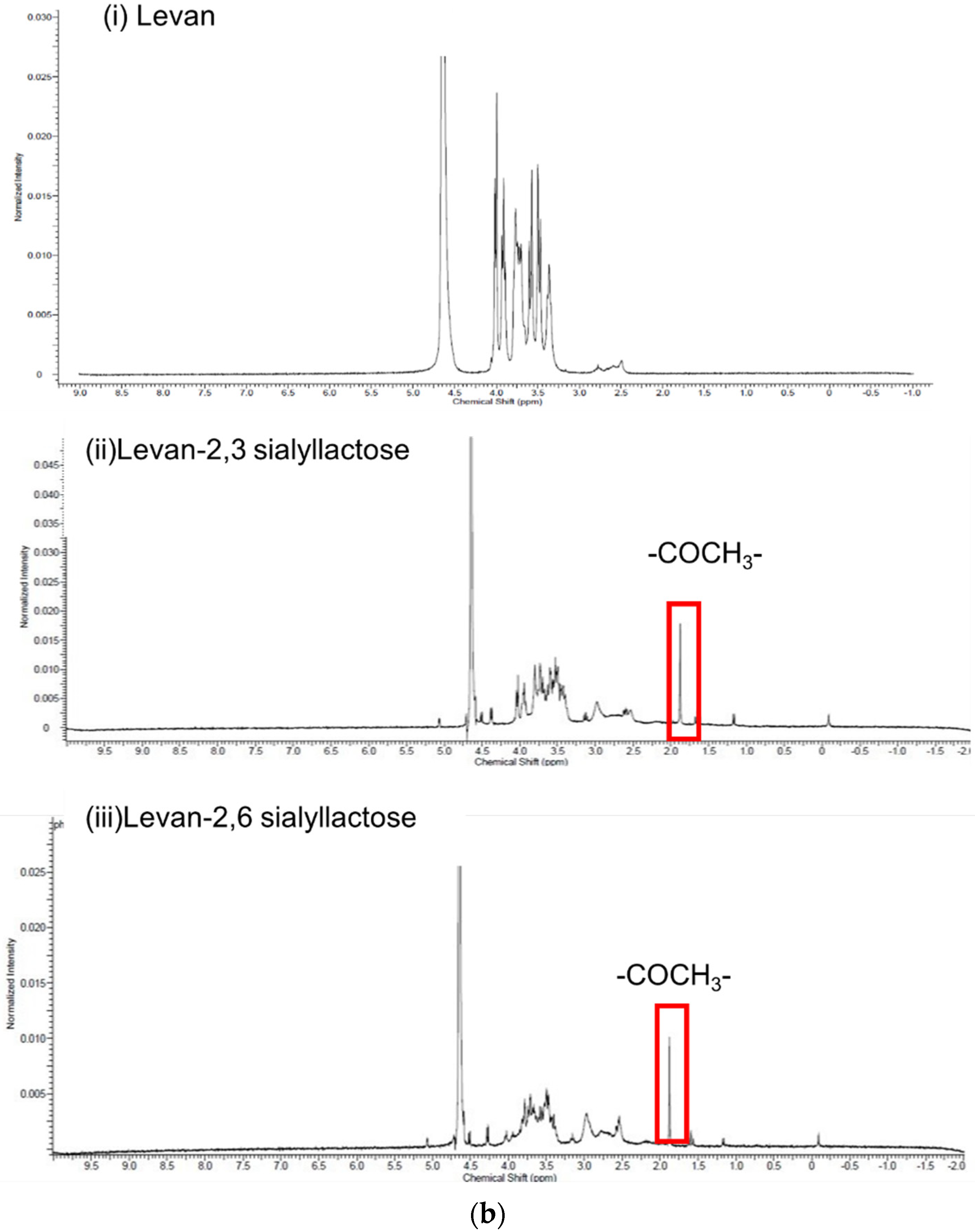
3.1. Conjugation of Levan to 2, 3- and 2, 6-Sialyllactose

To conjugate levan to sialyllactose, an amine functional group was introduced to levan by the reductive amination method. The amine group was successfully conjugated to levan, as confirmed by a fluorescamine assay. The amine group was modified with 0.098 µmol per 1 mg of levan. Levan was conjugated to sialyllactose by the EDC coupling method, and the conjugation was confirmed by FTIR and NMR analyses (Figure 1). The IR peaks of 2, 3-sialyllactose- and 2, 6-sialyllactose-conjugated levan appeared to indicate C-O stretching (amide I 1720 cm−1), which was attributed to the amide bond for conjugation of the amine group in levan and carboxylic acid in sialyllactose. The NMR results also showed that sialyllactose successfully conjugated to levan. The protons of the COCH3 in sialyllactose were assigned to 1.9 ppm in synthetic polymers. The percentage of 2, 3-siallylactose and 2, 6-sialyllactose attachment to the amine conjugation levan was calculated to be 6.6 and 6.9 mol%, respectively, through the ratio of proton integral between glycoside moiety (3.2–4.2 ppm) and COCH3 (1.9 ppm) by NMR analysis. These results demonstrated that a similar amount of each sialyllactose was conjugated to levan.

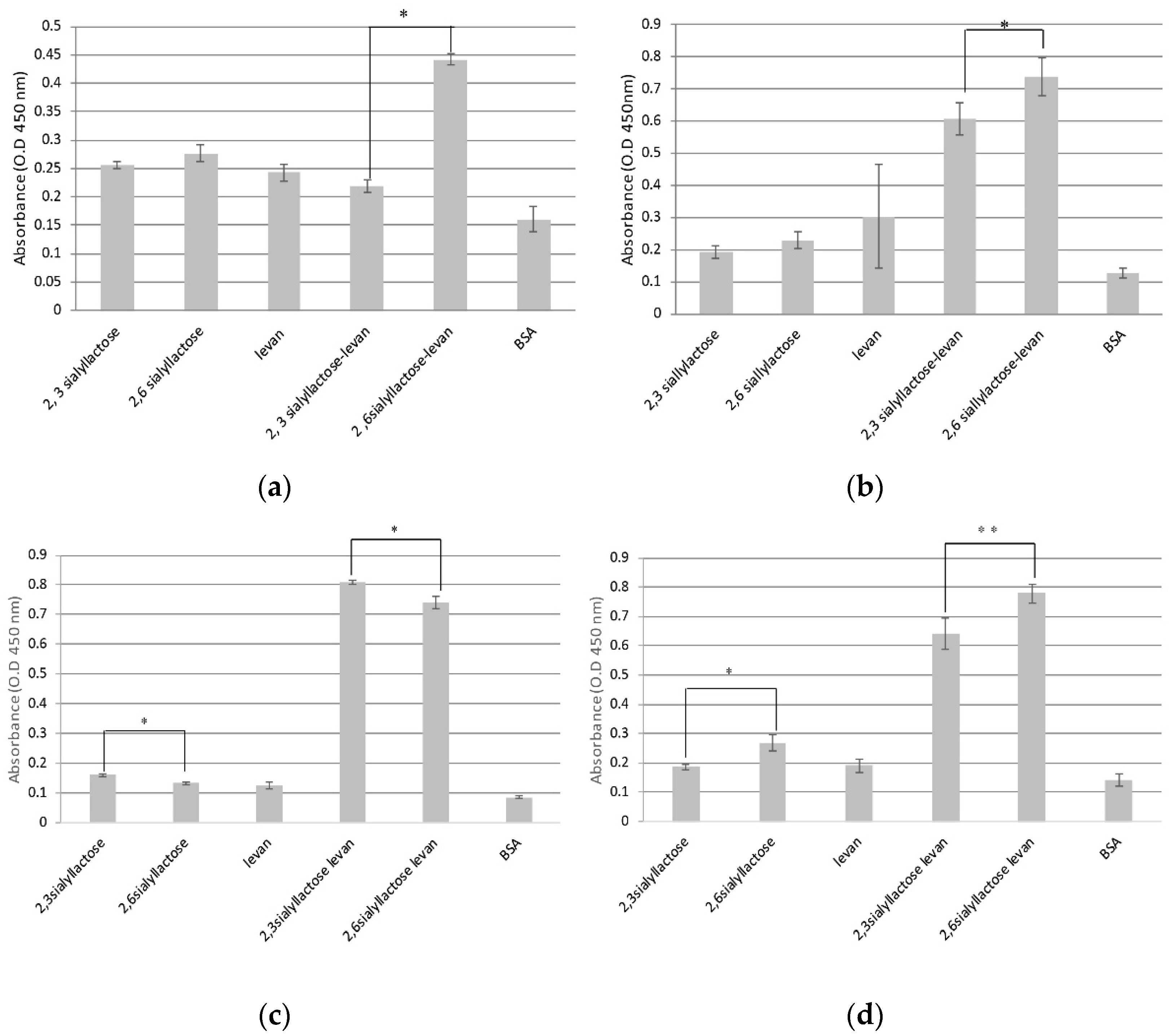
3.2. Interaction of the Sialyllactose-Conjugated Levan with HA Protein, Lectin and Influenza Virus

To confirm that the sialyllactose-conjugated levan interacted with HA and influenza virus, we performed a modified ELISA assay. We prepared sialyllactose-conjugated levan and sialyllactose-coated plates. The results showed that HA (Figure 2a; H1N1) and influenza virus (Figure 2b; A/California 07/2009 (H1N1)) were more strongly bound to 2, 6-sialyllactose-conjugated levan than 2, 3-sialyllactose-conjugated levan. Human influenza virus and HA are known to more specifically interact with 2, 6-sialyllactose than 2, 3-sialyllactose. To reconfirm that each sialyllactose (2, 3- and 2, 6-) successfully conjugated to levan, we also performed a lectin binding assay using MALII (Figure 2c) and SNL (Figure 2d), which are recognizable lectins that bind to 2, 3- and 2, 6-sialyllactose, respectively. The results demonstrated that MALII specifically interacted with 2, 3-sialyllactose-conjugated levan rather than 2, 6-sialyllactose-conjugated levan. In contrast, SNL was bound more preferentially to 2, 6-sialyllactose-conjugated levan than to 2, 3-sialyllactose-conjugated levan. The results demonstrate that each sialyllactose-conjugated levan successfully interacted with each recognizable lectin. These results also indicate that 2, 3- and 2, 6-sialyllactose successfully conjugated to levan. Moreover, sialyllactose-conjugated levan more highly interacted with each lectin than with sialyllactose alone via the multivalent effect. Therefore, sialyllactose-conjugated levan should efficiently bind the influenza virus.

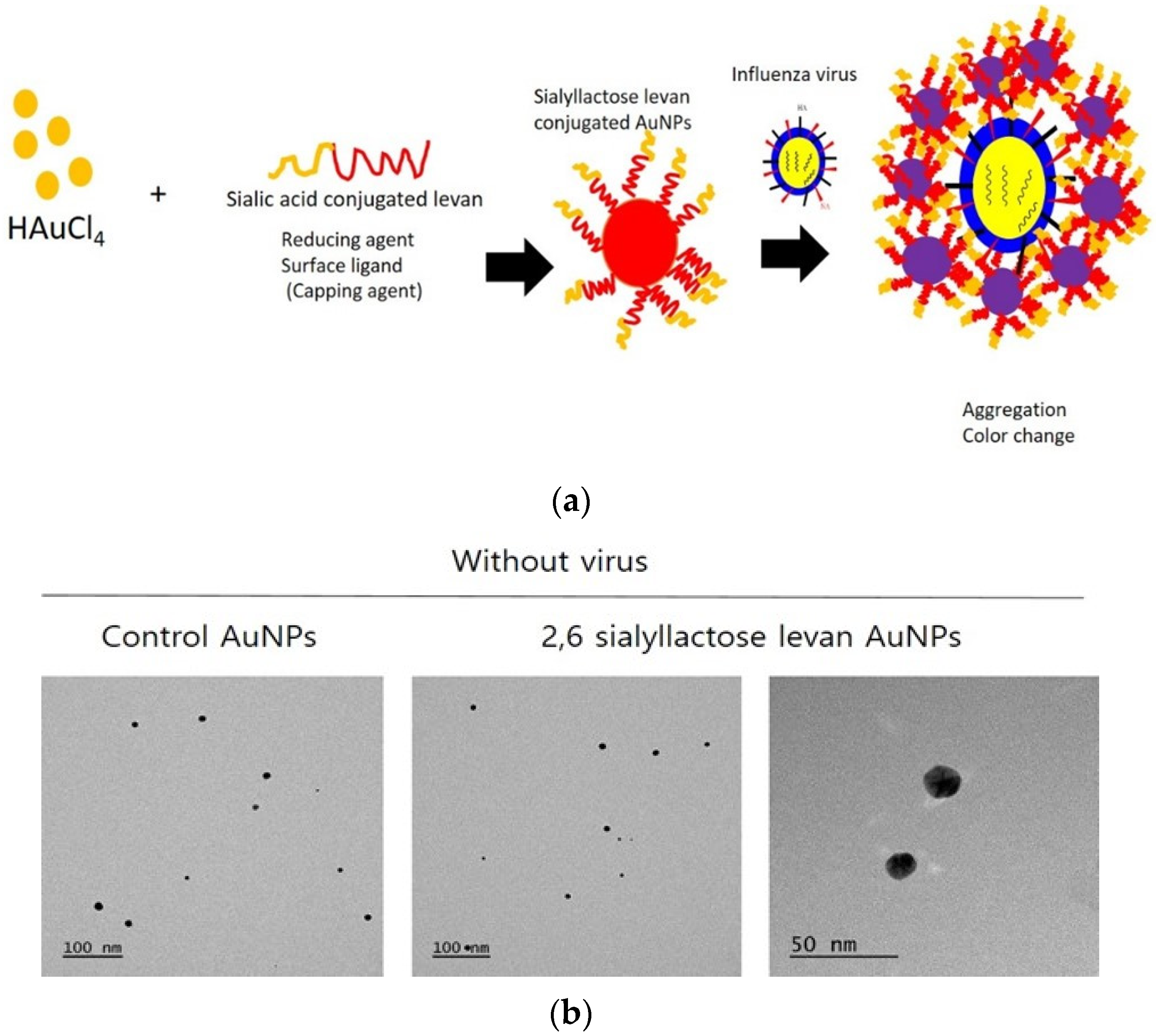
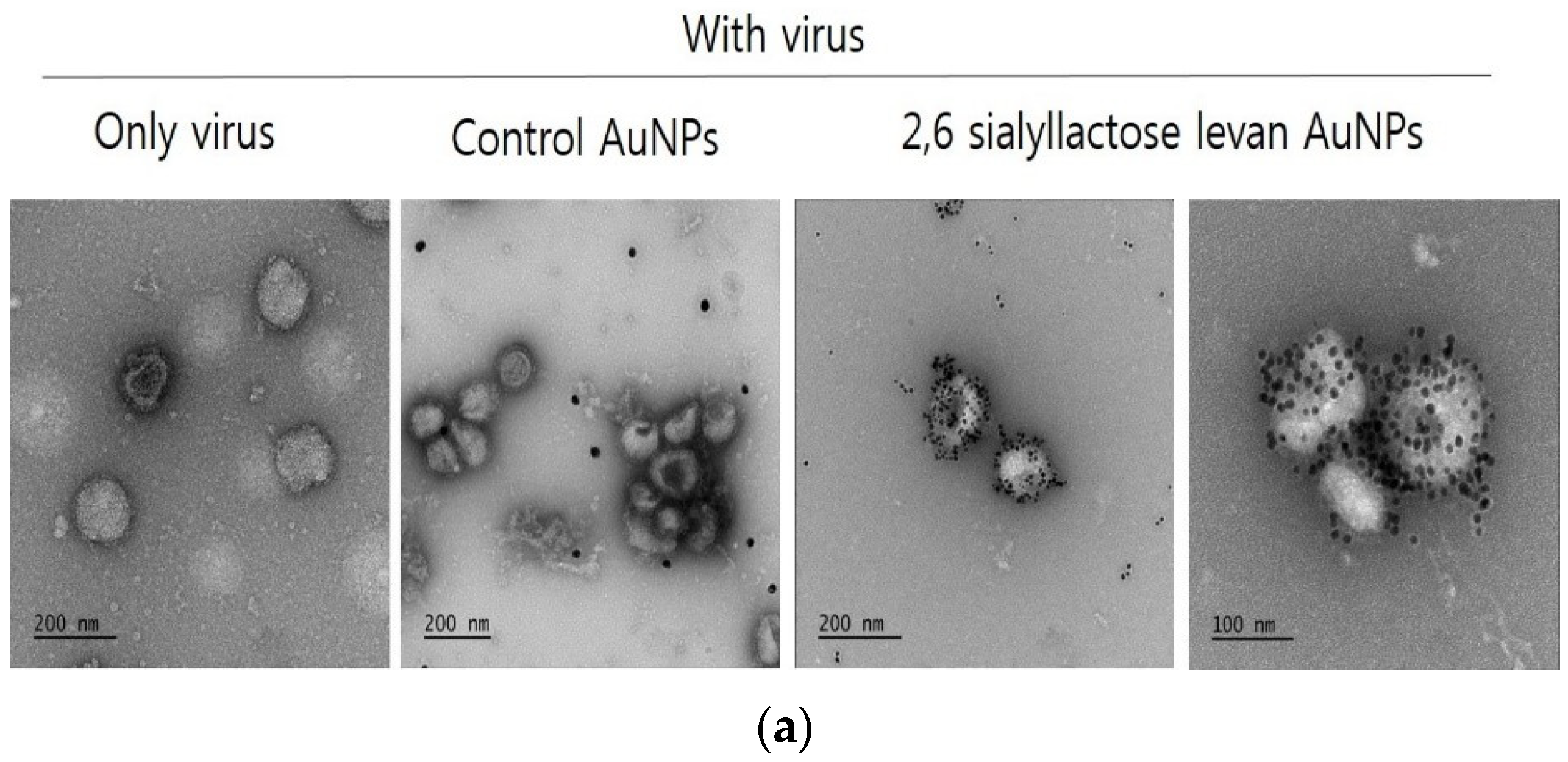
3.3. Synthesis of Levan-Sialyllactose-Coated AuNPs and Interaction with Influenza a Virus

Many studies have reported that 2, 6-sialyllactose has a high affinity for human influenza virus and HA protein. We hypothesized that sialyllactose-conjugated levan formed NPs in aqueous solution via the amphiphilicity of levan. Therefore, the conjugates have high multivalent glycocluster effects due to 2, 6-sialyllactose and strongly interact with human influenza virus and HA protein. We confirmed the high binding affinity of 2, 6-sialyllactose-conjugated levan with both human HA protein (H1N1) and influenza virus. To detect influenza virus using a colorimetric assay, we fabricated levan-sialyllactose-conjugated AuNPs. Synthetic polymers act as reducing and coating agents for the formation of the AuNPs. The AuNPs were not formed using only sialyllactose, which is a coating and reducing material (data not shown). For the synthesis of AuNPs with levan-sialyllactose, no other material was needed for the gold ion reduction. Therefore, AuNPs were simply formed in one step using levan-sialyllactose. The morphology and size distribution of NPs were observed by TEM (Figure 3a) and DLS. The sialyllactose-levan-conjugated AuNPs had a spherical morphology and were monodispersed. The hydrodynamic diameters of the control AuNPs, 2, 3-sialyllactose-levan-conjugated AuNPs, and 2, 6-sialyllactose-levan-conjugated AuNPs were 18.1 nm, 20.7 nm and 17.9 nm, respectively. Synthesized NPs had red colors induced by monodispersed particles. The levan-sialyllactose-conjugated AuNPs were well dispersed and stable in aqueous solution. Generally, monodispersed AuNPs show a reddish color; however, when target molecules, such as viruses and proteins, combine with AuNPs, the NPs aggregate and change to bluish colors (Figure 3b). Therefore, AuNPs are widely used in colorimetric assays for disease detection. Several polymer (control, 2, 3-sialyllactose-levan, 2, 6-sialyllactose-levan)-conjugated AuNP samples were incubated with human influenza virus (California 07/2009). The color of the 2, 6-sialyllactose-levan-conjugated AuNPs changed after incubation with influenza virus at 3.7 × 10⁴ pfu. However, for the 2, 3-sialyllactose-levan-conjugated AuNPs, the color of the NPs changed upon binding to virus at 7.4 × 10⁴ pfu. Moreover, the color of the control AuNPs did not change after incubation with the virus (Figure 3c). The UV absorbance peaks also became redshifted after the binding to human influenza virus. The relative binding affinity with HA was calculated using A 680/A 525. The results showed that the 2, 6-sialyllactose-levan-conjugated AuNPs had a high value of Abs 680/Abs 525. In the case of 2, 6-siallylactose-levan, it was confirmed that Abs 680/Abs 525 increased significantly from the 7.4 × 10³ PFU of H1N1 influenza virus of the control. Moreover, the value increased after incubation with higher virus concentrations. Therefore, we estimated that the limit of detection (L.O.D) of H1N1 influenza virus was 7.4 × 10³ PFU using 2, 6-siallylactose-levan-conjugated AuNPs.
In the TEM measurements, the 2, 6-sialyllactose-levan-conjugated AuNPs were aggregated around the virus after incubation with a high concentration of H1N1 virus. However, the control AuNPs did not aggregate around the viruses (Figure 4a). The size of NPs, which was determined by DLS, also changed after binding to the influenza virus. The 2, 6-sialyllactose-levan-conjugated AuNPs had a more dramatic size increase and aggregated upon binding to H1N1 influenza virus at 0, 7.4 × 10³, 3.7 × 10⁴, 7.4 × 10⁴ and 1.1 × 10⁵ pfu, with average hydrodiameters of 2, 6-sialyllactose-levan-conjugated AuNPs being 17.9 ± 3.4 nm, 24.9 ± 5.9 nm, 40.7 ± 9.8 nm, 116 ± 27.9 nm and 185.6 ± 48.2 nm, respectively (Figure 4b). The hydrodiameter growth indicated that the AuNPs specifically bound to the virus. These results demonstrated that the 2, 6-sialyllactose-levan-conjugated AuNPs more specifically bound to human influenza virus than did the 2, 3-sialyllactose-levan-conjugated and control AuNPs.
We also performed a colorimetric assay to detect H5N2 avian influenza virus (Figure 5). The results show that the 2, 3-sialyllactose-levan-conjugated AuNPs were highly bound to the avian virus, and the color rapidly changed from red to blue. The color of the 2, 3-sialyllactose-levan-conjugated AuNPs changed from red to blue after incubation with H5N2 virus at 4.2 × 10³ pfu. For 2, 6-sialyllactose-levan AuNPs, the color changed from red to purple with the same concentration of H5N2 virus. The size of 2, 3-sialyllactose-levan-conjugated AuNPs increased after avian virus incubation. After incubation with H5N2 influenza virus at 0, 4.2 × 10³, 6 × 10³, 1.2 × 10⁴ and 3 × 10⁴ pfu, the hydrodiameter of 2, 3-sialyllactose-levan-conjugated AuNPs were 20.7 ± 5 nm, 124.9 ± 33.4 nm, 157.3 ± 38.3 nm, 331.6 ± 89.3 nm and 397 ± 57.1 nm, respectively.
These results indicate that avian H5N2 virus interacted with 2, 3-sialyllactose-levan AuNPs than with 2, 6-sialyllactose-levan AuNPs and control AuNPs. Moreover, the ratio value of Abs 680/Abs 525 determined from the UV absorbance spectra of 2, 3-sialyllactose-levan AuNPs was higher than that for 2, 6-sialyllactose-levan AuNPs. In the case of 2, 3-siallylactose-levan, it was confirmed that Abs 680/Abs 525 increased in the presence of 4.2 × 10³ PFU of H5N2 influenza virus than the control. Moreover, the value increased after incubation with higher virus concentrations. Therefore, we estimated that limit of detection (L.O.D) of H5N2 influenza virus was 4.2 × 10³ pfu using 2, 3-siallylactose-levan- conjugated AuNPs. These results demonstrated that 2, 3-sialyllactose-levan-conjugated AuNPs are better for avian influenza detection.
We also examined the samples after exposure to FBS (Fetal bovine serum), which mimicked a blood serum protein (Figure 6). The results show that the color of sialyllactose-levan-conjugated AuNPs incubated with even a high concentration of FBS (50%) did not change, indicating that AuNPs do not interact with serum proteins. The results demonstrated that the sialyllactose-levan-conjugated AuNPs can be applied to blood samples. Moreover, we also evaluated the color change of AuNPs after incubation with real clinical swab sample buffer (universal transport medium, UTM). The results showed that the color of sialyllactose-levan-conjugated AuNPs did not change even for a high concentration of buffer. Therefore, these materials may be used to evaluate clinical samples.

4. Discussion

In the present study, we validated the sialyllactose-conjugated levan-coated gold nanoparticles for efficient interaction with and detection of influenza A virus. First, we conjugated 2, 3- and 2, 6-sialyllactose with levan for multivalent display of carbohydrate moieties. Many studies have reported that 2, 6-sialyllactose has a high affinity with human influenza virus hemagglutinin and 2, 3-siallyllactose highly interacted with avian influenza hemagglutinin. We hypothesized that sialyllactose-conjugated levan formed NPs in aqueous solution via the amphiphilicity of levan. Therefore, the conjugates have high multivalent glycocluster effects due to each sialyllactose, and interact with the targeted lectin and influenza more strongly. The results showed that 2, 6-sialyllactose-conjugated levan was more highly bound with SNL, while 2, 3-sialyllactose-conjugated levan interacted more with MAL. Moreover, in human influenza virus and hemagglutinin, 2, 6-sialyllactose-conjugated levan had a more effective binding ability than 2, 3-sialyllactose-modified levan. Therefore, these results indicate that sialyllactose was successfully conjugated with levan. Based on these results, we fabricated silalyllactose-levan-conjugated AuNPs. Gold nanoparticles have optical and plasmonic properties that can be deployed in colorimetric assays. When gold nanoparticles aggregate in the presence of biomolecules, their size and color change. Therefore, gold nanoparticles were applied for the detection of biomolecules via colorimetric assay. Levan sialyllactose plays a role as a reducing and coating agent during the formation of gold NPs. Because of the high molecular weight and biocompatibility of levan, gold nanoparticles can be well dispersed in water and maintain stability for a long time.
In the current study, the influenza virus can be detected and targeted by our system according to the origin of influenza virus. Our system detected influenza A virus in a short time using a simple colorimetric assay in which levan sialyllactose is conjugated to gold nanoparticles. In the current system, it is difficult to completely separate human and avian viruses. However, it may be possible to increase the specificity by modifying the binding sites on each sialyllactose. The advantage of this approach is that the gold nanoparticles can be functionalized for interaction with 2, 6 sialyllactose, dominant in human virus, or 2, 3-siallylactose found in avian virus.
The current influenza detection system, PCR, is the gold standard method; however, it is expensive and time consuming. Our system is advantageous in that the color change can simply be detected with the naked eye in a short time without any instrumentation. Moreover, since the commercially available influenza rapid diagnostic kit generally detects 104~105 TCID50/mL, the colorimetric assay described here is competitive [41].

5. Conclusions

In summary, we synthesized sialyllactose-conjugated levan for efficient interactions with and detection of influenza virus via the multivalent effect. Sialyllactose-conjugated levan specifically interact with influenza virus. Moreover, these synthesized polymer materials can act as reducing and stabilizer for the formation of the AuNPs. Gold nanoparticles functionalized with sialyllactose-conjugated levan were used to detect influenza virus using a colorimetric assay with either 2, 3- or 2, 6-sialyllactose-levan-conjugated AuNPs. Specifically, 2, 6-sialyllactose-levan-conjugated AuNPs interacted with human influenza virus, and 2, 3-sialyllactose-levan-conjugated AuNPs bind avian influenza virus. Our system provides a simple and fast method for detecting influenza virus via a color change in the colloidal functionalized AuNPs solution, and viruses with a specific origin can be detected via ligand-receptor interactions.

Supplementary Materials

The following are available online at https://0-www-mdpi-com.brum.beds.ac.uk/article/10.3390/chemosensors9070186/s1, Table S1: Comparison of PCR and sialyllactose levan AuNPs method, Table S2: An overview on recently reported nanomaterial-based optical methods for the determination of detection of influenza virus.

Author Contributions

Conceptualization, S.-J.K.; data curation, S.-J.K. and P.K.B.; formal analysis, S.-J.K.; funding acquisition, Y.-B.S. project administration, Y.-B.S.; writing—Original draft, S.-J.K.; writing—Review & editing, P.K.B. and Y.-B.S. All authors have read and agreed to the published version of the manuscript.

Funding

This work was supported by the BioNano Health Guard Research Center, which is funded by the Ministry of Science and ICT (MSIT) of Korea as a Global Frontier Project (Grant Number H-GUARD_2013M3A6B2078950) and by Nano Material Technology Development Program through the National Research Foundation of Korea (NRF) funded by the Ministry of Science and ICT (MSIT) of Korea (Grant Number 2021M3H4A4079382).

Institutional Review Board Statement

Not applicable.

Informed Consent Statement

Not applicable.

Data Availability Statement

Not applicable.

Acknowledgments

We acknowledge the KBSI for their TEM observations.

Conflicts of Interest

The authors declare no conflict of interest.

References

  1. Thompson, W.W.; Shay, D.K.; Weintraub, E.; Brammer, L.; Bridges, C.B.; Cox, N.J.; Fukuda, K. Influenza-Associated Hospitalizations in the United States. JAMA J. Am. Med. Assoc. 2004, 292, 1333–1340. [Google Scholar] [CrossRef] [PubMed]
  2. Garten, R.J.; Davis, C.T.; Russell, C.A.; Shu, B.; Lindstrom, S.; Balish, A.; Sessions, W.M.; Xu, X.; Skepner, E.; Deyde, V.; et al. Antigenic and Genetic Characteristics of Swine-origin 2009 A(H1N1) Influenza Viruses Circulating in Humans. Science 2009, 325, 197–201. [Google Scholar] [CrossRef] [PubMed] [Green Version]
  3. Skehel, J.J.; Wiley, D.C. Receptor Binding and Membrane Fusion in Virus Entry: The Influenza Hemagglutinin. Annu. Rev. Biochem. 2000, 69, 531–569. [Google Scholar] [CrossRef] [PubMed]
  4. Sauter, N.K.; Hanson, J.E.; Glick, G.D.; Brown, J.H.; Crowther, R.L.; Park, S.J.; Skehel, J.J.; Wiley, D.C. Binding of Influenza Virus Hemagglutinin to Analogs of Its Cell-Surface Receptor, Sialic Acid: Analysis by Proton Nuclear Magnetic Resonance Spectroscopy and X-ray Crystallography. Biochemistry 1992, 31, 9609–9621. [Google Scholar] [CrossRef]
  5. Imai, M.; Kawaoka, Y. The Role of Receptor Binding Specificity in Interspecies Transmission of Influenza Viruses. Curr. Opin. Virol. 2012, 2, 160–167. [Google Scholar] [CrossRef] [PubMed] [Green Version]
  6. Allen, T.M. Ligand-Targeted Therapeutics in Anticancer Therapy. Nat. Rev. Cancer 2002, 2, 750–763. [Google Scholar] [CrossRef]
  7. Mohanty, C.; Das, M.; Kanwar, J.R.; Sahoo, S.K. Receptor Mediated Tumor Targeting: An Emerging Approach for Cancer Therapy. Curr. Drug Deliv. 2011, 8, 45–58. [Google Scholar] [CrossRef] [PubMed]
  8. Cook, S.E.; Park, I.K.; Kim, E.M.; Jeong, H.J.; Park, T.G.; Choi, Y.J.; Akaike, T.; Cho, C.S. Galactosylated Polyethylenimine-Graft-Poly(vinyl pyrrolidone) as a Hepatocyte-Targeting Gene Carrier. J. Control. Release 2005, 105, 151–163. [Google Scholar] [CrossRef] [PubMed]
  9. Hashida, M.; Takemura, S.; Nishikawa, M.; Takakura, Y. Targeted Delivery of Plasmid DNA Complexed with Galactosylated Poly (L-lysine). J. Control. Release 1998, 53, 301–310. [Google Scholar] [CrossRef]
  10. Becer, C.R. The Glycopolymer Code: Synthesis of Glycopolymers and Multivalent Carbohydrate-Lectin Interactions. Macromol. Rapid. Commun. 2012, 33, 742–752. [Google Scholar] [CrossRef]
  11. Ting, S.R.S.; Chen, G.; Stenzel, M.H. Synthesis of Glycopolymers and Their Multivalent Recognitions with Lectins. Polym. Chem. 2010, 1, 1392–1412. [Google Scholar] [CrossRef]
  12. Mammen, M.; Choi, S.K.; Whitesides, G.M. Polyvalent Interactions in Biological Systems: Implications for Design and Use of Multivalent Ligands and Inhibitors. Angew. Chem. Int. Ed. 1998, 37, 2754–2794. [Google Scholar] [CrossRef]
  13. Vereyken, I.J.; Chupin, V.; Demel, R.A.; Smeekens, S.C.M.; De Kruijff, B. Fructans Insert between the Head groups of Phospholipids. Biochim. Biophys. Acta 2001, 1510, 307–320. [Google Scholar] [CrossRef] [Green Version]
  14. Sezer, A.D.; Kazak, H.; Öner, E.T.; Akbuğa, J. Levan-Based Nanocarrier System for Peptide and Protein Drug Delivery: Optimization and Influence of Experimental Parameters on the Nanoparticle Characteristics. Carbohydr. Polym. 2011, 84, 358–363. [Google Scholar] [CrossRef]
  15. Kim, S.J.; Bae, P.; Chung, B.H. Self- Assembled Levan Nanoparticles for Targeted Breast Cancer Imaging. Chem. Commun. 2015, 51, 107–110. [Google Scholar] [CrossRef]
  16. Kim, S.J.; Chung, B.H. Antioxidant Activity of Levan Coated Cerium Oxide Nanoparticles. Carbohydr. Polym. 2016, 150, 400–407. [Google Scholar] [CrossRef] [PubMed]
  17. Kim, S.J.; Bae, P.K.; Choi, M.; Keem, J.O.; Chung, W.; Shin, Y.B. Fabrication and Application of Levan-PVA Hydrogel for Effective Inflenza Virus Capture. ACS Appl. Mater. Interfaces 2020, 12, 29103–29109. [Google Scholar]
  18. Nichols, J. Point of Care Testing. Clin. Lab. Med. 2007, 27, 893. [Google Scholar] [CrossRef]
  19. Wood, W.G. Problems and Practical Solutions in the External Quality Control of Point of Care Devices with Respect to the Measurement of Blood Glucose. J. Diabetes Sci. Technol. 2007, 1, 158. [Google Scholar] [CrossRef]
  20. Kozel, T.R.; Burnham-Marusich, A.R. Point-of-Care Testing for Infectious Diseases: Past, and Future. J. Clin. Microbiol. 2017, 55, 2313–2320. [Google Scholar] [CrossRef] [Green Version]
  21. McRae, M.P.; Simmons, G.W.; Christodoulides, N.J.; Lu, Z.; Kang, S.K.; Fenyo, D.; Alcorn, T.; Dapkins, I.P.; Sharif, I.; Vurmaz, D.; et al. Clinical decision support tool and rapid point-of-care platform for determining disease severity in patients with COVID-19. Lab. Chip. 2020, 20, 2075–2085. [Google Scholar] [CrossRef]
  22. Nath, N.; Chilkoti, A. A Colorimetric Gold Nanoparticle Sensor to Interrogate Biomolecular Interactions in Real Time on a Surface. Anal. Chem. 2002, 74, 504–509. [Google Scholar] [CrossRef] [PubMed]
  23. Tsai, C.S.; Yu, T.B.; Chen, C.T. Gold Nanoparticle-Based Competitive Colorimetric Assay for Detection of Protein-Protein Interactions. Chem. Commun. 2005, 34, 4273–4275. [Google Scholar] [CrossRef]
  24. Sulaiman, I.S.C.; Chieng, B.W.; Osman, M.J.; Ong, K.K.; Rashid, J.I.A.; Wan Yunus, W.M.Z.; Noor, S.A.M.; Kasim, N.A.M.; Halim, N.A.; Mohamad, A. A review on colorimetric methods for determination of organophosphate pesticides using gold and silver nanoparticles. Microchim. Acta 2020, 187, 131. [Google Scholar] [CrossRef] [PubMed]
  25. Zhao, W.; Brook, M.A.; Li, Y. Design of Gold Nanoparticle-Based Colorimetric Biosensing Assays. ChemBioChem 2008, 9, 2363–2371. [Google Scholar] [CrossRef] [PubMed]
  26. Yen, C.W.; Puig, H.; Tam, J.O.; Gómez-Márquez, J.; Bosch, I.; Hamad-Schifferli, K.; Gehrke, L. Multicolored Silver Nanoparticles for Multiplexed Disease Diagnostics: Distinguishing Dengue, Yellow Fever, and Ebola Viruses. Lab Chip 2015, 15, 1638–1641. [Google Scholar] [CrossRef] [PubMed]
  27. Peng, X.H.; Qian, X.; Mao, H.; Wang, A.Y.; Chen, Z.; Nie, S.; Shin, D.M. Targeted Magnetic Iron Oxide Nanoparticles for Tumor Imaging and Therapy. Int. J. Nanomed. 2008, 3, 311–321. [Google Scholar]
  28. Chou, T.C.; Hsu, W.; Wang, C.H.; Chen, Y.J.; Fang, J.M. Rapid and specific influenza virus detection by functionalized magnetic nanoparticles and mass spectrometry. J. Nanobiotechnol. 2011, 9, 52. [Google Scholar] [CrossRef] [Green Version]
  29. Chen, L.; Sheng, Z.; Zhang, A.; Guo, X.; Li, J.; Han, H.; Jin, M. Quantum-dots-based fluoroimmunoassay for the rapid and sensitive detection of avian influenza virus subtype H5N1. Luminescence 2010, 25, 419–423. [Google Scholar] [CrossRef] [PubMed]
  30. Liu, Y.; Zhang, L.; Wei, W.; Zhao, H.; Zhou, Z.; Zhang, Y.; Liu, S. Colorimetric Detection of Influenza A Virus Using Antibody-Functionalized Gold Nanoparticles. Analyst 2015, 140, 3989–3995. [Google Scholar] [CrossRef]
  31. Wei, J.; Zheng, L.; Lv, X.; Bi, Y.; Chen, W.; Zhang, W.; Shi, Y.; Zhao, L.; Sun, X.; Wang, F.; et al. Analysis of Influenza Virus Receptor Specificity Using Glycan-Functionalized Gold Nanoparticles. ACS Nano 2014, 8, 4600–4607. [Google Scholar] [CrossRef]
  32. Medley, C.D.; Smith, J.E.; Tang, Z.; Wu, Y.; Bamrungsap, S.; Tan, W. Gold Nanoparticle-Based Colorimetric Assay for the Direct Detection of Cancerous Cells. Anal. Chem. 2008, 80, 1067–1072. [Google Scholar] [CrossRef]
  33. Lee, C.; Gaston, M.A.; Weiss, A.A.; Zhang, P. Colorimetric Viral Detection Based on Sialic Acid Stabilized Gold Nanoparticles. Biosens. Bioelectron. 2013, 42, 236–241. [Google Scholar] [CrossRef] [PubMed]
  34. Ogata, M.; Umemura, S.; Sugiyama, N.; Kuwano, N.; Koizumi, A.; Sawada, T.; Yanase, M.; Takaha, T.; Kadokawa, J.; Usui, T. Synthesis of Multivalent Sialyllactosamine-Carrying Glyco-Nanoparticles with High Affinity to the Human Influenza Virus Hemagglutinin. Carbohydr. Polym. 2016, 153, 96–104. [Google Scholar] [CrossRef]
  35. Poonthiyil, V.; Nagesh, P.T.; Husain, M.; Golovko, V.B.; Fairbanks, A.J. Gold Nanoparticles Decorated with Sialic Acid Terminated Bi-antennary N-Glycans for the Detection of Influenza Virus at Nanomolar Concentrations. ChemistryOpen 2015, 4, 708–716. [Google Scholar] [CrossRef] [PubMed]
  36. Azzam, T.; Eliyahu, H.; Shapira, L.; Liniai, M.; Berenholz, Y.; Domb, A.J. Polysaccharide-Oligoamine Based Conjugates for Gene Delivery. J. Med. Chem. 2002, 45, 1817–1824. [Google Scholar] [CrossRef]
  37. Azzam, T.; Eliyahu, H.; Makovitzki, A.; Liniai, M.; Berenholz, Y.; Domb, A.J. Hydrophobized Dextran-Spermine Conjugate as Potential Vector for in Vitro Gene Transfection. J. Control. Release 2004, 96, 309–323. [Google Scholar] [CrossRef]
  38. Birrell, G.B.; Hedberg, K.K.; Griffith, O.H. Pitfalls of immunogold labeling: Analysis by light microscopy, transmission electron microscopy, and photoelectron microscopy. J. Histochem. Cytochem. 1987, 35, 843–853. [Google Scholar] [CrossRef]
  39. Isaacs, C.E.; Wen, G.Y.; Xu, W.; Jia, J.H.; Rohan, L.; Corbo, C.; Di Maggio, V.; Jenkins, E.C., Jr.; Hillier, S. Epigallocatechin Gallate Inactivates Clinical Isolates of Herpes Simplex Virus. Antimicrob. Agents Chemother. 2008, 52, 962–970. [Google Scholar] [CrossRef] [Green Version]
  40. Kim, M.; Kim, S.Y.; Lee, H.Y.; Shin, J.S.; Kim, P.; Jung, Y.S.; Jeong, H.S.; Hyun, J.K.; Lee, C.K. Inhibition of Influenza Virus Internalization by (-)-Epigallocatechin-3-Gallate. Antivirl. Res. 2013, 100, 460–472. [Google Scholar] [CrossRef] [PubMed]
  41. Bouscambert, M.; Valette, M.; Lina, B. Rapid bedside tests for diagnosis, management, and prevention of nosocomial influenza. J. Hosp. Infect. 2015, 89, 314–331. [Google Scholar] [CrossRef] [PubMed]
Scheme 1. The synthesis of sialyllactose-conjugated levan.
Chemosensors 09 00186 sch001
Figure 1. Confirmation of sialyllactose conjugated levan determined by FTIR (3200–3300 cm−1, O-H; 2930 and 2880 cm−1, C-H; 1720 cm−1, C-O amide I; and 950–1200 cm−1, fingerprint region) (a) and NMR (b) ((i) levan, (ii) 2, 3-sialyllactose-conjugated levan, (iii) 2, 6-sialyllactose-conjugated levan) (3.2–4.2 ppm, glycoside moiety; 1.9 ppm COCH3).
Chemosensors 09 00186 g001aChemosensors 09 00186 g001b
Figure 2. Binding affinity of polymers to hemagglutinin (HA) (a), influenza A virus (A/California 07/2009 (H1N1)) (b), Maackia amurensis lectin II (c), and Sambucus nigra lectin (d), as determined using a modified ELISA assay (* p < 0.05, ** p < 0.005).
Chemosensors 09 00186 g002
Figure 3. The formation and interaction of AuNPs with influenza virus (a). TEM images of synthesized AuNPs (b). UV-vis and relative absorbance ratios (Abs 680/Abs 525) (H1N1 concentration: black, 0; red, 7.4 × 10³ pfu; green, 3.7 × 10⁴ pfu; blue, 7.4 × 10⁴ pfu; sky blue, 1.1 × 10⁵ pfu) (c). (* p < 0.05).
Chemosensors 09 00186 g003aChemosensors 09 00186 g003b
Figure 4. TEM images of virus-AuNP mixtures (a). The hydrodynamic diameter of nanoparticles through the reaction between each prepared nanoparticles and each concentration of influenza virus H1N1 as measured using DLS (b) (* p < 0.05).
Chemosensors 09 00186 g004aChemosensors 09 00186 g004b
Figure 5. Interaction of AuNPs with Avian H5N2 influenza virus. Colorimetric assay of AuNPs interacting with Avian H5N2 virus (a), UV-vis and relative absorbance ratios (Abs 680/Abs 525) (H5N2 virus concentration: black, 0; red, 4.2 × 10³ pfu; green, 6 × 10³ pfu; blue, 1.2 × 10⁴ pfu; sky blue, 3 × 10⁴ pfu). The hydrodynamic diameter of nanoparticles through the reaction between each prepared nanoparticles and each concentration of influenza virus H5N2 as measured using DLS (b) (* p < 0.05).
Chemosensors 09 00186 g005
Figure 6. Color images of sialyllactose-levan-conjugated AuNPs with FBS (a) and universal transport medium (UTM), a virus culture medium (b).
Chemosensors 09 00186 g006
Publisher’s Note: MDPI stays neutral with regard to jurisdictional claims in published maps and institutional affiliations.

