Communication

pH-Dependent Selective Colorimetric Detection of Proline and Hydroxyproline with Meldrum’s Acid-Furfural Conjugate

1
Department of Nanobiosystem Technology, Institute of Chemistry and Biotechnology, Technical University Ilmenau, Prof.-Schmidt-Straße 26, 98693 Ilmenau, Germany
2
Research Group Bioorganic Chemistry of Bioactive Surfaces, Institute of Chemistry and Biotechnology, Technical University Ilmenau, Prof.-Schmidt-Straße 26, 98693 Ilmenau, Germany
*
Author to whom correspondence should be addressed.
Chemosensors 2021, 9(12), 343; https://doi.org/10.3390/chemosensors9120343
Submission received: 30 September 2021 / Revised: 29 November 2021 / Accepted: 2 December 2021 / Published: 4 December 2021

Abstract

:
Activated 2-furfural gives intense color formation when reacted with amines, due to a ring opening reaction cascade that furnishes a conjugated molecular system. Unique colorimetric characteristic of this reaction makes it an interesting candidate for developing chemosensors operating in visible range. Among many activated 2-furfural derivatives, Meldrum’s acid furfural conjugate (MAFC) recently gained significant interest as colorimetric chemosensor. MAFC has been explored as selective chemosensor for detecting amines in solution, secondary amines on polymer surfaces and even nitrogen rich amino acids (AA) in aqueous solution. In this work, the pH dependency of MAFC-AA reaction is explored. It was found that proline gives an exceptionally fast colored reaction at pH 11, whereas at other pHs, no naked eye color product formation was observed. The reaction sequence including ring opening reaction upon nucleophilic addition of cyclic amine of proline resulting in a conjugated triene was confirmed by NMR titrations. The highly pH dependent reaction can e.g., potentially be used to detect proline presence in biological samples. An even more intense color formation takes place in the reaction of natural proline derivative 4-hydroxyproline. The detection limit of proline and 4-hydroxyproline with MAFC solution was found to be 11 µM and 6 µM respectively.

1. Introduction

In the last decade, donor acceptor stenhouse adducts (DASA) gained more and more significance due to their ability to offer a visible light-assisted photoreaction that can convert hydrophobic triene to hydrophilic cyclopentenone [1]. Apart from its excellent photoconversion, this reaction has gained fame due to ease of its preparation. The first step of the reaction can be performed simply by mixing activated furan with secondary amines, which results in the formation of highly colored conjugated triene species. The naked eye monitoring of the reaction makes it very simple to process [1,2]. This reaction can take place in liquids, solids, as well as in vapor phase. In the second step the colored conjugated triene can be converted to a colorless hydrophilic cyclopentenone upon exposure to visible light. This reaction can be reversed by heating [3,4]. Therefore, these molecules are highly interesting for developing new photosensitive material those can be processed with bio friendly visible light. However apart from its photoresponsive behavior, an important feature of this reaction is the spontaneous formation of colored conjugated triene upon addition of biogenic amines to the activated 2-furfural, which makes it an interesting candidate for developing chemosensors [3]. For the formation of colored DASA, an activated furfural is required, which can be made by condensation of 2-furfuraldehyde with active methylene compounds like Meldrum’s acid, barbituric acid, and indandione [5]. Among them Meldrum’s acid furfural conjugate 5-(furan-2-ylmethylene)-2,2-dimethyl-1,3-dioxane-4,6-dione (MAFC), was much more explored as chemosensor compared to the other two, due to its good solubility in ethanol, which in term offers reaction in aqueous solutions. It is an important property of this molecule because most of the biogenic amines and nutritionally important molecules like amino acids are soluble in aqueous solution.
Dietary imbalance of proteinogenic AA can have severe impact on human health. New detection methods of measuring essential AA content are thus always searched in biotech, medical, and food industry, by which diseases resulting from AA imbalance can be prevented. Moreover, essential AA, which cannot be produced by the human body, are added to the food products for balanced nutritional value. Methods of quantifying such essential AA are therefore also important to assess the quality of food and drug supplements. Recently, we were able to utilize MAFC-amine color chemistry for selective detection of nitrogen rich amino acids (AA) in aqueous medium [6]. Therefore, MAFC appears to be a potential candidate for colorimetric detection of biogenic amines and nutritionally important substances like AA.
From literature it is known that certain AA can be detected selectively through a variety of fluorimetric or colorimetric methods like detection with metal complexes [7], polymers [8], nanoparticles [9], and DNA [10] based sensors [10,11]. Colorimetric sensors based on metal complexes mainly utilize copper [7,12] or zinc [13] and are able to detect a wide variety of AA. Li et al. used a polyfluorene polymer for a “turn-on” approach detection of alpha-AA in concentrations as low as 33 µM [14]. In case of nanoparticle-based probing of AA, a chemosensor based on calyx-capped gold nanoparticles can be named among others [15]. Lysine, arginine, and histidine were detectable through a color change from red to purple at concentrations as low as 1 µM.
Typically, an AA consists of one carboxy group and a primary amine with the only exception of proline where the amine group is integrated into a pyrrolidine ring and therefore has secondary amine characteristic. Proline with its exceptional rigidity is not only important for collagen triple helix stability in vertebrates but also plays a role in stress response of plants [16,17,18]. Selective detection of proline is therefore important to quantify plant stress as well as the nutritional value of food or drug supplements. Unfortunately, there are only a few methods available where proline can be detected exclusively. Isatin [19] and ninhydrine [20] were explored for the detection of proline, however these reagents lack specificity. The ninhydrine approach was improved by using a paper-based microfluidic method [21]. In another method, an enzymatic assay was explored by utilizing the proline dependent reduction to enhance the proline detection [22]. Another type of chemosensors for proline was developed by utilizing the aldol reaction catalyzation potential of proline either with coumarinyl aldehyde [23] or [Ir(ppy)2(5-CHOphen)]PF6 [24]. However, complex synthesis protocols, relatively fast decomposition, and purification needs hinder their wide spread use. Table 1 compares the aforementioned methods regarding their limit of detection (LOD) and linear measurement range.
Despite of the availability of these methods it is necessary to discover new molecular probes that are easy to synthesize and derived from inexpensive materials. Therefore, small organic chemosensors are of high interest due to their ease of synthesis and structural understandability.
In this report, we explore MAFC as selective sensor for AA proline under selective conditions. Fast, selective and naked eye color appearance characteristics of the reaction of MAFC with amine groups make them suitable to be explored as colorimetric sensor for AA that contain a free amine group. To our knowledge, only our group used this class of molecules as specific chemosensor for lysine and arginine under particular conditions so far. It is known that MAFC reacts more selectively to secondary amines compared to primary amines [3,25]. However, in our last study we have noticed that when MAFC is reacted with essential AA at neutral pH primary amine or guanidino side chain group containing AA, it shows much more prominent and faster color reaction as compared to proline which contains an innate secondary amine group. This gives the opportunity to use MAFC as selective chemosensor for lysine and arginine at neutral pH [6]. This surprising characteristic with reversal of selectivity between primary and secondary amine group encouraged us to search appropriate conditions for understanding this behavior. A possible reason for the effect could be the pKa values of AA, which are between 1.7 and 3 for the carboxy functionality and 8.9 and 11 for the amine group [26]. Under neutral conditions, carboxyl groups are negatively charged whereas N-terminus are positively charged. Therefore, it became obvious to investigate the color reaction of all essential AA at different pH, which might extend the chemosensory potential of the MAFC to specific AA. An overview of the study design and results can be found in Figure A1. For our pH-dependent investigations studies, we have used standard UV-Vis spectrometer and the underlying mechanism of the reaction was investigated with NMR and LCMS measurements.

2. Materials and Methods

Chemicals if not otherwise mentioned, were used as supplied, without any further purification. Most of the solvents were purchased from Carl Roth (Karlsruhe, Germany) and Fisher Scientific (Hampton, NH, USA). AA were purchased from either Carl Roth, Sigma Aldrich (St. Louis, MO, USA) or FLUKA. Deuterated solvents were bought from Deutero GmbH Germany (Kastellaun, Germany). The supplier of the NMR tube was company Duran wheaton kimble. Deionized water was obtained from lab water purification system from Merck Millipore (Burlington, MA, USA). All analytical measurements were done in triplicate.

2.1. UV-Vis Measurements

UV-Vis measurements were recorded with a portable spectrometer device “Fluidlab R-300” from Anvajo GmbH (Dresden, Germany). Total of 100 mM stock solutions of AA in Millipore water and MAFC in ethanol were prepared in advance. Tryptophan and tyrosine stock solutions contain 1:1 water and ethanol to improve the solubility. For UV-Vis measurements 3 µL of respective AA and MAFC was added to 980 µL solution of 1:1 water and ethanol to obtain a final concentration of 0.3 mM and the solutions were measured after 15 min reaction time. The pH of solutions was adjusted between 1 and 14 by addition of HCl or NaOH to water before mixing with ethanol. The time dependent studies of MAFC-AA reaction were performed at pH 11 with all AA at 0.3 mM 15 min, 1 h, 3 h, 6 h, and 24 h after mixing. The graph of UV-Vis spectra was post processed using Origin Pro 2017G. Measurements for limit of detection (LOD) and limit of quantification (LOQ) calculations were performed by diluting proline or hydroxyproline and MAFC with the respective solvent to the desired concentration before mixing them together in a 1:1 ratio and measuring them with UV-Vis. Concentrations of reactants were lowered until no peak could be detected in the absorption spectrum after 15 min reaction time. For both molecules, the concentration, which showed the highest noise in the UV-Vis graph, was chosen by visual analysis (0.2 mM and 0.02 mM respectively) and measured 10 times for the calculation of the signal standard deviation. LOD and LOQ were then calculated with 3 × σ k and 10 × σ k with σ being the standard deviation of the low concentration signal and k equaling the slope of the respective calibration curve. Calculations were experimentally proven by measuring MAFC-proline and –hydroxyproline at concentrations close to the calculated LOQ, and evaluating if an adequate absorbance signal is detectable. Additionally, Job plot data were acquired by 1:1 mixing as explained before. Fractions of MAFC of 0.1, 0.2, 0.3, …, 0.9 were measured with the sum of molar concentration of reactants being held constant at 0.6 mM.

2.2. NMR-Measurements

1H-NMR (300 MHz; CDCl3) spectra were recorded using commercially available deuterated solvents on multinuclear spectrometer Bruker 300 MHz. Data analysis was done by using MestReNova v8.1.0-11315 software. NMR is reported as follows: chemical shifts, (multiplicity [singlet (s), doublet (d), double of doublet (dd), doublet of doublet of doublet (ddd), triplet (t), multiplet (m), doublet of multiplet (dm)], integration, J and j values). High resolution mass spectra (HRMS) were recorded on Bruker Daltonics MicroTof Focus spectrometer using Li and Na-formate solution as calibrant. NMR measurements were performed 1 h and 24 h after sample preparation. Samples were prepared by dissolving the respective AA (9.5 mg) and MAFC (20 mg) in D2O (0.25 mL) or aceton-d6 (0.5 mL) respectively and mixing both solutions together.

2.3. MAFC-Synthesis

Synthesis of MAFC was performed as previously described [25]. Briefly, 2,2-dimethyl-1,3-dioxane-4,6-dione (1.51 g, 10.5 mmol) and 2-furaldehyde (961 mg, 10 mmol) were mixed in 30 mL H2O. The suspension was stirred at 70 °C for 2 h. After completion of the reaction (TLC) the precipitated solid was filtered. The collected solid was dissolved in dichloromethane and washed with 30 mL saturated aqueous NaHSO3, 30 mL H2O, 30 mL saturated aqueous NaHCO3, 30 mL brine. The organic layer was dried over NaSO4, filtered and evaporated to obtain 1.67 g of MAFC as a bright yellow powder. The spectroscopic data has shown good comparison with literature values.

3. Results and Discussion

Synthesis of MAFC via Knoevenagel condensation of 2-furfural and Meldrum’s acid was first described by Read de Alaniz et al. in 2014 [1]. When reacted with secondary amines these furan conjugates show a very fast and prominent color reaction due to formation of conjugated triene species. MAFC is especially interesting in this regard since it has a high solubility in ethanol, which allows it to be readily mixed with aqueous solutions of AA. Additionally, high yield and ease of synthesis of MAFC by simply mixing 2,2-dimethyl-1,3-dioxane-4,6-dione and 2-furfural in water makes it practically simple, so much so that Helmy et al. recently proposed to use this reaction for demonstrating photochromism to undergraduate practical courses [27].

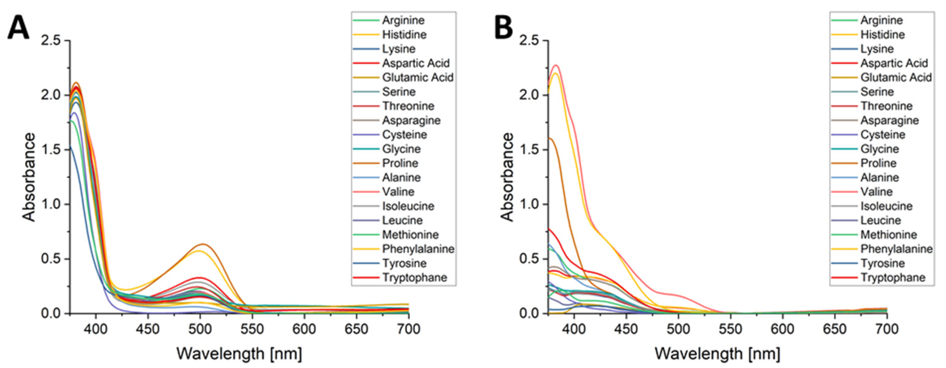
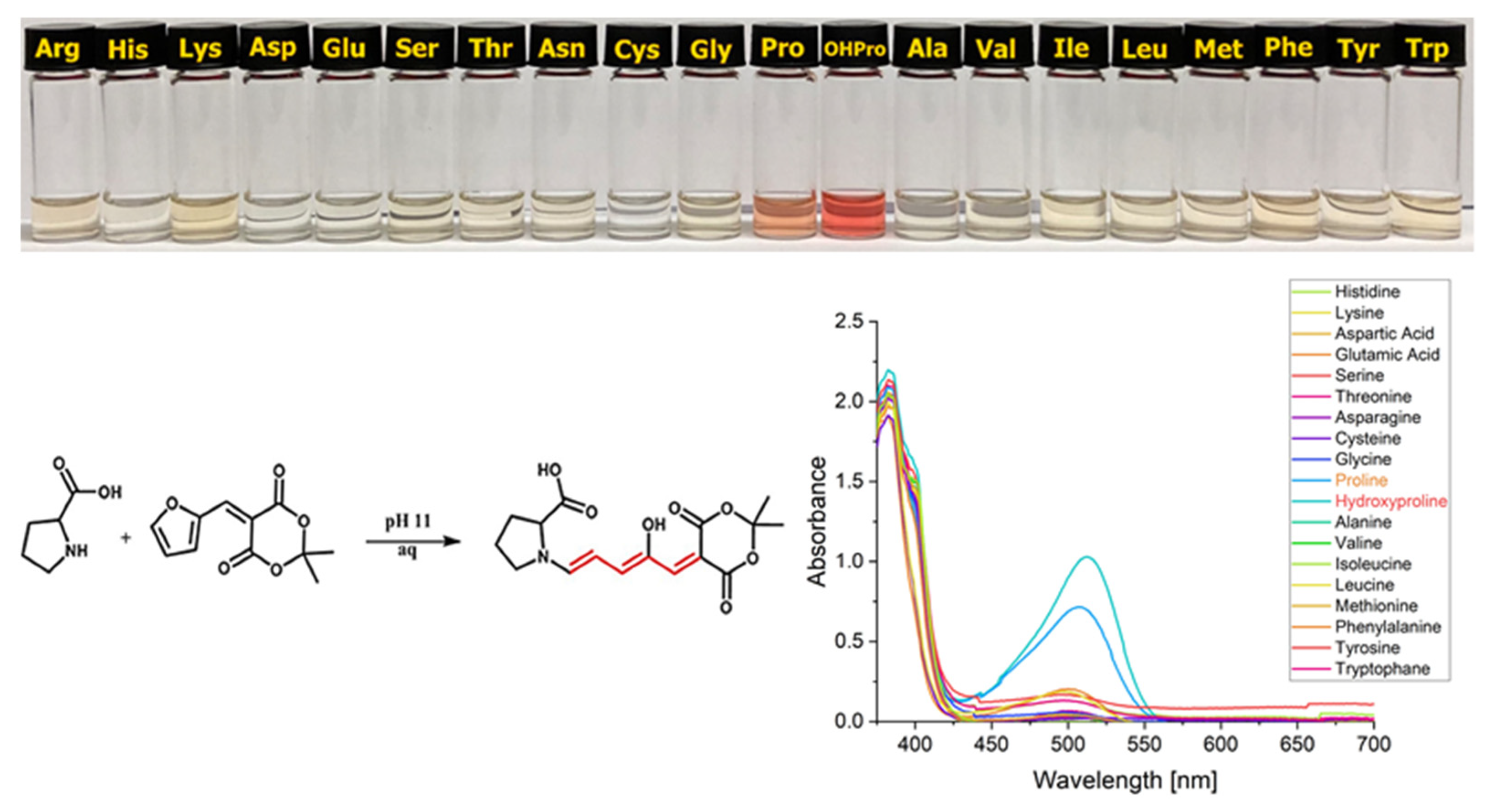
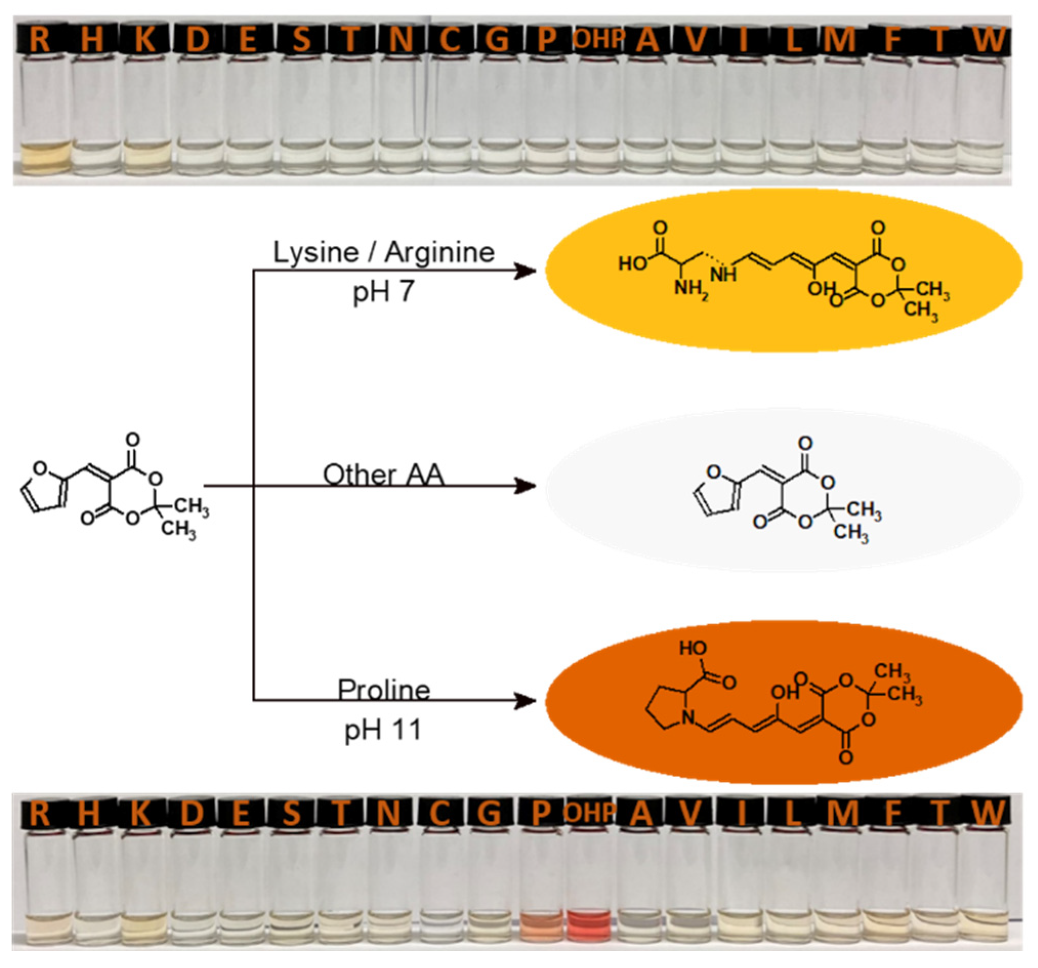
3.1. pH-Dependency of MAFC Reaction with All AA at Neutral and Alkaline pH

In our previous study, we were able to detect AA lysine and arginine by mixing them with ethanoic solution of MAFC over a wide range of pH from 4 to 11 and at concentrations as low as 100 µM [6]. However, in this study we have investigated behavior of all essential amino acids in presence of sensor molecule MAFC at different pHs, because it is known that pH can influence sensing capabilities of probes [28]. Moreover, different pKa values of amine either on side chain or on the chiral carbon of amino acids could result in different reaction patterns. As expected from previous results no other AA led to any color reaction at pH 7 except for lysine and arginine (see Figure 1A). Exceptionally, samples containing proline showed a steady increase in absorption at 510 nm, when the pH was rising from 7 onwards (see Figure 1B). Surprisingly, all AA have shown a color reaction with MAFC at pH 11 (see Figure 1C). However, no color formation was observed when AA cysteine and aspartic acid were mixed with MAFC. These results indicate that at pH 11 the amine group of most AA gets deprotonated and are available for a nucleophilic addition to MAFC, irrespective of side chain functionalization.
Considering the hypothesis of higher selectivity of MAFC to secondary amine compared to primary amine [2] a similar trend has been shown by AA reaction with MAFC at pH 11. As shown in Figure 1C, AA proline containing a secondary amine functional group shows a prominent absorption at 510 nm, which is much higher than all primary amine group containing amino acids. The obvious reason for not showing color reaction with MAFC at pH 7 is the protonated state of amine groups of amino acids, which has a pKa value mostly above 9, therefore this group cannot react with MAFC to form conjugated triene species. On the other hand, lysine and arginine can react with MAFC due to their primary amine side chain functional groups, due to their high nucleophilicity and less steric hindrance. Proline shows special characteristics due to it being the only proteinogenic secondary amino acid. Therefore, instead of having a primary amine functionality proline inhibits a secondary amine group with a high pKa of 10.6 [26] which is thus also in its cationic state at pH 7 and is responsible for the slow DASA formation. Yet at pH 11, proline has now normal secondary amine behavior toward MAFC resulting in the already known faster and much more prominent color formation with MAFC than it is the case with primary amines. The next surprising behavior of colored product formation reaction of proline with MAFC was the sudden disappearance of the color when pH is raised above 11, which might be due to the deprotonation of hydroxyl proton present on conjugated triene. As a result, the conjugation of the triene between donor and acceptor end could be influenced and ultimately lead to the formation of colorless cyclopentenone derivative [29].

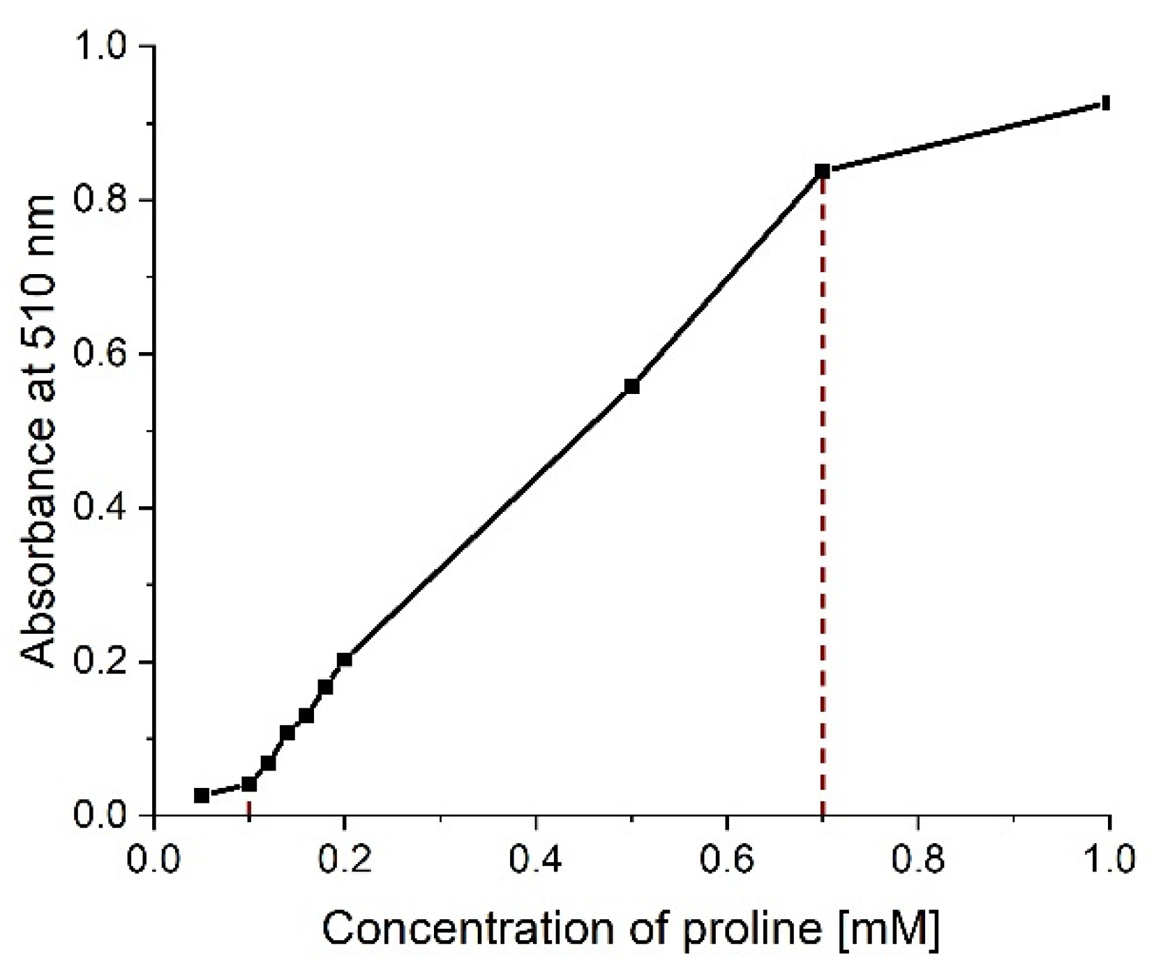
3.2. LOD in MAFC based Proline Sensing

After observing prominent selective color reaction between MAFC and proline at pH 11, it became obvious to explore the sensing capabilities of our chemosensor. For this, MAFC-proline solutions with different concentrations ranging from 50–1000 µM were prepared and measured with the UV-Vis spectrometer. An absorption-concentration graph was prepared (see Figure 2) for finding the linear measurement range and calculating LOD and LOQ. The linear measurement range was found to be 100–700 µM, LOD and LOQ were calculated to be 11 µM and 37 µM respectively. This result fits the experimental data (see Supporting Information Figure S1).

3.3. Job Plot of MAFC-Proline Reaction

Following LOD studies, Job Plot measurements were performed to gain insight in the stoichiometry of the MAFC-proline reaction. The resulting graph (see Figure 3) showed a linear correlation between MAFC-proline ratio and absorbance that increases from MAFC fraction of 0.2 to 0.5 and decreases from 0.5 to 0.9. As expected from previous experiments, maximum absorption of MAFC-proline conjugate is reached at molar fraction of 0.5, indicating a 1:1 reaction of MAFC and proline.

3.4. Time-Dependent Study of MAFC-AA Reaction

A time-dependent study of all MAFC-AA conjugates at pH 11 (see Supporting Information Figure S2) reveals that AA-MAFC adducts are not stable in these conditions after a particular period of time, which limit the applicability of this method when long-term measurements are done. However, the adduct is stable over a suitable period of time for at least 3 h (see Figure 4A), which is usually enough for measurements. Whereas no AA-DASA adducts were visible after 24 h reaction time even when stored in darkness, under established set of conditions (see Figure 4B). Most probable reason for disappearance of the color could be due to the formation of base-assisted cyclopentenone formation [29]. The UV-Vis spectra shows that arginine-, histidine-, and proline-MAFC conjugates undergo another reaction sequence, which is not similar to the rest of the amino acids. However, we have not investigated it in detail because, when color disappears the chemosensor characteristic of the molecule is lost and hence it became inessential for the present work.

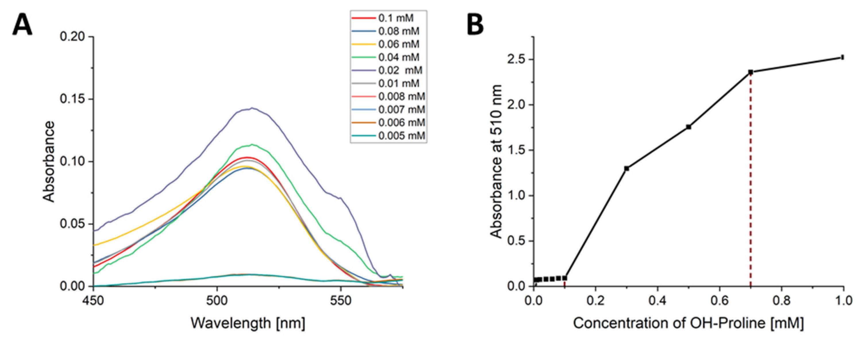
3.5. pH Dependency of MAFC-Hydroxyproline Reaction

After evaluating the selectivity and sensing capabilities of our chemosensor, where proline came up as prominent candidate, we were curious to see how biologically important derivatives of proline such as hydroxyproline react with MAFC. Hydroxyproline plays an important role in collagen stability by stabilizing collagens triple helical formation [30]. Due to its relevance in collagen, hydroxyproline can be used as a biomarker in urine for detection of liver disease or measurement of bone and tissue turnover and is therefore a relevant AA derivative for our MAFC chemosensor [31,32,33]. It was amazing to observe that hydroxyproline is a wonderful candidate for reaction with MAFC resulting in a very fast and prominent color formation especially at pH 10 and 11. The intensity of color highly outperformed all other AA tested so far. Typical DASA triene absorbance peak at 510 nm was visible from pH 6–11 with exceptionally raised absorbance at pH 11 compared to proline (see Figure 5). At pH 12 a distinct hypsochromic shift from 510 nm to 420 nm was observed when using higher concentrations (1 mM) (see Figure 5B) indicating the formation of a different product with significantly reduced intensity for detection and hence this pH became irrelevant for our study.

3.6. LOD in MAFC Based Hydroxyproline Sensing

Encouraged by the incredibly intense color formation of MAFC-hydroxyproline reaction we observed if the detection limit of our chemosensor could be enhanced for reaction with hydroxyproline at pH 11 in comparison to original proline analog. Different concentrations ranging from 5–100 µM of MAFC-hydroxyproline solutions were prepared and analyzed with the UV-Vis spectrometer (see Figure 6A). As before, LOD and LOQ were calculated mathematically from absorption-concentration plot (see Figure 6B). The linear measurement range for MAFC-hydroxyproline reaction was found again to be 100–700 µM. LOD and LOQ equaled 6 µM and 22 µM respectively, which is nearly factor 2 better than for MAFC-proline reaction.

3.7. NMR Studies on MAFC-AA Reaction

Essentially, we expect that the color formation in this reaction is due to the triene formation as reported before [6], however, to give confident statement on the structure, we have performed 1H-NMR titrations and LCMS of MAFC-proline conjugates (see Figure 7; for full spectrum see Supporting Information Figure S3). Initially, we tried to replicate the conditions of sensing solution by using deuterated methanol and alkaline water for dissolving MAFC and AA respectively. However, adjusting the pH exactly to 11 by adding NaOH to deuterated solution was tedious. Therefore, we planned to screen some other comparable deuterated solvent systems where a prominent color reaction can be observed without the need of adjusting pH. After recording, the 1H-NMR in various deuterated solvent acetone-d6 appears to be the most suitable solvent due to complete disappearance of MAFC peaks at 6.9 ppm, 8.2 ppm, and 8.4 ppm in a given time (see Figure 7C). At the same time the appearance of peaks at 4.61 ppm, 6.03 ppm, 6.27 ppm, and 7.30 ppm in MAFC-proline sample indicates the formation of typical open chain triene. A comparative structural analog of pyrrolidine, was chosen to confirm if similar peaks of triene can be observed as well. As expected signature peaks of triene were observed at 6.13 ppm, 6.28 ppm, 7.37 ppm, and 7.56 ppm in MAFC-pyrrolidine. Similarly, in case of MAFC-hydroxyproline 1H peaks at 4.57 ppm, 6.03 ppm, 6.28 ppm, and 7.3 ppm corresponding to the triene protons confirm the fact that color appearance is due to the formation of triene upon reaction of activated furan ring with free secondary amine of proline (for complete proton assignments of each spectrum see Supporting Information Figures S5–S7). Similarly, LCMS product peaks of both MAFC-hydroxyproline and MAFC-proline at 354+ (MH+) and 338+ (MH+) respectively confirmed 1:1 adduct of MAFC and AA via ring opening reaction resulting in triene formation (see Supporting Information Figure S10).
Moreover, decreasing the ratio of proline to MAFC was investigated to determine if secondary reactions are happening on the MAFC-proline adduct (see Supporting Information Figure S4). As expected no additional reactions were visible and incomplete reaction was obvious through typical furan ring signals of MAFC at 6.9 ppm, 8.2 ppm, and 8.4 ppm. This also supports our hypothesis of MAFC-proline product being a 1:1 adduct of proline and MAFC.

4. Conclusions

In this work, we extended our understanding of sensing capabilities of our previously published Meldrum’s acid based colorimetric sensor MAFC for the selective detection of proline and hydroxyproline. By testing the reaction of MAFC with all proteinogenic AA over a wide range of pH we were able to find impressive behavioral changes at alkaline conditions. At neutral pH the color reaction is favorable for amine side groups containing AA like lysine and arginine. Whereas, when pH is increased to 11, proline and hydroxyproline react much faster with MAFC than any other AA and give intense color immediately. Reaction with hydroxyproline is even more intense than with proline and an impressive detection limit of 6 µM was found. 1H-NMR titrations and LCMS analysis were used to elucidate the structure of adduct. We conclude, that while our MAFC colorimetric sensor is capable of detecting lysine and arginine at neutral pH, as previously published, it is also able to sense proline and hydroxyproline at pH 11 at low concentrations due to their intense coloration. Minimal synthetic requirement, low cost, and synthetic simplicity for production of MAFC make this chemosensor an ideal candidate for easy amino acid detection. Figure 8 shows a schematic presentation of the presented findings.

Supplementary Materials

The following are available online at https://0-www-mdpi-com.brum.beds.ac.uk/article/10.3390/chemosensors9120343/s1. Figure S1: UV-Vis spectra of MAFC-proline at concentration of 40–1000 µM and photo image of solutions showing increase of color intensity with increasing concentrations from 100–500 µM. Figure S2: UV-Vis analysis of AA-MAFC adducts after (A) 15 min, (B) 1h, (C) 3h, (D) 6h, (E) 24h reaction time, concentration of AA was 0.3 mM. Figure S3: 1H-NMR-spectrum of (A) MAFC-hydroxyproline, (B) MAFC-pyrrolidine, (C) MAFC-proline and (D) MAFC crude reaction mixture in acetone-d6/D2O. Figure S4: 1H-NMR-spectra of MAFC-proline crude reaction mixture in different molar ratios of 1:1–6:1 in acetone-d6/D2O. Figure S5: 1H-NMR-spectrum of MAFC-hydroxyproline crude reaction mixture in acetone-d6/D2O. Figure S6: 1H-NMR-spectrum of MAFC-proline crude reaction mixture in acetone-d6/D2O. Figure S7: 1H-NMR-spectrum of MAFC in acetone-d6. Figure S8: 1H-NMR-spectrum of proline in D2O. Figure S9: 1H-NMR-spectrum of hydroxyproline in D2O. Figure S10: LCMS of MAFC-proline (top) and MAFC-hydroxyproline reaction (bottom).

Author Contributions

S.S. was contributing to conceptualization, data interpretation, supervision, review and editing. A.S. made contributions to conceptualization of measurement methods and data interpretation. C.A. was responsible for methodology, validation of experiments and visualization. L.Z. prepared original draft and contributed to visualization, methodology and data interpretation. All authors have read and agreed to the published version of the manuscript.

Funding

This research was funded by Federal Ministry of Education and Research (BMBF) within the Initiative “Centre for Innovation Competence” Meta-ZIK (BioLithoMorphie: FKZ 03Z1M511, FKZ 03Z1M512) and BMBF VIP Platox.

Institutional Review Board Statement

Not applicable.

Informed Consent Statement

Not applicable.

Data Availability Statement

The data presented in this study are available on request from the corresponding author.

Acknowledgments

L. Zeußel thanks the Thuringian State Graduate Support and QuantenHub for financing. We thank Katrin Risch, Susann Guenther for measuring the NMR, UV-Vis spectra and Alexander Groß for measuring LCMS and valuable discussions.

Conflicts of Interest

The authors declare no conflict of interest.

Appendix A. Table of Content

In this work the pH dependency of the previously reported reaction of Meldrum’s acid furfural conjugate (MAFC) with amino acids (AA) was observed and AA proline and hydroxyproline were examined further due to their outstanding reaction at pH 11. Our colorimetric sensor is able to selectively detect proline and hydroxyproline concentrations in aqueous solutions at pH 11 as low as 11 µM and 6 µM respectively. 1H-NMR, LCMS, and job plot data strengthened our believe in the proposed 1:1 addition reaction mechanism.
Figure A1. Overview of the study design and results.
Chemosensors 09 00343 g0a1

References

  1. Helmy, S.; Leibfarth, F.A.; Oh, S.; Poelma, J.E.; Hawker, C.J.; Read de Alaniz, J. Photoswitching using visible light: A new class of organic photochromic molecules. J. Am. Chem. Soc. 2014, 136, 8169–8172. [Google Scholar] [CrossRef] [PubMed] [Green Version]
  2. Singh, S.; Mai, P.; Borowiec, J.; Zhang, Y.; Lei, Y.; Schober, A. Donor-acceptor Stenhouse adduct-grafted polycarbonate surfaces: Selectivity of the reaction for secondary amine on surface. R. Soc. Open Sci. 2018, 5, 180207. [Google Scholar] [CrossRef] [PubMed] [Green Version]
  3. Diaz, Y.J.; Page, Z.A.; Knight, A.S.; Treat, N.J.; Hemmer, J.R.; Hawker, C.J.; Read de Alaniz, J. A Versatile and Highly Selective Colorimetric Sensor for the Detection of Amines. Chemistry 2017, 23, 3562–3566. [Google Scholar] [CrossRef] [Green Version]
  4. Jia, S.; Du, J.D.; Hawley, A.; Fong, W.-K.; Graham, B.; Boyd, B.J. Investigation of Donor-Acceptor Stenhouse Adducts as New Visible Wavelength-Responsive Switching Elements for Lipid-Based Liquid Crystalline Systems. Langmuir 2017, 33, 2215–2221. [Google Scholar] [CrossRef] [PubMed]
  5. Helmy, S.; Oh, S.; Leibfarth, F.A.; Hawker, C.J.; Read de Alaniz, J. Design and synthesis of donor-acceptor Stenhouse adducts: A visible light photoswitch derived from furfural. J. Org. Chem. 2014, 79, 11316–11329. [Google Scholar] [CrossRef] [PubMed] [Green Version]
  6. Zeußel, L.; Mai, P.; Sharma, S.; Schober, A.; Ren, S.; Singh, S. Colorimetric Method for Instant Detection of Lysine and Arginine Using Novel Meldrum’s Acid-Furfural Conjugate. ChemistrySelect 2021, 6, 6834–6840. [Google Scholar] [CrossRef]
  7. Dean, K.E.S.; Klein, G.; Renaudet, O.; Reymond, J.-L. A green fluorescent chemosensor for amino acids provides a versatile high-throughput screening (HTS) assay for proteases. Bioorg. Med. Chem. Lett. 2003, 13, 1653–1656. [Google Scholar] [CrossRef]
  8. Li, X.; Wang, K.; Ma, N.; Jia, X. Poly(ionic liquid) Based Chemosensors for Detection of Basic Amino Acids in Aqueous Medium. Front. Chem. 2017, 5, 69. [Google Scholar] [CrossRef] [Green Version]
  9. Shahrajabian, M.; Ghasemi, F.; Hormozi-Nezhad, M.R. Nanoparticle-based Chemiluminescence for Chiral Discrimination of Thiol-Containing Amino Acids. Sci. Rep. 2018, 8, 14011. [Google Scholar] [CrossRef] [Green Version]
  10. Pu, F.; Huang, Z.; Ren, J.; Qu, X. DNA/ligand/ion-based ensemble for fluorescence turn on detection of cysteine and histidine with tunable dynamic range. Anal. Chem. 2010, 82, 8211–8216. [Google Scholar] [CrossRef]
  11. Zhou, Y.; Yoon, J. Recent progress in fluorescent and colorimetric chemosensors for detection of amino acids. Chem. Soc. Rev. 2012, 41, 52–67. [Google Scholar] [CrossRef] [PubMed]
  12. Corradini, R.; Paganuzzi, C.; Marchelli, R.; Pagliari, S.; Sforza, S.; Dossena, A.; Galaverna, G.; Duchateau, A. Design and synthesis of fluorescent beta-cyclodextrins for the enantioselective sensing of alpha-amino acids. Chirality 2003, 15, S30–S39. [Google Scholar] [CrossRef]
  13. Kwong, H.-L.; Wong, W.-L.; Lee, C.-S.; Yeung, C.-T.; Teng, P.-F. Zinc(II) complex of terpyridine-crown macrocycle: A new motif in fluorescence sensing of zwitterionic amino acids. Inorg. Chem. Commun. 2009, 12, 815–818. [Google Scholar] [CrossRef]
  14. Li, Z.; Lou, X.; Li, Z.; Qin, J. A new approach to fluorescence “turn-on” sensing of alpha-amino acids. ACS Appl. Mater. Interfaces 2009, 1, 232–234. [Google Scholar] [CrossRef] [PubMed]
  15. Patel, G.; Menon, S. Recognition of lysine, arginine and histidine by novel p-sulfonatocalix4arene thiol functionalized gold nanoparticles in aqueous solution. Chem. Commun. 2009, 24, 3563–3565. [Google Scholar] [CrossRef]
  16. Morgan, A.A.; Rubenstein, E. Proline: The distribution, frequency, positioning, and common functional roles of proline and polyproline sequences in the human proteome. PLoS ONE 2013, 8, e53785. [Google Scholar] [CrossRef] [Green Version]
  17. Szabados, L.; Savouré, A. Proline: A multifunctional amino acid. Trends Plant Sci. 2010, 15, 89–97. [Google Scholar] [CrossRef] [PubMed]
  18. Adams, E.; Frank, L. Metabolism of proline and the hydroxyprolines. Annu. Rev. Biochem. 1980, 49, 1005–1061. [Google Scholar] [CrossRef] [PubMed]
  19. Boctor, F.N. An improved method for colorimetric determination of proline with isatin. Anal. Biochem. 1971, 43, 66–70. [Google Scholar] [CrossRef]
  20. Bates, L.S.; Waldren, R.P.; Teare, I.D. Rapid determination of free proline for water-stress studies. Plant Soil 1973, 39, 205–207. [Google Scholar] [CrossRef]
  21. Choi, Y.-S.; Im, M.K.; Lee, M.R.; Kim, C.S.; Lee, K.-H. Highly sensitive enclosed multilayer paper-based microfluidic sensor for quantifying proline in plants. Anal. Chim. Acta 2020, 1105, 169–177. [Google Scholar] [CrossRef]
  22. Forlani, G.; Funck, D. A Specific and Sensitive Enzymatic Assay for the Quantitation of L-Proline. Front. Plant Sci. 2020, 11, 582026. [Google Scholar] [CrossRef] [PubMed]
  23. Kim, G.-J.; Kim, H.-J. Highly selective and sensitive fluorescence turn-on probe for proline. Tetrahedron Lett. 2010, 51, 4670–4672. [Google Scholar] [CrossRef]
  24. Liu, J.-B.; Liu, L.-J.; Dong, Z.-Z.; Yang, G.-J.; Leung, C.-H.; Ma, D.-L. An Aldol Reaction-Based Iridium(III) Chemosensor for the Visualization of Proline in Living Cells. Sci. Rep. 2016, 6, 36509. [Google Scholar] [CrossRef] [Green Version]
  25. Singh, S.; Friedel, K.; Himmerlich, M.; Lei, Y.; Schlingloff, G.; Schober, A. Spatiotemporal Photopatterning on Polycarbonate Surface through Visible Light Responsive Polymer Bound DASA Compounds. ACS Macro Lett. 2015, 4, 1273–1277. [Google Scholar] [CrossRef]
  26. Fan, G.-L.; Liu, Y.-L.; Wang, H. Identification of thermophilic proteins by incorporating evolutionary and acid dissociation information into Chou’s general pseudo amino acid composition. J. Theor. Biol. 2016, 407, 138–142. [Google Scholar] [CrossRef]
  27. Roberts, C.A.; Allen, S.; Helmy, S. Using Donor–Acceptor Stenhouse Adducts to Teach Photochromism in the Undergraduate Laboratory. J. Chem. Educ. 2021, 98, 1736–1740. [Google Scholar] [CrossRef]
  28. Doumani, N.; Bou-Maroun, E.; Maalouly, J.; Tueni, M.; Dubois, A.; Bernhard, C.; Denat, F.; Cayot, P.; Sok, N. A New pH-Dependent Macrocyclic Rhodamine B-Based Fluorescent Probe for Copper Detection in White Wine. Sensors 2019, 19, 4514. [Google Scholar] [CrossRef] [Green Version]
  29. Cai, Y.-D.; Chen, T.-Y.; Chen, X.Q.; Bao, X. Multiresponsive Donor-Acceptor Stenhouse Adduct: Opportunities Arise from a Diamine Donor. Org. Lett. 2019, 21, 7445–7449. [Google Scholar] [CrossRef] [PubMed]
  30. Berg, R.A.; Prockop, D.J. The thermal transition of a non-hydroxylated form of collagen. Evidence for a role for hydroxyproline in stabilizing the triple-helix of collagen. Biochem. Biophys. Res. Commun. 1973, 52, 115–120. [Google Scholar] [CrossRef]
  31. Stoilov, I.; Starcher, B.C.; Mecham, R.P.; Broekelmann, T.J. Measurement of elastin, collagen, and total protein levels in tissues. Methods Cell Biol. 2018, 143, 133–146. [Google Scholar] [CrossRef] [PubMed]
  32. Pohlídal, A.; Husek, P.; Palicka, V.; Slabík, D.; Hill, M.; Matucha, P. Novel and traditional biomarkers of bone turnover in postmenopausal women. Clin. Chem. Lab. Med. 2003, 41, 74–78. [Google Scholar] [CrossRef] [PubMed]
  33. Gabr, S.A.; Alghadir, A.H.; Sherif, Y.E.; Ghfar, A.A. Hydroxyproline as a Biomarker in Liver Disease. In Biomarkers in Liver Disease; Patel, V.B., Preedy, V.R., Eds.; Springer: Dordrecht, The Netherlands, 2017; pp. 471–491. ISBN 978-94-007-7675-3. [Google Scholar]
Figure 1. UV-Vis absorbance spectra of all AA reacted with MAFC at (A) pH 7 and (C) pH 11, (B) UV-Vis spectrum of MAFC-proline at pH 1–14, concentration of amino acids for all graphs was 0.3 mM, (D) photo image of MAFC-proline samples with different pH from 1–14, Close-Up of pH 10 and 11, c = 1 mM.
Chemosensors 09 00343 g001
Figure 2. Absorption-concentration plot of MAFC-proline reaction with concentrations ranging from 50–1000 µM. Linear measurement range from 100–700 µM is depicted by dashed brown lines.
Chemosensors 09 00343 g002
Figure 3. Job Plot of MAFC-proline reaction indicating 1:1 reaction. Sum of molar concentrations of the two binding partners was 0.6 mM.
Chemosensors 09 00343 g003
Figure 4. Absorbance spectra of all AA at pH 11 (A) 1 h, (B) 24 h after mixing with MAFC, concentration of AA is 0.3 mM.
Chemosensors 09 00343 g004
Figure 5. UV-Vis absorbance spectrum of hydroxyproline (A) at pH 1–14, concentration 0.3 mM, (B) at pH 12 and 1 mM hydroxyproline.
Chemosensors 09 00343 g005
Figure 6. (A) UV-Vis spectrum of MAFC-hydroxyproline at pH 11, (B) absorption-concentration graph of MAFC-hydroxyproline at 510 nm; concentrations ranging from 5–100 µM; linear measurement range from 100–700 µM is depicted by dashed brown lines.
Chemosensors 09 00343 g006
Figure 7. 1H-NMR spectrum of (A) MAFC-hydroxyproline, (B) MAFC-pyrrolidine, (C) MAFC-proline and (D) MAFC, peaks for open chain triene are highlighted. All spectra were recorded in deuterated acetone.
Chemosensors 09 00343 g007
Figure 8. Schematic presentation of herein presented findings.
Chemosensors 09 00343 g008
Table 1. Comparison of proline sensors regarding assay method, sensor substance, limit of detection (LOD), linear measurement range (LMR), and advantages/disadvantages.
Assay MethodSensor SubstanceLOD [µM]LMR [µM]Advantages/Disadvantages
ColorimetricIsatin-<220- stability 1 h
- pH 4.1
- inconsistent results through slightes contamination
- colored product extraction necessary
+ no interference with other amino acids
[19]
ColorimetricNinhydrine100100–36,000- interferences in biological samples
- low pH
- harsh chemical
- reagent stability 24 h
t = 1 h, 100 °C
+ interferences with other amino acids
[20]
Fluorescence turn-onAldol functionalized coumarin1.2-- t = 6 h
+ high selectivity
[23]
Luminescence[Ir(ppy)2(5-CHOphen)]PF60.752–100- metal based
- t = 40 min
- interferences with other amino acids
+ detection in living cells & blood
+ detection in highly autofluorescent samples
[24]
EnzymaticRecombinant enzymes from Arabidopsis thaliana and Oryza sativa-100–350+ higher specifity than ninhydrin methods
- bacterial cultures necessary
- narrow pH range
+ t = 10–15 min
+ no harmful chemicals
+ low volume
[22]
ColorimetricNinhydrin2323–100- lengthy preparation
- strong acids needed
+ Sample volume 100 µL
[21]
Publisher’s Note: MDPI stays neutral with regard to jurisdictional claims in published maps and institutional affiliations.

