Article

A Model System for Sensitive Detection of Viable E. coli Bacteria Combining Direct Viability PCR and a Novel Microarray-Based Detection Approach

1
Leibniz Institute of Photonic Technology, Member of the Research Alliance “Leibniz Health Technologies”, Albert-Einstein-Straße 9, 07745 Jena, Germany
2
Institute of Physical Chemistry and Abbe Centre of Photonics, Friedrich Schiller University Jena, Helmholtzweg 4, 07743 Jena, Germany
3
InfectoGnostics Forschungscampus Jena, Zentrum für Angewandte Forschung, Philosophenweg 7, 07743 Jena, Germany
4
fzmb GmbH-Forschungszentrum für Medizintechnik und Biotechnologie, Geranienweg 7, 99947 Bad Langensalza, Germany
5
Ernst-Abbe-Hochschule Jena, University of Applied Sciences, Carl-Zeiss-Promenade 2, 07745 Jena, Germany
6
IST Innuscreen GmbH, Robert-Rössle-Straße 10, 13125 Berlin, Germany
*
Author to whom correspondence should be addressed.
These authors contributed equally to this work.
Submission received: 28 October 2021 / Revised: 6 December 2021 / Accepted: 9 December 2021 / Published: 13 December 2021

Abstract

:
We established an innovative approach that included direct, viability, and nested PCR for rapid and reliable identification of the fecal indicator organism Escherichia coli (E. coli). Direct PCR enabled successful amplification of the target uidA gene, omitting a prior DNA isolation or purification step. Furthermore, we applied viability PCR (v-PCR) to ensure the detection of only relevant viable bacterial cells. The principle involves the binding of propidium monoazide (PMA), a selective nucleic acid intercalating dye, to accessible DNA of heat killed bacteria cells and, consequently, allows viable and heat killed E. coli cells to be discriminated. To ensure high sensitivity, direct v-PCR was followed by a nested PCR step. The resulting amplicons were analyzed by a rapid 30 min microarray-based DNA hybridization assay for species-specific DNA detection of E. coli. A positive signal was indicated by enzymatically generated silver nanoparticle deposits, which served as robust endpoint signals allowing an immediate visual readout. The presented novel protocol allows the detection of 1 × 101 viable E. coli cells per PCR run.

1. Introduction

Globally, at least two billion people use a drinking water source contaminated with feces. Microbiological contamination can cause gastrointestinal disorders, as well as symptoms of poisoning [1]. During the last decade, the number of human diseases caused by pathogens in drinking water has increased. Contaminated water can transmit diseases such as cholera, dysentery, typhoid, and polio [2,3,4]. In addition to common bacteria, such as Salmonella and Shigella, other pathogens, such as Campylobacter, enterohemorrhagic E. coli (EHEC) or Norovirus, have emerged. In many cases, these microorganisms are spread through human or animal feces into the aquatic environment. For that reason, microbiological monitoring of the quality of drinking water is crucial to ensure that these bacteria and viruses exist in concentrations that are harmless to humans. These pathogens usually appear with increased quantities of E. coli or other innocuous bacteria. Therefore, it is necessary to routinely monitor the appearance of E. coli or other enterococci in drinking water samples. E. coli serves as a ‘fecal indicator organism’ for water monitoring and is defined as the best biological drinking water indicator for public health protection [5]. According to the drinking water ordinance in Germany, not a single E. coli cell is allowed in 100 mL of water. Only when the analysis reveals zero colony-forming units is the water declared drinkable [https://www.dvgw.de] (accessed on 25 October 2021).
Thus, suitable detection methods for specific identification and sensitive quantification of these indicator organisms are mandatory, with the additional challenge of precisely detecting viable cells. Common techniques for routine diagnosis of E. coli in drinking water rely upon culture-based approaches. According to the drinking water ordinance, three different detection procedures are approved and ISO (International Organization for Standardization) certified [DIN EN ISO 9308-1, DIN EN ISO 9308-2, DIN EN ISO 9308-3]. Primarily, traditional cultivation for quantifying bacteria in drinking water is applied (membrane filtration combined with subculture on different media). Nevertheless, this method has several disadvantages. First and foremost, it takes 18–24 h for the sample-to-answer process [3]. Furthermore, nutrient-rich traditional media support the growth of non-targets that can mimic or suppress target organisms, yielding false negative results. Second, water quality is evaluated by the “most probable number” method which is based on the growth of target organisms in a liquid medium and the subsequent calculation of the “most probable number” (MPN) of organisms with reference to MPN tables. This approach for quantifying is used in the Bluewater Biosciences ColiPlate™ kit [6]. In addition, the US Environmental Protection Agency (EPA)-approved Colilert Quanti-Tray is used to monitor the presence or absence of coliforms and E. coli, as well as to quantify their levels within 18 to 21 h [http://www.idexx.de/water/water-testing-solutions.html] (accessed on 25 October 2021) [6].
To circumvent these long verification procedures, molecular-based methods, such as polymerase chain reaction (PCR), could be applied to obtain results within a few hours. The classical PCR approach does not differentiate between viable and heat killed cells since DNA persists after cell death. However, only reproducible or metabolically active bacteria produce toxins, which pose a health hazard to humans. Therefore, viability PCR was introduced, which inhibits amplification of DNA originating from dead cells [7]. This approach represents one of the most successful techniques to detect only viable bacterial cells and has been effectively established and evaluated for live-dead discrimination in multiple microorganisms (bacteria [8,9,10,11,12,13,14,15,16], protozoa [17,18,19], viruses [20,21,22], fungi [23]). The principle is based upon supplementation of PMA, a membrane-impermeable, photoreactive dye, which is unable to permeate intact cell membranes of viable bacteria [24]. PMA can only penetrate damaged membranes of dead bacteria and covalently modifies the DNA. This cross-linkage is induced by photoactivation. As a consequence, PMA-modified template DNA cannot be amplified by PCR [7,13].
To realize a simple and rapid sample preparation, direct PCR was implemented, which has been previously described [25]. In this approach, bacterial cell lysate is directly added to the PCR mixture after simple heat lysis. DNA sequences coding for the lacZ gene (β-galactosidase), the uidA gene (β-D-glucuronidase), and the ycjM gene (glucosyltransferase) have been used to detect total coliforms and E. coli [26]. In the approach presented herein, the uidA gene was chosen as a potential target gene for PCR amplification.
Our work focused on establishing a molecular biology-based platform technology for sensitive and reliable detection of living E. coli cells. By combining direct, viability and nested PCR, rapid and precise analysis of viable bacteria is possible, omitting time-consuming sample preparation. Subsequent precise specification was realized using on-chip DNA hybridization, enabling easy visual signal readout. Innovative spoon-shaped polypropylene substrates were designed to avoid evaporation of the hybridization solution, enable a targeted and permanent incubation of the PCR product with the immobilized capture probes, and facilitate an easy-to-manage, rapid washing mode. We highlight the optimization of the complete process chain for the reliable and rapid detection of viable E. coli cells in drinking water, beginning from DNA extraction to on-chip identification of the bacteria by naked eye (Figure 1).

2. Materials and Methods

2.1. Culturing and Harvesting Defined Cell Numbers of E. coli Bacteria

The E. coli strain (DSM-423, Leibniz Institute DSMZ–German Collection of Microorganisms and Cell Culture, Braunschweig, Germany) was grown on Lysogeny Broth (LB) medium (Carl Roth, Karlsruhe, Germany) at 37 °C shaking at 160 rpm. Growth was monitored by measuring the optical density at 600 nm (OD600) using a biophotometer (Eppendorf AG, Hamburg, Germany). Bacterial cells were harvested when they reached the exponential growth phase. Cell numbers (1 × 10⁶–1 × 100 CFU/mL) were calculated assuming that OD600 = 1 corresponds to 8 × 10⁸ CFU/mL [27]. The cell sediment was washed twice with 1× phosphate buffered saline (1× PBS) and treated with PMA. To generate dead bacteria, the cell sediment was incubated at 70 °C for 10 min and immediately cooled down before treatment with PMA.

2.2. PMA Treatment for Viability PCR

In the present study, combination of direct, viability and nested PCR was performed. The viability PCR approach was applied to discriminate between viable and heat killed E. coli cells. PMA (Biotrend chemicals GmbH, Köln, Germany) was dissolved in nuclease-free water to create a stock solution of 10 mM and stored at −20 °C until usage. Viable and heat killed bacterial cells were treated with 10 µM PMA followed by thorough mixing. Afterward, tubes were first kept in the dark for 10 min (occasionally inverting). Then, the PMA reagent was light activated for 10 min using an in-house constructed photoactivator (Figure 2). Cells were centrifuged at 10,000× g for 5 min, washed twice in 1× PBS, and finally resuspended in 1× PBS with the pellets stored at −20 °C until DNA extraction.

2.3. Thermal Cell Lysis for Direct PCR

To circumvent laborious genomic DNA extraction and purification, a direct PCR approach was employed. PMA treated bacterial cell sediments were mixed with 10 µL nuclease-free water and incubated for 5 min at 95 °C for thermal cell lysis. Emerging lysates containing the E. coli gDNA were cleared by centrifugation at 12,000× g for 10 min, and PCR was subsequently performed [28].

2.4. gDNA Extraction

The Qiagen DNeasy Blood and Tissue Kit was used as a positive control for bacterial DNA extraction. Genomic DNA was extracted using the protocol for pretreatment of gram-positive and gram-negative bacteria.

2.5. Direct, Viability, and Nested PCR Amplification

For nested PCR, two primer sets were constructed to target different regions of the uidA gene (Table 1). One primer pair amplified a large fragment of E. coli in the first round (uidA_long; 1.37 kb) and a short DNA fragment (uidA_short; 96 bp), which is located within the first product, in the second round. Primers and capture probes for the uidA amplification were designed using the “primer3”-program (https://primer3.ut.ee/, accessed on 25 October 2021). All primers and capture probes were purchased from Eurofins MWG Operon (Ebersberg, Germany).
The reaction mix for direct v-PCR contained 2 mM MgCl2, 1 × KCl buffer, 0.25 mM dNTPs, 0.25 µM of each primer (uidA_long, Table 1), 2.5 units Taq polymerase (innuTaq DNA Polymerase Kit, Analytik Jena AG, Germany) and 10 µL heat-lysed E. coli cells in a final volume of 20 μL. For nested PCR, a second round of amplification was conducted utilizing the same conditions but with 0.25 µM of each primer (uidA_short, Table 1) and 1 µL of 1:100 diluted PCR product from the first round of PCR. All PCR reactions were performed in a FlexCycler2 (Analytik Jena AG, Jena, Germany). PCR reactions had the following temperature-time profile: initial denaturation at 94 °C for 180 s, 35 cycles (first round)/20 cycles (second round) of denaturation at 94 °C for 30 s, annealing at 60 °C (first round)/58 °C (second round) for 30 s, elongation at 72 °C for 90 s (first round) or 30 s (second round) and a final elongation at 72 °C for 10 min (first round) or 5 min (second round).
The resulting PCR products were either verified on a 1% (w/v) agarose gel (Carl Roth, Karlsruhe, Germany) for long amplicons (1.37 kb) or on a 2% (w/v) agarose gel for short amplicons (96 bp). DNA was stained with 1:50,000 diluted GelRed (VWR International GmbH, Darmstadt, Germany). Alongside the samples, the molecular weight marker GeneRuler (1 kb or 100 bp) DNA Ladder (Fisher Scientific, Germany) was run on the agarose gels.

2.6. Generation of Single-Stranded DNA

To generate single-stranded DNA (ssDNA) amplicons were heat-incubated for 5 min at 95 °C and immediately cooled on ice for 2 min.

2.7. Microarray-Based DNA Hybridization and Signal Detection

Specificity of the short DNA amplicons was evaluated by microarray-based DNA hybridization experiments [29].
White polypropylene (PP) sheets (Modulor GmbH, Berlin, Germany) were used to generate solid chip substrates. The flexible material was stamped in a spoon-like shape with 1.3 cm diameter of the cavity. Thereafter, PP spoons were successively cleaned in an ultrasonic bath with acetone, ethanol and water for 10 min each.
The uidA gene region was chosen for design of species-specific capture probes. The capture probes (Eurofins MWG Operon, Ebersberg, Germany) were dissolved in 1 × Micro Spotting Solution (ArrayIt Corporation, Synnyvale, CA, USA) to a final concentration of 20 µM and spotted (Nanoplotter 2.1, GeSim, Grosserkmannsdorf, Germany) in a microarray format (Figure S2) in the PP spoon cavity. Afterward UV light exposure at 254 nm for 10 min ensured optimal binding of the capture probe nucleic acids to the surface [30]. PP spoons were washed with 0.1 × saline-sodium citrate (SSC)/0.5% (w/v) sodium dodecyl sulphate (SDS) to remove unbound DNA molecules and dried. A biotin-labelled non-complementary probe was immobilized as a process control to verify binding of the streptavidin-labelled enzyme and subsequent silver deposition.
Specific biomolecule interaction on the PP spoon microarray was achieved using a combined protocol that encompassed DNA hybridization and enzyme binding (streptavidin horseradish peroxidase complex; Sigma Aldrich Chemie GmbH, Taufkirchen, Germany) in one step. For this purpose, 80 µL reagent solution (20 µL single-stranded PCR (ssPCR) product and 1 µL enzyme in 5 × SSC/0.1% (w/v) SDS) were placed in the PP spoon cavity and subsequently covered with PCR foil (VWR International GmbH, Darmstadt, Germany) to avoid evaporation. The reagent solution was incubated for 30 min at 50 °C (INCUCELL, MMM Medcentre Einrichtungen GmbH, München, Germany).
Afterward PP spoon cavities were washed five times with solution 1 (2× SSC/0.1% (w/v) SDS), solution 2 (2× SSC), solution 3 (0.2× SSC) and nuclease-free water. Finally, enzymatic silver deposition on the microarray was performed by applying the EnzMet™ HRP detection kit according to the manufacturer’s recommendations (Nanoprobes Inc., Yaphank, NY, USA; component A–C) [29,31,32,33]. In addition, the silver deposits were quantified by their grey values. Respective spots were scanned (ProScan 7200, reflecta GmbH, Rottenburg, Germany) and analysed using the software ImageJ (National Institutes of Health, Bethesda, MD, USA). The mean grey value of the biotin-positive control was set at 100%, and the grey value signals of the capture probes are presented as grey value percentages of the positive control.

3. Results

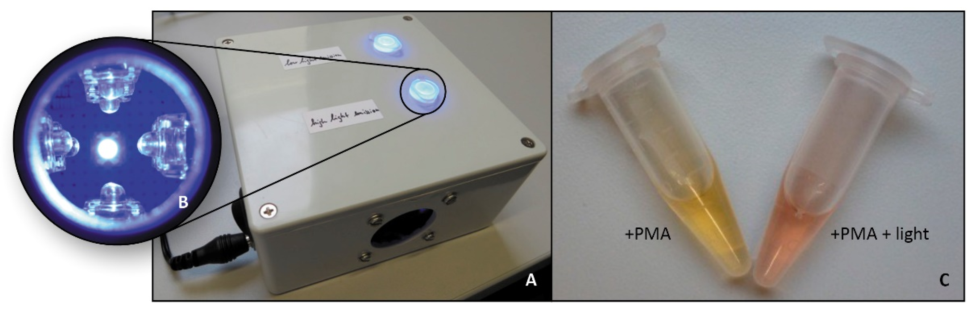
3.1. Viability PCR Using an in-House Constructed PMA-Photoactivation Device

First, for photoactivation of PMA and consequent covalent binding of the dye to accessible DNA of nonviable bacteria, a photoactivator is mandatory. Therefore, we constructed an LED device (Figure 2). Compared to commercial systems, the in-house designed and custom-build photoactivator offers several advantages. Creation of the mentioned device was inexpensive and allowed for the illumination of samples in 1.5 mL microreaction tubes. The device uses LED lights with 470 nm emission for an efficient activation of PMA or other similar dyes. The device was set up with two different LED types. The first layout contained 16 Standard 5 mm oval LEDs (each 37.5 mW) and the second nine Kingbright SuperFlux LEDs (each 86 mW). To prevent heating of the sample, a 40 mm cooling fan (3 W) was installed. The price for all components was less than 100 €.

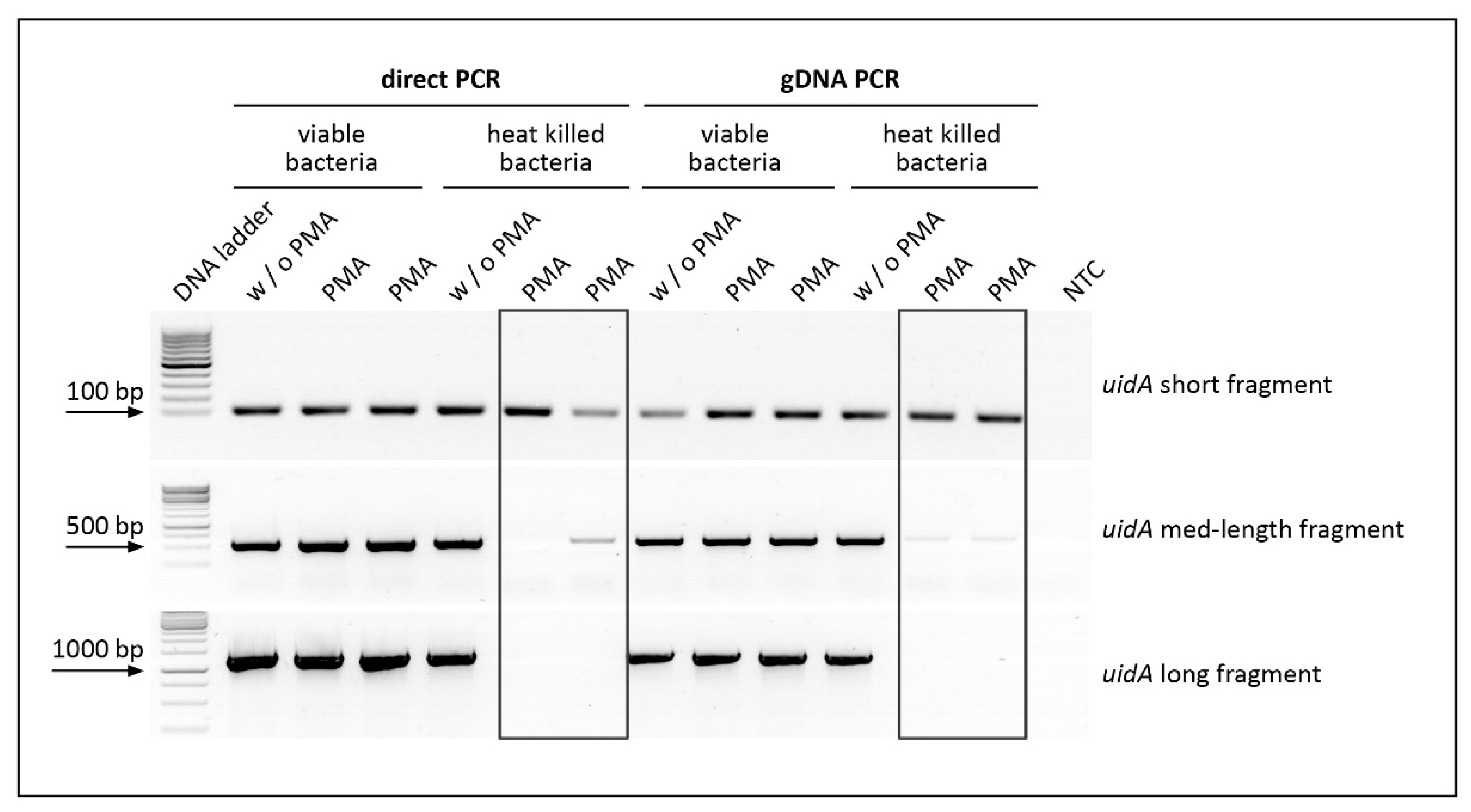
3.2. Impact of Amplicon Length on PMA Pretreatment of Heat Killed E. coli Cells

Although PMA pretreatment selects DNA templates from viable bacteria, one major drawback of this technique remains: the exclusion of heat killed cell signals can be incomplete, leading to false-positive results. Consequent overestimation of the viable cell population can occur by choosing a too short amplicon length and is problematic for reliable pathogen detection. Therefore, in preliminary experiments, the impacts of fragment length and the concentration of PMA were investigated to reliably amplify DNA from viable E. coli and exclude heat killed bacteria (data not shown). Optimal PMA concentrations were determined by testing a range of different PMA concentrations (10–100 µM) on 1 × 10⁸ viable and heat killed bacteria by amplifying a 1.5 kb fragment of the 16S ribosomal DNA (rDNA). The usage of 10 µM PMA resulted in effective suppression of the heat killed cell signal without affecting viable cells.
To determine the influence of amplicon size on suppression of the heat killed cell signal, uidA DNA fragments from viable and heat killed bacteria were amplified using three different primer pairs (uidA short fragment 96 bp, uidA medium-length fragment 556 bp and uidA long fragment 1370 bp). Resulting DNA fragments were analysed on an analytical agarose gel (Figure 3).
Treatment with the photoactive dye PMA followed by amplification of short (96 bp) and medium-length DNA fragments (556 bp) resulted in incomplete signal inhibition for heat killed bacteria. As seen in Figure 3, there are distinct bands for the short fragments, and weak bands remain for the medium-length fragments. Only PCR amplification of the long fragment (1.37 kb) entirely suppressed the heat killed cell signal after PMA treatment, which is vital for effective detection of viable bacterial cells. Thus, optimal amplicon size resulted in the suppression of the heat killed cell signal without affecting viable cells.

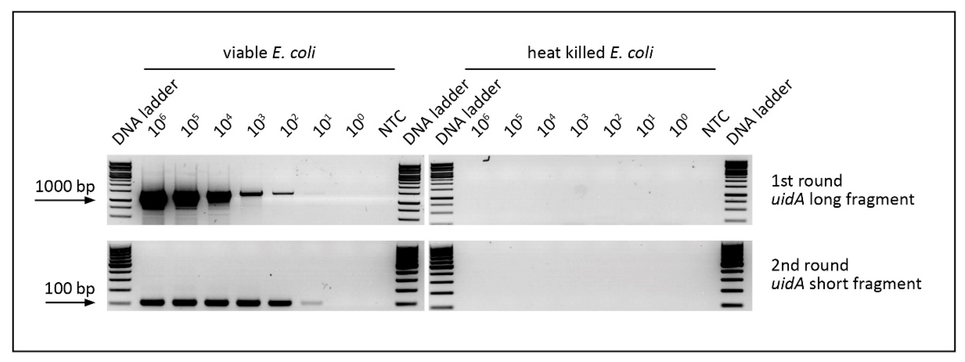
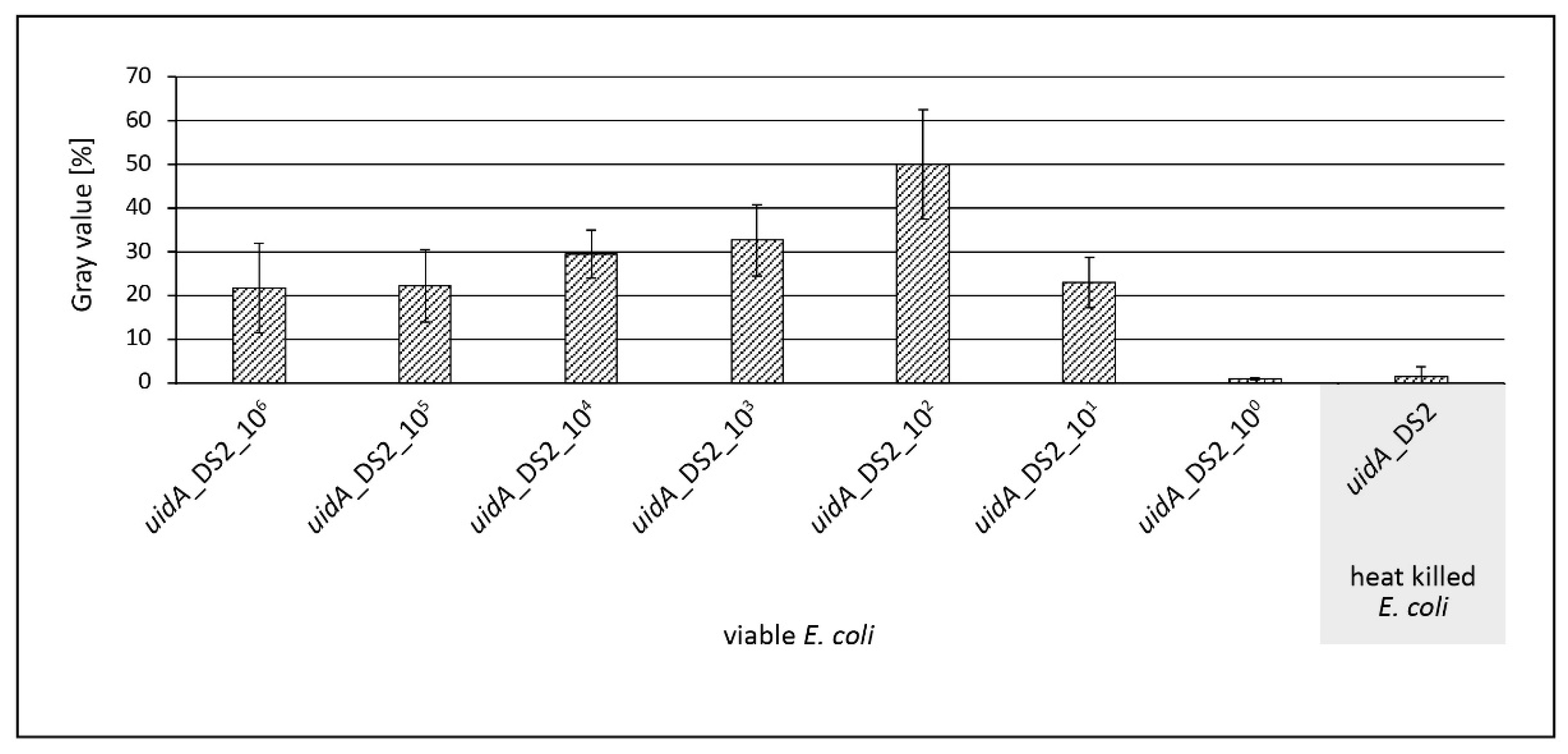
3.3. Evaluation of v-PCR

Since the efficiency of v-PCR was limited to longer DNA fragments, a nested PCR approach was necessary to create short PCR products that allow for subsequent detection by microarray-based DNA hybridization. In the first round of PCR, a 1.37 kb fragment of the uidA gene was amplified by direct v-PCR. In the subsequent second round of PCR, a 96 bp fragment of the target uidA gene was generated. First, optimal conditions for the nested PCR were elucidated (Supplement Figure S1). It was determined that 1 µL of a 1:100 dilution of the first round PCR product was optimal as a template nucleic acid in the following second round PCR approach.
To verify the limit of detection (LOD) for nested PCR, different amounts of E. coli cells were prepared, treated with PMA, and uidA fragments of DNA from viable and heat killed bacteria were subsequently amplified by nested PCR. As depicted in Figure 4, amplification was successful for up to 10 viable bacteria per PCR run (20 µL).
To evaluate the newly designed primers and capture probes for the E. coli uidA gene, genomic DNA of other relevant pathogens in drinking water was used for nested PCR amplification (Figure 5). Only genomic DNA from E. coli served as a template for the amplification of uidA fragments. No amplicons were generated using DNA extracted from Enterococcus faecalis (E. faecalis), Pseudomonas aeruginosa (P. aeruginosa), Clostridium perfrigens (C. perfrigens), Yersinia enterocolitica (Y. enterocolitica), Legionella pneumophila (L. pneumophila) or Bacillus subtilis (B. subtilis). The last mentioned is not a relevant water pathogen but served as negative control.
The next step in the analytical chain is verification of amplified PCR products by microarray-based DNA hybridization on novel solid support substrates. The mentioned substrates have a spoon-shaped format for easy handling performance (Figure 6).

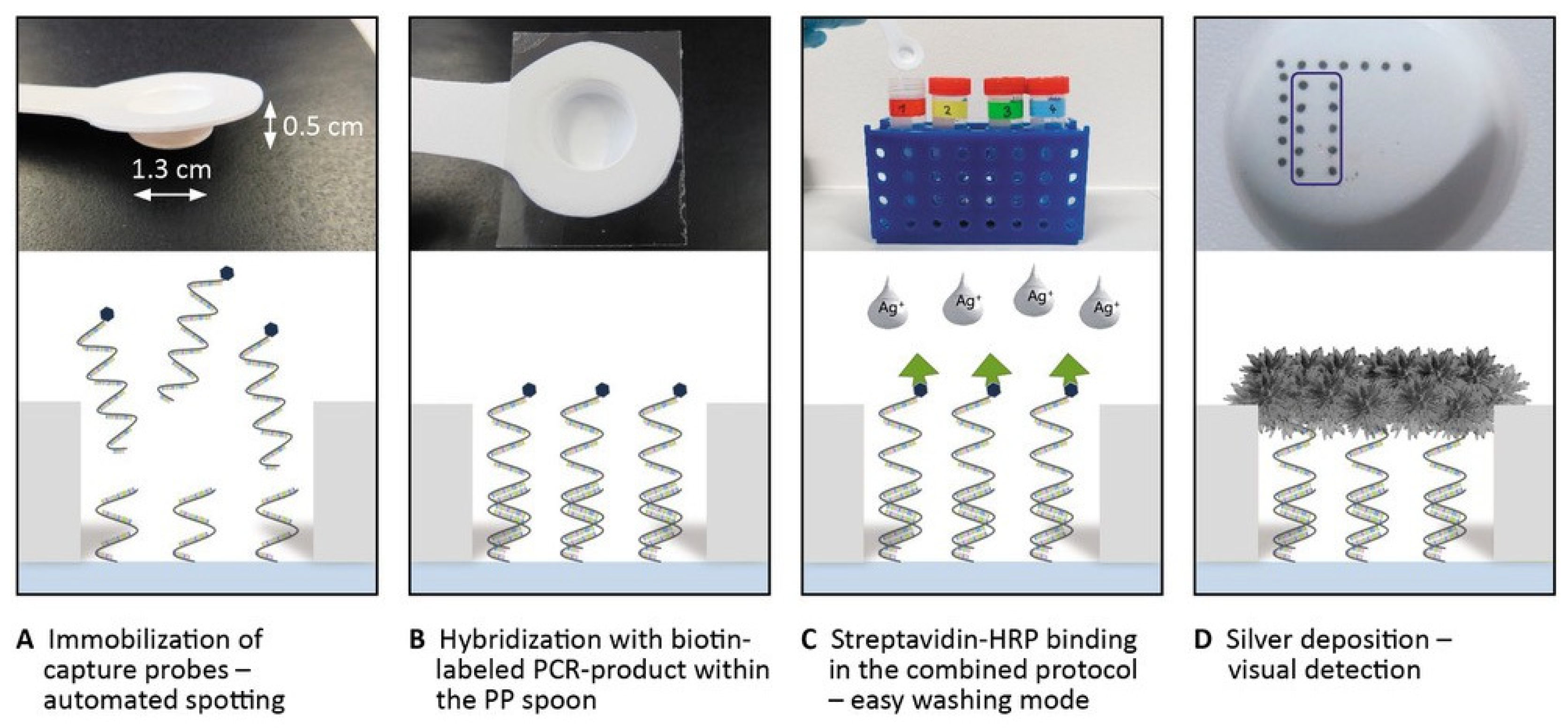
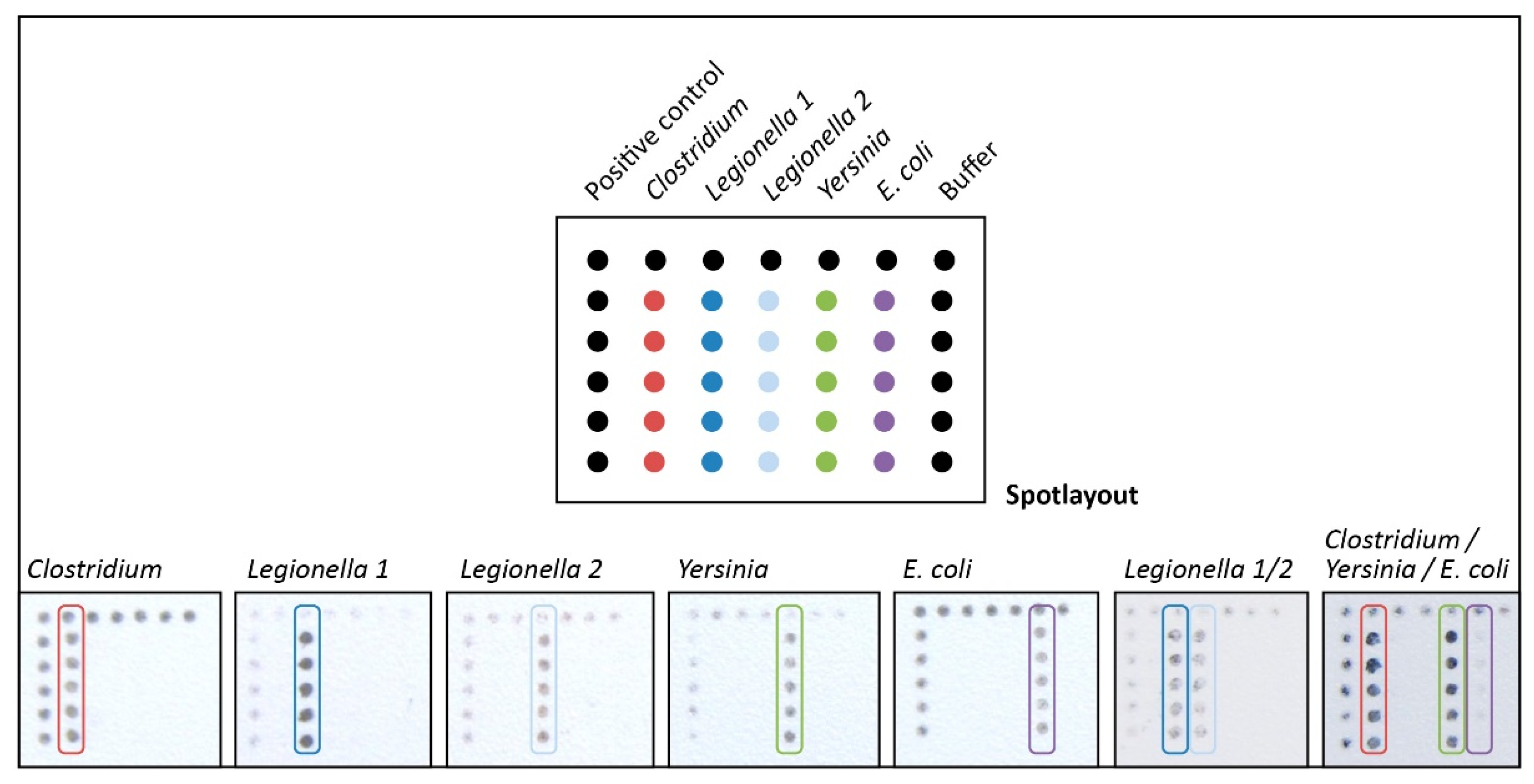
3.4. Microarray-Based DNA Detection on a Novel Designed Spoon-Shaped Substrate

An innovative DNA hybridization assay was created that enabled reliable and specific identification of E. coli separate from other relevant water pathogens due to immobilized capture probes based on the uidA gene. First, specific capture probes for the indicator organism E. coli were designed that hybridize with sequence segments within the uidA gene (Table 1). These capture probes were spotted in a microarray format (five replicate pattern for each capture probe) on novel easy-to-handle PP spoons.
Prior to DNA hybridization, conventional heat denaturation of dsPCR product was conducted (5 min) to generate single-stranded PCR products. The presence of ssDNA is a crucial and limiting factor for efficient DNA hybridization [29]. The well-established hybridization protocol was improved to significantly shorten the assay time. To this end, DNA hybridization and enzyme binding were combined. Therefore, different buffers and incubation intervals had to be tested and validated (data not shown). We successfully established a protocol that reduces the hands-on-time to only 30 min. Moreover, identification of E. coli was realizable using a simple visual readout of black silver spots on the microarray. In accordance with PCR product levels elucidated by analytical agarose gels, microarray detection allowed for the quantification of 10 viable E. coli cells per PCR run (Figure 7).
Regarding chip-based analysis of many relevant water pathogens, additional capture probes of appropriate bacteria were investigated. Moldiax GmbH provided those and the required PCR primers (Table 1). Figure 8 illustrates the reliable detection of three further relevant indicators in drinking water, namely Clostridium, Legionella, and Yersinia. In addition to the particular detection of a single species, we also mixed PCR fragments of various pathogens and successfully identified them on the microarray.

4. Discussion

Within this study, a model system comprising the complete process for the reliable and rapid detection of viable E. coli cells is highlighted, beginning from DNA extraction to on-chip identification of the bacteria by naked eye. The optimized protocol enabled the reduction of the total assay time to approximately five hours.
The focus was on the reliable detection of viable E. coli cells by discriminating between living and heat killed bacteria. Due to their higher potential health risk, viable bacterial cells are of great interest for water monitoring. Therefore, v-PCR was performed, which allows for thermal amplification of genomic DNA fragments only from viable bacterial cells possessing an intact cell membrane. PMA serves as a photoreactive and cell membrane-impermeable dye, which selectively and covalently binds to accessible double-stranded DNA (dsDNA) released by heat killed bacterial cells with compromised membranes. As a consequence of this PMA pretreatment step, the PMA-modified DNA cannot be amplified by PCR. Thus, only genomic DNA from viable cells serves as a template for thermal amplification [7,24,34]. Nonviable/dead bacteria were generated by heat exposure at 70 °C for 10 min. Other inactivation conditions were not considered at this stage of investigation.
For the illumination of samples, an innovative and cost-minimized LED photolysis device was developed in-house (Figure 2). The device uses LED lights with 470 nm emission for efficient activation of PMA. The price for all components was less than 100€.
To avoid a time-consuming gDNA extraction, isolation and purification protocol, direct PCR was performed. For this purpose, bacterial cells were lysed by heat for 5 min and directly applied in the amplification reaction. Direct PCR is increasingly utilized for clinical, forensic, agricultural and genetic applications since it saves time and considerably reduces costs by lowering the number of necessary chemicals [35].
Some studies have elucidated that treatment of heat killed bacterial cells with PMA does not completely suppress the amplification signal [12,36,37,38]. For that reason, the effect of the fragment length on PMA treatment was evaluated to target only viable E. coli cells in drinking water samples to suppress interference from heat killed bacteria. The results confirmed that efficient PMA treatment followed by DNA amplification of short or medium-length uidA fragments did not entirely suppress the signal from heat killed cells (Figure 3). Only long amplicons led to complete signal suppression of heat killed E. coli cells. The results indicate that fragment length is a crucial factor in v-PCR design. For efficient detection of only relevant viable cells, it is necessary to amplify a long PCR fragment to exclude dead bacteria. These findings for E. coli are in accordance with previous studies in different organisms [15,36,39,40]. Since the operation of v-PCR was limited to long DNA fragments, nested PCR was necessary to generate short amplicons for subsequent specificity testing. Compared to standard PCR methods, nested PCR enhances the sensitivity of amplification by several orders of magnitude due to exploitation of the product from the first amplification round as a template [41]. In addition, nested PCR was favorable in the current study for preparing short amplicons for subsequent on-chip hybridization experiments. Using this approach, process time for hybridization was considerably reduced. By applying nested PCR, amplification was successful for up to 10 viable bacterial cells per PCR run (Figure 4).
In addition to E. coli, several other relevant pathogens in cold drinking water, E. faecalis, P. aeruginosa, C. perfrigens, and Y. enterocolitica, as well as L. pneumophila in warm water, have to be considered. Thus, the nested PCR primers for E. coli genomic DNA needed to be verified. In the nested PCR approach with template DNA in other than E. coli, no amplicon was generated (Figure 5). Therefore, suitability of the nested PCR primers for E. coli as a water contaminant was demonstrated.
Often underestimated but still a major requirement for the hybridization with complementary capture probes, is the generation of single-stranded DNA [29,42]. After PCR amplification, the PCR product is typically double-stranded. To realize subsequent successful DNA hybridization, it is crucial to generate ssDNA [29,42]. In the present study, heat denaturation of the short-nested PCR product (96 bp) was adequate for efficient hybridization between target and capture DNA molecules. No elaborate post-amplification treatment, such as lambda exonuclease incubation or magnetic bead-based separation, was necessary to generate single-stranded DNA. In addition, heat denaturation favored the on-site operation purpose and additionally reduced the risk for contamination.
To further verify the functionality and sequence-specificity of the generated amplicons, a rapid and easy-to-handle on-chip hybridization assay was performed. First, specific capture probes for the indicator organism E. coli were designed that hybridized within the uidA gene. This gene, encoding the intracellular enzyme β-D-glucuronidase, was chosen as target gene for PCR amplification since it is present in all E. coli and Shigella species but not in other coliform bacteria [43,44,45,46]. Therefore, it enables a selective detection of E. coli in water.
White polypropylene (PP) was used as a planar low-cost chip substrate. The major advantage of PP is that no prior chemical surface modification or plasma activation is necessary, and capture probes can be directly immobilized on the chip surface after cleaning [47,48]. Use of PP ensures consistent quality of the microarray surface without other environmental influences. Furthermore, the white color simplifies visual detection of the black silver spots in our experiments [47].
To avoid time-consuming and laborious washing procedures, PP chips were shaped as spoons (‘cavity with a stick’—Figure 6). This innovative design was selected to avoid evaporation of the hybridization solution from the microarray area due to covering the spoon cavity harboring the spotted microarray with PCR foil. The cavities enabled a targeted and permanent incubation of the PCR product with the immobilized capture probes. Moreover, the spoon shape enabled an easy-to-manage, rapid washing mode. Instead of laborious error-prone handling steps, the spoons can be easily dipped into the washing solutions, one after another. The format allows a simple switch between dipping in buffers and incubation of the limited DNA hybridization solution for highly convenient assay performance.
To further minimize the detection time, a combined protocol, including both hybridization and enzyme immobilization, was examined [47]. Figure 7 and Figure 8 illustrate the limit of detection and specificity obtained through the on-chip hybridization experiments. Up to 10 viable E. coli cells per PCR run (volume = 20 µL) were detectable.
For each sample, results of the array-based hybridization were specific and no false positive signals were obtained at the positions of the non-complementary probes. These experiments indicate the rather easy adaption of our assay to additional bacteria species. So, the main advantage of the microarray-based detection relies in the parallel testing of broader bacterial panels, which will be an important part of ongoing investigations. Further species of interest are for example in the group of legionella, salmonella or Vancomycin-resistant enterococci.
Due to the increasing demand for rapid, culture-independent molecular detection methods, a plethora of multiplex PCR detection systems for E. coli and total coliform bacteria have been developed during the last decade [49,50]. Recently, Krapft et al. revealed a clear correlation between viable cell counts and real-time quantitative PCR (qPCR) data [51]. Nevertheless, only intercalating dyes allow differentiation between viable and dead bacteria cells. Therefore, coupling of PMA with quantitative amplification methods, such as qPCR or quantitative loop-mediated isothermal amplification (qLAMP), was recently highlighted [52,53,54]. Salam et al. combined PMA with quantitative PCR (PMA-qPCR assay) for the detection and quantification of viable E. faecalis [55]. Duarte-Guevare et al. presented a microfluidic device for viable qPCR and qLAMP, with a limit of detection of 1.1 × 10³ colony forming unit (CFU)/mL for E. coli [53]. The approach presented herein allowed the detection of 10 viable E. coli cells per PCR run.

5. Conclusions

We report on the development of a model system for the sensitive, specific and reliable detection of living E. coli cells. By combining a direct, viability and nested PCR, a rapid, precise analysis of viable bacteria is realizable omitting time-consuming sample preparation steps. The subsequent specification of the targets was realized by on-chip DNA hybridization on innovative spoon-shaped substrates, enabling an easy visual signal readout. Due to a growing interest in user friendly and robust agriculture diagnostic tools for rapid and reliable identification of viable cells, we developed a solid assay design for the indicator organism E. coli. This detection system displays a robust foundation and can easily be adapted to additional targets.

Supplementary Materials

The following are available online at https://0-www-mdpi-com.brum.beds.ac.uk/article/10.3390/chemosensors9120357/s1, Figure S1: Analytical agarose gel showing the investigation of different parameters for optimizing nested PCR conditions; Figure S2: Spot layout for the capture probes.

Author Contributions

Conceptualization, S.P., K.W. (Karina Weber) and J.P.; methodology, A.R., L.L. and S.P.; validation, A.R., L.L. and S.P.; investigation, A.R., L.L., S.P., K.W. (Kristin Wessel), M.B. and N.G.; resources, K.W. (Kristin Wessel) and M.B.; writing—original draft preparation, L.L; writing—review and editing, L.L., S.P., K.W. (Karina Weber) and J.P.; visualization, L.L.; supervision, L.L., S.P., K.W. (Karina Weber) and J.P.; project administration, K.W. (Karina Weber); funding acquisition, K.W. (Karina Weber) and J.P. All authors have read and agreed to the published version of the manuscript.

Funding

Funding of the research projects ‘EXASENS’ (13N13856) and ‘InfectoGnostics’ (13GW0096F) by the Federal Ministry of Education and Research, Germany and by the Thüringer Aufbaubank for the research project ’RowDix’ (2013FE9083) are gratefully acknowledged.

Institutional Review Board Statement

Not applicable.

Informed Consent Statement

Not applicable.

Conflicts of Interest

The authors declare no conflict of interest.

References

  1. World Health Organization. Progress on Drinking Water, Sanitation and Hygiene: 2017 Update and SDG Baselines; World Health Organization: Geneva, Switzerland, 2017. [Google Scholar]
  2. Ailes, E.; Budge, P.; Shankar, M.; Collier, S.; Brinton, W.; Cronquist, A.; Chen, M.; Thornton, A.; Beach, M.J.; Brunkard, J.M. Economic and Health Impacts Associated with a Salmonella Typhimurium Drinking Water Outbreak−Alamosa, CO, 2008. PLoS ONE 2013, 8, e57439. [Google Scholar] [CrossRef]
  3. World Health Organization. A Global Overview of National Regulations and Standards for Drinking-Water Quality; World Health Organization: Geneva, Switzerland, 2018. [Google Scholar]
  4. Sarkar, R.; Prabhakar, A.T.; Manickam, S.; Selvapandian, D.; Raghava, M.V.; Kang, G.; Balraj, V. Epidemiological investigation of an outbreak of acute diarrhoeal disease using geographic information systems. Trans. R. Soc. Trop. Med. Hyg. 2007, 101, 587–593. [Google Scholar] [CrossRef] [PubMed]
  5. Edberg, S.; Rice, E.; Karlin, R.; Allen, M. Escherichia coli: The best biological drinking water indicator for public health protection. J. Appl. Microbiol. 2000, 88, 106S–116S. [Google Scholar] [CrossRef]
  6. Gibson, C.; Maritim, A.; Marion, J. Comparison of the ColiPlate™ Kit with Two Common E. coli Enumeration Methods for Water. Water 2021, 13, 1804. [Google Scholar] [CrossRef]
  7. Nogva, H.K.; Drømtorp, S.M.; Nissen, H.; Rudi, K. Ethidium Monoazide for DNA-Based Differentiation of Viable and Dead Bacteria by 5′-Nuclease PCR. Biotechniques 2003, 34, 804–813. [Google Scholar] [CrossRef] [PubMed]
  8. Bae, S.; Wuertz, S. Discrimination of Viable and Dead Fecal Bacteroidales Bacteria by Quantitative PCR with Propidium Monoazide. Appl. Environ. Microbiol. 2009, 75, 2940–2944. [Google Scholar] [CrossRef] [Green Version]
  9. Cawthorn, D.-M.; Witthuhn, R. Selective PCR detection of viableEnterobacter sakazakiicells utilizing propidium monoazide or ethidium bromide monoazide. J. Appl. Microbiol. 2008, 105, 1178–1185. [Google Scholar] [CrossRef]
  10. Delgado-Viscogliosi, P.; Solignac, L.; Delattre, J.-M. Viability PCR, a Culture-Independent Method for Rapid and Selective Quantification of Viable Legionella pneumophila Cells in Environmental Water Samples. Appl. Environ. Microbiol. 2009, 75, 3502–3512. [Google Scholar] [CrossRef] [Green Version]
  11. Nocker, A.; Mazza, A.; Masson, L.; Camper, A.K.; Brousseau, R. Selective detection of live bacteria combining propidium monoazide sample treatment with microarray technology. J. Microbiol. Methods 2009, 76, 253–261. [Google Scholar] [CrossRef] [PubMed] [Green Version]
  12. Pan, Y.; Breidt, F. Enumeration of Viable Listeria monocytogenes Cells by Real-Time PCR with Propidium Monoazide and Ethidium Monoazide in the Presence of Dead Cells. Appl. Environ. Microbiol. 2007, 73, 8028–8031. [Google Scholar] [CrossRef] [PubMed] [Green Version]
  13. Rudi, K.; Moen, B.; Drømtorp, S.M.; Holck, A.L. Use of Ethidium Monoazide and PCR in Combination for Quantification of Viable and Dead Cells in Complex Samples. Appl. Environ. Microbiol. 2005, 71, 1018–1024. [Google Scholar] [CrossRef] [Green Version]
  14. Soejima, T.; Iida, K.-I.; Qin, T.; Taniai, H.; Seki, M.; Yoshida, S.-I. Method to Detect Only Live Bacteria during PCR Amplification. J. Clin. Microbiol. 2008, 46, 2305–2313. [Google Scholar] [CrossRef] [Green Version]
  15. Soejima, T.; Schlitt-Dittrich, F.; Yoshida, S.-I. Polymerase chain reaction amplification length-dependent ethidium monoazide suppression power for heat-killed cells of Enterobacteriaceae. Anal. Biochem. 2011, 418, 37–43. [Google Scholar] [CrossRef]
  16. Agustí, G.; Codony, F.; Fittipaldi, M.; Adrados, B.; Morató, J. Viability Determination of Helicobacter pylori Using Propidium Monoazide Quantitative PCR. Helicobacter 2010, 15, 473–476. [Google Scholar] [CrossRef]
  17. Rawsthorne, H.; Dock, C.N.; Jaykus, L.A. PCR-Based Method Using Propidium Monoazide to Distinguish Viable from Nonviable Bacillus subtilis Spores. Appl. Environ. Microbiol. 2009, 75, 2936–2939. [Google Scholar] [CrossRef] [Green Version]
  18. Brescia, C.C.; Griffin, S.M.; Ware, M.W.; Varughese, E.A.; Egorov, A.I.; Villegas, E.N. Cryptosporidium Propidium Monoazide-PCR, a Molecular Biology-Based Technique for Genotyping of Viable Cryptosporidium Oocysts. Appl. Environ. Microbiol. 2009, 75, 6856–6863. [Google Scholar] [CrossRef] [PubMed] [Green Version]
  19. Fittipaldi, M.; Rodriguez, N.J.P.; Adrados, B.; Agustí, G.; Peñuela, G.; Morató, J.; Codony, F. Discrimination of Viable Acanthamoeba castellani Trophozoites and Cysts by Propidium Monoazide Real-Time Polymerase Chain Reaction. J. Eukaryot. Microbiol. 2011, 58, 359–364. [Google Scholar] [CrossRef]
  20. Graiver, D.A.; Saunders, S.E.; Topliff, C.L.; Kelling, C.L.; Bartelt-Hunt, S.L. Ethidium monoazide does not inhibit RT-PCR amplification of nonviable avian influenza RNA. J. Virol. Methods 2010, 164, 51–54. [Google Scholar] [CrossRef]
  21. Fittipaldi, M.; Rodriguez, N.J.P.; Codony, F.; Adrados, B.; Peñuela, G.A.; Morató, J. Discrimination of infectious bacteriophage T4 virus by propidium monoazide real-time PCR. J. Virol. Methods 2010, 168, 228–232. [Google Scholar] [CrossRef]
  22. Sánchez, G.; Elizaquível, P.; Aznar, R. Discrimination of Infectious Hepatitis A Viruses by Propidium Monoazide Real-Time RT-PCR. Food Environ. Virol. 2012, 4, 21–25. [Google Scholar] [CrossRef]
  23. Vesper, S.; McKinstry, C.; Hartmann, C.; Neace, M.; Yoder, S.; Vesper, A. Quantifying fungal viability in air and water samples using quantitative PCR after treatment with propidium monoazide (PMA). J. Microbiol. Methods 2008, 72, 180–184. [Google Scholar] [CrossRef]
  24. Nocker, A.; Cheung, C.-Y.; Camper, A.K. Comparison of propidium monoazide with ethidium monoazide for differentiation of live vs. dead bacteria by selective removal of DNA from dead cells. J. Microbiol. Methods 2006, 67, 310–320. [Google Scholar] [CrossRef]
  25. Loaiza, Ó.A.; Campuzano, S.; Pedrero, M.; García, P.; Pingarrón, J.M. Ultrasensitive detection of coliforms by means of direct asymmetric PCR combined with disposable magnetic amperometric genosensors. Analyst 2009, 134, 34–37. [Google Scholar] [CrossRef]
  26. Maheux, A.F.; Boudreau, D.K.; Bisson, M.-A.; Dion-Dupont, V.; Bouchard, S.; Nkuranga, M.; Bergeron, M.G.; Rodriguez, M.J. Molecular Method for Detection of Total Coliforms in Drinking Water Samples. Appl. Environ. Microbiol. 2014, 80, 4074–4084. [Google Scholar] [CrossRef] [PubMed] [Green Version]
  27. Christensen, T.; Trabbic-Carlson, K.; Liu, W.; Chilkoti, A. Purification of recombinant proteins from Escherichia coli at low expression levels by inverse transition cycling. Anal. Biochem. 2007, 360, 166–168. [Google Scholar] [CrossRef] [Green Version]
  28. Beyer, A.; Pollok, S.; Rudloff, A.; Cialla-May, D.; Weber, K.; Popp, J. Fast-Track, One-Step E. coli Detection: A Miniaturized Hydrogel Array Permits Specific Direct PCR and DNA Hybridization while Amplification. Macromol. Biosci. 2016, 16, 1325–1333. [Google Scholar] [CrossRef]
  29. Schwenkbier, L.; König, S.; Wagner, S.; Pollok, S.; Weber, J.; Hentschel, M.; Popp, J.; Werres, S.; Weber, K. On-site detection of Phytophthora spp.—single-stranded target DNA as the limiting factor to improve on-chip hybridization. Microchim. Acta 2014, 181, 1669–1679. [Google Scholar] [CrossRef]
  30. Schüler, T.; Nykytenko, A.; Csaki, A.; Möller, R.; Fritzsche, W.; Popp, J. UV cross-linking of unmodified DNA on glass surfaces. Anal. Bioanal. Chem. 2009, 395, 1097–1105. [Google Scholar] [CrossRef] [PubMed]
  31. Schüler, T.; Asmus, T.; Fritzsche, W.; Möller, R. Screen printing as cost-efficient fabrication method for DNA-chips with electrical readout for detection of viral DNA. Biosens. Bioelectron. 2009, 24, 2077–2084. [Google Scholar] [CrossRef] [PubMed]
  32. Schüler, T.; Kretschmer, R.; Jessing, S.; Urban, M.; Fritzsche, W.; Möller, R.; Popp, J. A disposable and cost efficient microfluidic device for the rapid chip-based electrical detection of DNA. Biosens. Bioelectron. 2009, 25, 15–21. [Google Scholar] [CrossRef] [PubMed]
  33. Seise, B.; Brinker, A.; Kretschmer, R.; Schwarz, M.; Rudolph, B.; Kaulfuß, T.; Urban, M.; Henkel, T.; Popp, J.; Möller, R. Chip-based detection system for the on-site analysis of animal diseases. Eng. Life Sci. 2011, 11, 148–156. [Google Scholar] [CrossRef]
  34. Fittipaldi, M.; Nocker, A.; Codony, F. Progress in understanding preferential detection of live cells using viability dyes in combination with DNA amplification. J. Microbiol. Methods 2012, 91, 276–289. [Google Scholar] [CrossRef] [PubMed]
  35. Vondrakova, L.; Turonova, H.; Scholtz, V.; Pazlarova, J.; Demnerova, K. Impact of various killing methods on EMA/PMA-qPCR efficacy. Food Control. 2018, 85, 23–28. [Google Scholar] [CrossRef]
  36. Contreras, P.J.; Urrutia, H.; Sossa, K.; Nocker, A. Effect of PCR amplicon length on suppressing signals from membrane-compromised cells by propidium monoazide treatment. J. Microbiol. Methods 2011, 87, 89–95. [Google Scholar] [CrossRef]
  37. Nocker, A.; Sossa-Fernandez, P.; Burr, M.D.; Camper, A.K. Use of Propidium Monoazide for Live/Dead Distinction in Microbial Ecology. Appl. Environ. Microbiol. 2007, 73, 5111–5117. [Google Scholar] [CrossRef] [Green Version]
  38. Kralik, P.; Nocker, A.; Pavlik, I. Mycobacterium avium subsp. paratuberculosis viability determination using F57 quantitative PCR in combination with propidium monoazide treatment. Int. J. Food Microbiol. 2010, 141, S80–S86. [Google Scholar] [CrossRef]
  39. Banihashemi, A.; Van Dyke, M.; Huck, P. Long-amplicon propidium monoazide-PCR enumeration assay to detect viable Campylobacter and Salmonella. J. Appl. Microbiol. 2012, 113, 863–873. [Google Scholar] [CrossRef] [PubMed]
  40. Luo, J.-F.; Lin, W.-T.; Guo, Y. Method to detect only viable cells in microbial ecology. Appl. Microbiol. Biotechnol. 2010, 86, 377–384. [Google Scholar] [CrossRef]
  41. Juck, D.; Ingram, J.; Prévost, M.; Coallier, J.; Greer, C. Nested PCR protocol for the rapid detection of Escherichia coli in potable water. Can. J. Microbiol. 1996, 42, 862–866. [Google Scholar] [CrossRef] [PubMed]
  42. Brinker, A.; Schulze, H.; Bachmann, T.; Möller, R. Lambda exonuclease pre-treatment for improved DNA-chip performance depends on the relative probe-target position. Biosens. Bioelectron. 2010, 26, 898–902. [Google Scholar] [CrossRef]
  43. Farnleitner, A.H.; Kreuzinger, N.; Kavka, G.G.; Grillenberger, S.; Rath, J.; Mach, R.L. Simultaneous Detection and Differentiation of Escherichia coli Populations from Environmental Freshwaters by Means of Sequence Variations in a Fragment of the β- d -Glucuronidase Gene. Appl. Environ. Microbiol. 2000, 66, 1340–1346. [Google Scholar] [CrossRef] [PubMed] [Green Version]
  44. Khan, I.U.; Gannon, V.; Kent, R.; Koning, W.; Lapen, D.R.; Miller, J.; Neumann, N.; Phillips, R.; Robertson, W.; Topp, E.; et al. Development of a rapid quantitative PCR assay for direct detection and quantification of culturable and non-culturable Escherichia coli from agriculture watersheds. J. Microbiol. Methods 2007, 69, 480–488. [Google Scholar] [CrossRef]
  45. Maheux, A.F.; Picard, F.J.; Boissinot, M.; Bissonnette, L.; Paradis, S.; Bergeron, M.G. Analytical comparison of nine PCR primer sets designed to detect the presence of Escherichia coli/Shigella in water samples. Water Res. 2009, 43, 3019–3028. [Google Scholar] [CrossRef] [PubMed]
  46. Tryland, I.; Fiksdal, L. Enzyme characteristics of beta-D-galactosidase- and beta-D-glucuronidase-positive bacteria and their interference in rapid methods for detection of waterborne coliforms and Escherichia coli. Appl. Environ. Micro-Biol. 1998, 64, 1018–1023. [Google Scholar] [CrossRef] [Green Version]
  47. Schwenkbier, L.; Pollok, S.; Rudloff, A.; Sailer, S.; Cialla-May, D.; Weber, K.; Popp, J. Non-instrumented DNA isolation, amplification and microarray-based hybridization for a rapid on-site detection of devastating Phytophthora kernoviae. Analyst 2015, 140, 6610–6618. [Google Scholar] [CrossRef] [PubMed] [Green Version]
  48. Wünscher, S.; Seise, B.; Pretzel, D.; Pollok, S.; Perelaer, J.; Weber, K.; Popp, J.; Schubert, U.S. Chip-on-foil devices for DNA analysis based on inkjet-printed silver electrodes. Lab Chip 2014, 14, 392–401. [Google Scholar] [CrossRef] [Green Version]
  49. Beckman, A.K.; Ferrieri, P. Prospective Investigation of an Automated PCR/Nucleic Acid Microarray-Based Platform for Enteric Pathogen Testing. Lab. Med. 2019, 50, 390–395. [Google Scholar] [CrossRef]
  50. Brennecke, J.; Kraut, S.; Zwadlo, K.; Gandi, S.K.; Pritchard, D.; Templeton, K.; Bachmann, T. High-yield extraction of Escherichia coli RNA from human whole blood. J. Med。 Microbiol. 2017, 66, 301–311. [Google Scholar] [CrossRef]
  51. Krapf, T.; Kuhn, R.M.; Kauf, P.; Gantenbein-Demarchi, C.H.; Fieseler, L. Quantitative real-time PCR does not reliably detect single fecal indicator bacteria in drinking water. Water Supply 2016, 16, 1674–1682. [Google Scholar] [CrossRef] [Green Version]
  52. Ahmad, F.; Stedtfeld, R.D.; Waseem, H.; Williams, M.R.; Cupples, A.M.; Tiedje, J.M.; Hashsham, S.A. Most probable number—Loop mediated isothermal amplification (MPN-LAMP) for quantifying waterborne pathogens in <25 min. J. Microbiol. Methods 2017, 132, 27–33. [Google Scholar] [CrossRef] [PubMed] [Green Version]
  53. Duarte-Guevara, P.; Duarte-Guevara, C.; Ornob, A.; Bashir, R. On-chip PMA labeling of foodborne pathogenic bacteria for viable qPCR and qLAMP detection. Microfluid. Nanofluidics 2016, 20, 114. [Google Scholar] [CrossRef]
  54. Kibbee, R.J.; Örmeci, B. Development of a sensitive and false-positive free PMA-qPCR viability assay to quantify VBNC Escherichia coli and evaluate disinfection performance in wastewater effluent. J. Microbiol. Methods 2017, 132, 139–147. [Google Scholar] [CrossRef]
  55. Salam, K.W.; El-Fadel, M.; Barbour, E.K.; Saikaly, P.E. A propidium monoazide–quantitative PCR method for the detection and quantification of viable Enterococcus faecalis in large-volume samples of marine waters. Appl. Microbiol. Biotechnol. 2014, 98, 8707–8718. [Google Scholar] [CrossRef] [PubMed]
Figure 1. Scheme, illustrating individual steps of the v-PCR based detection approach.
Chemosensors 09 00357 g001
Figure 2. In-house constructed photoactivator for covalent binding of the photoactive dye PMA to accessible DNA from heat killed bacteria (A). The LED diodes allow for high or low blue light emission (A,B). Exposure of blue light to a PMA containing solution leads to a color change from yellow (only PMA without light) to red (PMA + light) (C).
Chemosensors 09 00357 g002
Figure 3. Impact of different fragment lengths on signal reduction of heat killed cells, displayed on an analytical agarose gel. Viable and heat killed E. coli cells were treated with or without (w/o) PMA. Direct PCR or classical PCR was performed using isolated gDNA as a template with three different primer pairs for three different target regions of the uidA gene (long 1370 bp, medium 556 bp, short 96 bp). Two independent PMA-treatments and PCR amplifications were performed for each condition. The No Template Control (NTC) includes all PCR reagents except for DNA.
Chemosensors 09 00357 g003
Figure 4. LOD tests applying direct viability nested PCR shown for viable and heat killed E. coli cells. Bacteria were treated with PMA, and DNA was subsequently amplified. PCR products from the first round (uidA long fragment; 1.37 kb) served as templates for the second PCR round (uidA short fragment; 96 bp). The No Template Control (NTC) for ensuring the purity of PCR reactions was also amplified by PCR. All PCR products were verified using analytical agarose gels.
Chemosensors 09 00357 g004
Figure 5. Primer test for amplification of the E. coli uidA gene by nested PCR displayed on an analytical agarose gel. The results of the second round are shown for E. coli, E. faecalis, P. aeruginosa, C. perfrigens, Y. enterocolitica, L. pneumophila, and B. subtilis.
Chemosensors 09 00357 g005
Figure 6. Schematic principle of the chip-based detection system using spoon-shaped PP chips: automated spotting of the capture probe array within the PP spoon cavity (A), hybridization of immobilized capture probes with biotin-labelled (blue dot) ssPCR products in the spoon cavity covered with PCR foil (B), interaction of the streptavidin horseradish peroxidase complex (green arrow) with biotin (C), and enzymatic silver deposition and visual detection (D).
Chemosensors 09 00357 g006
Figure 7. Grey value analysis of PP microarrays to verify the LOD of the newly designed assay. 1 × 10⁶–1 × 100 viable and 1 × 10⁶ heat killed E. coli cells were amplified by direct, nested v-PCR. PCR products from the second round (uidA short fragment; 96 bp) were heat denaturized to generate ssPCR products. For specific biomolecule interaction in the PP spoon cavity, DNA hybridization and enzyme binding were combined. Enzymatic silver deposition on the microarrays was performed to generate robust endpoint signals.
Chemosensors 09 00357 g007
Figure 8. Specific on-chip detection of E. coli, Legionella and Yersinia using a combined protocol encompassing DNA hybridization and enzyme binding. DNA of the pathogens was amplified by classical PCR using isolated gDNA as a template. For on-chip hybridization experiments, PCR products were used singly and mixed.
Chemosensors 09 00357 g008
Table 1. Primers and capture probes.
SpeciesPrimers and ProbesSequence 5′ → 3′Tm (°C)ModificationAmplicon Length
(bp)
Target Gene
E. coliuidA_long_FATT TGA AGC CGA TGT CAC GC60.01 1.37 kbpuidA
uidA_long_RTCC CTT TCT TGT TAC CGC CA59.71
uidA_mid_FCCG ACG AAA ACG GCA AGA AA60.155′-biotin556 bp
uidA_mid_RTCA GCG TAA GGG TAA TGC GA59.655′-phosphate
uidA_short_FAGT CAA CGG GGA AAC TCA GCA A56.435′-biotin96 bp
uidA_short_RGCA ATA CTC CAC ATC ACC ACG CTT57.795′-phosphate
process controlAGA ATC AAG GAG CAG ATG CTG AAA AAA 5′-NH2, 3′-biotin
uidA_DM1GTC CAC CCA GGT GTT CGG C 5′-NH2-C12
uidA_DM2TTT TTT TTT TTT TTT GTC CAC CCA GGT GTT CGG C 5′-NH2-C12
uidA_DS1TGG TTT TTG TCA CGC GCT ATC AGC 5′-NH2-C12
uidA_DS2TTT TTT TTT TTT TTT TGG TTT TTG TCA CGC GCT ATC AGC 5′-NH2-C12
Eco4TTT TTT TTT TTT TTT GAATCACAAAGTGGTAAGCG 5′-NH2-C12
Legionella 1forwardGAGGGTTGATAGGTTAAG525′-biotin167 bplegU
reverseCCAGGAATTTCACAGATA495′-phosphate
mdx74CTTAATCAACCACCTACGCAC685′-NH2-C12-Poly-T
Legionella 2forwardCCGATGCCACATCATTAGC575′-biotin150 bpmipN
reverseCCAATTGAGCGCCACTCATAG615′-phosphate
mdx84TGCCTTTAGCCATTGCTTCCG695′-NH2-C12-Poly-T
ClostridiumforwardATGATTGGGATTATGCAGCAAAGGT635′-biotin112 bpcpa
reverseCCAACTGATGGATCATTACCCTCTG665′-phosphate
mdx40TCTATAAATATATCCTGCTGTTCCTT675′-NH2-C12-Poly-T
YersiniaforwardAACAGTTTCAGGGCAGTTCAGTG635′-biotin128 bpyst
reverseAACATACATCGCAGCAATCCCAAT625′-phosphate
mdx43CGACACCAATAACCGCTGAG685′-NH2-C12-Poly-T
Publisher’s Note: MDPI stays neutral with regard to jurisdictional claims in published maps and institutional affiliations.

