Review

Managing Skin Ageing as a Modifiable Disorder—The Clinical Application of Nourella® Dual Approach Comprising a Nano-Encapsulated Retinoid, Retilex-A® and a Skin Proteoglycan Replacement Therapy, Vercilex®

by
Jan Wadstein
1,*,
Israel Sánchez Alvarez
2 and
Lidia Bernal López
3
1
Research and Development, Wadlund A/S, 21147 Malmö, Sweden
2
Dermatology Clinic, Azcapotzalco Centro, Azcapotzalco, Ciudad de Mexico 02000, Mexico
3
Dermatology Clinic, Letran Valle, Benito Juarez, Ciudad de Mexico 03650, Mexico
*
Author to whom correspondence should be addressed.
Submission received: 2 February 2022 / Revised: 8 March 2022 / Accepted: 11 March 2022 / Published: 15 March 2022
(This article belongs to the Special Issue Novel Cosmetic Ingredients, Formulations and Devices)

Abstract

:
Skin ageing is a progressive, but modifiable, multi-factorial disorder that involves all the skin’s tissues. Due to its wide range of physiological and psychosocial complications, skin ageing requires rigorous clinical attention. In this review, we aim to encourage clinicians to consider skin ageing as a disorder and suggest a novel, dual approach to its clinical treatment. Topical retinoids and per-oral proteoglycans are promising, non-invasive, therapeutic modalities. To overcome the low bioavailability of conventional free retinoids, Nourella® cream with Retilex-A® (Pharma Medico, Aarhus, Denmark) was developed using a proprietary nano-encapsulation technology. The nano-encapsulation is a sophisticated ‘permeation/penetration enhancer’ that optimises topical drug delivery by increasing the surface availability and net absorption ratio. Treatment adherence is also improved by minimising skin irritation. Interventional evidence suggests the greater efficacy of Retilex-A® in improving skin thickness and elasticity compared with conventional free forms. It is also reported that the rejuvenating efficacy of Retilex-A® and tretinoin are comparable. Another skin anti-ageing approach is proteoglycan replacement therapy (PRT) with Vercilex®. Vercilex® in Nourella® tablet form has the potential to ameliorate proteoglycan dysmetabolism in aged skin by activating skin cells and improving collagen/elastin turnover. Replicated clinical trials evidenced that PRT can significantly enhance the density, elasticity and thickness of both intrinsically aged and photoaged skin. Evidently, Vercilex® and Retilex-A® share a range of bioactivities that underlie their synergistic activity, as observed in a clinical trial. Dual therapy with Nourella® tablets and cream produced greater effects on skin characteristics than monotherapy with each of the two treatments. In conclusion, Nourella® cream and tablets are safe and effective treatments for skin ageing; however, combining the two in a ‘dual skin rejuvenation system’ significantly improves treatment outcomes.

1. Chronological (Intrinsic) and Accelerated (Extrinsic) Skin Ageing

Ageing is a complex, multi-factorial and progressive process driven by both genetic programming and cumulative environmental damage. Detectable cellular ageing starts in cutaneous tissues somewhat earlier than in most other tissues, probably due to the fact that skin is exposed to environmental stressors and hazards. Visible signs of skin ageing gradually appear during the third decade of life onwards at a rate that accelerates with age [1]. Two interacting and overlapping processes of skin ageing are recognised: intrinsic (chronological) and extrinsic (accelerated or premature) skin ageing. These processes run in parallel in most areas of the skin. Premature skin ageing is estimated to affect as much as 83% of adults under the age of 55 [2].
Intrinsic aetiologies that lead to chronological skin ageing include cellular ageing, characterised by increased genomic instability, epigenetic defects, altered metabolism, shortening of telomeres, mutations of mitochondrial DNA, oxidative stress, genetic mutations, loss of cellular regenerative potential (Hayflick-Limit), and a reduced level of several hormones. The molecular and cellular mechanisms of ageing and ageing-associated diseases are exhaustively discussed elsewhere [3,4].
The rate and intensity of chronological processes can be accelerated by superimposed factors, such as ultraviolet (UV)-light exposure, tobacco smoking, infrared radiation, environmental pollution, psychological stress, and malnutrition. The major molecular mechanisms through which these factors accelerate the disintegration rate of skin components are presented in Figure 1. In clinic, chronologically aged skin is manifested by a deepened frown and wrinkle lines of the forehead, glabella and lateral periorbital area and increased intensity of creasing on the chin, upper lip, nasolabial folds, nasal flare, and platysma neck bands. The skin becomes conspicuously thinner, transparent, and fragile and age spots begin to spread and magnify over decades. Expressions of accelerated skin ageing (photoageing) largely depend on the Fitzpatrick skin type and the cumulative exposure to stressors [5].
The distinctive clinical symptoms of skin ageing are external presentations of the underlying structural and functional deteriorations that occur to all histological layers of the skin. The outermost layer, the epidermis, declines in thickness at about 6.4% per decade, and its turnover rate plummets to half during the eighth decade of life. Rete pegs at the epidermal–dermal junction gradually flatten, which contributes to wrinkle formation and leaves the skin less resistant to shearing forces. In the dermal parts, vascularity, cellularity, and the volume of extracellular matrix (ECM) decrease over time. Secondary to the lowered number/activity of fibroblasts, the turnover of collagen and elastin, and thus skin flexibility and elasticity, also decrease in aged skin. Vascular network deterioration leads to a considerable decline in cutaneous skin blood perfusion [1]. However, being overly exposed to UV light and other risk factors can extensively influence the course and path of skin histological alterations during ageing. For example, in sun-exposed areas, the epidermis is thicker with higher degrees of solar elastosis, perivascular inflammation and perifollicular fibrosis, whereas severe photodamage can elicit epidermal atrophy [6]. Characteristic histological changes of intrinsically and extrinsically aged skin and their effects on the biomechanical function of the skin were investigated in a comparative human study by Langton et al. [7]. According to their report, modest effacement of rete ridges and disorganisation of papillary dermal elastic fibres in intrinsically aged skin caused a significant decline in both the resilience and the elasticity of the skin. In comparison, the photo-exposed skin of both young and aged individuals exhibited exaggerated forms of these pathological alterations, in addition to severe truncation of microfibrils, a reduction in organised collagen, and the accumulation of amorphous elastin in the dermis. The higher intensity of structural deteriorations naturally resulted in more profound biomechanical declines in the photoaged areas of the skin [7]. In the following sections, we aim to provide a scientific rationale that positions skin ageing as a disorder, rather than a natural consequence of ageing, and suggest a novel dual approach to its clinical treatment.

2. Approaching Skin Ageing as a Modifiable Disorder

Whether a specific condition is recognised as a ‘disease/disorder’ is greatly influenced by several cultural and historical factors, as well as the existing level of scientific understanding of its pathophysiology and complications. Several conditions, such as osteoporosis and senile Alzheimer’s, that are incontrovertibly known as disorders at present, were in the past labelled as normal results of ageing [8]. Major geriatric conditions, such as congestive heart failure, chronic obstructive pulmonary disease, and chronic kidney disease, are in fact the end results of a chronological or accelerated decline in the function of a specific organ due to ageing. Surprisingly, degenerative changes to the skin are still considered by some as components of the ‘normal ageing process’; whereas skin ageing fully meets the accepted criteria for a ‘disorder’, which is a disruption of the normal arrangement or functioning of the body. In terms of the skin, as long as there is no reference range for each skin function in a given age range, no loss of function or disrupted structure at any age can be marked as normal. Skin ageing involves several parallel pathological mechanisms that produce considerable loss of function and clinically significant complications (see below). This is particularly true in the case of premature skin ageing (dermatoheliosis), with recognised underlying risk factors and a high risk of comorbidities [9]. During the past decade, it has become more common to refer to the chronic skin fragility syndrome of ageing as ‘dermatoporosis’, in order to encourage timely treatment and to provide a nosological tool for the classification of this under-recognised condition [10].
Skin ageing involves a significant and progressive decline in almost all the physiological functions of the skin, ranging from biochemical to neurosensory alterations. Aged skin is drier, with decreased lipid content and higher cutaneous PH; cell renewal, synthetic capacity and vascularisation also wane in aged skin, leading to impaired wound-healing capability. Adding this to the gradual deterioration of superficial neurosensory perception makes the aged skin exceedingly vulnerable to injury, which brings significant indirect risk of morbidity [11]. In addition to physical complications, skin ageing disorder can inflict a significant psychosocial burden on affected individuals. Research shows that the discoloured, blemished, and slack appearance of the aged skin can negatively affect one’s body-image and self-esteem, setting the stage for anxiety and depression. Skin ageing can also repel physical contact by others, which may end in social withdrawal and the disturbance of interpersonal relationships [12], with profound effects on overall quality of life. It can therefore be concluded that skin ageing qualifies for recognition as a ‘disorder’ that requires targeted medical attention. In the same manner as other senile disorders, the process of skin ageing does not follow a predetermined, invariable time course; instead, its underlying pathologies and presentations may be delayed and/or reversed by avoiding modifiable risk factors (e.g., unprotected sun exposure and smoking) [13] and using proper pharmaceutical intervention [14].
A rich variety of medicinal and cosmetic anti-ageing products have been introduced to the market, with variable efficacy profiles and action mechanisms [15]. The main conventional, non-invasive treatments are sunscreens, topical and systemic antioxidants, topical retinoids, topical and systemic glycosaminoglycans and proteoglycans. Experimental evidence indicates that despite their claims, few anti-ageing ingredients have the capacity to penetrate far enough into the dermis to ameliorate deep wrinkles [16]. Minimally invasive resurfacing techniques, such as chemical peeling, laser/radiofrequency tightening and filler injections and invasive surgical procedures are also used in selected cases [17]. However, the effective prevention of skin ageing disorder is a long-term endeavour that requires interventions that are easy to apply, skin friendly, well tolerated, affordable and devoid of side effects. Accordingly, topical retinoids [10] and oral proteoglycans appear to be among the most appropriate candidates for long-term anti-ageing therapy of the skin. In this paper, we strive to sketch an overview of these two successful approaches, which, in combination, form a targeted dual treatment known as the ‘Nourella® Skin Rejuvenation System’ (developed by Pharma Medico, Aarhus, Denmark).

3. Natural Retinoids as the ‘Gold Standard’ Treatment of Skin Ageing

Retinoids are a group of natural and synthetic vitamers that share either molecular or functional similarities with all-trans retinol (vitamin A). Topical formulations of retinoids have been in cosmetic and clinical use since the 1940s; however, their prominent anti-ageing effects were not scientifically reported until the early 1980s [18]. Since then, a growing body of clinical research has clearly demonstrated their ability to improve fine wrinkling and skin elasticity and lighten uneven skin pigmentation [19]; thus, natural retinoids have become the ’gold standard’ for both the prevention and treatment of skin ageing.
Retinol and its storage form, retinyl ester (e.g., retinyl palmitate and acetate), are the most abundant, naturally occurring retinoids present in the body. Retinol is a transport form and a precursor that is converted via a two-step oxidation process to its bioactive metabolites, retinoic acids. Through interaction with several cytoplasmic and nuclear receptors, retinoids regulate a range of vital molecular pathways and cellular activities, from embryogenesis to skin regeneration and maintenance. The skin is a major retinoid-responsive tissue, in which retinoic acid directly and indirectly regulates the expression of multiple key genes [20]. This multifarious, gene-level bioactivity is employed in the clinical prevention and management of the skin ageing disorder. According to available evidence [21,22,23,24,25,26], long-term topical treatment with natural retinoids promotes pervasive histopathological improvements in all layers of the skin, as listed below:
  • A significant rise in the epidermal thickness of the aged skin via the amplification of the proliferation of epidermal keratinocytes by several folds [21,22,25]
  • A marked upturn in the proliferation rate of dermal stromal cells, particularly in the papillary dermis, compared to placebo-treated skin [22]
  • Improvement of the dermoepidermal ECM microenvironment by two mechanisms. First, retinoids stimulate the activity of fibroblasts and boost the production of type-I and III collagen, fibronectin and tropoelastin in both chronologically aged and photoaged skin [21,22]; and second, these compounds can inhibit the expression of matrix metallopeptidases and thereby diminish the degradation of ECM components [26].
  • A considerable increase in the proliferation of dermal endothelial cells and the formation of blood vessels, thus improving the skin’s microcirculation and blood supply [22,23]
  • Efficient anti-inflammatory effects through suppressing the release of proinflammatory cytokines and the activity of leukocytes [24]
In conclusion, after a sufficient course of retinoid therapy, the skin becomes thicker, with improved fibroelastic properties, enhanced blood circulation and reduced inflammatory reactions. These cellular-level enhancements by natural retinoids give rise to the clinical improvements observed in short- and long-term controlled clinical trials, as detailed by Mukherjee et al. [21]. Based on published reports, 3 to 4 months of retinoid therapy can compact the stratum corneum and fade both fine and coarse wrinkles, making the skin appear rejuvenated. In addition, concentrations of glycosaminoglycans increase [27,28]. As evidenced by long-term trials, extended courses of retinoid application, i.e., for six months or more, result in higher degrees of improvement in skin wrinkling, which plateaus after around 10 months and is maintained afterwards [29]. However, it appears that for appreciable dermal-level improvement and the formation of new collagen, more than six months of retinoid treatment are required. It is of practical interest to note that in clinical studies, the skin condition of the participants continued to improve even after the cessation of active treatment [21]. Accordingly, both prescription (tretinoin, adapalene) and over-the-counter retinoids (retinol) are recommended as the first choice to treat and prevent skin aging [15].
At present, there are two commonly used natural retinoids approved for the management of skin ageing: all-trans retinol (and its derivatives) and tretinoin. Despite the fact that retinol is theoretically less potent than tretinoin, comparative clinical studies suggest that its efficiency at producing ‘retinoid-mediated histological changes’ is comparable with tretinoin. Of note, retinol and its derivatives induce much less skin irritation than tretinoin and are preferable in the long-term treatment of skin ageing [30,31].

3.1. Using Nano-Encapsulation to Enhance the Bioavailability and Safety of Retinoids

Despite their proven molecular efficacy, the ‘dose efficiency’ and ‘long-term patient compliance’ of topical free retinoids (the conventional form) is compromised by two major impediments. The first challenge is to penetrate deep enough into the skin to reach the primary action site of retinoids, i.e., the epidermal basal layer and upper papillary dermis, with a high density of cells and collagen fibres. Experiments by Roos et al. revealed that topically applied retinoids penetrate poorly into or through the skin. This is specifically true with gel-based formulations, which tend to trap active drug molecules [32]. Poor penetration causes around 80% of the applied active retinoid to remain on the skin surface [33]. The long-term entrapment of unprotected retinoid molecules on the skin surface exposes them to environmental factors, such as heat and UV radiation. Since retinoids are chemically and thermally unstable and sensitive to photoisomerization and degradation [34], a large share of the applied dosage is lost to degradation. This reduces the overall ‘dose efficiency’ and ‘bioavailability’ of the treatment. In order to overcome this critical shortcoming and improve the topical absorption of retinoids, several techniques have been attempted over decades, such as developing retinol derivatives [35] and chemical penetration enhancers. However, the rate of success with penetration enhancers has been limited owing to their tendency to irritate the skin and weak enhancement effects [36].
The second impediment that hampers the effective use of topical free retinoids is their common irritation side-effects. Retinoid-induced irritation is clinically similar to mildly irritant dermatitis and presents with excessive skin dryness, burning sensation, erythema, scaling and pruritus. Retinoid reaction is time- and dose-dependent and frequently occurs when using an effective dosage of either free retinol or tretinoin for extended periods of time. The induced release of pro-inflammatory cytokines, e.g., TNF-α and IL-8, is reported to be a triggering factor in retinoid reaction [37]. The likelihood of skin reactions is higher with conventional products containing a free form of retinoid since these products release their active ingredients in relatively high concentrations and within short periods of time. This unmodified release profile leads to a cycle of short-term overmedication followed by long-term undermedication, which maximises side-effects and undermines clinical efficacy [38]. It is conceivable that by transforming the release and absorption profile of retinoids into a more gradual and steady form, the overload of biological membranes can be attenuated, thereby minimising the release of cytokines and the likelihood of skin irritation.
An efficient, nanomolecular skin delivery system that works as a ‘permeation and penetration enhancer’ of the transdermal delivery of pharmaceuticals is nano-encapsulation with biomaterials. Examples of materials frequently used in biomedical industries are non-conjugated peptides, nanostructured lipid carriers, dendrimers, and cyclodextrins. Based upon this practical knowledge and on years of research and development, Retilex-A® has been developed through the proprietary technique of nano-encapsulation and optimised emulsification of retinyl ester, by scientists at Pharma Medico (Aarhus, Denmark). This novel complex is a next-generation topical retinoid with modified pharmacokinetic characteristics used as the main active ingredient in Nourella® anti-ageing cream (Pharma Medico, Aarhus, Denmark). This smart drug delivery system aims to overcome both of the aforementioned impediments of topical retinoid therapy of skin ageing. The improved efficacy and safety of Retilex-A® in Nourella® cream have been corroborated by controlled clinical trials (described in the next section).
There is enough evidence to establish that the specific form of nano-encapsulation used in the formulation of Retilex-A® enhances topical drug bioavailability and treatment efficacy by:
  • Increasing drug availability at the skin surface. It is known that in aqueous solutions, lipophilic molecules, e.g., retinyl esters, compete for a space in carrier cavity, which forms a dynamic equilibrium between encapsulated and free drug molecules. At the surface, retinoid molecules that partition from the carrier cavity and penetrate the lipophilic skin barrier are replaced by newly partitioned active molecules (a buffering effect), such that a fresh pool of free, intact retinoid molecules is continuously available for absorption while the rest of the pool is protected from environmental factors inside the nano-capsules (a protective effect). The significant action of nano-encapsulation in producing a controlled, constant drug release profile has previously been documented [39].
  • Augmenting retinoid net absorption ratio. Whether the result of improved availability or of an independent effect, nano-encapsulated retinyl ester exhibits augmented absorbability, as demonstrated by a comparative study on a model barrier system (Franz Diffusion Cell). The measurements demonstrated that retinoid molecules from Retilex-A® had a markedly higher penetration ratio into a skin model compared to a conventional, commercial formula in both water and isopropyl alcohol media [40].
  • Reducing skin irritation side-effects. As explained earlier in this section, it is well established that specific forms of nano-encapsulation can alleviate the skin irritation caused by chemicals. Clinical studies with various skin irritants have verified this integral benefit of nano-encapsulation [36].
In conclusion, nano-encapsulated Retilex-A® improves skin bioavailability and tolerance due to a diminished molecular disintegration rate, constant release profile, increased absorption ratio and reduced skin irritation compared to conventional free forms of retinoid.

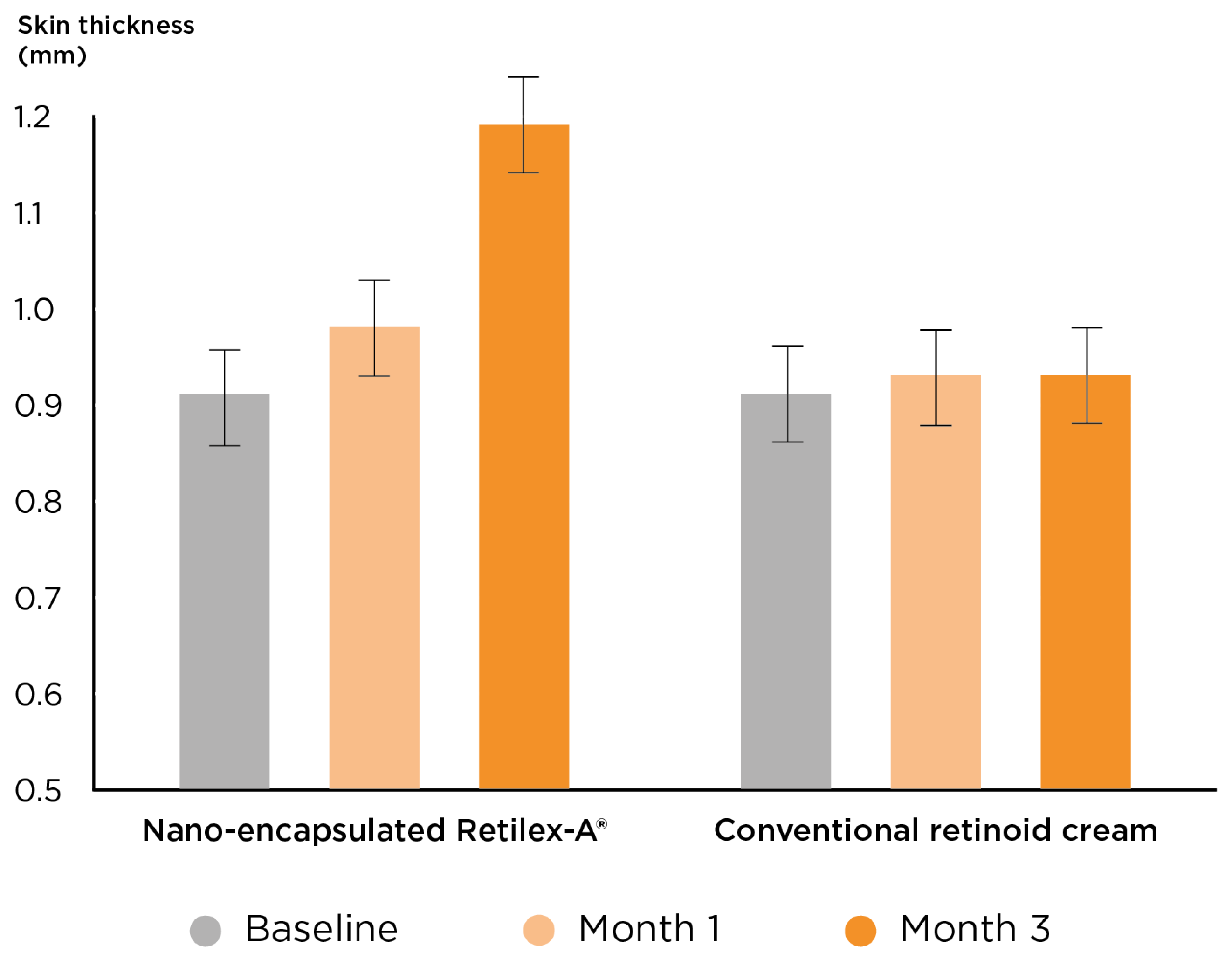
3.2. Supporting Evidence for the Anti-Ageing and Skin-Rejuvenating Efficacy of Retilex-A®

Since its development around the turn of the 21st century, Retilex-A® has been subjected to several clinical studies on subjects with chronological and premature skin ageing, with promising outcomes [31,41,42,43]. The role of nano-encapsulation in the anti-ageing efficacy characteristics of Retilex-A® was clinically studied in a double-blind clinical trial. Both encapsulated (Retilex-A®) and conventional test preparations contained 0.2% W/W of retinyl palmitate in an identical vehicle and were applied b.i.d. (in the morning and the evening) on a random side of the volar aspect of each participant’s forearm (age range = 40–60 years). The skin thickness and elasticity were objectively measured by ultrasonic dermal scanning. No topical treatment or emollient was used by the subjects shortly before performing the skin measurements. After just 12 weeks of treatment, RetileX-A® significantly improved both skin thickness and elasticity (+31% and +18%, respectively, p < 0.01), while the conventional free form induced negligible effects (+2% and +1%, respectively) (Figure 2). The participants’ self-evaluation of the treatment results on a visual analogue scale (VAS) also confirmed the objective assessments and showed a significant improvement only after using the nano-encapsulated RetileX-A® [41].
A follow-up evaluation revealed that as much as 25% of the therapeutic effects of RetileX-A® on skin thickness and 23% on skin elasticity were still present and measurable after one year of treatment cessation. This led the individuals treated with RetileX-A® to have significantly thicker (p < 0.05) and more flexible (p < 0.05) skin 12 months after the study [42]. Overall, this trial signifies that the specific nano-encapsulation technique in RetileX-A® can intensify and accelerate the anti-ageing effects of retinyl ester. A subsequent trial re-evaluated the clinical efficacy and safety of Nourella® with Retilex-A® and compared it with an equivalent dosage of tretinoin. Twenty females, with an average age of 51.4 years, participated in this clinical study. Each used both Nourella® and tretinoin on the volar aspect of their right or left forearm (chosen at random) b.i.d. for 3 months. Ultrasonic measurements of skin thickness and elasticity detected comparable significant increases in skin thickness and elasticity index at the endpoint in both groups. The treated individuals also judged both formulations to be equally effective, whereas the rate of side effects was much lower with RetileX-A® [31]. These findings suggest that, in the clinical treatment of skin ageing, nano-encapsulated retinyl ester (RetileX-A®) is as effective as the prescription medication tretinoin. However, more concrete conclusions require more evidence from larger clinical studies.
An unpublished clinical research project by Erling Thom and Allan Bertil Lassus [44], demonstrated the efficacy of RetileX-A® in treating premature skin ageing (photoageing). The study’s subjects were middle-aged volunteers with moderate-to-severe degrees of facial solar elastosis who applied Nourella® cream twice a day on affected parts of their skin for 12 weeks. Monthly clinical assessments pointed to a continuous improvement in skin hydration, wrinkling and mottling in the affected areas. Objective measurements consistently reported significant enhancements of 6%, 11% and 16% in lesional skin elasticity after 1, 2 and 3 months of Nourella® therapy, respectively. No clinically important side effects were reported during the course of this trial. These observations provide evidence for using Nourella® with Retilex-A® for the rejuvenation of chronologically and prematurely aged skin.

4. Proteoglycan Dysmetabolism in Ageing Skin

Proteoglycans are bioactive macromolecules with a multiplicity of integral mechanical and biological functions. Skin proteoglycans are highly bioactive and contribute to tissue hydration, resistance and resilience by forming super-molecular structures with matrix proteins and regulating fibrillogenesis. They also control cell behaviour and interactions and form a major biological reservoir for various cytokines and growth factors [45,46]. Over 40 different proteoglycans have been discovered, several of which are expressed in cutaneous tissues. Among the important skin proteoglycans, versican, decorin and biglycan are the most abundant [47,48]. Versican is a large chondroitin sulphate (CS) proteoglycan, which binds to and modifies several ECM components via its active domains. Versican co-localises with elastic fibres in the dermis and influences cell proliferation and migration. Decorin and Biglycan are small, leucine-rich proteoglycans with vital roles in modifying the organisation of the dermal matrix. These proteoglycans bind to several types of collagen and elastic fibre components and regulate their fibrillogenesis and interfibrillar distance [49,50]. Therefore, any disruption in their turnover or structure may significantly interrupt the homeostasis and structure of dermal tissues. This fact is best demonstrated by decorin/biglycan knockout mice models, which exhibit thin and fragile skin with reduced tensile strength, caused by irregular collagen fibrillogenesis [51,52].
The degradative transformation of proteoglycans is an inherent part of the pathogenesis of both intrinsic and extrinsic skin ageing disorders. Naturally, the synthesis of extracellular matrix proteins by senescent fibroblasts tends to reduce. Research findings show a considerable decline in the total amount of sulphated glycosaminoglycans in the dermis (in both sexes) and epidermis (only in women) of intrinsically aged skin [50]. Tzellos et al. reported lower levels of versican, decorin and biglycan mRNA in sun-protected aged skin compared to juvenile skin [53]. The relative composition of proteoglycans also undergoes radical changes as the skin ages. Observations evidence that concomitant to a decline in the proportion of large CS proteoglycans (e.g., versican), the proportion of small dermatan sulphate proteoglycans (e.g., decorin) increases during ageing [54]. Parallel with these quantitative deviations, the molecular characteristics of certain proteoglycans, i.e., the number and size of their side chains and cleavage products, are affected as a function of age [49,54]. This phenomenon is allegedly a by-product of the age-associated functional loss of glycosaminoglycan synthesising enzymes. The decline in the secretion of sex hormones may also play a role in the dysmetabolism of skin proteoglycans [49,50].
Proteoglycan dysmetabolism is presented differently in photo-aged skin that occurs together with the deposition of abnormal elastic tissue. The total amounts of dermal sulphated glycosaminoglycans and hyaluronic acid are elevated as a compensatory recovery response to the cumulative damage of overexposure to UV light. However, the reactions of different proteoglycans to photoageing are not identical [50]. For example, although versican, which co-distributes with elastic fibres, accumulates in the regions of solar elastosis, it does so with an abnormal distribution pattern. While glycans are normally distributed diffusely in the dermis, these are predominantly deposited on the abnormal elastotic material in affected areas. This abnormal localisation renders the glycans incapable of enhancing cell activities or serving as sources of hydration. Furthermore, similar to chronologically aged skin, the size and structure of glycosaminoglycans in sun-damaged skin is deviated, affecting their water-binding properties and ability to interact with other ECM components [55]. By contrast, the expression of decorin is greatly decreased in areas of solar elastosis [56]. These abnormalities in the expression, structure and distribution of skin proteoglycans are suggested to be primary factors that contribute to the development of accelerated skin ageing.

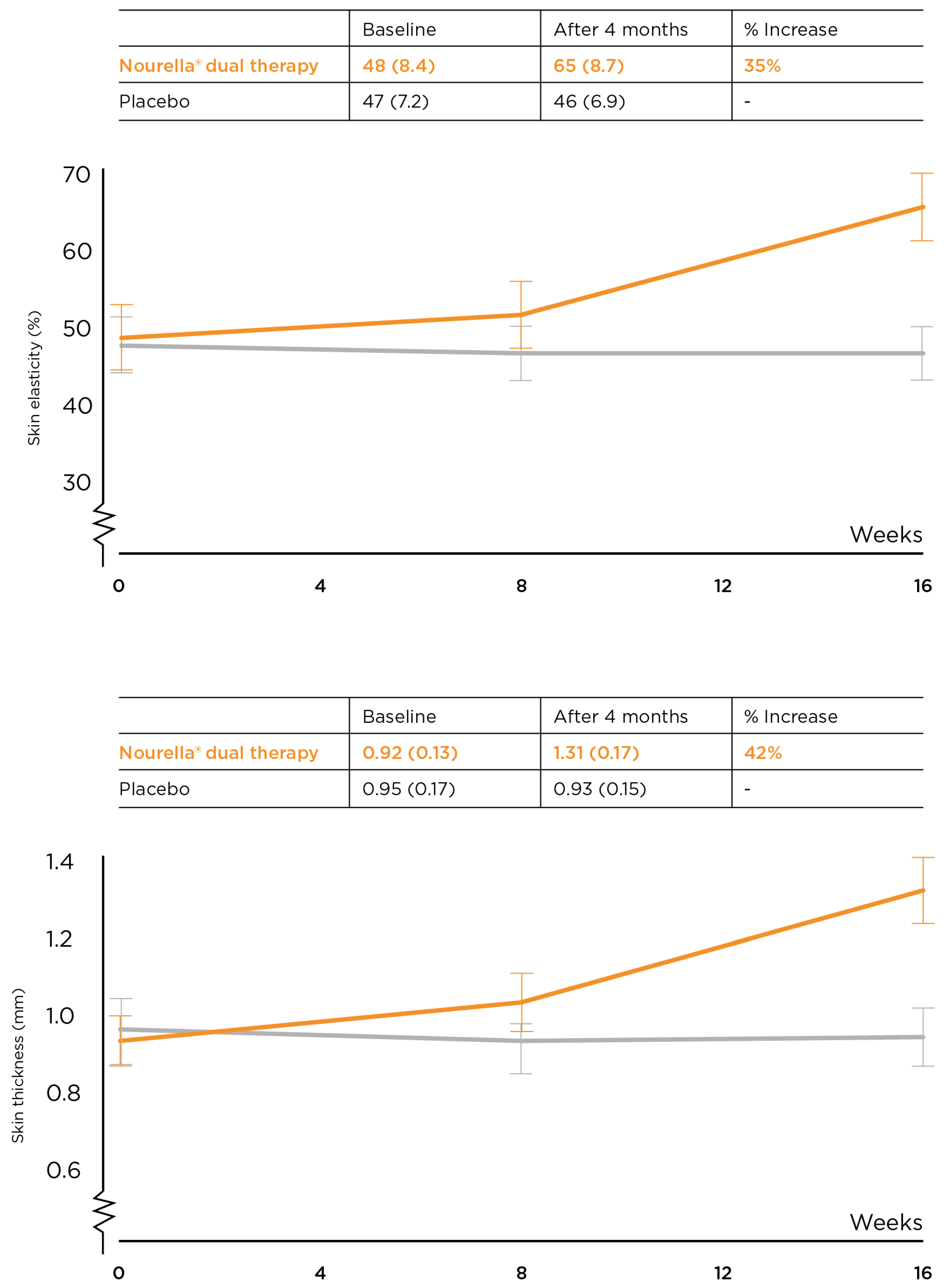
4.1. Proteoglycan Replacement Therapy (PRT) of Aged Skin Using Vercilex®

The oral administration of natural proteoglycans and glycosaminoglycans is an emerging therapeutic method that has been tried for the treatment of a variety of human disorders including, but not limited to, hair loss disorders [57], osteoarthritis [58] and skin psoriasis [59]. Pertaining to the integral role of ‘proteoglycan dysmetabolism’ in the pathogenesis of both chronological and accelerated skin ageing, the oral administration of specific bioactive proteoglycans is deemed to be a relevant anti-ageing approach. Certain proteoglycans and their moieties have proven oral bioavailability and the ability to reach peripheral tissues after oral ingestion. In vivo pharmacokinetic studies on other animals and humans have shown gastric acid survivability and gastrointestinal absorption of glycosaminoglycans, such as CS [60] and hyaluronic acid [61]. Evidence suggests that a portion of orally administered proteoglycans is absorbed intact via endocytosis, mostly in distal parts of the small intestine [62].
The concept of treating skin ageing with the oral administration of proteoglycans stems from the works of Wadstein et al. in the 1980s and early 1990s, which has since been developed into proteoglycan replacement therapy (PRT). Throughout the past few decades, several independent research groups have presented supporting evidence for the efficacy of treating the skin with natural proteoglycans of marine origin. Both in vitro and in vivo research confirmed the enhancement of cellular function and improvement of skin integrity with this treatment approach. However, the proteoglycan-based treatments currently on the market are of diverse composition, origin and, conceivably, efficacy levels. Some, such as salmon nasal cartilage extract, have a simple composition, mainly of aggrecan, while Nourella® tablets with Vercilex® (Pharma Medico, Aarhus, Denmark) have a more complex, multi-molecular nature, rich in several skin-specific proteoglycans. Sano et al. reported that a crude salmon nasal cartilage extract (mainly aggrecan) amplified the proliferation of human dermal fibroblasts through the activation of Erk1/2. This growth-stimulating effect of aggrecan is presumably related to its epidermal growth factor-like module [63]. Another study confirmed that this complex has the ability to cause prominent the upregulation of both the proliferation and migration activity of skin fibroblasts [64]. Other lines of research propose that certain proteoglycans and their moieties provide cell protection against oxidative stress. For example, CS retains an ability to reduce the generation of free radicals, while aggregating CS proteoglycans with negatively charged side chains can contribute to reducing local oxidative stress by scavenging and binding redox-active iron [65]. The importance of this feature can be clearly understood in light of the fact that oxidative stress has determining roles in both intrinsic [66] and extrinsic skin ageing [67]. The in vivo therapeutic potential of oral proteoglycan therapy was studied in a model of accelerated skin ageing. Treatment with either a proteoglycan extract (composed mainly of aggrecan) or CS was started 3 weeks prior to skin UV irradiation and continued for 11 weeks thereafter. The proteoglycan treatment appeared to mitigate various photoaging effects of UV-B irradiation, including skin erythema, increased water loss and decreased skin hydration levels. Compared with the controls, a dose-dependent suppressing effect on epidermal and dermal hypertrophy was also observed, while CS alone had no effect [68]. These significant findings were subsequently verified in humans through a randomised, controlled study. A two-week treatment course could improve skin elasticity, periocular wrinkling, conspicuous pores and blotches and induce positive corneum structural changes in both men and women [69]. Since Vercilex® is a multi-molecular extract rich in several bioactive proteoglycans, such as versican, decorin and biglycan, its level and spectrum of clinical efficacy are higher and wider compared to the more basic aggrecan-based cartilage extracts (see below).
Nourella® tablets are a per-oral anti-ageing treatment for both chronological and accelerated skin ageing. The primary active ingredient of Nourella® tablets is Vercilex®, a proteoglycan complex, refined and optimised over decades to prevent and ameliorate the degenerative changes associated with ageing in human skin. One of its crude initial versions (branded, at the time, as Imedeen) was used by Lassus et al. to treat premature skin ageing in an open-label pilot followed by a controlled, blinded trial. Subjects with photoaged skin were treated with 500 mg/day for 3 months. The majority of the participants experienced improvements in wrinkling, mottles and skin dryness from the second month of treatment onwards. The administration of PRT with Vercilex® increased both the skin thickness and elasticity indices of patients with an average age of ≈50 years such that, after treatment, these parameters were comparable to those of individuals in their 20s–30s. Before treatment, the skin characteristics of the patients were at the level of 60+-year-old individuals [70]. In a subsequent controlled trial, the clinical efficacy of a first-generation form of Vercilex® (with the working title of Nourelle) was studied on volunteers aged 35 to 65 years with UV-induced premature facial skin ageing. Three months of full-dose PRT with Vercilex® (500 mg/day) significantly reduced skin thickening due to elastosis by 55% (compared to no change with placebo) and mitigated skin laxity by 76% (compared to 4% with placebo) and dryness by 78% (compared to 23% with placebo). All the objectively measured skin parameters, i.e., skin density, elasticity, corneometer and erythemal indices, were enhanced after 3 months of Vercilex® therapy, demonstrating an all-around structural improvement. The participants reported positive changes in their skin consistency after 1–2 months of treatment with a full dose and after 3 months with a low dose of Vercilex® [71]. Per-oral treatment with Vercilex® was also shown to be effective at treating chronological skin ageing. A group of subjects, of whom 55% had moderate-to-severe symptoms of skin ageing, received a modified version of the Nourella® tablet with Vercilex® for 6 months. Subjective and objective assessments performed every second month demonstrated a prominent incremental improvement in skin structure throughout the intervention period. At the endpoint, skin thickness and elasticity were enhanced by 34% and 32%, respectively, in contrast with the decline in both parameters in the placebo group. In line with this, the participants’ self-evaluation of the treatment outcomes using VAS revealed positive changes that reached statistical significance after 4 months compared to the placebo [72]. These pieces of evidence support the conclusion that PRT with Vercilex® may be utilised for the treatment of intrinsic and extrinsic skin ageing and to prevent their complications. This method is also proven to be safe, with no related side-effects reported in published clinical trials. Nonetheless, there is still much to be explored and understood with regards to this unconventional therapeutic method, which calls for more clinical and mechanistic research.
In explaining the mechanism of action of PRT with Vercilex®, both direct and indirect pharmacodynamic mechanisms have been proposed. As mentioned above, intact proteoglycans and the absorption of their degradation products through the intestine are likely to cause direct modifications in the bioactivity of skin cells and skin fibrillogenesis. There is evidence, however, that natural proteoglycans may also influence cutaneous cells indirectly by changing the composition of the gut microbiota and their active metabolites [73].

4.2. Potential Synergistic Anti-Aging Effects of Vercilex® with Retilex-A®

In an earlier section, on the bioactivities of natural retinoids, we mentioned that the topical application of Retilex-A® can stimulate the proliferation of epidermal keratinocytes, as well as the activity of fibroblasts, thereby boosting the production of ECM components. According to the evidence summarised above, PRT shares this bioactivity with Retilex-A® and exhibits a significant ability to activate skin cells and improve the synthesis and arrangement of ECM microfibres. Furthermore, as Wolf et al. demonstrated [24], topical retinoids possess a range of anti-inflammatory properties. This key pharmacological effect has also been reported with specific marine proteoglycans [74,75]. Therefore, combining per-oral Vercilex® with topical Retilex-A® may potentially produce substantial synergistic efficacy in activating skin cells and improving the quality of dermal fibrillogenesis, two primary anti-ageing properties. This original concept was tested in a long-term, double-blinded, randomised, controlled, parallel-group study. The subjects in the intervention group received Nourella® cream with Retilex-A® together with Nourella® tablets with Vercilex® twice daily for 16 weeks. The rejuvenating effects of Nourella® dual therapy were first observed after 8 weeks. At the endpoint, the treated individuals displayed significant improvements in both objectively measured skin thickness (42% increase) and skin elasticity (35% improvement), while these parameters worsened in the placebo group (Figure 3). Before treatment, the volunteers in both groups (with an average age of around 48 years) had facial skin characteristics typical of individuals in their 60s, indicating the presence of premature skin ageing. Through a comparison with an age-stratified sample of persons of similar ethnicity to the participants, the authors concluded that dual therapy with Nourella® cream and tablets induced a prominent skin-age-reversing effect of over 20 years [43].
The efficacy of this novel approach was also reflected in the results of the participants’ subjective evaluations. When asked to express their overall clinical evaluation of the treatment outcomes on a VAS, the participants gave a score of 8.1/10 (±3.1) to the active treatment and 0.2/10 (±0.8) to the placebo after 4 months (p = 0.0001) [43]. The higher effect sizes of the treatment in this trial compared with those achieved through monotherapy with Vercilex® [72] suggest the possibility of a complementary and synergistic effect between Retilex-A® and Vercilex®. Head-to-head controlled trials are required to confirm this conclusion.

5. Conclusions

Skin ageing is a multi-factorial condition that leads to a considerable loss of skin functionality and a broad range of somatic and psychosocial complications. Thus, it deserves to be recognised as a clinical disorder. Evidence demonstrates that the progression rate and quality of skin ageing disorder can be modified by effective topical and/or systemic treatments. The topical application of natural retinoids, as the gold standard, and the per-oral administration of proteoglycans, as a safe approach, are among the treatments of choice for this condition. Retilex-A® is a proprietary nano-encapsulated retinoid with enhanced bioavailability and skin penetration profiles and improved skin compatibility. It is indicated for the prevention and treatment of intrinsic and extrinsic skin ageing. Comparative research demonstrated Nourella® cream with Retilex-A® to be more effective than a conventional formulation of retinoid at counteracting the pathological changes associated with skin ageing and causing a more lasting therapeutic effect. The clinical effects of RetileX-A® were on par with tretinoin, and RetileX-A® caused fewer side effects. Overall, the aggregation of clinical evidence signifies that the specific nano-encapsulation technique used in RetileX-A® significantly intensifies and accelerates the anti-ageing efficacy of retinol ester.
According to recent evidence, the turnover of skin proteoglycans undergoes conspicuous alterations as a function of age, leading to proteoglycan dysmetabolism in aged skin. Clinical studies revealed that PRT with Vercilex® ameliorates the symptoms of skin ageing, i.e., skin dryness, elastosis and laxity. This treatment is hypothesised to work directly, by upregulating the activity of skin cells and normalising the rate of fibrillogenesis and anti-inflammatory actions; and indirectly, through changing the composition of the gut microbiota. This range of pharmacological properties shares features with the bioactivity of RetileX-A® in Nourella® cream in a synergistic fashion. Combining topical and per-oral anti-ageing treatments in the ‘Nourella® Skin Rejuvenation System’ is a novel approach that covers several aspects of skin ageing, whose efficacy is supported by controlled clinical research. Further basic and clinical research using topical nano-encapsulated retinoids and per-oral proteoglycans is warranted.

Author Contributions

Conceptualization, J.W.; methodology and literature search, J.W. and L.B.L.; validation and analysis, J.W. and I.S.A.; writing—original draft preparation, J.W.; writing—review and editing, J.W., I.S.A. and L.B.L.; visualization, J.W., I.S.A. and L.B.L.; supervision, I.S.A. and L.B.L.; project administration, J.W. All authors have read and agreed to the published version of the manuscript.

Funding

The authors have not received any funding or other support for the preparation of the current review.

Institutional Review Board Statement

The unpublished study included in this review was conducted in accordance with the Declaration of Helsinki and approved by the local Ethics Committee of the Institute for Dermatological and Venereological Research, Helsinki, Finland.

Informed Consent Statement

Informed consent was obtained from all subjects involved in the unpublished study included in this review.

Data Availability Statement

The unpublished data cited in this review are available upon request. Please contact the corresponding author.

Acknowledgments

We would like to honour the memory of Erling Thom, a renowned clinical researcher, who provided us with his research data and expertise throughout the preparation of this paper.

Conflicts of Interest

The authors have no conflict of interest to declare.

References

  1. Farage, M.A.; Miller, K.W.; Elsner, P.; Maibach, H.I. Structural Characteristics of the Aging Skin: A Review. Cutan. Ocul. Toxicol. 2007, 26, 343–357. [Google Scholar] [CrossRef] [PubMed]
  2. Green, A.C.; Hughes, M.C.; McBride, P.; Fourtanier, A. Factors Associated with Premature Skin Aging (Photoaging) before the Age of 55: A Population-Based Study. Dermatology 2011, 222, 74–80. [Google Scholar] [CrossRef] [PubMed]
  3. Kubben, N.; Misteli, T. Shared molecular and cellular mechanisms of premature ageing and ageing-associated diseases. Nat. Rev. Mol. Cell Biol. 2017, 18, 595–609. [Google Scholar] [CrossRef] [PubMed]
  4. Zhang, S.; Duan, E. Fighting against Skin Aging: The Way from Bench to Bedside. Cell Transplant. 2018, 27, 729–738. [Google Scholar] [CrossRef]
  5. Kohl, E.; Steinbauer, J.; Landthaler, M.; Szeimies, R.M. Skin ageing. J. Eur. Acad. Dermatol. Venereol. 2011, 25, 873–884. [Google Scholar] [CrossRef]
  6. Bhawan, J.; Andersen, W.; Lee, J.; Labadie, R.; Solares, G. Photoaging versus intrinsic aging: A morphologic assessment of facial skin. J. Cutan. Pathol. 1995, 22, 154–159. [Google Scholar] [CrossRef]
  7. Langton, A.K.; Graham, H.K.; Griffiths, C.E.M.; Watson, R.E.B. Ageing significantly impacts the biomechanical function and structural composition of skin. Exp. Dermatol. 2019, 28, 981–984. [Google Scholar] [CrossRef] [Green Version]
  8. Bulterijs, S.; Hull, R.S.; Björk, V.C.E.; Roy, A.G. It is time to classify biological aging as a disease. Front. Genet. 2015, 6, 205. [Google Scholar] [CrossRef] [Green Version]
  9. Tsatsou, F.; Trakatelli, M.; Patsatsi, A.; Kalokasidis, K.; Sotiriadis, D. Extrinsic aging: UV-mediated skin carcinogenesis. Dermato-Endocrinology 2012, 4, 285–297. [Google Scholar] [CrossRef] [Green Version]
  10. Kaya, G.; Saurat, J.-H. Dermatoporosis: A Chronic Cutaneous Insufficiency/Fragility Syndrome. Clinicopathological features, mechanisms, prevention and potential treatments. Dermatology 2007, 215, 284–294. [Google Scholar] [CrossRef]
  11. Farage, M.A.; Miller, K.W.; Elsner, P.; Maibach, H.I. Functional and physiological characteristics of the aging skin. Aging Clin. Exp. Res. 2008, 20, 195–200. [Google Scholar] [CrossRef] [PubMed]
  12. Gupta, M.A.; Gilchrest, B.A. Psychosocial Aspects Of Aging Skin. Dermatol. Clin. 2005, 23, 643–648. [Google Scholar] [CrossRef] [PubMed]
  13. Asakura, K.; Nishiwaki, Y.; Milojevic, A.; Michikawa, T.; Kikuchi, Y.; Nakano, M.; Iwasawa, S.; Hillebrand, G.; Miyamoto, K.; Ono, M.; et al. Lifestyle Factors and Visible Skin Aging in a Population of Japanese Elders. J. Epidemiol. 2009, 19, 251–259. [Google Scholar] [CrossRef] [PubMed] [Green Version]
  14. Shin, J.-W.; Kwon, S.-H.; Choi, J.-Y.; Na, J.-I.; Huh, C.-H.; Choi, H.-R.; Park, K.-C. Molecular Mechanisms of Dermal Aging and Antiaging Approaches. Int. J. Mol. Sci. 2019, 20, 2126. [Google Scholar] [CrossRef] [Green Version]
  15. Baumann, L. How to Use Oral and Topical Cosmeceuticals to Prevent and Treat Skin Aging. Facial Plast. Surg. Clin. N. Am. 2018, 26, 407–413. [Google Scholar] [CrossRef]
  16. Baumann, L. Skin ageing and its treatment. J. Pathol. 2007, 211, 241–251. [Google Scholar] [CrossRef] [PubMed]
  17. Ganceviciene, R.; Liakou, A.I.; Theodoridis, A.; Makrantonaki, E.; Zouboulis, C.C. Skin anti-aging strategies. Derm-Endocrinology 2012, 4, 308–319. [Google Scholar] [CrossRef] [Green Version]
  18. Kligman, L.H.; Duo, C.H.; Kligman, A.M. Topical Retinoic Acid Enhances the Repair of Ultraviolet Damaged Dermal Connective Tissue. Connect. Tissue Res. 1984, 12, 139–150. [Google Scholar] [CrossRef] [PubMed]
  19. Gilman, R.H.; Buchanan, P.J.; Gilman, R.H. Retinoids: Literature review and suggested algorithm for use prior to facial resurfacing procedures. J. Cutan. Aesthetic Surg. 2016, 9, 139–144. [Google Scholar] [CrossRef]
  20. Lee, D.-D.; Stojadinovic, O.; Krzyzanowska, A.; Vouthounis, C.; Blumenberg, M.; Tomic-Canic, M. Retinoid-responsive transcriptional changes in epidermal keratinocytes. J. Cell. Physiol. 2009, 220, 427–439. [Google Scholar] [CrossRef] [Green Version]
  21. Mukherjee, S.; Date, A.; Patravale, V.; Korting, H.C.; Roeder, A.; Weindl, G. Retinoids in the treatment of skin aging: An overview of clinical efficacy and safety. Clin. Interv. Aging 2006, 1, 327–348. [Google Scholar] [CrossRef] [PubMed]
  22. Shao, Y.; He, T.; Fisher, G.J.; Voorhees, J.J.; Quan, T. Molecular basis of retinol anti-ageing properties in naturally aged human skin in vivo. Int. J. Cosmet. Sci. 2016, 39, 56–65. [Google Scholar] [CrossRef] [PubMed] [Green Version]
  23. Suzuki, Y.; Komi, Y.; Ashino, H.; Yamashita, J.; Inoue, J.; Yoshiki, A.; Eichmann, A.; Amanuma, H.; Kojima, S. Retinoic acid controls blood vessel formation by modulating endothelial and mural cell interaction via suppression of Tie2 signaling in vascular progenitor cells. Blood 2004, 104, 166–169. [Google Scholar] [CrossRef] [PubMed]
  24. Wolf, J.E., Jr. Potential anti-inflammatory effects of topical retinoids and retinoid analogues. Adv. Ther. 2002, 19, 109–118. [Google Scholar] [CrossRef]
  25. Duell, E.A.; Kang, S.; Elder, J.T.; Voorhees, J.J.; Derguini, F. Extraction of Human Epidermis Treated with Retinol Yields Retro-Retinoids in Addition to Free Retinol and Retinyl Esters. J. Investig. Dermatol. 1996, 107, 178–182. [Google Scholar] [CrossRef] [Green Version]
  26. Quan, T.; Qin, Z.; Shao, Y.; Xu, Y.; Voorhees, J.J.; Fisher, G.J. Retinoids suppress cysteine-rich protein 61 (CCN1), a negative regulator of collagen homeostasis, in skin equivalent cultures and aged human skin in vivo. Exp. Dermatol. 2011, 20, 572–576. [Google Scholar] [CrossRef] [Green Version]
  27. Lever, L.; Kumar, P.; Marks, R. Topical retinoic acid for treatment of solar damage. Br. J. Dermatol. 1990, 122, 91–98. [Google Scholar] [CrossRef]
  28. Weiss, J.S.; Ellis, C.N.; Headington, J.T.; Tincoff, T.; Hamilton, A.T.; Voorhees, J.J. Topical tretinoin improves photoaged skin. A double-blind vehicle-controlled study. JAMA 1988, 259, 527–532. [Google Scholar] [CrossRef]
  29. Ellis, C.N.; Weiss, J.S.; Hamilton, T.A.; Headington, J.T.; Zelickson, A.S.; Voorhees, J.J. Sustained improvement with prolonged topical tretinoin (retinoic acid) for photoaged skin. J. Am. Acad. Dermatol. 1990, 23 Pt 1, 629–637. [Google Scholar] [CrossRef]
  30. Kong, R.; Cui, Y.; Fisher, G.J.; Wang, X.; Chen, Y.; Schneider, L.M.; Majmudar, G. A comparative study of the effects of retinol and retinoic acid on histological, molecular, and clinical properties of human skin. J. Cosmet. Dermatol. 2016, 15, 49–57. [Google Scholar] [CrossRef] [Green Version]
  31. Thom, E.A. Comparative double-blind within subject study of the efficacy and tolerability of two different derivatives of vitamin a on skin thickness and elasticity: Retinoic acid and conjugated retinyl palmitate. J. Appl. Cosmetol. 1997, 15, 133–138. [Google Scholar]
  32. Roos, T.C.; Jugert, F.K.; Merk, H.F.; Bickers, D.R. Retinoid metabolism in the skin. Pharmacol. Rev. 1998, 50, 315–333. [Google Scholar]
  33. Lehman, P.A.; Slattery, J.T.; Franz, T.J. Percutaneous Absorption of Retinoids: Influence of Vehicle, Light Exposure, and Dose. J. Investig. Dermatol. 1988, 91, 56–61. [Google Scholar] [CrossRef] [PubMed] [Green Version]
  34. Tolleson, W.H.; Cherng, S.-H.; Xia, Q.; Boudreau, M.; Yin, J.J.; Wamer, W.G.; Howard, P.C.; Yu, H.; Fu, P.P. Photodecomposition and Phototoxicity of Natural Retinoids. Int. J. Environ. Res. Public Health 2005, 2, 147–155. [Google Scholar] [CrossRef] [Green Version]
  35. Han, H.-S.; Kwon, Y.-J.; Park, M.-S.; Park, S.-H.; Cho, S.-M.K.; Rho, Y.-S.; Kim, J.-W.; Sin, H.-S.; Um, S.-J. Efficacy validation of synthesized retinol derivatives In vitro: Stability, toxicity, and activity. Bioorg. Med. Chem. 2003, 11, 3839–3845. [Google Scholar] [CrossRef]
  36. Chen, Y.; Wang, M.; Fang, L. Biomaterials as novel penetration enhancers for transdermal and dermal drug delivery systems. Drug Deliv. 2013, 20, 199–209. [Google Scholar] [CrossRef] [Green Version]
  37. Kim, B.H.; Lee, Y.S.; Kang, K.S. The mechanism of retinol-induced irritation and its application to anti-irritant development. Toxicol. Lett. 2003, 146, 65–73. [Google Scholar] [CrossRef]
  38. Date, A.; Naik, B.; Nagarsenker, M. Novel Drug Delivery Systems: Potential in Improving Topical Delivery of Antiacne Agents. Ski. Pharmacol. Physiol. 2005, 19, 2–16. [Google Scholar] [CrossRef]
  39. Orienti, I.; Zecchi, V.; Bertasi, V.; Fini, A. Release of ketoprofen from dermal bases in presence of cyclodextrins: Effect of the affinity constant determined in semisolid vehicles. Arch. Der Pharm. 1991, 324, 943–947. [Google Scholar] [CrossRef]
  40. Wadstein, J. Penetration of different retinoid formulations in a model barrier system (Franz diffusion cell). In Proceedings of the Nordic Symposium on Focus on the Anti-Ageing Treatment of the Skin, Oslo, Norway, 24 March 1993. [Google Scholar]
  41. Thom, E. Skin treatment with two different galenical formulations of retinyl palmitate in humans. J. Appl. Cosmetol. 1993, 11, 71–76. [Google Scholar]
  42. Thom, E. Long-term effects after topical application of Active Retinyl Palmitate. J. Appl. Cosmetol. 1994, 12, 25–30. [Google Scholar]
  43. Wadstein, J.T.; Thom, E. A randomized, placebo-controlled doubleblind parallel group study in the Treatment of Aging Symptoms of the skin using Topical and Oral Treatments. J. Appl. Cosmetol. 2013, 31, 31–40. [Google Scholar]
  44. Lassus, A.E.T. Sincera in the treatment of solar elastosis in females—A pilot study. 1990; Unpublished. [Google Scholar]
  45. Iozzo, R.V. Matrix proteoglycans: From Molecular Design to Cellular Function. Annu. Rev. Biochem. 1998, 67, 609–652. [Google Scholar] [CrossRef] [Green Version]
  46. Couchman, J.R.; Pataki, C.A. An introduction to proteoglycans and their localization. J. Histochem. Cytochem. 2012, 60, 885–897. [Google Scholar] [CrossRef]
  47. Naba, A.; Hoersch, S.; Hynes, R.O. Towards definition of an ECM parts list: An advance on GO categories. Matrix Biol. 2012, 31, 371–372. [Google Scholar] [CrossRef] [PubMed] [Green Version]
  48. Smith, M.M.; Melrose, J. Proteoglycans in Normal and Healing Skin. Adv. Wound Care 2015, 4, 152–173. [Google Scholar] [CrossRef] [Green Version]
  49. Nomura, Y. Structural Change in Decorin with Skin Aging. Connect. Tissue Res. 2006, 47, 249–255. [Google Scholar] [CrossRef]
  50. Lee, D.H.; Oh, J.-H.; Chung, J.H. Glycosaminoglycan and proteoglycan in skin aging. J. Dermatol. Sci. 2016, 83, 174–181. [Google Scholar] [CrossRef]
  51. Corsi, A.; Xu, T.; Chen, X.-D.; Boyde, A.; Liang, J.; Mankani, M.; Sommer, B.; Iozzo, R.; Eichstetter, I.; Robey, P.; et al. Phenotypic Effects of Biglycan Deficiency Are Linked to Collagen Fibril Abnormalities, Are Synergized by Decorin Deficiency, and Mimic Ehlers-Danlos-Like Changes in Bone and Other Connective Tissues. J. Bone Miner. Res. 2002, 17, 1180–1189. [Google Scholar] [CrossRef]
  52. Danielson, K.G.; Baribault, H.; Holmes, D.F.; Graham, H.; Kadler, K.E.; Iozzo, R.V. Targeted Disruption of Decorin Leads to Abnormal Collagen Fibril Morphology and Skin Fragility. J. Cell Biol. 1997, 136, 729–743. [Google Scholar] [CrossRef] [Green Version]
  53. Tzellos, T.G.; Sinopidis, X.; Kyrgidis, A.; Vahtsevanos, K.; Triaridis, S.; Printza, A.; Klagas, I.; Karakiulakis, G.; Papakonstantinou, E. Differential hyaluronan homeostasis and expression of proteoglycans in juvenile and adult human skin. J. Dermatol. Sci. 2011, 61, 69–72. [Google Scholar] [CrossRef] [PubMed]
  54. Carrino, D.A.; Sorrell, J.; Caplan, A. Age-related Changes in the Proteoglycans of Human Skin. Arch. Biochem. Biophys. 2000, 373, 91–101. [Google Scholar] [CrossRef] [PubMed]
  55. Bernstein, E.F.; Underhill, C.B.; Hahn, P.J.; Brown, D.B.; Uitto, J. Chronic sun exposure alters both the content and distribution of dermal glycosaminoglycans. Br. J. Dermatol. 1996, 135, 255–262. [Google Scholar] [CrossRef] [PubMed]
  56. Bernstein, E.F.; Fisher, L.W.; Li, K.; LeBaron, R.; Tan, E.M.; Uitto, J. Differential expression of the versican and decorin genes in photoaged and sun-protected skin. Comparison by immunohistochemical and northern analyses. Lab. Investig. 1995, 72, 662–669. [Google Scholar]
  57. Wadstein, J.; Thom, E.; Gadzhigoroeva, A. Integral Roles of Specific Proteoglycans in Hair Growth and Hair Loss: Mechanisms behind the Bioactivity of Proteoglycan Replacement Therapy with Nourkrin(R) with Marilex(R) in Pattern Hair Loss and Telogen Effluvium. Dermatol. Res. Pract. 2020, 2020, 8125081. [Google Scholar] [CrossRef]
  58. Tomonaga, A.; Takahashi, T.; Tanaka, Y.T.; Tsuboi, M.; Akihito, T.; Nagaoka, I. Evaluation of the effect of salmon nasal proteoglycan on biomarkers for cartilage metabolism in individuals with knee joint discomfort: A randomized double-blind placebo-controlled clinical study. Exp. Ther. Med. 2017, 14, 115–126. [Google Scholar] [CrossRef] [Green Version]
  59. Vergés, J.; Montell, E.; Herrero, M.; Perna, C.; Cuevas, J.; Dalmau, J.; Pérez, M.; Möller, I. Clinical and histopathological improvement of psoriasis with oral chondroitin sulfate: A serendipitous finding. Dermatol. Online J. 2005, 11. [Google Scholar] [CrossRef]
  60. Barthe, L.; Woodley, J.; Lavit, M.; Przybylski, C.; Philibert, C.; Houin, G. In vitro Intestinal Degradation and Absorption of Chondroitin Sulfate, a Glycosaminoglycan Drug. Arzneimittelforschung 2004, 54, 286–292. [Google Scholar] [CrossRef]
  61. Balogh, L.; Polyak, A.; Mathe, D.; Kiraly, R.; Thuroczy, J.; Terez, M.; Janoki, G.; Ting, Y.; Bucci, L.R.; Schauss, A.G. Absorption, Uptake and Tissue Affinity of High-Molecular-Weight Hyaluronan after Oral Administration in Rats and Dogs. J. Agric. Food Chem. 2008, 56, 10582–10593. [Google Scholar] [CrossRef]
  62. Tsuchiya, Y.; Tomita, M.; Tsuboi, M.; Takahashi, T.; Yonezuka, M.; Kikuchi, S.; Nagasawa, S.; Kumazawa, A.; Kubota, J. Absorption of proteoglycan via clathrin-mediated endocytosis in the small intestine of rats. Biosci. Biotechnol. Biochem. 2013, 77, 654–656. [Google Scholar] [CrossRef]
  63. Sano, M.; Shang, Y.; Nakane, A.; Saito, T. Salmon nasal cartilage proteoglycan enhances growth of normal human dermal fibroblast through Erk1/2 phosphorylation. Biosci. Biotechnol. Biochem. 2017, 81, 1379–1385. [Google Scholar] [CrossRef] [PubMed] [Green Version]
  64. Ito, G.; Kobayashi, T.; Takeda, Y.; Sokabe, M. Proteoglycan from salmon nasal cartridge [corrected] promotes in vitro wound healing of fibroblast monolayers via the CD44 receptor. Biochem. Biophys. Res. Commun. 2015, 456, 792–798. [Google Scholar] [CrossRef] [PubMed]
  65. Egea, J.; Garcia, A.G.; Verges, J.; Montell, E.; Lopez, M.G. Antioxidant, antiinflammatory and neuroprotective actions of chondroitin sulfate and proteoglycans. Osteoarthr. Cartil. 2010, 18 (Suppl. 1), S24–S27. [Google Scholar] [CrossRef] [PubMed] [Green Version]
  66. Poljšak, B.; Dahmane, R.G.; Godić, A. Intrinsic skin aging: The role of oxidative stress. Acta Dermatovenerol. Alp. Panon. Adriat. 2012, 21, 33–36. [Google Scholar]
  67. Poljšak, B.; Dahmane, R. Free Radicals and Extrinsic Skin Aging. Dermatol. Res. Pr. 2012, 2012, 135206. [Google Scholar] [CrossRef] [PubMed] [Green Version]
  68. Katagata, Y.; Goto, M.; Ito, S.; Kato, Y.; Yamazaki, S.; Yamamoto, K. Anti-aging effects of extracts prepared from salmon nasal cartilage in hairless mice. Mol. Med. Rep. 2011, 4, 779–784. [Google Scholar] [CrossRef]
  69. Takahashi, T.M.J.; Wakamatsu, K.; Tanaka, Y.T.; Masutani, T.; Yonezuka, M.; Kenichi Ito, I.; Tsuji-Takayama, K.; Tsuboi, M. Ingestion of Salmon Nasal Cartilage-Derived Proteoglycan Improves Skin Condition: A Randomized, Double-Blind, Controlled Study. Immun. Endo. Metabol. Agen. Medicin. Chem. 2015, 15, 120773. [Google Scholar] [CrossRef]
  70. Lassus, A.; Jeskanen, L.; Happonen, H.; Santalahti, J. Imedeen® for the Treatment of Degenerated Skin in Females. J. Int. Med. Res. 1991, 19, 147–152. [Google Scholar] [CrossRef]
  71. Majass, M.P.O. A double-blind, placebo-controlled study of Nourella for treatment of sun-damaged skin in females. Euro. J. Clin. Res. 1997, 9, 123–127. [Google Scholar]
  72. Thom, E. A randomized, double-blind, placebo-controlled study on the clinical efficacy of oral treatment with DermaVite on ageing symptoms of the skin. J. Int. Med. Res. 2005, 33, 267–272. [Google Scholar] [CrossRef]
  73. Asano, K.; Yoshimura, S.; Nakane, A. Alteration of Intestinal Microbiota in Mice Orally Administered with Salmon Cartilage Proteoglycan, a Prophylactic Agent. PLoS ONE 2013, 8, e75008. [Google Scholar] [CrossRef] [PubMed] [Green Version]
  74. Hirose, S.; Asano, K.; Nakane, A. Attenuation of obesity-induced inflammation in mice orally administered with salmon cartilage proteoglycan, a prophylactic agent. Biochem. Biophys. Res. Commun. 2017, 484, 480–485. [Google Scholar] [CrossRef] [PubMed]
  75. Sashinami, H.; Takagaki, K.; Nakane, A. Salmon cartilage proteoglycan modulates cytokine responses to Escherichia coli in mouse macrophages. Biochem. Biophys. Res. Commun. 2006, 351, 1005–1010. [Google Scholar] [CrossRef] [PubMed] [Green Version]
Figure 1. A simplified illustration of the main pathogenetic factors leading to major clinical features of accelerated skin ageing. Abbreviations: AP-1: activator protein 1; NF-κB: nuclear factor-κB; TGF-β: transforming growth factor beta; MMPs: matrix metallopeptidases.
Cosmetics 09 00031 g001
Figure 2. Mean changes in skin thickness (mm) of individuals using either Nourella® cream containing nano-encapsulated RetileX-A® (left) or a matching cream containing a similar concentration of free retinyl palmitate (right); Error bars represent SD [41].
Cosmetics 09 00031 g002
Figure 3. Mean skin elasticity (up) and thickness index (down) (measured objectively on both sides of the face in duplicates) in 60 individuals randomly assigned at a 1:1 ratio to receive either oral proteoglycans plus topical nano-encapsulated retinoid or placebo after 8 and 16 weeks; error bars represent SD [43].
Cosmetics 09 00031 g003
Publisher’s Note: MDPI stays neutral with regard to jurisdictional claims in published maps and institutional affiliations.

