
Application of Reactive Oxygen Species-Based Nanomaterials in Dentistry: A Review

1
Hunan Key Laboratory of Oral Health Research, Hunan 3D Printing Engineering Research Center of Oral Care, Hunan Clinical Research Center of Oral Major Diseases and Oral Health, Academician Workstation for Oral-Maxillofacial and Regenerative Medicine, Xiangya School of Stomatology, Central South University, Changsha 410008, China
2
Xiangya School of Pharmaceutical Sciences, Central South University, Changsha 410013, China
3
School of Pharmacy, Changzhou University, Changzhou 213164, China
4
School of Physics and Electronic Science, Changsha University of Science and Technology, Changsha 410114, China
*
Author to whom correspondence should be addressed.
Crystals 2021, 11(3), 266; https://0-doi-org.brum.beds.ac.uk/10.3390/cryst11030266
Submission received: 13 February 2021
/
Revised: 2 March 2021
/
Accepted: 3 March 2021
/
Published: 8 March 2021
(This article belongs to the Special Issue Crystalline Micro- and Nano-Materials for Medical and Other Biochemical Applications)
Abstract
:Maintenance of dental health has attracted attention of researchers at present. Various materials have been constructed and applied for curing different dental diseases, although limitation of biocompatibility and safety is still a big challenge. To overcome these limitations, nanomaterials with unique properties are incorporated into various dental treatment materials used in dental applications, including endodontic treatment, periodontal treatment, implant treatment, oral surgery, and restorative treatment, etc. Especially, reactive oxygen species-based nanomaterials equipped with nanoscale properties and reactive oxygen activities can be used as sterilization agents in dentistry, along with being used as good fillers in the dental field. This review concludes the common reactive oxygen species (ROS) nanomaterials and reviews the utilization of ROS in dentistry, highlighting the potential application and safety in clinical treatment. The future prospect will also be proposed to conduct the clinic dental cure.
1. Introduction
Dental diseases like dental caries and periodontal diseases are the widespread issues at present caused by bacteria. Bacteria are adhered to the tooth surface leading to destruction of tooth structure presented by means of the aggregation of the microorganism known as oral biofilm, dental plaque [1,2]. A number of bacterial species of streptococci and lactobacilli produce acids and break the balance between mineral ions in tooth and dental plaque fluid, and normally cause demineralization and remineralization [3]. Tooth demineralization is always followed by the loss of calcium and phosphate from enamel and dentin, while the calcium and phosphate lost by enamel will be deposited again into the tooth during remineralization process [4]. If this accumulation and/or mineral dissolution process is not stopped, serious oral disease will be caused [5]. Consequently, scientists try to use conventional unltrahydrophobic surface coating layer with lotus effect to reduce and control dental problems by inhibiting the demineralization process [6,7]. The surface wear and equilibration of surface topography from the coating layer impeded the application in dentistry. Fluoropolymer matrix integrating inorganic particles can achieve easy-to-clean surface properties; nevertheless, fluoride has deficiency of limited penetration [8]. Thus, to date, fabrication of novel materials and devices to prevent and manage oral disease is still a big challenge for dental research [9]. Nanoscaled particles with a size approximately 5 to 100 nm have drawn scientists’ attention with numerous biomedical science applications [10]. As superior antimicrobial activity and comparable physical properties, nanomaterials with small size and high surface area have been widely applied to the dental field as innovative materials. Various nanoparticles are used in the dentistry field according to the chemical and physical properties of nanoparticles. In recent years, the biocompatibility and bioactivity were improved using nanoscaled materials [11,12]. Especially, reaction oxide species-based nanoparticles are applied to the dental field to enhance antimicrobial performance and biocompatibility.
Reaction oxide species (ROS) are also called oxygen free radicals, which are byproducts of sites on the mitochondrial complexes I and III of the electron transmitter chain. ROS normally include hydroxyl radicals (•OH), hydrogen peroxides (H2O2), and superoxides (O2•−) [13,14]. In dentistry, ROS are generated from lasers, photosensitizers, bleaching agent, cold plasma, and resin cement. Nonthermal plasma used as a source of ROS generation for biomedical applications has potential in use of dental stem cells. Dental stem cells have various types at present, but their therapeutic uses are still largely untapped. Periodontal ligament stem cell is the current focus of dental research study. Further study in this field is necessary, including the mechanism of ROS with dental cells, and the utilization of ROS in redox medicine. Such a study will supply instructions to the clinic treatment for various diseases. As the application of ROS in many diseases has been elucidated, ROS-based materials gained increasing importance. So far, ROS-based materials are still relatively new, and emerging research for study.
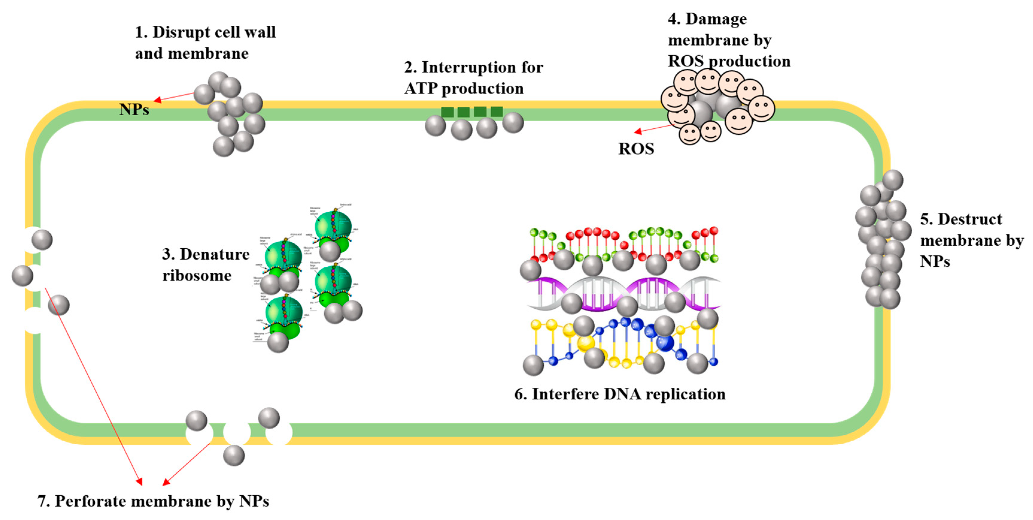
Recently, researchers found that ROS-based silver particles have outstanding antibacterial properties. Ramkumar team reported that silver nanoparticles can generate reactive oxygen species, thereby interrupting adenosine triphosphate production, stimulating cell membrane destruction. Meanwhile, silver has self-extinction of microorganisms by interacting with sulfur and phosphorus in DNA and inhibits protein synthesis [15]. Upon excitation with dental offices light (blue light), doped TiO2 nanoparticles can generate antibacterial ROS for the long-term and repeatedly, while conventional organic antibiotics are consumed when used [16]. Thus, the potential efficacy of ROS-based nanoparticles on extending useful lifetime as addition of antibacterial functionality was determined. Generally, the mechanism of nanoparticles for sterilization in dentistry is elaborated in Figure 1.
In this review, we conclude the ROS-based nanoparticles used in dentistry, as well as their performance and potential dental application for the future. The functional mechanism will be discussed. We also review superiority and deficiency of current ROS-based nanoparticles used in dentistry. Furthermore, the possible challenge in developing ROS-nanomaterials is concluded in this work.
2. Reaction Oxide Species
2.1. ROS in Dentistry
ROS are free radicals involving oxygen with high chemical reactivity. Since the 1980s, ROS was known as toxic agents altering biochemical balance of cells by producing irreversible oxidation species. However, recently, studies about ROS showed that they can have positive effects as chemical mediators in intercellular communication. In cells, ROS are metabolic byproducts in numerous diseases, associated with inflammation, injury, and cell ischemia [17]. In inflammation of dental pulp, ROS may be released. Reduced nicotinamide adenine dinucleotide phosphate (NADPH) oxidase, cytochrome P450, and endothelial nitric oxide synthase are common sources in cells. At low or moderate concentration, ROS have positive effects such as tissue repair, cell differentiation, angiogenesis, and vascular endothelial growth factor (VEGF) induced cell migration, while at a high concentration, uncontrolled cell activity may cause cell death and disease, carcinogenesis, mitochondrial dysfunction. Therefore, ROS are not only generated as a cellular response, but are also products of normal metabolism in cells [18].
2.2. Source of ROS Generation in Dentistry
ROS can be used in wound healing, immunological response generation, and antibacterial properties during dental treatment. In dentistry, the common sources of ROS are generated from nonthermal plasma, lasers, photosensitizers, resin cements, and ionizing radiation when exposed to right irradiation. We have listed the source of ROS generation in Table 1 in details.
2.2.1. Nonthermal Plasma (NTP) for ROS Generation
The sterilization mechanism of NTP used in dentistry mainly depends on the generation of ROS, electromagnetic fields, ions, and electrons. Nowadays, as the stability and safety of nonthermal plasma, it has become the most common and novel method applied in dentistry [19]. Li et al. suggested that NTP became the more popular method than traditional sterilization methods, which has superior safety, thoroughness, fastness, and low temperature [20]. Some studies found that an NTP device has better efficiency on deactivation of both Bacillus subtilis and E. coli than UV sterilizer. NTP devices can generate reaction oxide species leading to deactivate the bacteria, but keep the tissue intact since it can work under room temperature. Furthermore, NTP exert their function of treatment and sterilization on the surface of tooth without drilling [21]. Recently, tiny and easy to use plasma jet has been developed and can produce NTP in the root canal. Laroussi et al. introduced a miniature jet (plasma pencil), which can be used in the treatment of Escherichia coli, leukemia cells, and Porphyromonas gingivalis [22]. In addition, NTP can also be used for the treatment of oral candidiasis, linear gingival erythema, and angular stomatitis. The adhesion strength of the interface between dentin and composites can be enhanced by about 60% using NTP treatment, thereby improving the performance, life durability of restoration composites. Table 1 is the source of reactive oxygen species (ROS) in dentistry.
Cold atmospheric plasma (CAP) is one common type of NTP, which is a patient-friendly and painless treatment of dental diseases. It can cause the inactivation of pathogenic bacteria and effectively modify non-inflammatory tissues. It is an effective tool for the treatment of dental caries and composite repair. In dentistry, it is widely used in root canal sterilization, disinfection of dental instruments and equipment, removal of dental plaque, teeth whitening (bleaching), and enhancing adhesion strength [33]. Plasma dental treatment is patient-friendly and appropriate to children and elder [34]. Thus, NTP has promising prospects in the dental field. However, further research works are necessary to clarify the mechanism of function and application in dentistry.
2.2.2. Laser for ROS Generation
In dentistry, laser is extensively used in tissue repair and gingivectomy procedures [35,36]. The advantage of laser compared to a traditional method is low pain feeling and sensitivity. When exposed to laser therapy, the reactive oxygen species are activated. At a low level, ROS can speed the growth factor and tissue repair processes, while the excessive ROS may cause damage to the protein components of normal cells. Thus, the appropriate intensity and exposure time of the laser are very critical during dental therapy process. Cold laser therapy, the common low-level laser therapy, works under low levels of infrared light [37]. In addition, laser for ROS generation is an effective approach to treat pulpal wound and facilitate the formation of reparative dentin. Satoshi reported that laser irradiation generating ROS can promote the calcification ability of human dental pulp cells [38].
2.2.3. ROS Generating from Bleaching Agents
In tooth whitening procedure, various oxidizing agents are applied to bleach the tooth. Hydrogen peroxide, sodium hypochlorite, and ozone are the common oxidizing agents in dental bleaching. During the oxidization process, they can produce ROS, which penetrate through dentin and cause the decomposition of organic materials to achieve cleaning and sterilization functions. Eimar’s study found that bleaching treatment was more effective using hydrogen peroxide than others (NaOH and EDTA cleaning agents) [39]. However, the utilization of hydrogen peroxide may also have unfavorable effects on soft and hard oral tissues. When studied the effect of H2O2 on odontoblasts, Lee’s team discovered that ROS fascinated cells differentiation, when the cells were treated with H2O2 at concentrations below 0.3 mmol/L [29]. In clinical bleaching, the solution of 30% H2O2 or H2O2/TiO2 combination is commonly used. Makiko used TiO2 coated with hydroxyapatite and produced a high level of ROS and showed superior bleaching effects without any change of temperature and pH [28]. To maximize cleaning and sterilization functions, and to avoid the deficiency in dentistry, the moderate generation of ROS is worthy of further discussion.
2.2.4. Photodynamic Therapy and Light Sources for ROS Generation
Photodynamic therapy and light sources are also forms to generate ROS. In a dental office, blue light is the most common equipment for dental treatment. As we know, photosensitizers can adsorb light when exposed to light irradiation, then, produce free radicals to deactivate the microbial species. This process normally includes two steps. This first process is the electron transformation and free radicals generation by the reaction between photosensitizers and substrates. The second step is the formation of singlet oxide (active species) by the reaction between photosensitizers and oxygen [40]. The photodynamic treatment method is usually used as therapy for the periodontal disease, while the utilization of blue light irradiation may accelerate aging of pulpal blood vessel [41]. Furthermore, nucleus-targeting photodynamic therapy can generate ROS through introducing active photosensitizers for oral cancer treatment. In oral cancer theranostics, nucleus-targeting photodynamic therapy is a useful tool for destruction of cancer cells by directly attacking the DNA. Aklima synthesized noble carbon dots using a combination of curcumin and folic acid to enhance efficacy of photodynamic therapy for oral cancer treatment. Within the nucleus upon two-photon excitation, enhanced ROS generation was observed [42].
2.2.5. Other Sources for ROS Generation
In addition, ionizing radiation and UV rays are also used to produce ROS to deal with clinic dental problems [43]. In dentistry, the detection, identification, and completion removal of resin cement can be accomplished by ultraviolet illumination. Germicidal lamps used to identify and kill bacteria have been successfully adopted [44]. As a new mode of surface treatment for dental implant, ultraviolet photofunctionalization has been attracted researchers’ attention. Generally, surface contaminants can be removed by UV irradiation and photocatalysis. UV irradiation may result in skin aging and even cancer, which impedes its large-scale utilization in clinic dentistry.
3. ROS-Based Nanomaterials Used in Dentistry
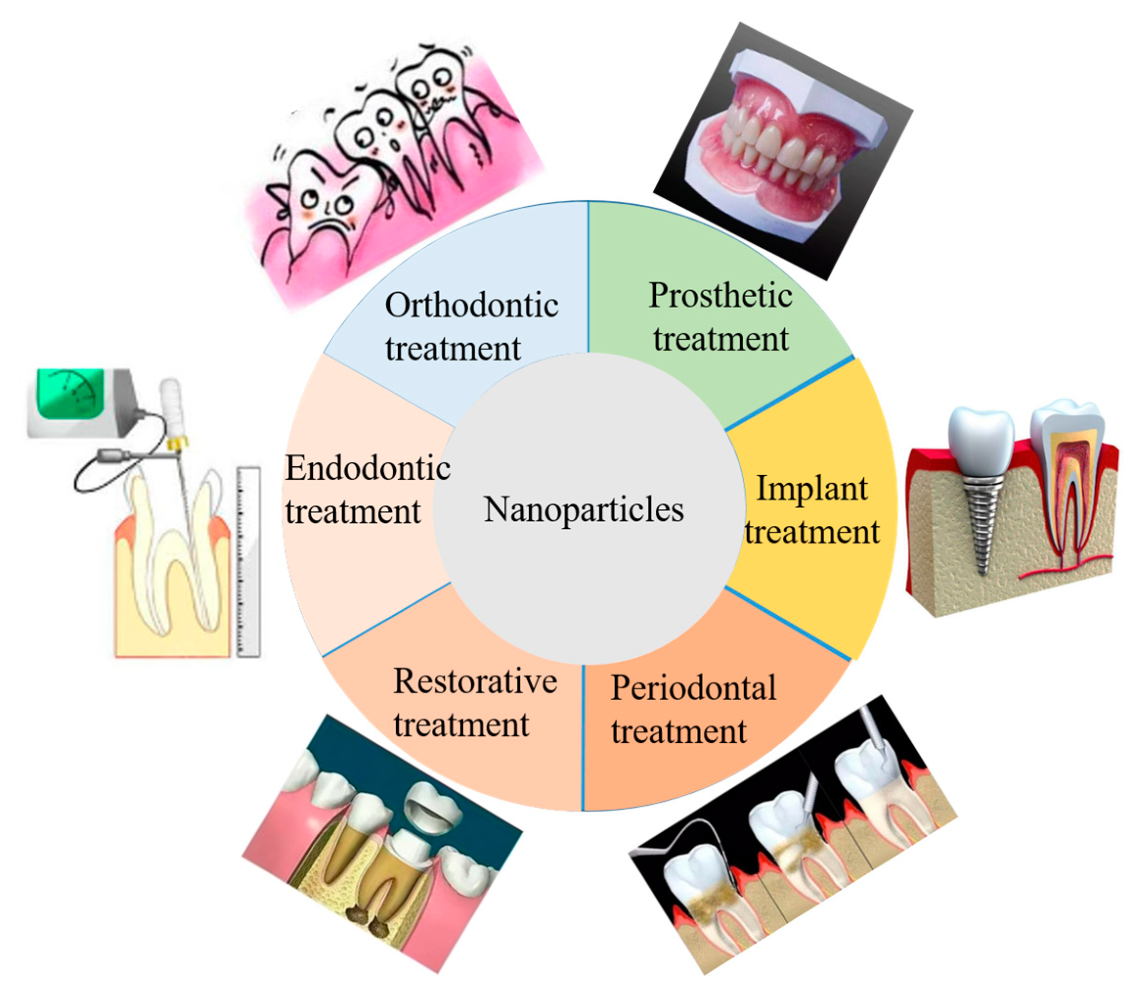
ROS-based nanoparticles have been adopted as efficient antibacterial materials and sterilization agents with no side effects. The nano-size and big ratio between surface area and volume of nanoparticles results in excellent efficacy with high antibacterial properties. In Table 2, we have discussed the common nanomaterials, their inferiorities and superiorities used in dentistry. It can improve the penetrability through the cell membrane; at the same time it can produce reactive oxide species. Many works have been done to study ROS-based nanoparticles used in dentistry. For instance, ROS-based silver nanoparticles can be used in prosthetic treatment, restorative treatment, endodontic treatment, adhesive materials in orthodontic treatment, as well as tissue regeneration in periodontal treatment [45]. The summary of nanoparticles used in dentistry is concluded in Figure 2.
3.1. Metal Nanoparticles in Dental Treatment
Metal nanoparticles with high surface area have gained significant focus due to their remarkable antimicrobial properties over the past few decades [46]. The excellent antimicrobial efficiency is mainly attributed to the small size and high surface area, which enable to provide maximum contact with target objects. Small size particles can easily penetrate through cell membranes, thus, improving the reactivity and antimicrobial activity by affecting intracellular processes [47].
3.1.1. ROS-Based Silver Nanoparticles
Silver nanoparticles are widely applied in prosthetic treatment, restorative treatment, endodontic treatment, and orthodontic treatment as a remarkable antibacterial material with low cytotoxicity and immunological response. In dentistry, silver nanoparticles are incorporated to acrylic resins to fabricate removable dentures and adhesive materials [48]. They can increase the permeability of cell membranes, producing reactive oxygen species, and block the replication of DNA by releasing silver ions during dental treatment. However, this method also has detrimental effects. When incorporated in dental materials, Ag nanoparticles may cause cosmetic issue specially tooth colored changing. The safe concentration of Ag nanoparticles when used in dentistry is between 0.05 and 0.70% incorporated in polymers [49]. As reached to limitation of 10%, Ag nanoparticles can influence monomer conversion from dental materials, which cause an increase in the degree of hardened monomer composite, or cause allergic reactions [50].
3.1.2. The Antibacterial Mechanism of ROS-Based Silver Nanoparticles in Dentistry
Nano-silver particles can release silver ions continuously, which is considered a main component to microbes. Silver ions have electrostatic attraction and affinity to sulfur proteins leading to adhere to cell wall and cytoplasmic membrane. The adhesive ions result in destruction of bacterial envelope through enhancing the permeability of cytoplasmic membrane. Then silver ions into cell promote the producing of reactive oxygen species, but interrupting the production of adenosine triphosphate. As previous mentioned, reactive oxygen species can cause cell membrane rupture and deoxyribonucleic acid (DNA) modification. In addition, silver ions can interact with sulfur- and phosphorous- containing compounds such as DNA, playing a role in impeding DNA replication, cell reproduction, leading to death of microorganisms. Moreover, silver ions can inhibit the synthesis of proteins by denaturing ribosomes in the cytoplasm [51].
3.1.3. ROS-Based Au Nanoparticles
Au nanoparticles used as a vehicle is popular in medical and dental application. Studies reported that chitosan-Au nanocomposites were used for wound curing, adhesive bandages, and coatings. Au nanoparticles not only have excellent antibacterial performance, but also can enhance the mechanical strength of composites. Innovative chirosan-based Au nanoparticles have been constructed by Regiel-Futyra with no cytotoxicity that showed high sterilization against of pseudomonas aeruginosa and staphylococcus aureus [52]. Au NPs can also be used as osteogenic agents. Heo used AuNPs as coating on the surface of Ti implants to promote bone regeneration [53]. AuNPs can provide a non-invasive approach to visualize dentin structure. Thereby, AuNPs is very useful as an antibacterial composite and coating layer on the surface of the tooth. In addition, AuNPs can also be utilized for multifunctional nanocarriers with antimicrobial effect and used in imaging technologies in dentistry. However, under the high concentration, the agglomeration has a negative effect when used in the dental field. Thus, the concentration of used AuNPs is desired discussed in future research work.
3.2. ROS-Based Metal Oxide Nanoparticles in Dentistry
The metal oxide nanoparticles are mostly used in medical and dental field [54]. Metal oxide nanoparticles have a strong effect on teeth healing. It can avoid formation of plaque inside the oral cavity caused by roughness and chemical decomposition from conventional materials. Surface modification by metal oxide nanoparticles can improve dental hardness and strength.
3.2.1. ROS-Based Titanium Oxide Nanoparticles
TiO2 has attracted a great deal of interest recently as a traditional excellent photocatalyst with strong bactericidal activity. It is also widely used in dentistry. Monteiro used TiO2 as additive incorporated into 3.5% H2O2 bleaching agent to increase the release of ROS, and found that after adding TiO2, the bleaching effect was improved compared without additive and the enamel color was kept unchanged [55]. Komatsu reported that TiO2 nanotubes used for tooth whitening can generate larger amounts of ROS than TiO2, which are promising materials for dental bleaching [56]. Zane synthesized TiO2 nanoparticles (20–30 nm size) with nitrogen doping using sol-gel and used as addition of antibacterial functionality to dental resins to extend useful lifetime by reducing secondary [57]. However, TiO2 can only absorb UV light. Researchers made many efforts to extend the light absorption to visible-light by doping. In dentistry, doping TiO2 with transition metal ions and anions has been studied. In addition, the toxicity and risks in vivo of TiO2 is the vital consideration when used in the dental field. Kurzmann’s evaluated experiments on TiO2 in vivo confirmed that bleaching agent containing TiO2/Ag or TiO2 nanoparticles showed lower cytotoxicity than H2O2 [58]. Acrylic acid-functionalized TiO2 nanoparticles was incorporated into resin adhesives, which can generated more ROS and showed superior photopolymerization as well as biocompatible properties with low cytotoxicity and genotoxicity for dental applications [59].
3.2.2. ROS-Based ZnO Nanoparticles
Similar to the silver nanoparticles, ZnO nanoparticles have selective antibacterial effect on bacteria. ZnO nanoparticles can effectively sterilize gram-negative bacteria and gram-positive bacteria. ZnO nanoparticles have selective toxicity to bacteria with minimal effects on human cells [60]. Its mechanism is related to the change of cell membrane activity and reactive oxygen species. ZnO nanoparticles can generate active substances such as H2O2 to inhibit the growth of microorganisms. Another potential antibacterial mechanism of ZnO is the penetration of Zn2+ into cell medium, reducing the formation of biofilms by inhibiting the active transport and metabolism of carbohydrates, and replacing magnesium to disrupt the enzyme system, which is the vital for enzyme activity of oral biofilms [61]. Its sterilization effect on streptococci is better than silver nanoparticles. However, studies showed that the right concentration of nanofiller is critical, and 10% ZnO nanoparticles used in dental composite resin can effectively kill bacteria. To keep original mechanical properties, achieving the maximum antibacterial activities by controlling the proportion of dental composite resins will be the focus for the future research work.
3.3. Other Nanoparticles Used in Dentistry
Organic nanoparticles like chitosan nanoparticles as a novel drug delivery carriers have also been used in dentistry for sterilization. With the functionality and cytocompatibility, the chitosan can increase the release of drug, which is ideal for therapeutic interventions for daily oral care [62]. Because of the stable physiochemical properties and good sterilization, CuO was used in oral care. However, the potential adverse effect impedes the application caused by residual materials. Khan synthesized CuO NPs with a size of 40 nm and assessed their sterilization for oral microbe and biofilm on diverse matrices [63]. The extensive application of CuO in dentistry is still a big challenge because of the adverse effects. Generally, the types of ROS-based nanoparticles contain metal nanoparticles, metal oxide nanoparticles, and polymer or organic nanoparticles. We conclude on other nanoparticles and summarize the advantages and disadvantages used in the dental field, as well as the mechanism of killing microbacterials, in Table 2.
4. Clinical Applications of ROS-Nanoparticles in Dentistry
Nanoparticles with nano-scale size have prominent influence in molecular restoration and regeneration of necrotic tissues. Recently, with low toxicity, good sterilization, and enhancing interaction with protein, nanomaterials were widely utilized for periodontal treatment and management. For instance, nanoparticles were incorporated into composite resins and binding materials to improve the tensile strength and compressive strength for oral restoration treatment. Nanoparticles were also used as a novel method for canal space irrigation, removal of pulp debris. The addition of nanoparticles has also been proven to be an effective method for root canal organisms, preventing the recurrence of infection. Used in implant treatment, nanoparticles can be used as osteogenesis agents with good biocompatibility and adaptability of oral tissue. Developments of nanoparticles for periodontal apparatus, dentine, bone, cementum, and periodontal ligaments have huge commercial applications. In recent studies, Li’s team used gold nanoparticles with 45nm particle size for the treatment of periodontitis, and the results showed that gold nanoparticles have good anti-inflammatory effects and can enhance the periodontal environments [73]. Fe3O4-silane@Ce6/C6 nanoparticles with amphiphilic property have been prepared to inhibit the occurrence of periodontal disease in Sun’s work [74]. Impregnated nanoparticles along with tissue can mimic formation of host tissues. The formation of biomaterials in different forms can be used for various dental applications. The continuous improvement for treatment method and advances for clinic application of nanoparticles is promising way to dental care [75].
5. Safety Evaluation of Nanoparticles in Dental Application
Indeed, nanomaterials have different biological effects on cellular, subcellular, and proteins. Some of them are easy to adhere to organs, penetrating cell membranes, or staying in mitochondria, then, causing different adverse reactions. Recently, many studies have shown that nanomaterials can accumulate in the heart, liver, spleen, lungs, and kidneys of animals. Generally, the blood–brain barrier can prevent this substance from entering the brain, leading to disease. However, due to its small size and high surface activity, nanomaterials can pass through the brain barrier, and may even reach the brain through the olfactory and sensory nerve centers, causing pathological changes.
In general, the biological toxicity of nanomaterials can be summarized as: oxidative stress and inflammation. Thus, the safety of nanoparticles needs to consider these two aspects. However, the accuracy and reliability of traditional methods and techniques for analyzing the toxicity of nanomaterials need to be explored and strengthened. About whether the nanomaterials with unique physicochemical properties have introduced new damage mechanisms and whether these new mechanisms will lead to new pathologies need further verification. Even though nanomaterials do not introduce new pathology, there may be new damage mechanisms that have not been realized. Thus, special tools and characterization, assessment approaches, and methods are necessary to evaluate their toxicity. Therefore, it is too early to draw a clear conclusion on their inherent harm to the human body. So far, the toxicity mechanism of nanomaterials is still obscured [76].
6. Conclusions and Future Prospect
With the industrialization development of nanotechnology, dental nanomaterials have been widely used. Especially in the dental field, the utilization of nanomaterials for daily life has greatly increased. Nanotechnology-based materials have intensively improved clinical treatment and promoted the innovation of many conventional dental materials. This review summarized the main types of commonly used ROS-based nanomaterials in the dental field, and summarizes their possible mechanisms for dental antibacterial applications. The current challenges and safety performance evaluations of these materials in this field are concluded, which may provide feasible clinical guidance for future dental research.
Nanotechnology has attained prominent achievement in tissue sterilization, modification on the surface of materials, and dental whitening, but this method used in dentistry is still in the initial stage. The mechanism and long-term effects and non-toxicity still need to be clarified for a long time when used in clinic oral medicine. It is desirable to further study how to make it more human-friendly for use, easier to operate, how to make the process more simplified, reduce operating time, and make required dosage more precise. Future work will focus on reducing the operating time, enhancing the antibacterial efficiency, while minimizing the adverse impact on oral tissue. Some studies reported that the toxic of nanoparticles can be decreased through controlling nanoparticles diameter and surface modification. Thus, these aspects of studying nanoparticles in the dental field may be valued for the next research work.
Author Contributions
F.L. wrote the paper. T.H. participated in the design and drafting of this manuscript. J.X. and X.Z. performed the literature search and revised the paper. Y.W. guided the writing of the paper and reviewed the manuscript. All authors have read and agreed to the published version of the manuscript.
Funding
This work was supported by National Natural Science Foundation of China (81901020), Natural Science Foundation of Hunan Province, China (2020JJ4458), Hunan Provincial Health and Family Planning Commission’s 2017 Scientific Research Plan Project (No.: B2017036)
Institutional Review Board Statement
Not applicable.
Informed Consent Statement
Not applicable.
Data Availability Statement
Not applicable.
Conflicts of Interest
The authors declare no conflict of interest. The funders had no role in the design of the study; in the collection, analyses, or interpretation of data; in the writing of the manuscript, or in the decision to publish the results.
References
- Marsh, P.D.; Martin, M.V.; Lewis, M.A.; Williams, D. Oral Microbiology e-Book; Elsevier: Amsterdam, The Netherlands, 2009. [Google Scholar]
- Filoche, S.; Wong, L.; Sissons, C.H. Oral biofilms: Emerging concepts in microbial ecology. J. Dent. Res. 2010, 89, 8–18. [Google Scholar] [CrossRef] [PubMed]
- Melo, M.A.; Guedes, S.F.; Xu, H.H.; Rodrigues, L.K. Nanotechnology-based restorative materials for dental caries management. Trends Biotechnol. 2013, 31, 459–467. [Google Scholar] [CrossRef] [Green Version]
- Cury, J.A.; Tenuta, L.M.A. Enamel remineralization: Controlling the caries disease or treating early caries lesions? Braz. Oral Res. 2009, 23, 23–30. [Google Scholar] [CrossRef] [Green Version]
- Fontana, M.; Young, D.A.; Wolff, M.S.; Pitts, N.B.; Longbottom, C. Defining dental caries for 2010 and beyond. Dent. Clin. 2010, 54, 423–440. [Google Scholar] [CrossRef]
- Engel, E.; Michiardi, A.; Navarro, M.; Lacroix, D.; Planell, J.A. Nanotechnology in regenerative medicine: The materials side. Trends Biotechnol. 2008, 26, 39–47. [Google Scholar] [CrossRef] [PubMed] [Green Version]
- Solga, A.; Cerman, Z.; Striffler, B.F.; Spaeth, M.; Barthlott, W. The dream of staying clean: Lotus and biomimetic surfaces. Bioinspiration Biomim. 2007, 2, S126. [Google Scholar] [CrossRef]
- Watson, P.S.; Pontefract, H.A.; Devine, D.A.; Shore, R.C.; Nattress, B.R.; Kirkham, J.; Robinson, C. Penetration of fluoride into natural plaque biofilms. J. Dent. Res. 2005, 84, 451–455. [Google Scholar] [CrossRef]
- Hannig, M.; Kriener, L.; Hoth-Hannig, W.; Becker-Willinger, C.; Schmidt, H. Influence of nanocomposite surface coating on biofilm formation in situ. J. Nanosci. Nanotechnol. 2007, 7, 4642–4648. [Google Scholar] [CrossRef]
- Hajipour, M.J.; Fromm, K.M.; Ashkarran, A.A.; de Aberasturi, D.J.; de Larramendi, I.R.; Rojo, T.; Serpooshan, V.; Parak, W.J.; Mahmoudi, M. Antibacterial properties of nanoparticles. Trends Biotechnol. 2012, 30, 499–511. [Google Scholar] [CrossRef] [Green Version]
- Li, X.; Cui, R.; Liu, W.; Sun, L.; Yu, B.; Fan, Y.; Feng, Q.; Cui, F.; Watari, F. The use of nanoscaled fibers or tubes to improve biocompatibility and bioactivity of biomedical materials. J. Nanomater. 2013, 2013, 16. [Google Scholar] [CrossRef]
- Li, X.M.; Lee, S.C.; Zhang, S.M.; Akasaka, T. Biocompatibility and Toxicity of Nanobiomaterials 2014. J. Nanomater. 2015, 2015, 259264. [Google Scholar] [CrossRef]
- Kalyanaraman, B.; Darley-Usmar, V.; Davies, K.J.; Dennery, P.A.; Forman, H.J.; Grisham, M.B.; Mann, G.E.; Moore, K.; Roberts, L.J., II; Ischiropoulos, H. Measuring reactive oxygen and nitrogen species with fluorescent probes: Challenges and limitations. Free Radic. Biol. Med. 2012, 52, 1–6. [Google Scholar] [CrossRef] [Green Version]
- Atashi, F.; Modarressi, A.; Pepper, M.S. The role of reactive oxygen species in mesenchymal stem cell adipogenic and osteogenic differentiation: A review. Stem. Cells Dev. 2015, 24, 1150–1163. [Google Scholar] [CrossRef] [Green Version]
- Park, H.J.; Park, S.; Roh, J.; Kim, S.; Choi, K.; Yi, J.; Kim, Y.; Yoon, J. Biofilm-inactivating activity of silver nanoparticles: A comparison with silver ions. J. Ind. Eng. Chem. 2013, 19, 614–619. [Google Scholar] [CrossRef]
- Ashkarran, A.A.; Hamidinezhad, H.; Haddadi, H.; Mahmoudi, M. Double-doped TiO2 nanoparticles as an efficient visible-light-active photocatalyst and antibacterial agent under solar simulated light. Appl. Surf. Sci. 2014, 301, 338–345. [Google Scholar] [CrossRef]
- Baumgardner, K.R.; Sulfaro, M.A. The anti–inflammatory effects of human recombinant copper–zinc superoxide dismutase on pulp inflammation. J. Endod. 2001, 27, 190–195. [Google Scholar] [CrossRef]
- Krifka, S.; Spagnuolo, G.; Schmalz, G.; Schweikl, H. A review of adaptive mechanisms in cell responses towards oxidative stress caused by dental resin monomers. Biomaterials 2013, 34, 4555–4563. [Google Scholar] [CrossRef]
- Nasir, N.M.; Lee, B.K.; Yap, S.S.; Thong, K.L.; Yap, S.L. Cold plasma inactivation of chronic wound bacteria. Arch. Biochem. Biophys. 2016, 605, 76–85. [Google Scholar] [CrossRef]
- Yang, H.L.; Liu, S.; Hu, T. Application of low-temperature plasma in dental clinical sterilization. Foreign Med. Sci. Stomatol. 2013, 40, 483–485. [Google Scholar]
- Laroussi, M. Low temperature plasma-based sterilization: Overview and state-of-the-art. Plasma Process. Polym. 2005, 2, 391–400. [Google Scholar] [CrossRef]
- Laroussi, M.; Tendero, C.; Lu, X.; Alla, S.; Hynes, W.L. Inactivation of bacteria by the plasma pencil. Plasma Process. Polym. 2006, 3, 470–473. [Google Scholar] [CrossRef]
- Sung, S.J.; Huh, J.B.; Yun, M.J.; Chang, B.M.W.; Jeong, C.M.; Jeon, Y.C. Sterilization effect of atmospheric pressure non-thermal air plasma on dental instruments. J. Adv. Prosthodont. 2013, 5, 2–8. [Google Scholar] [CrossRef] [Green Version]
- Kumar, S.C.; Sarada, P.; Reddy, S.C.; Reddy, S.M.; Dsv, N. Plasma torch toothbrush a new insight in fear free dentistry. J. Clin. Diagn. Res. JCDR 2014, 8, 7–10. [Google Scholar]
- Chen, M.; Zhang, Y.; Driver, M.S.; Caruso, A.N.; Yu, Q.; Wang, Y. Surface modification of several dental substrates by non-thermal, atmospheric plasma brush. Dent. Mater. 2013, 29, 871–880. [Google Scholar] [CrossRef] [PubMed] [Green Version]
- Kwon, J.H.; Park, H.C.; Zhu, T.; Yang, H.C. Inhibition of odontogenic differentiation of human dental pulp cells by dental resin monomers. Biomater. Res. 2015, 19, 1–7. [Google Scholar] [CrossRef] [PubMed] [Green Version]
- Geurtsen, W.; Spahl, W.; Leyhausen, G. Residual monomer/additive release and variability in cytotoxicity of light-curing glass-ionomer cements and compomers. J. Dent. Res. 1998, 77, 2012–2019. [Google Scholar] [CrossRef] [PubMed]
- Saita, M.; Kobatashi, K.; Yoshino, F.; Hase, H.; Nonami, T.; Kimoto, K.; Masaichi, C.I. ESR investigation of ROS generated by H2O2 bleaching with TiO2 coated Hap. Dent. Mater. J. 2012, 31, 458–464. [Google Scholar] [CrossRef] [Green Version]
- Lee, D.H.; Lim, B.S.; Lee, Y.K.; Yang, H.C. Effects of hydrogen peroxide (H2O2) on alkaline phosphatase activity and matrix mineralization of odontoblast and osteoblast cell lines. Cell Biol. Toxicol. 2006, 22, 39–46. [Google Scholar] [CrossRef]
- Andersen, R.; Loebel, N.; Hammond, D.; Wilson, M. Treatment of periodontal disease by photodisinfection compared to scaling and root planning. J. Clin. Dent. 2007, 18, 34. [Google Scholar]
- Wilson, M. Lethal photosensitisation of oral bacteria and its potential application in the photodynamic therapy of oral infections. Photochem. Photobiol. Sci. 2004, 3, 412–418. [Google Scholar] [CrossRef]
- Jha, N.; Ryu, J.J.; Choi, E.H.; Kaushik, N.K. Generation and role of reactive oxygen and nitrogen species induced by plasma, lasers, chemical agents, and other systems in dentistry. Oxidative Med. Cell. Longev. 2017, 2017, 7542540. [Google Scholar] [CrossRef] [PubMed]
- Hoffmann, C.; Berganza, C.; Zhang, J. Cold Atmospheric Plasma: Methods of production and application in dentistry and oncology. Med. Gas Res. 2013, 3, 2045–9912. [Google Scholar] [CrossRef] [Green Version]
- Gherardi, M.; Tonini, R.; Colombo, V. Plasma in dentistry: Brief history and current status. Trends Biotechnol. 2018, 36, 583–585. [Google Scholar] [CrossRef]
- Rosa, D.S.A.; Aranha, A.C.C.; de Paula Eduardo, C.; Aoki, A. Esthetic treatment of gingival melanin hyperpigmentation with Er: YAG laser: Short-term clinical observations and patient follow-up. J. Periodontol. 2007, 78, 2018–2025. [Google Scholar] [CrossRef]
- Sobouti, F.; Rakhshan, V.; Chiniforush, N.; Khatami, M. Effects of laser-assisted cosmetic smile lift gingivectomy on postoperative bleeding and pain in fixed orthodontic patients: A controlled clinical trial. Prog. Orthod. 2014, 15, 1–5. [Google Scholar] [CrossRef] [PubMed] [Green Version]
- Farivar, S.; Malekshahabi, T.; Shiari, R. Biological effects of low level laser therapy. J. Lasers Med Sci. 2014, 5, 58. [Google Scholar] [PubMed]
- Matsui, S.; Takahashi, C.; Tsujimoto, Y.; Matsushima, K. Stimulatory effects of low-concentration reactive oxygen species on calcification ability of human dental pulp cells. J. Endodontia 2009, 35, 67–72. [Google Scholar] [CrossRef] [PubMed]
- Eimar, H.; Siciliano, R.; Abdallah, M.N.; Abi Nader, S.; Amin, W.M.; Martinez, P.P.; Celemin, A.; Cerruti, M.; Tamimi, F. Hydrogen peroxide whitens teeth by oxidizing the organic structure. J. Dent. 2012, 40, e25–e33. [Google Scholar] [CrossRef]
- Diogo, P.; Gonçalves, T.; Palma, P.; Santos, J.M. Photodynamic antimicrobial chemotherapy for root canal system asepsis: A narrative literature review. Int. J. Dent. 2015, 2015, 269205. [Google Scholar] [CrossRef] [Green Version]
- Yoshida, A.; Iwata, S.; Iizuka, J.; Takahashi, S.S.; Wada-Takahashi, S.; Miyamoto, C.; Maehata, Y.; Ogura, Y.; Lee, M.; Yo, F. Blue light from dental resin curing unit causes light-induced vasoconstriction in isolated rat aorta. Oral Health Dent. Manag. 2014, 13, 1147–1151. [Google Scholar]
- Nasrin, A.; Hassan, M.; Gomes, V.G. Two-photon active nucleus-targeting carbon dots: Enhanced ROS generation and photodynamic therapy for oral cancer. Nanoscale 2020, 12, 20598–20603. [Google Scholar] [CrossRef]
- Desouky, O.; Ding, N.; Zhou, G. Targeted and non-targeted effects of ionizing radiation. J. Radiat. Res. Appl. Sci. 2015, 8, 247–254. [Google Scholar] [CrossRef] [Green Version]
- Bush, M.A.; Hermanson, A.S.; Yetto, R.J.; Wieczkowski, G. The use of ultraviolet LED illumination for composite resin removal: An in vitro study. Gen. Dent. 2010, 58, 214–218. [Google Scholar]
- Yin, I.X.; Zhang, J.; Zhao, I.S.; Mei, M.L.; Li, Q.; Chu, C.H. The Antibacterial Mechanism of Silver Nanoparticles and Its Application in Dentistry. Int. J. Nanomed. 2020, 15, 2555. [Google Scholar] [CrossRef] [Green Version]
- Jandt, K.D.; Watts, D.C. Nanotechnology in dentistry: Present and future perspectives on dental nanomaterials. Dent. Mater. 2020, 36, 1365–1378. [Google Scholar] [CrossRef]
- Radad, K.; Al-Shraim, M.; Moldzio, R.; Rausch, W.D. Recent advances in benefits and hazards of engineered nanoparticles. Environ. Toxicol. Pharm. 2012, 34, 661–672. [Google Scholar] [CrossRef]
- Bapat, R.A.; Chaubal, T.V.; Joshi, C.P.; Bapat, P.R.; Choudhury, H.; Pandey, M.; Gorain, B.; Kesharwani, P. An overview of application of silver nanoparticles for biomaterials in dentistry. Mater. Sci. Eng. C 2018, 91, 881–898. [Google Scholar] [CrossRef]
- Martínez-Gutiérrez, F.; Guajardo-Pacheco, J.M.; Noriega-Trevino, M.E.; Thi, E.P.; Reiner, N.; Orrantia, E.; Av-Gay, Y.; Ruiz, F.; Bach, H. Antimicrobial activity, cytotoxicity and inflammatory response of novel plastics embedded with silver nanoparticles. Future Microbiol. 2013, 8, 403–411. [Google Scholar] [CrossRef] [Green Version]
- Durner, J.; Stojanovic, M.; Urcan, E.; Hickel, R.; Reichl, F.X. Influence of silver nano-particles on monomer elution from light-cured composites. Dent. Mater. 2011, 27, 631–636. [Google Scholar] [CrossRef]
- Durán, N.; Nakazato, G.; Seabra, A.B. Antimicrobial activity of biogenic silver nanoparticles, and silver chloride nanoparticles: An overview and comments. Appl. Microbiol. Biotechnol. 2016, 100, 6555–6570. [Google Scholar] [CrossRef]
- Regiel-Futyra, A.; Kus-Liśkiewicz, M.; Sebastian, V.; Irusta, S.; Arruebo, M.; Stochel, G.; Kyzioł, A. Development of noncytotoxic chitosan–gold nanocomposites as efficient antibacterial materials. ACS Appl. Mater. Interfaces 2015, 7, 1087–1099. [Google Scholar] [CrossRef]
- Braz, A.K.S.; de Araujo, R.E.; Ohulchanskyy, T.Y.; Shukla, S.; Bergey, E.J.; Gomes, A.S.; Prasad, P.N. In situ gold nanoparticles formation: Contrast agent for dental optical coherence tomography. J. Biomed. Opt. 2012, 17, 066003. [Google Scholar] [CrossRef] [Green Version]
- Mcintyre, R.A. Common nano-materials and their use in real world applications. Sci. Prog. 2012, 95, 1–22. [Google Scholar] [CrossRef] [PubMed]
- Monteiro, N.R.; Basting, R.T.; Amaral, F.L.B.D.; França, F.M.G.; Turssi, C.P.; Gomes, O.P.; Lisboa Filho, P.N.; Kantovitz, K.R.; Basting, R.T. Titanium dioxide nanotubes incorporated into bleaching agents: Physicochemical characterization and enamel color change. J. Appl. Oral Sci. 2020, 28. [Google Scholar]
- Komatsu, O.; Nishida, H.; Sekino, T.; Yamamoto, K. Application of titanium dioxide nanotubes to tooth whitening. Nano Biomed. 2014, 6, 63–72. [Google Scholar]
- Argueta-Figueroa, L.; Torres-Gómez, N.; Scougall-Vilchis, R.J.; García-Contreras, R. Biocompatibility and antibacterial activity of nitrogen-doped titanium dioxide nanoparticles for use in dental resin formulations. Int. J. Nanomed. 2016, 11, 6459. [Google Scholar]
- Kurzmann, C.; Verheyen, J.; Coto, M.; Kumar, R.V.; Divitini, G.; Shokoohi-Tabrizi, H.A.; Verheyen, P.; De Moor, R.J.G.; Moritz, A.; Agis, H. In vitro evaluation of experimental light activated gels for tooth bleaching. Photochem. Photobiol. Sci. 2019, 18, 1009–1019. [Google Scholar] [CrossRef] [PubMed]
- Sun, J.; Petersen, E.J.; Watson, S.S.; Sims, C.M.; Kassman, A.; Frukhtbeyn, S.; Skrtic, D.; Ok, M.T.; Jacobs, D.S.; Reipa, V.; et al. Biophysical characterization of functionalized titania nanoparticles and their application in dental adhesives. Acta Biomater. 2017, 53, 585–597. [Google Scholar] [CrossRef] [PubMed] [Green Version]
- Sharma, V.; Shukla, R.K.; Saxena, N.; Parmar, D.; Das, M.; Dhawan, A. DNA damaging potential of zinc oxide nanoparticles in human epidermal cells. Toxicol. Lett. 2009, 185, 211–218. [Google Scholar] [CrossRef] [PubMed]
- Gu, H.; Fan, D.; Gao, J.; Zou, W.; Peng, Z.; Zhao, Z.; Ling, J.; LeGeros, R.Z. Effect of ZnCl2 on plaque growth and biofilm vitality. Arch. Oral Biol. 2012, 57, 369–375. [Google Scholar] [CrossRef]
- Chronopoulou, L.; Nocca, G.; Castagnola, M.; Paludetti, G.; Ortaggi, G.; Sciubba, F.; Bevilacqua, M.; Lupi, A.; Gambarini, G.; Palocci, C. Chitosan based nanoparticles functionalized with peptidomimetic derivatives for oral drug delivery. New Biotechnol. 2016, 33, 23–31. [Google Scholar] [CrossRef]
- Khan, S.T.; Ahamed, M.; Al-Khedhairy, A.; Musarrat, J. Biocidal effect of copper and zinc oxide nanoparticles on human oral microbiome and biofilm formation. Mater. Lett. 2013, 97, 67–70. [Google Scholar] [CrossRef]
- Misawa, M.; Takahashi, J. Generation of reactive oxygen species induced by gold nanoparticles under X-ray and UV Irradiations. Nanomed. Nanotechnol. Biol. Med. 2011, 7, 604–614. [Google Scholar] [CrossRef] [PubMed]
- Priyadarsini, S.; Mukherjee, S.; Mishra, M. Nanoparticles used in dentistry: A review. J. Oral Biol. Craniofacial Res. 2018, 8, 58–67. [Google Scholar] [CrossRef] [Green Version]
- Dwivedi, S.; Wahab, R.; Khan, F.; Mishra, Y.K.; Musarrat, J.; Al-Khedhairy, A.A. Reactive oxygen species mediated bacterial biofilm inhibition via zinc oxide nanoparticles and their statistical determination. PLoS ONE 2014, 9, e111289. [Google Scholar] [CrossRef] [PubMed]
- Guerreiro-Tanomaru, J.M.; Trindade-Junior, A.; Cesar Costa, B.; da Silva, G.F.; Drullis Cifali, L.; Basso Bernardi, M.I.; Tanomaru-Filho, M. Effect of zirconium oxide and zinc oxide nanoparticles on physicochemical properties and antibiofilm activity of a calcium silicate-based material. Sci. World J. 2014, 2014, 975213. [Google Scholar] [CrossRef] [Green Version]
- Tsai, Y.Y.; Oca-Cossio, J.; Lin, S.M.; Woan, K.; Yu, P.C.; Sigmund, W. Reactive oxygen species scavenging properties of ZrO2–CeO2 solid solution nanoparticles. Future Med. 2008, 637–645. [Google Scholar]
- Toodehzaeim, M.H.; Zandi, H.; Meshkani, H.; Firouzabadi, A.H. The effect of CuO nanoparticles on antimicrobial effects and shear bond strength of orthodontic adhesives. J. Dent. 2018, 19, 1. [Google Scholar]
- Bitar, A.; Ahmad, N.M.; Fessi, H.; Elaissari, A. Silica-based nanoparticles for biomedical applications. Drug Discov. Today 2012, 17, 1147–1154. [Google Scholar] [CrossRef]
- Timpe, N.; Fullriede, H.; Borchers, L.; Stiesch, M.; Behrens, P.; Menzel, H. Nanoporous silica nanoparticles with spherical and anisotropic shape as fillers in dental composite materials. BioNanoMaterials 2014, 15, 89–99. [Google Scholar] [CrossRef]
- Akasaka, T.; Nakata, K.; Uo, M.; Watari, F. Modification of the dentin surface by using carbon nanotubes. Bio-Med Mater. Eng. 2009, 19, 179–185. [Google Scholar] [CrossRef] [Green Version]
- Ni, C.; Zhou, J.; Kong, N.; Bian, T.; Zhang, Y.; Huang, X.; Xiao, Y.; Yang, W.; Yan, F. Gold nanoparticles modulate the crosstalk between macrophages and periodontal ligament cells for periodontitis treatment. Biomaterials 2019, 206, 115–132. [Google Scholar] [CrossRef]
- Sun, X.; Wang, L.; Lynch, C.D.; Sun, X.; Li, X.; Qi, M.; Ma, C.; Li, C.; Dong, B.; Zhou, Y.; et al. Nanoparticles having amphiphilic silane containing Chlorin e6 with strong anti-biofilm activity against periodontitis-related pathogens. J. Dent. 2019, 81, 70–84. [Google Scholar] [CrossRef] [PubMed]
- Bapat, R.A.; Joshi, C.P.; Bapat, P.; Chaubal, T.V.; Pandurangappa, R.; Jnanendrappa, N.; Gorain, B.; Khurana, S.; Kesharwani, P. The use of nanoparticles as biomaterials in dentistry. Drug Discov. Today 2019, 24, 85–98. [Google Scholar] [CrossRef] [PubMed]
- Feng, X.; Chen, A.; Zhang, Y.; Wang, J.; Shao, L.; Wei, L. Application of dental nanomaterials: Potential toxicity to the central nervous system. Int. J. Nanomed. 2015, 10, 3547. [Google Scholar]
Figure 1.
The mechanism of nanoparticles for sterilization in dentistry.

Figure 2.
The application of nanoparticles in dentistry.


Table 1.
Source of reactive oxygen species (ROS) in dentistry.
| ROS Source in Dentistry | Application in Dentistry | Ref. |
|---|---|---|
| Nonthermal Plasma | Wound healing, sterilization in gingival crevices, biofilm removal, dental implants, and cleaning dentin during periodontal regeneration. | [23,24,25] |
| Laser | Tissue repair, gingivectomy | [26,27] |
| Bleaching Agents | Destroy root pathogens, sterilization | [28,29] |
| Photodynamic Therapy and Light Sources | Cause cellular damage, break biofilm, act as adhensive materials | [30,31] |
| Ionizing Radiation and UV rays | Break DNA molecule of bacteria, radiotherapy in dentistry, elimination cancer cells | [32] |
Table 2.
Summary of ROS-based nanoparticles used in dentistry.
| Nanoparticles | Dental Application | Mechanisms/ROS Generation | Superiorities | Inferiorities | Ref. |
|---|---|---|---|---|---|
| Silver | Antibacterial agents, dental restorative materials, dental implants, root canal irrigation, orthodontic treatment. | Producing ROS, Releasing free silver ions leads to interruption of ATP molecules, preventing DNA replication or silver ions (Ag+) directly damage cell membranes. | Reduce bacterial colonization and improve oral health. biocompatibility, low toxicity and long-term antibacterial activity. | AgNPs are toxic, may cause silver-poisoning, lacking of research data in vivo to prove its safety. | [48] |
| Gold | Gingival inflammation, dental caries treatment, tissue engineering, dental implants, cancer diagnosis. | ROS production. | Small size, large specific surface area, with minimal adverse to mammals. | Toxic. Particles distribution in various organs. Larger gold nanoparticles scattered throughout the liver and spleen, harmful effects on various systems. | [64] |
| TiO2 | Preparing dental adhesives, dental restoration. | ROS production. | Compared with traditional dental adhesives, dental adhesives with acrylic functionalized nano-titanium dioxide have a stronger binding effect on human teeth. | The cytotoxicity and genotoxicity cannot be warranted. | [59,65] |
| ZnO | Deformation and damage of cells, surface coating material with antibacterial activity. | ROS production, inhibition on bacterial growth and biofilm formation. | Cell wall of bacteria can be destroyed at low concentration of ZnO-NPs. | Its mechanism of inhibition on bacterial growth and biofilm formation is still obscure. | [66] |
| Zirconia | Reducing bacterial adhesion to the tooth surface, providing protection against dental caries, effective polishing agent. | Yield ROS. | Similar mechanical properties and color like a tooth, have low cytotoxicity, sensible biocompatibility, and high fracture resistance. | Cytotoxic, inducing oxidative stress in cells, leading to cell death. | [67,68] |
| CuO | CuO NPs can improve the antibacterial performance and binding strength as orthodontic adhesives. | ROS production. | Low-priced and more stable, have strong antimicrobial effects on inhibition of bacterial colonization and plaque development. | Exhibit toxic effects. | [69] |
| Silica | Tooth filler, tooth whitening, prevention for tooth cavity, antibacterial agent. | By polishing prevents dental caries, which acts as a primary defense mechanism against the cariogenic bacteria. | Good biocompatibility, low toxicity, low density, significant adsorption capacity and low cost. | Silica nanoparticles, like crystal particles, can induce silicosis and lung cancer. SiNPs are cytotoxic. In addition, it can also induce oxidative stress and mediate cell apoptosis. SiNPs also have genotoxic effects (DNA damage, gene regulation involving apoptosis and autophagy) and immunotoxic effects. | [70,71] |
| Carbon nanotube | Teeth filling, coating layer on the teeth surface. | Carbon nanotubes can adhere to the surface of dentin and cementum by interacting with exposed collagen fibers. | Large surface area, easy to adhere to the surface of teeth, dentin and cementum. | May induce inflammation and fibrosis under certain conditions. | [72] |
Publisher’s Note: MDPI stays neutral with regard to jurisdictional claims in published maps and institutional affiliations. |
© 2021 by the authors. Licensee MDPI, Basel, Switzerland. This article is an open access article distributed under the terms and conditions of the Creative Commons Attribution (CC BY) license (http://creativecommons.org/licenses/by/4.0/).
Share and Cite
MDPI and ACS Style
Liu, F.; Hong, T.; Xie, J.; Zhan, X.; Wang, Y. Application of Reactive Oxygen Species-Based Nanomaterials in Dentistry: A Review. Crystals 2021, 11, 266. https://0-doi-org.brum.beds.ac.uk/10.3390/cryst11030266
AMA Style
Liu F, Hong T, Xie J, Zhan X, Wang Y. Application of Reactive Oxygen Species-Based Nanomaterials in Dentistry: A Review. Crystals. 2021; 11(3):266. https://0-doi-org.brum.beds.ac.uk/10.3390/cryst11030266
Chicago/Turabian StyleLiu, Fenglin, Tingting Hong, Jingxian Xie, Xuehui Zhan, and Yuehong Wang. 2021. "Application of Reactive Oxygen Species-Based Nanomaterials in Dentistry: A Review" Crystals 11, no. 3: 266. https://0-doi-org.brum.beds.ac.uk/10.3390/cryst11030266
Note that from the first issue of 2016, this journal uses article numbers instead of page numbers. See further details here.