
Seed-Mediated Preparation of Ag@Au Nanoparticles for Highly Sensitive Surface-Enhanced Raman Detection of Fentanyl


Abstract
:1. Introduction
2. Materials and Methods
2.1. Materials
2.2. Synthesis of Ag@Au Nanoparticles
2.3. Characterization and SERS Test
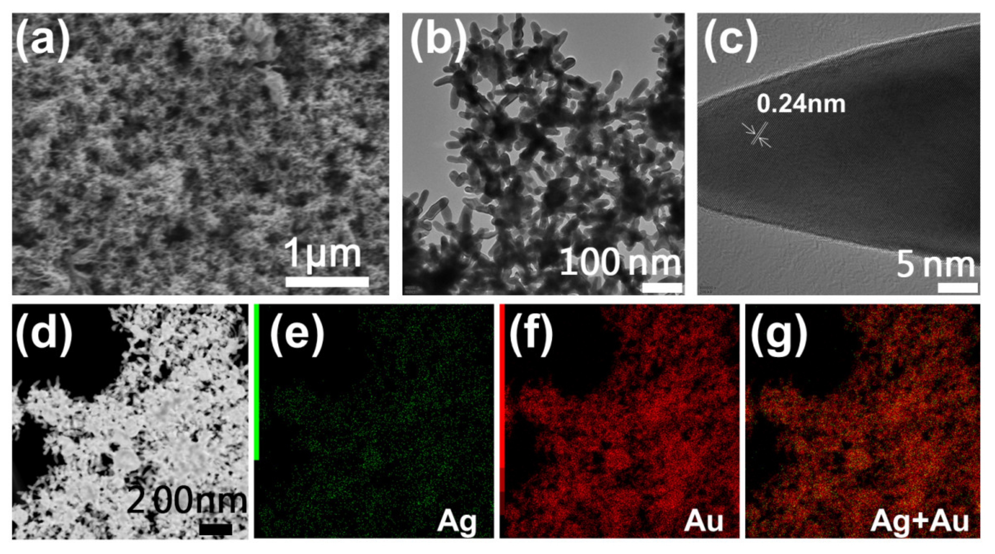
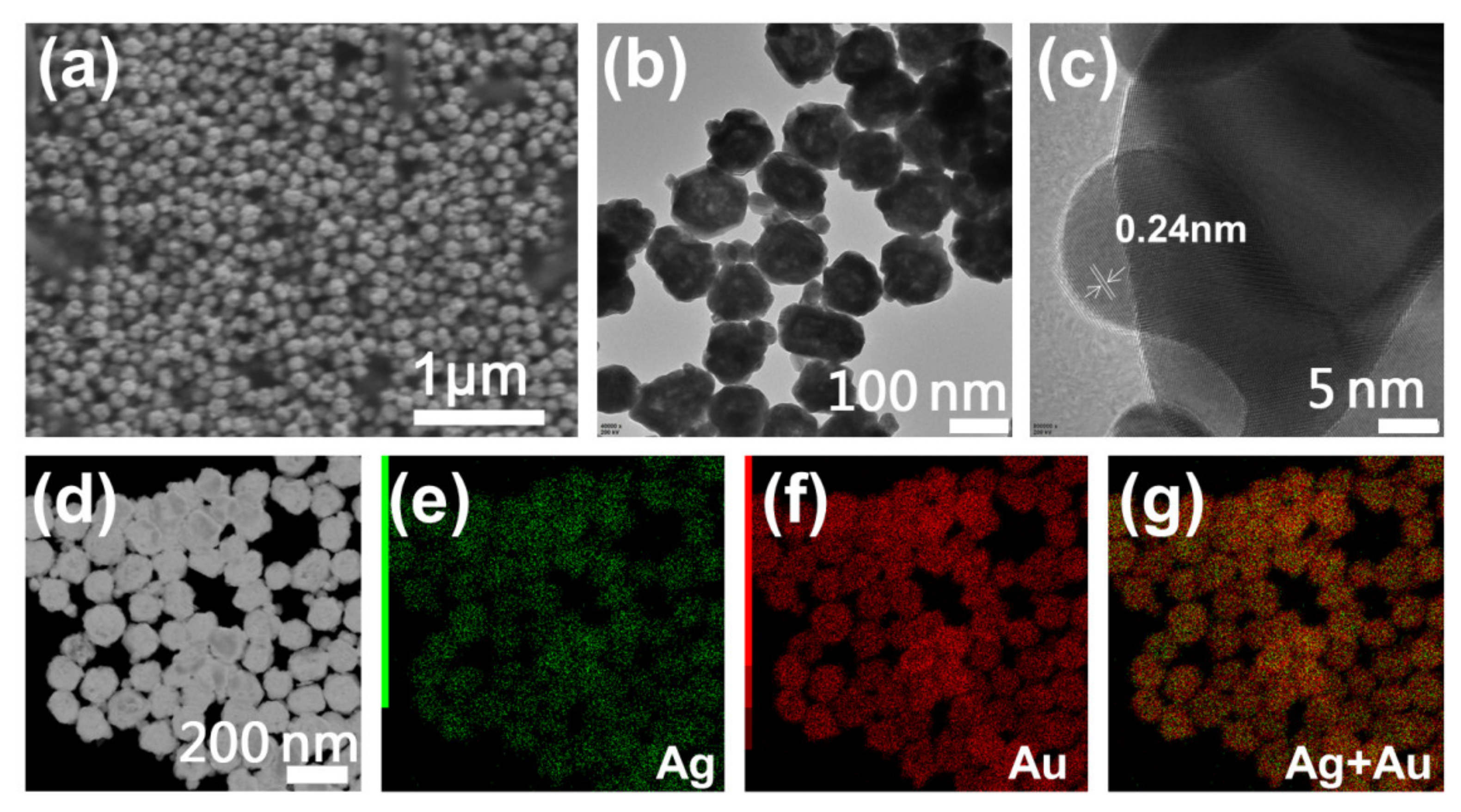
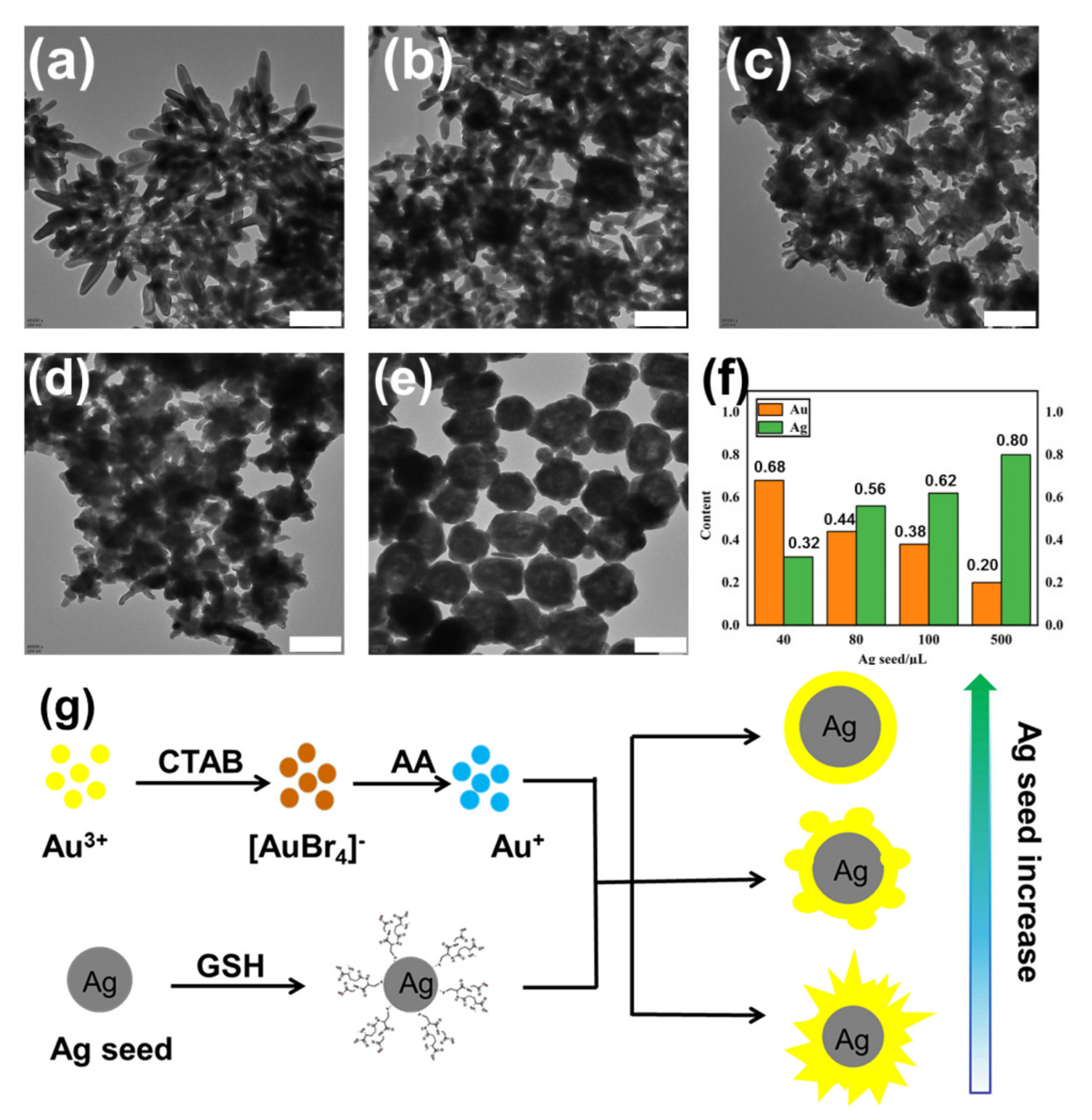
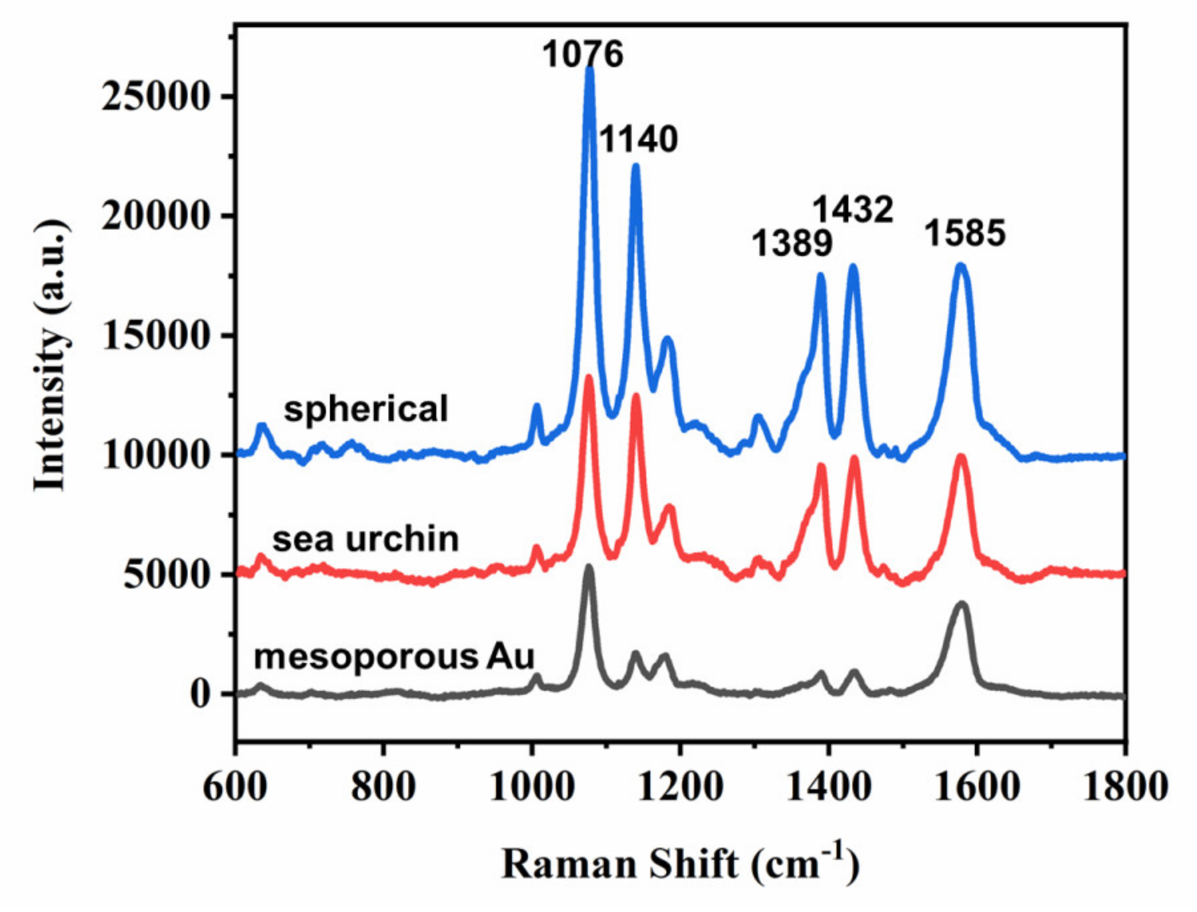
3. Results and Discussion
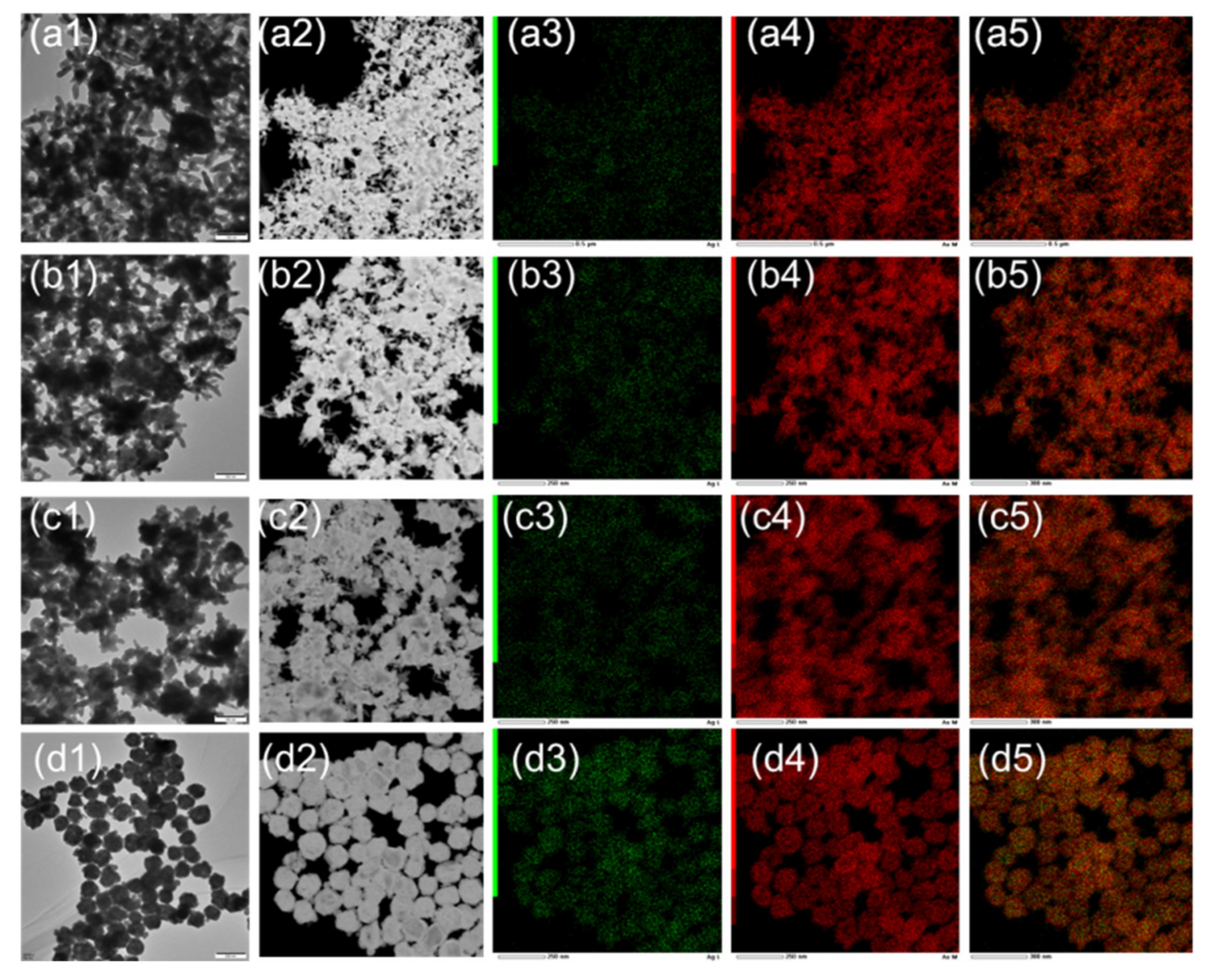
Characterization of Ag@Au NPs
4. Conclusions
Author Contributions
Funding
Institutional Review Board Statement
Informed Consent Statement
Data Availability Statement
Acknowledgments
Conflicts of Interest
Appendix A


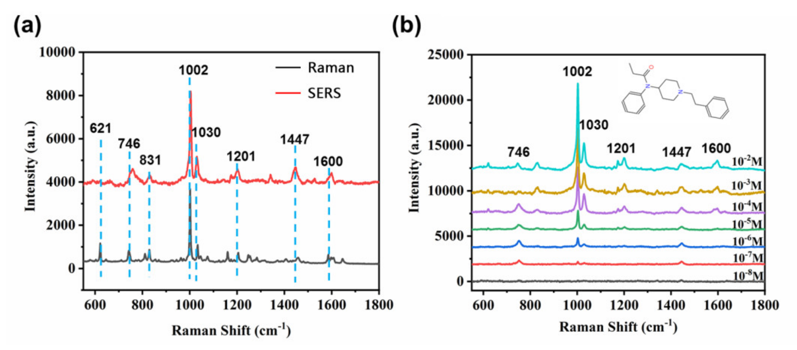
| Fentanyl | |
|---|---|
| 746 | δ(C–C–C), δ(N–CH3) |
| 831 | ν(N–C–C–C) |
| 1002 | δ(CH) Ar |
| 1030 | ν(C=C), δ(CH2) twisting in aliphatic ring |
| 1201 | δ(C–C) benzyl stretch |
| 1447 | δ(CH2) scissoring |
| 1600 | ν(C=C) Ar |
References
- Ding, S.Y.; Yi, J.; Li, J.F.; Ren, B.; Wu, D.Y.; Panneerselvam, R.; Tian, Z.Q. Nanostructure-based plasmon-enhanced Raman spectroscopy for surface analysis of materials. Nat. Rev. Mater. 2016, 1, 16021–16036. [Google Scholar] [CrossRef]
- Li, J.F.; Zhang, Y.J.; Ding, S.Y.; Panneerselvam, R.; Tian, Z.Q. Core–Shell Nanoparticle-Enhanced Raman Spectroscopy. Chem. Rev. 2017, 117, 5002–5069. [Google Scholar] [CrossRef]
- Dong, D.; Yap, L.W.; Smilgies, D.M.; Si, K.J.; Shi, Q.; Cheng, W. Two-dimensional gold trisoctahedron nanoparticle superlattice sheets: Self-assembly, characterization and immunosensing applications. Nanoscale 2018, 10, 5065–5071. [Google Scholar] [CrossRef]
- Qin, Y.; Lu, Y.; Pan, W.; Yu, D.D.; Zhou, J.G. One-pot synthesis of hollow hydrangea Au nanoparticles as a dual catalyst with SERS activity for in situ monitoring of a reduction reaction. RSC Adv. 2019, 9, 10314–10319. [Google Scholar] [CrossRef] [Green Version]
- Ha, M.J.; Kim, J.H.; You, M.; Li, Q.; Fan, C.H.; Nam, J.M. Multicomponent Plasmonic Nanoparticles: From Heterostructured Nanoparticles to Colloidal Composite Nanostructures. Chem. Rev. 2019, 119, 12208–12278. [Google Scholar] [CrossRef]
- Qin, Y.; Pan, W.F.; Yu, D.D.; Lu, Y.X.; Wu, W.H.; Zhou, J.G. Stepwise evolution of Au micro/nanocrystals from an octahedron into a truncated ditetragonal prism. Chem. Commun. 2018, 54, 3411–3414. [Google Scholar] [CrossRef]
- Qin, Y.Z.; Lu, Y.X.; Yu, D.D.; Zhou, J.G. Controllable synthesis of Au nanocrystals with systematic shape evolution from an octahedron to a truncated ditetragonal prism and rhombic dodecahedron. CrystEngComm 2019, 21, 5602–5609. [Google Scholar] [CrossRef]
- Litti, L.; Reguera, J.; Abajo, F. Manipulating chemistry through nanoparticle Morphology. Nanoscale Horiz. 2020, 5, 102–108. [Google Scholar] [CrossRef]
- Tian, N.; Zhou, Z.Y.; Yu, N.F.; Wang, L.Y.; Sun, S.G. Direct electrodeposition of tetrahexahedral Pd nanocrystals with high-index facets and high catalytic activity for ethanol electrooxidation. J. Am. Chem. Soc. 2010, 132, 7580–7581. [Google Scholar] [CrossRef]
- Du, J.H.; Sheng, T.; Xiao, C.; Tian, N.; Xiao, J.; Xie, A.; Liu, S.; Zhou, Z.; Sun, S.G. Shape transformation of {hk0}-faceted Pt nanocrystals from a tetrahexahedron into a truncated ditetragonal prism. Chem. Commun. 2017, 22, 3236–3238. [Google Scholar] [CrossRef] [PubMed]
- Guillerm, G.R.; Pablo, D.N.; Antonio, R.; Alejandro, P.; Gloria, T.; Jesús, G.; Luis, B.; Pablo, L.; Luis, G.M.; Mauricio, A.P.; et al. Femtosecond laser reshaping yields gold nanorods with ultranarrow surface plasmon resonances. Science 2017, 358, 640–644. [Google Scholar]
- Langille, M.R.; Zhang, J.; Personick, M.L.; Li, S.; Mirkin, C.A. Stepwise evolution of spherical seeds into 20-fold twinned icosahedra. Science 2012, 337, 954–957. [Google Scholar] [CrossRef] [PubMed]
- Cai, Z.; Hu, Y.; Sun, Y.; Gu, Q.; Wu, P.; Cai, C.; Yan, Z. Plasmonic SERS Biosensor Based on Multibranched Gold Nanoparticles Embedded in Polydimethylsiloxane for Quantification of Hematin in Human Erythrocytes. Anal. Chem. 2021, 93, 1025–1032. [Google Scholar] [CrossRef] [PubMed]
- Zhao, M.; Chen, Z.; Shi, Y.; Hood, Z.D.; Lyu, Z.; Xie, M.; Chi, M.; Xia, Y. Kinetically Controlled Synthesis of Rhodium Nanocrystals with Different Shapes and a Comparison Study of Their Thermal and Catalytic Properties. J. Am. Chem. Soc. 2021, 143, 6293–6302. [Google Scholar] [CrossRef] [PubMed]
- Zhao, J.; Pinchuk, A.O.; McMahon, J.M.; Li, S.; Ausman, L.K.; Atkinson, A.L.; Schatz, G.C. Methods for Describing the Electromagnetic Properties of Silver and Gold Nanoparticles. Acc. Chem. Res. 2008, 41, 1710–1720. [Google Scholar] [CrossRef]
- Abajo, F.J. Colloquium: Light scattering by particle and hole arrays. Rev. Mod. Phys. 2007, 79, 1267–1290. [Google Scholar] [CrossRef] [Green Version]
- Murphy, C.J.; Gole, A.M.; Stone, J.W.; Sisco, P.N.; Alkilany, A.M.; Goldsmith, E.C.; Baxter, S.C. Gold Nanoparticles in Biology: Beyond Toxicity to Cellular Imaging. Acc. Chem. Res. 2008, 41, 1721–1730. [Google Scholar] [CrossRef] [PubMed]
- Schlücker, S. Surface-enhanced Raman spectroscopy: Concepts and chemical applications. Angew. Chem. Int. Ed. 2014, 53, 4756–4795. [Google Scholar] [CrossRef]
- Sperling, R.A.; Gil, P.R.; Zhang, F.; Zanella, M.; Parak, W.J. Biological applications of gold nanoparticles. Chem. Soc. Rev. 2008, 37, 1896–1908. [Google Scholar] [CrossRef]
- Guisbiers, G.; Mendoza-Cruz, R.; Bazán-Díaz, L.; Elázquez-Salazar, J.J.; Mendoza-Perez, V.R.; Torres, J.A.; Lopez, J.L.; Carrizales, J.M.; Whetten, R.L.; Yacamán, M.J. Response to “Comment on ‘Electrum, the Gold–Silver Alloy, from the Bulk Scale to the Nanoscale: Synthesis, Properties, and Segregation Rules’”. ACS Nano 2016, 10, 188–198. [Google Scholar] [CrossRef] [PubMed]
- Cardinal, M.F.; González, B.R.; Puebla, R.A.; Juste, J.P.; Marzán, L.M. Modulation of Localized Surface Plasmons and SERS Response in Gold Dumbbells through Silver Coating. J. Phys. Chem. C 2010, 114, 10417–10423. [Google Scholar] [CrossRef]
- Gao, Z.; Shao, S.; Gao, W.; Tang, D.; Tang, D.; Zou, S.; Kim, M.J.; Xia, X. Morphology-Invariant Metallic Nanoparticles with Tunable Plasmonic Properties. ACS Nano 2021, 15, 2428–2438. [Google Scholar] [CrossRef]
- Dong, D.; Shi, Q.; Sikdar, D.; Zhao, Y.; Liu, Y.; Fu, R.; Premaratne, M.; Cheng, W. Site-specific Ag coating on concave Au nanoarrows by controlling the surfactant concentration. Nanoscale Horiz. 2019, 4, 940–946. [Google Scholar] [CrossRef]
- Liao, H.; Fisher, A.; Xu, Z.J. Surface Segregation in Bimetallic Nanoparticles: A Critical Issue in Electrocatalyst Engineering. Small 2015, 11, 3221–3246. [Google Scholar] [CrossRef]
- Sun, Y.; Mayers, B.T.; Xia, Y. Template-Engaged Replacement Reaction: A One-Step Approach to the Large-Scale Synthesis of Metal Nanostructures with Hollow Interiors. Nano Lett. 2002, 2, 481–485. [Google Scholar] [CrossRef]
- Sun, Y.; Xia, Y. Shape-Controlled Synthesis of Gold and Silver Nanoparticles. Science 2002, 298, 2176–2179. [Google Scholar] [CrossRef] [Green Version]
- Xuan, H.; Nguyen, D.D.; Thi, T.H.P.; Nguyen, V.T.; Nguyen, X.C.; Vu, V.T. Tunable LSPR of silver/gold bimetallic nanoframes and their SERS activity for methyl red detection. RSC Adv. 2021, 11, 14596–14606. [Google Scholar]
- Ahn, J.; Wang, D.; Ding, Y.; Zhang, J.; Qin, D. Site-Selective Carving and Co-Deposition: Transformation of Ag Nanocubes into Concave Nanocrystals Encased by Au–Ag Alloy Frames. ACS Nano 2017, 12, 298–307. [Google Scholar] [CrossRef] [PubMed]
- Yang, M.; Hood, Z.D.; Yang, X.; Chi, M.; Xia, Y. Facile synthesis of Ag@Au core–sheath nanowires with greatly improved stability against oxidation. Chem. Commun. 2017, 53, 1965–1968. [Google Scholar] [CrossRef]
- Choi, S.; Han, S.I.; Jung, D.; Hwang, H.J.; Lim, C.; Bae, S.; Park, O.K.; Tschabrunn, C.M.; Lee, M.; Bae, S.Y.; et al. Highly conductive, stretchable and biocompatible Ag–Au core–sheath nanowire composite for wearable and implantable bioelectronics. Nat. Nanotech. 2018, 13, 1748–3387. [Google Scholar] [CrossRef]
- Irene, C.; Ramon, A.A.; Nicolas, P.P. Gold-spiked coating of silver particles through cold nanowelding. Nanoscale 2021, 13, 4530–4536. [Google Scholar]
- Lee, H.; Ahn, H.Y.; Mun, J.; Lee, Y.Y.; Kim, M.; Cho, N.H.; Chang, K.; Kim, W.S.; Rho, J.; Nam, K. Amino-acid-and peptide-directed synthesis of chiral plasmonic gold nanoparticles. Nature 2018, 556, 360–365. [Google Scholar] [CrossRef] [PubMed]
- Qin, Y.Z.; Wu, Y.; Wang, B.; Wang, J.; Zong, X.; Yao, W. Controllable preparation of sea urchin-like Au NPs as a SERS substrate for highly sensitive detection of the toxic atropine. RSC Adv. 2021, 11, 19813–19818. [Google Scholar] [CrossRef]
- Huang, Y.F.; Zhu, H.P.; Liu, G.K.; Wu, D.Y.; Ren, B.; Tian, Z.Q. When the signal is not from the original molecule to be detected: Chemical transformation of para-aminothiophenol on Ag during the SERS measurement. J. Am. Chem. Soc. 2010, 132, 9244–9246. [Google Scholar] [CrossRef] [PubMed]
- Haddad, A.; Greencand, O.; Lombardi, J.R. Detection of fentanyl in binary mixtures with cocaine by use of surface-enhanced Raman spectroscopy. Spectrosc. Lett. 2019, 8, 462–472. [Google Scholar] [CrossRef]
- Matas, M.D.; Edwards, H.G.; Lawson, E.E.; Shields, L.; York, P. FT-Raman spectroscopic investigation of a pseudopolymorphic transition in caffeine hydrate. J. Mol. Struct. 1998, 440, 97–104. [Google Scholar] [CrossRef]


Publisher’s Note: MDPI stays neutral with regard to jurisdictional claims in published maps and institutional affiliations. |
© 2021 by the authors. Licensee MDPI, Basel, Switzerland. This article is an open access article distributed under the terms and conditions of the Creative Commons Attribution (CC BY) license (https://creativecommons.org/licenses/by/4.0/).
Share and Cite
Qin, Y.; Wang, B.; Wu, Y.; Wang, J.; Zong, X.; Yao, W. Seed-Mediated Preparation of Ag@Au Nanoparticles for Highly Sensitive Surface-Enhanced Raman Detection of Fentanyl. Crystals 2021, 11, 769. https://0-doi-org.brum.beds.ac.uk/10.3390/cryst11070769
Qin Y, Wang B, Wu Y, Wang J, Zong X, Yao W. Seed-Mediated Preparation of Ag@Au Nanoparticles for Highly Sensitive Surface-Enhanced Raman Detection of Fentanyl. Crystals. 2021; 11(7):769. https://0-doi-org.brum.beds.ac.uk/10.3390/cryst11070769
Chicago/Turabian StyleQin, Yazhou, Binjie Wang, Yuanzhao Wu, Jiye Wang, Xingsen Zong, and Weixuan Yao. 2021. "Seed-Mediated Preparation of Ag@Au Nanoparticles for Highly Sensitive Surface-Enhanced Raman Detection of Fentanyl" Crystals 11, no. 7: 769. https://0-doi-org.brum.beds.ac.uk/10.3390/cryst11070769