Article

Luminescent Color-Adjustable Europium and Terbium Co-Doped Strontium Molybdate Phosphors Synthesized at Room Temperature Applied to Flexible Composite for LED Filter

Research and Business Development Foundation, Engineering Building, Silla University, Busan 45985, Korea
Submission received: 6 April 2022 / Revised: 13 April 2022 / Accepted: 13 April 2022 / Published: 15 April 2022
(This article belongs to the Topic Optoelectronic Materials)

Abstract

:
In this study, terbium and europium rare-earth ions were single-doped and co-doped to synthesized SoMoO4 phosphor at room temperature. The samples prepared synthesized crystalline SrMoO4 powder by the co-precipitation. Samples had a tetragonal structure in XRD analysis and d(112) spacing was changed by rare-earth doping. As the amount of rare earth added increased, a secondary phase appeared, and the structure changed. The synthesized SrMoO4:Tb3+ phosphors showed a green light emission at 544 nm under 287 nm, SrMoO4:Eu3+ phosphors showed a red light emission at 613 nm under 290 nm, and SrMoO4:[Eu3+]/[Tb3+] phosphor showed a yellow-white light emission at 544 and 613 nm when excited at 287 nm. The synthesized phosphor exhibited a change in green and red luminescence intensity based on the amount of Eu3+ doped and showed strong red luminescence as the Eu3+ doping increased. To use the SrMoO4:[Eu3+]/[Tb3+] phosphor with these characteristics in an LED color filter, a flexible composite prepared by mixing with PDMS showed green, red, and yellow-white emission under a UV-lamp.

1. Introduction

Recently, considerable research has been conducted to develop a white-light-emitting (wLED) diode with a high color-rendering index and excellent color reproducibility to replace incandescent and fluorescent lamps [1,2,3]. In general, wLEDs are manufactured in three ways. There is a method of applying yellow phosphor on the upper part of the blue LED chip or applying red and green phosphors on the upper part of the blue LED chip with the same method, and white light emission is achieved by applying red, green, and blue phosphor powder on the surface of the near-ultraviolet LED chip [4,5,6]. Among the emission wavelength ranges of the phosphors used in an LED chip, the intensity of red and green light emission is known as an important factor to increase the color-rendering index of the white light emission [7]. Oxidized compounds including molybdate, aluminate, borate, and silicate exhibit strong light absorbance in the ultraviolet region [8,9]. Among them, molybdate has a scheelite-type crystal structure and is considered as a good host material for light-emitting materials due to its excellent chemical and thermal stability [10,11,12]. In addition, molybdate compounds are important materials that can be applied to wLEDs, lasers, optical fibers, catalysts, etc., in addition to phosphors. Among molybdate compounds with these advantages, strontium molybdate (SrMoO4) is doped with rare-earth ions and has a high luminous efficiency and chemical stability, thereby emerging as a phosphor material, and extensive research has been conducted on it [13,14,15,16]. Wang et al. produced a CaMoO4:Eu3+ phosphor with a three-dimensional walnut-shaped shape by hydrothermal synthesis and reported that the size of the crystal grains increased as the heat treatment temperature was increased. In addition, when excited at 394 nm, red light emission was observed with a peak at 615 nm [17]. Du et al. synthesized a phosphor by doping rare-earth (RE) Eu3+ ions into AMoO4 (A = Mg, Ca, Sr, Ba) oxide with a tetragonal crystal structure by a solid-state reaction method. Among them, the CaMoO4 phosphor doped with Eu3+ ions exhibited the strongest red emission intensity [18]. Li et al. produced SrMoO4 green phosphor doped with Tb3+ and Na3+ ions by sintering at 600~750 °C for 3 h using the solid-state reaction method. It has been reported that the maximum emission intensity of the phosphor occurs at 548 nm due to the 5D47F5 transition of the Tb3+ ions when excited at 375 and 488 nm [19]. Jung et al. reported on producing a thin film and disk-shaped material capable of detecting deep UV by co-doping Ce3+ and Dy3+ ions into hexagonal boron nitride to synthesized white-light-emitting nanocrystal phosphors [20]. In this study, a high-efficiency green-emitting phosphor was synthesized at room temperature by co-precipitation by doping the host crystal SrMoO4 with activator ions Tb3+ and Eu3+ single-doped and [Eu3+]/[Tb3+] co-doped at different Eu3+ concentrations. In particular, the optimal concentration of [Eu3+]/[Tb3+] co-doped ions with the maximum yellow-white emission intensity suitable for application to wLEDs was determined, and the crystallinity, particle shape, and luminescence characteristics of the phosphors according to the change in the concentration of the activator ions were investigated. In addition, a filter that can be applied to a UV-LED was manufactured by fabricating a flexible composite.

2. Materials and Methods

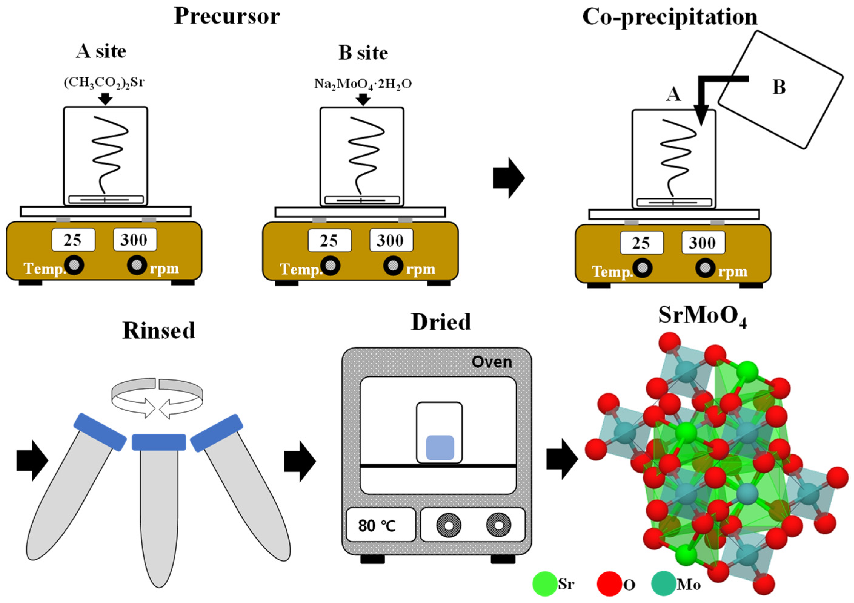
2.1. Synthesis of the Crystalline CaMoO4 and CaMoO4:RE3+ Phosphors via Co-Precipitation

Starting materials: strontium acetate ((CH3COO)2Sr), sodium molybdate dihydrate (Na2MoO4·2H2O), Terbium (III) nitrate hydrate (Tb(NO3)3·xH2O, Tb3+), and europium (III) nitrate hydrate (Eu(NO3)3·xH2O, Eu3+).
Two beakers were prepared as shown in Figure 1. Next, 50 mL of distilled water and 1 mmol of barium acetate were added to the ‘A’ beaker and stirred. In the other ‘B’ beaker, 1 mmol of sodium molybdate and 50 mL of distilled water were added and stirred. The solution completely dissolved in the ‘B’ beaker was slowly poured into beaker ‘A’ and stirred for 2 h to react. After the 2 h, centrifugation was performed at 4000 rpm for 5 min to recover the white powder produced by the reaction. To remove any unreacted substances from the recovered powder, it was washed with 50 mL of distilled water, and the powder obtained after centrifugation under the same conditions was dried at 80 °C for 18 h. In the same way, a phosphor to which Tb3+ and Eu3+ ions single-doped and co-doped were added was synthesized, and the phosphor was prepared by single-doped (0.025 mmol) and changing the amount of [Eu3+]/[Tb3+]~(0.1, 0.2, 0.3, 0.5, 0.8) added to beaker ‘A’.

2.2. Fabricated Flexible Composite for an LED

The flexible composite was prepared by mixing polydimethylsiloxane (PMDS) 10 wt.% and phosphor powder 1 wt.%, pouring them into a square-shaped frame and curing them at 80 °C for 2 h.

2.3. Characterization

The crystal structure of the SrMoO4 and SrMoO4:RE3+ powders synthesized by coprecipitation was measured at a diffraction angle of 10~70° at a rate of 4° per minute using an X-ray diffraction device (Cu-Kα radiation; 0.15406 nm, 40 kV and 20 mA, X’Pert PROMPD, XRD). Optical characteristics were irradiated at a photomultiplier voltage of 300 V using a fluorescence spectrometer (FS-2, Scinco) at room temperature. The fine shape of the surface and the size of the crystal particles were photographed by a field radial scanning electron microscope (FE-SEM, acceleration voltage of 3 kV, a working distance of 6.1 mm, and a magnification of 5 to 30 k), and the components were analyzed by energy dispersion X-ray spectroscopy (EDS, 5 min at an acceleration voltage of 15 kV with surface mapping).

3. Results and Discussion

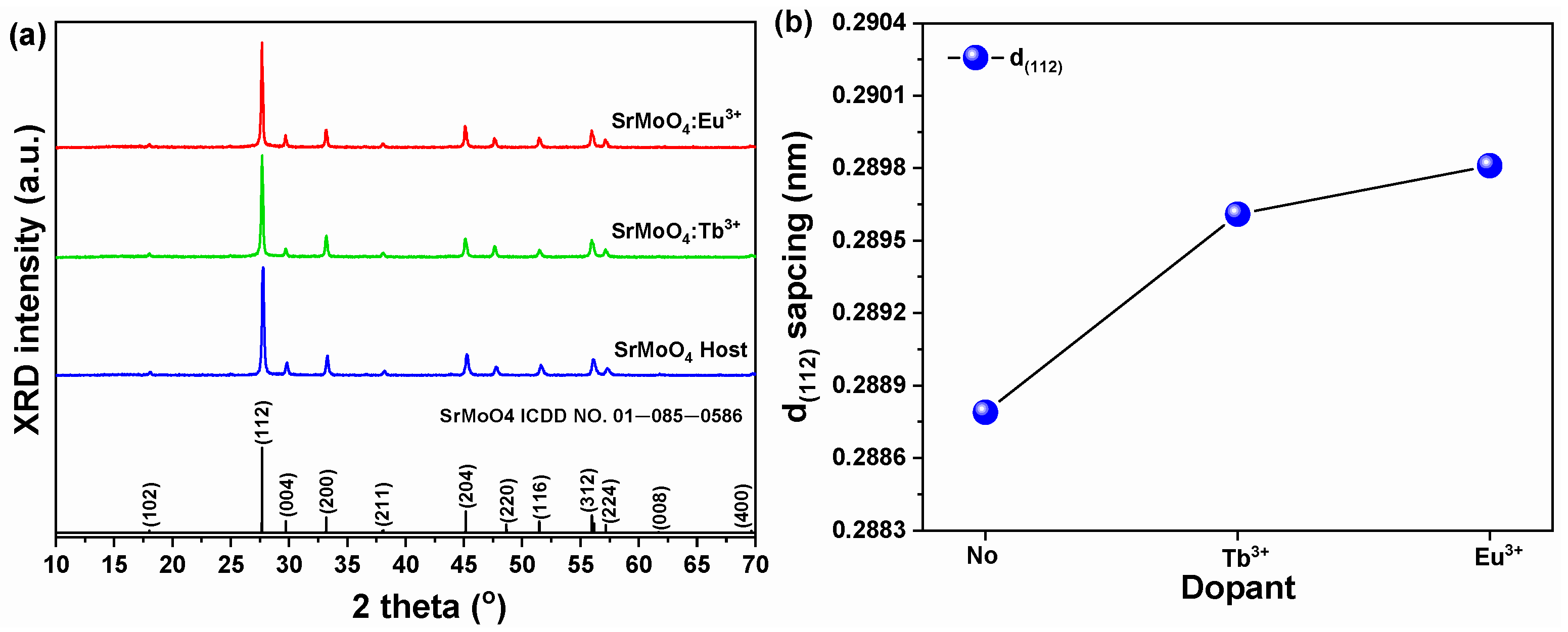
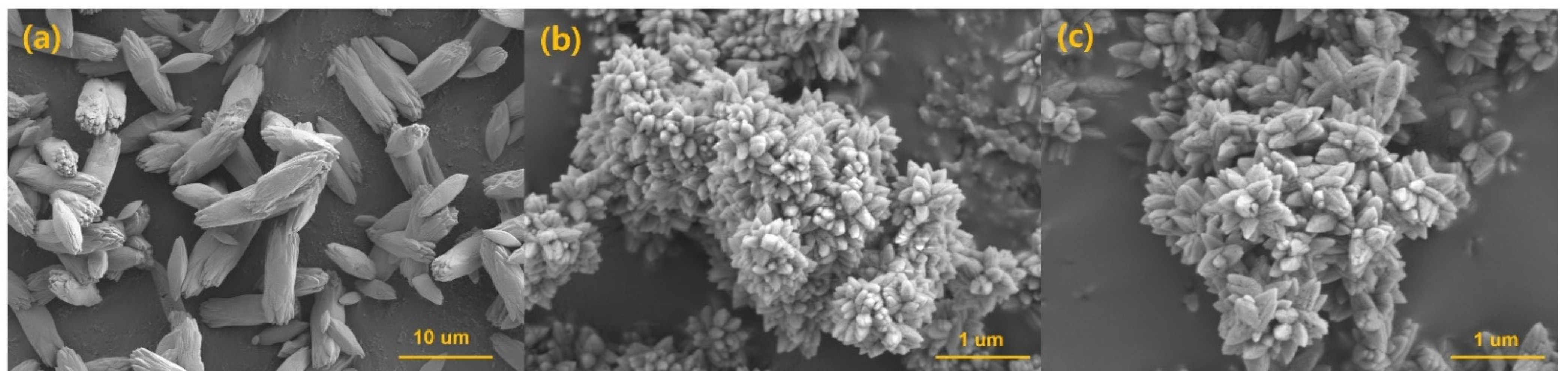
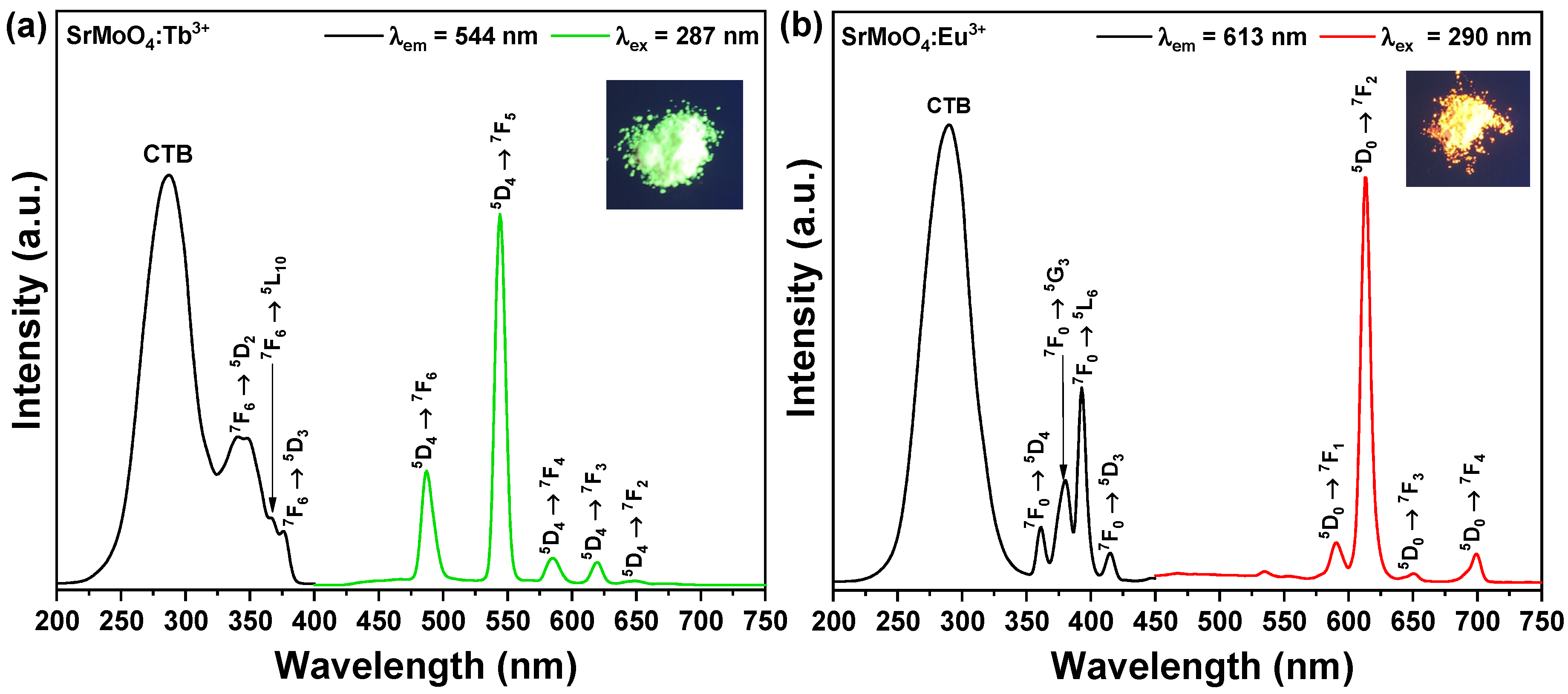
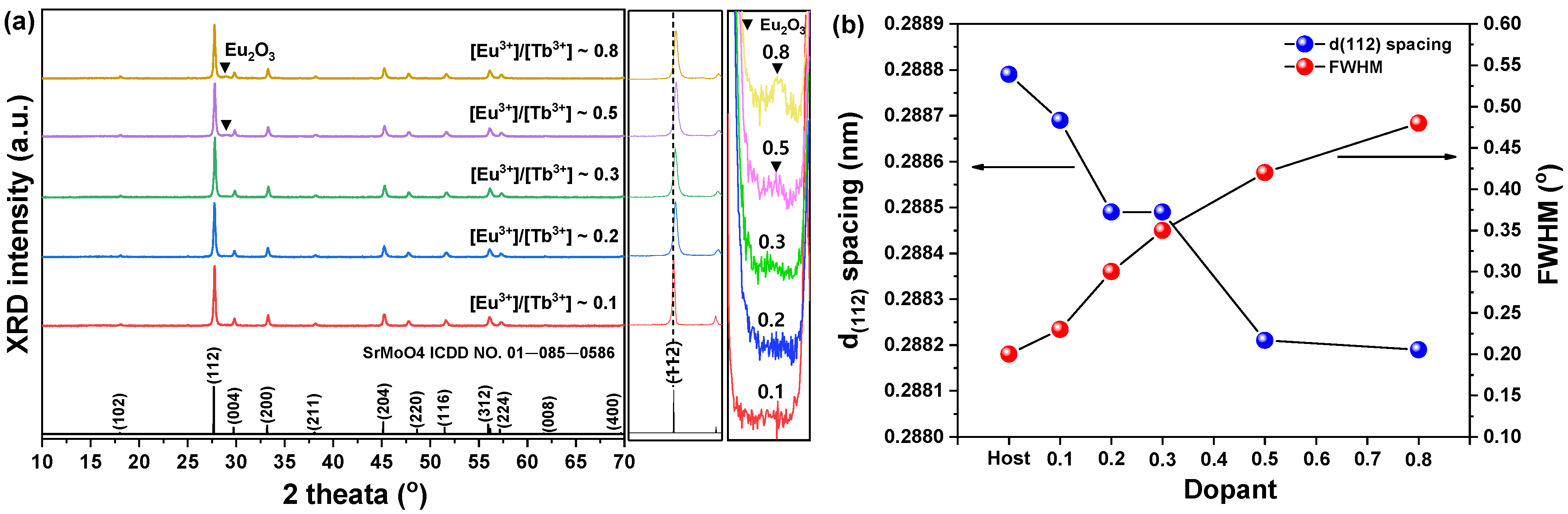
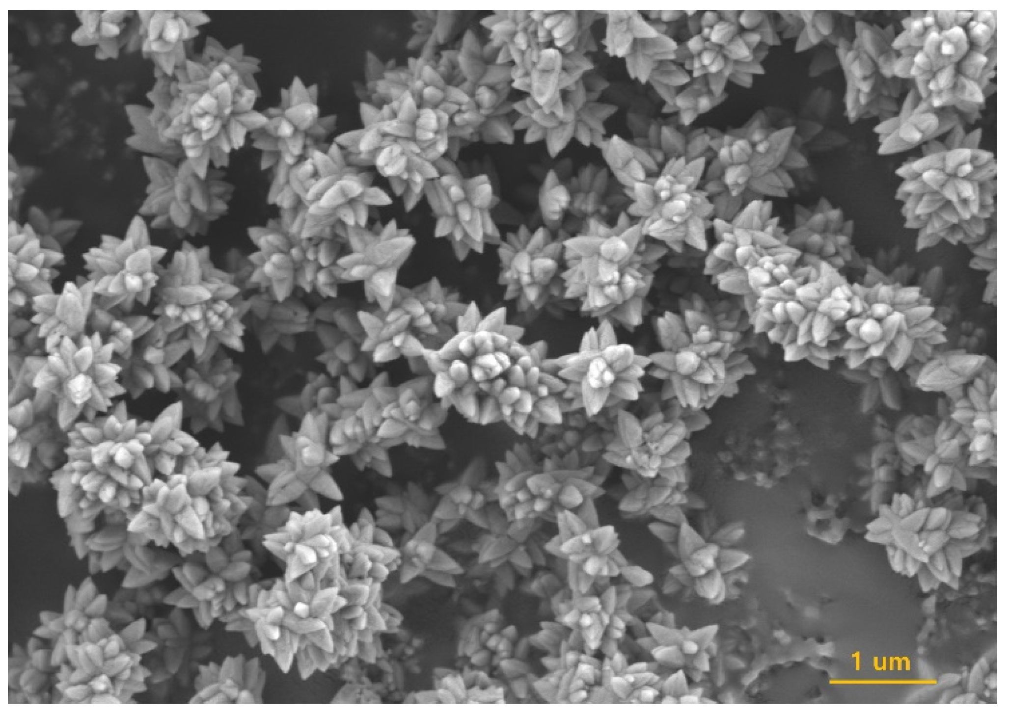
3.1. Characteristics of SrMoO4 and Single-Doped SrMoO4

Figure 2a shows that the XRD data of SrMoO4 and SrMoO4:RE3+ phosphor powder samples synthesized at room temperature by co-precipitation had a tetragonal (a = 5.394, b = 5.394, c = 12.020 Å) structure with main peaks at (112), (004), (200), (204), (220), (116), (312), and (224) which were found to be consistent with ICDD card No. 01-085-0586 [21]. The MXO4 (M = Sr, X = Mo) particle precipitation –M2+ cations act as electron pair receptors (Lewis acids) and XO42− anions as electron pair donors (Lewis bases). The reaction between the two substances (M2+ ← :XO42−) continues to bond. The lowest molecular orbital energy of the Lewis acid interacts with the highest molecular orbital energy of the Lewis base and eventually synthesizes the MXO4 particles [22,23]. As the amount of Tb3+ and Eu3+ ions added was increased, the interplanar distance on the main peak (112) increased and then decreased (Figure 2b). When the amount of rare-earth ions added is small, it is considered that the structure of the lattice is distorted when a space between the grids is opened, and the quantity of added ions is continuously increased due to rare-earth ions having a relatively large ionic radius [24,25].
The particle size of the SrMoO4 and SrMoO4:RE3+ samples was calculated using the (112) phase, which is the main peak, and the Scherrer’s equation [26] as follows:
D = n λ / B c o s θ
Here, n is Scherrer’s constant 0.9; λ is the X-ray wavelength (0.15406 nm); θ is the diffraction angle, and B is the FWHM. SrMoO4 was calculated to be about 100 nm, SrMoO4:Tb3+ was calculated to be about 32 nm, and SrMoO4:Eu3+ was calculated to be about 38 nm. Figure 3 shows the images taken by FE-SEM to observe the actual particle size and surface shape of the synthesized powder. SrMoO4 was about 6.91 μm in the longitudinal direction and about 2.15 μm in the transverse direction. SrMoO4:Tb3+ was about 0.31 μm and about 0.13 μm in the transverse direction. SrMoO4:Eu3+ was about 0.41 μm and about 0.15 μm in the transverse direction. Both these samples had an octahedron-like shape and showed a longitudinal growth. According to Bharat et al., BaMoO4 synthesized by reacting with MoO4−2, a monomer of an oxyanion, grows in the longitudinal direction immediately after mixing the Ba aqueous solution and Mo aqueous solution. It was reported that the shuttle shape appeared clearly due to the growing Oswald’s ripping. It was synthesized using a basic material and explained by the action of the base [27]. Cavalcante et al. reported that, in the case of particles synthesized by a high-energy reaction such as co-precipitation, aggregation between small particles is induced, and octahedral growth is induced in a basic atmosphere [28]. In Figure S1 (see Supplementary Materials), Sr, Mo, and O components and the addition of Tb and Eu were confirmed through SEM-EDS component analysis.
SrMoO4 synthesized by co-precipitation as a basic material in this study is considered to have such a shape. The photoluminescence excitation (PLE) and photoluminescence (PL) spectra of the SrMoO4:Tb3+ and SrMoO4:Eu3+ phosphor are shown in Figure 4. Figure 4a shows the PLE spectrum (black line) of the SrMoO4:Tb3+ phosphor monitored at an emission wavelength of 544 nm. The PLE spectrum of the SrMoO4:Tb3+ phosphors contain a charge transfer state band (CTB) [29] and f–f transitions due to the presence of the Tb3+ ions. CTB includes a broad band in the wavelength range of 220–325 nm. This band could rise due to the Mo-O and Tb–O charge transfer [30]. The f–f transitions associated with a number of weak peaks are located at 339, 350, 368, and 376 nm which are attributed to the 7F65D1, 7F65D2, 7F65L10, and 7F65D3 transitions of the Tb3+ ions, respectively. The PL spectrum (green line) was observed for the excitation wavelength 287 nm, stemming from Tb–O CTB [31]. The emission spectra contain five peaks centered at 487, 544, 585, 619, and 649 nm wavelengths, corresponding to the 5D47F6, 5D47F5, 5D47F4, 5D47F3, and 5D47F2 transitions [32]. In addition, among the above five components of emission spectra, the intensity of the green emission spectrum due to the 5D47F5 (544 nm) magnetic dipole transition was the strongest, and this emission intensity was the strongest due to the 5D47F6 (487 nm) electric dipole transition of the Tb3+ ion. It was 3.29 times that of the blue emission intensity. From this result, it can be confirmed that the Tb3+ ion located in the host crystal is located at an inversion symmetry site [33]. Figure 4b shows the results of the PLE (black line) and PL (red line) spectra when measured in the SrMoO4:Eu3+ phosphor powder. SrMoO4:Eu3+ phosphor controlled to an emission wavelength of 613 nm of PLE spectra was CTB observed between Mo-O ions with a wide bandgap over the region of 220–320 nm and a peak at 292 nm, and the other type of absorption signal is at 320~450 nm. These absorption peaks were 4f–4f transition signals of Eu3+ ions. The PLE spectra monitored at the peaks at 361 (7F05D4), 381 (7F05G3), 393 (7F05L6), and 415 (7F05D3) nm are correspondingly transition signals of Eu3+ ions located in the host crystal [34]. The PL spectrum by which the phosphor was measured excited 290 nm, showed the strongest absorption intensity. The phosphor powder was composed of an emission spectrum having the strongest emission intensity with a peak at a wavelength of 613 nm, a peak emission spectrum at 590 nm having relatively weak emission intensity, and an emission spectrum having peaks at 651 nm and 700 nm. Here, 613 nm is the signal induced by the 5D07F2 electric dipole transition, 590 nm is the 5D07F1 magnetic dipole transition, and the two red emission spectra (651 and 700 nm) are the 5D07F3 and 5D07F4 electric dipole signals. In this experiment, because the intensity of the red emission (613 nm) spectrum stemming from the 5D07F2 electric dipole transition was 9.89 times greater than the intensity of the orange emission (590 nm) from the 5D07F1 magnetic dipole transition, Eu3+ ions in the host lattice were found to be located at the sites of inversion symmetry, not inversion symmetry [35,36].

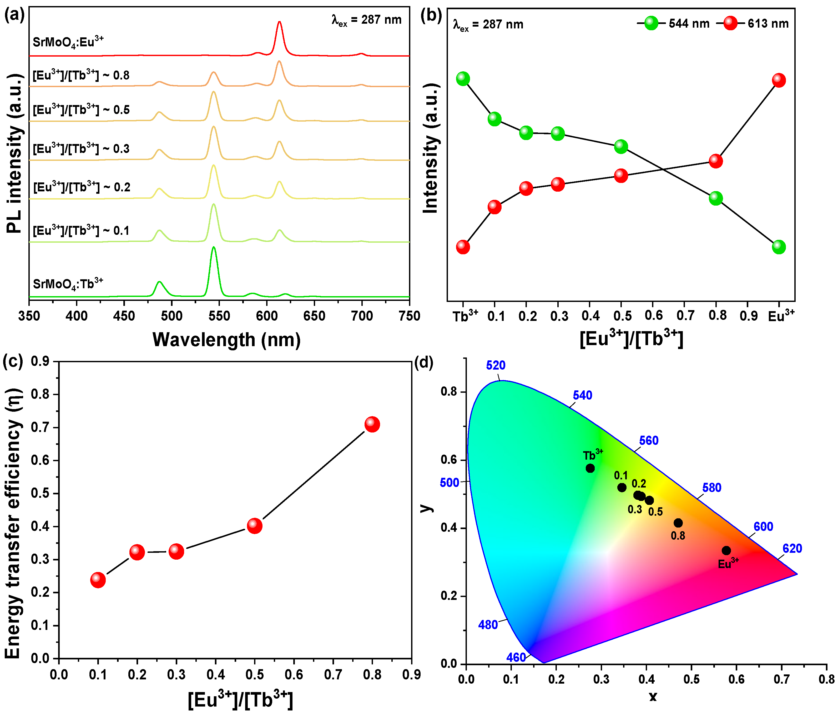
3.2. Characteristics of [Eu3+]/[Tb3+] Co-Doped SrMoO4

The XRD pattern of the sample prepared by fixing Tb3+ (2.5 mol%) ions and changing the amount of Eu3+ ions added is shown in Figure 5. The XRD patterns of the SrMoO4:[Eu3+]/[Tb3+] samples synthesized by doping with rare-earth ions were shown to be consistent with the ICDD card No. 01-085-0586. However, from the [Eu3+]/[Tb3+]~0.5 samples, the Eu2O3 secondary phase was observed as the Eu3+ addition amount increased. As the amount of rare-earth ions added increased, a single oxide phase was detected without doping in the lattice, so the optimal rare-earth doping condition was considered to be [Eu3+]/[Tb3+]~0.3. In addition, excessive doping of rare-earth ions showed that d(112) spacing changed excessively (Figure 5b), so it was thought that excessive doping of rare-earth ions changed the crystal structure of SrMoO4 and adversely affected the expression of a secondary phase. It was also thought to be the cause of the changes in the lattice constant due to the doping of rare-earth ions with relatively large ionic radii (r(Sr2+ = 1.26 Å), r(Mo6+ = 0.59 Å), r(Tb3+ = 1.18 Å), r(Eu3+ = 1.06 Å)) [37,38]. Additionally, as full width at half maximum (FWHM) tends to increase as the amount of rare-earth ions added increases, it is considered that excessive doping of rare earths affects the crystal structure and crystallinity.
Figure 6 shows an FE-SEM image of the SrMoO4:[Eu3+]/[Tb3+] phosphor doped with two kinds of rare earths. It had the same octahedron shape as the single-doped specimen above and had a size of 0.35 um in the longitudinal direction and 0.12 um in the transverse direction. In the EDS component analysis, Sr, Mo, O, Eu, and Tb were detected, and each component could be identified (see Supplementary Materials Figure S2).
Figure 7 shows the PL spectrum measured by excitation of the synthesized SrMoO4:[Eu3+]/[Tb3+] phosphor powder with a wavelength of 287 nm while fixing Tb3+ ions and changing the doping amount of Eu3+ ions. In the [Eu3+]/[Tb3+]~0.1 samples, emission spectra of two types of activator ions were observed. In the first case, a green light emission signal with a peak at 544 nm and a blue light emission signal at 487 nm appeared due to electric dipole transition (5D47F5) and magnetic dipole transition (5D47F6) of the activator Tb3+ ion [39], and the second was 619 nm due to the electric dipole transition of Eu3+ ion. A red emission spectrum having peaks at 651 nm and 700 nm, due to the 5D07F3 and 5D07F4 transition of Eu3+ ions with relatively weak emission intensity, was observed [40]. In this case, the intensity of green emission at 544 nm by Tb3+ ions was about 3.5 times stronger than that of red emission at 619 nm by Eu3+ ions. When the addition amount of Eu3+ was increased, the intensity of the emission spectrum due to the transition of Tb3+ ions decreased, the intensity of the emission spectrum due to the transition of Eu3+ ions increased, and the intensity of the emission spectrum was reversed at [Eu3+]/[Tb3+]~0.8.
Figure 7b shows the change in the intensity of the emission wavelength according to the change in the amount of Eu3+ ions added. As the addition amount of Eu3+ ions increased, the main emission signal shifted from green to red emission, and it is believed that the emission energy was converted from Tb3+ ions to Eu3+ ions. The energy transfer efficiency from Tb3+ ions to Eu3+ ions can be calculated using the following equation [41].
η = 1 I / I 0
Here, I0 is the intensity of green and blue emission emitted by Tb3+ ions from a phosphor doped with single Tb3+ ions, and I is the intensity of emission from Tb3+ ions from a phosphor doped with Tb3+ and Eu3+ ions simultaneously. As shown in Figure 7c, the energy transfer efficiency [42,43,44] from Tb3+ ions to Eu3+ ions increases as the doping concentration of Eu3+ ions increases. The energy transfer efficiency increased from 23% to 70%, but the luminescence tax decreased due to the concentration-quenching phenomenon due to the addition of excessive rare earths. In addition, in the XRD data, a secondary phase was found from [Eu3+]/[Tb3+]~0.5 and the crystal structure was changed. It is believed that the addition of excessive rare earths changes the structure and luminescence properties. In the Commission Internationale de L’Eclairage 1931 (CIE 1931) [45] color coordinates, Tb3+ and Eu3+ single-doped specimens are in the green (0.2741, 0.5819) and red (0.4689, 0.4253) coordinates, respectively. Samples doped with Tb3+ and Eu3+ simultaneously moved from green to red as the addition amount of Eu3+ ions increased, and it was found that samples [Eu3+]/[Tb3+]~0.3 (0.3904, 0.4925) and [Eu3+]/[Tb3+]~0.5 (0.4068, 0.4839) were in yellow-white.

3.3. Fabricated Flexible Composite for LED Filter

Figure 8 shows a photograph of a flexible composite made by mixing the synthesized phosphor powder with a PDMS polymer for use as an LED filter. The square-shaped complex appeared white in everyday light but showed unique luminescence properties depending on the doped rare-earth ion in the UV lamp. The complex of SrMoO4:Tb3+ phosphor doped with Tb3+ ions showed green, the complex of SrMoO4:Eu3+ phosphor showed red, and the complex of SrMoO4:[Eu3+]/[Tb3+] phosphor showed yellow-white. SrMoO4 phosphors, which have flexibility due to the PDMS polymer and can be used as various LED filters depending on the type of doped rare earth, were synthesized and their applications were presented. To find out the resistance of the fabricated composite to temperature and humidity, water was put in a beaker and the composite was immersed. The beaker was placed in an 80-degree oven for 72 h and the change in light-emitting characteristics was examined through PL measurement. As shown in Figure 8b, there was no significant change in the light emission intensity, so it is thought to be able to withstand temperature and humidity.

4. Conclusions

Crystalline SrMoO4 was synthesized at room temperature using the coprecipitation method. The synthesized SrMoO4 was able to confirm the main peak (112) phase in XRD analysis and was consistent with ICDD card No. 01-085-0586. Tb3+ and Eu3+ ions were doped, respectively, to make the synthesized SrMoO4 a host phosphor. The doped sample showed green and red light emission as rare-earth ions with a large ion radius were doped, and the lattice constant of the surface of the main peak (112) was increased. For use as a white-light-emitting phosphor, SrMoO4 was co-doped with Tb3+ and Eu3+. The phosphor having two rare earths co-doped with each other had a fixed Tb3+ addition amount, the crystal structure of SrMoO4 changed according to a change in the Eu3+ addition amount, and an E2O3 phase, which was a secondary phase, was found when the Eu3+ ion added amount was increased. In addition, as the amount of Eu3+ ions added increased, the emission characteristics changed from green to red, and yellow-white emission was shown when [Eu3+]/[Tb3+]~0.5. The synthesized phosphor had an octahedron shape and a size of about 0.4 um. The composite manufactured by mixing the synthesized phosphor and PDMS together showed green, red, and yellow-white luminescence and had flexible characteristics. In addition, it showed resistance that the emission characteristics did not change even when exposed to temperature and humidity.

Supplementary Materials

The following supporting information can be downloaded at: https://0-www-mdpi-com.brum.beds.ac.uk/article/10.3390/cryst12040552/s1, Figure S1: SEM-EDS results (a) SrMoO4, (b) SrMoO4:Tb3+, and (c) SrMoO4:Eu3+ powders, Figure S2: SEM-EDS results of SrMoO4:[Eu3+]/[Tb3+].

Funding

This research received no external funding.

Institutional Review Board Statement

Not applicable.

Informed Consent Statement

Not applicable.

Data Availability Statement

The data presented in this study are available on request from the corresponding author.

Conflicts of Interest

The author declares no conflict of interest.

References

  1. Takahashi, H.; Matsushima, Y. Investigation on Luminescent Properties of Rare Earth Doped Mullite Phosphors and the Occupation Site of the Doped Rare Earths. J. Electrochem. Soc. 2019, 166, B3209–B3217. [Google Scholar] [CrossRef]
  2. Wang, X.; Xu, J.; Yu, J.; Bu, Y.; Marques-Hueso, J.; Yan, X. Morphology control, spectrum modification and extended optical applications of rare earth ion doped phosphors. Phys. Chem. Chem. Phys. 2020, 22, 15120–15162. [Google Scholar] [CrossRef] [PubMed]
  3. Secu, M.; Secu, C.; Bartha, C. Optical Properties of Transparent Rare-Earth Doped Sol-Gel Derived Nano-Glass Ceramics. Materials 2021, 14, 6871. [Google Scholar] [CrossRef] [PubMed]
  4. Zhang, H.; Liu, T.; Zhang, Q.; Wang, X.; Yin, J.; Song, M.; Guo, X. First-principles study on electronic structures of BaWO4 crystals containing F-type color centers. J. Phys. Chem. Solids 2008, 69, 1815–1819. [Google Scholar] [CrossRef]
  5. Wang, M.; Liu, J.; Zhang, Y.; Hou, W.; Wu, X.; Xu, S. Two-phase solvothermal synthesis of rare-earth doped NaYF4 upconversion fluorescent nanocrystals. Mater. Lett. 2009, 63, 325–327. [Google Scholar] [CrossRef]
  6. Maia, A.S.; Stefani, R.; Kodaira, C.A.; Felinto, M.C.F.C.; Teotonio, E.E.S.; Brito, H.F. Luminescent nanoparticles of MgAl2O4:Eu,Dy prepared by citrate sol–gel method. Opt. Mater. 2008, 31, 440–444. [Google Scholar] [CrossRef]
  7. Jain, U.K. Structural and Photoluminescence Characterization of MgAl2O4:Eu Phosphor Synthesized by Combustion Method. Int. J. Adv. Res. 2017, 5, 1887–1890. [Google Scholar] [CrossRef] [Green Version]
  8. Wu, H.; Yang, J.; Wang, X.; Gan, S.; Li, L. Solvent directed morphologies and enhanced luminescent properties of BaWO4:Tm3+,Dy3+ for white light emitting diodes. Solid State Sci. 2018, 79, 85–92. [Google Scholar] [CrossRef]
  9. Botelho, G.; Nogueira, I.C.; Moraes, E.; Longo, E. Study of structural and optical properties of CaMoO4 nanoparticles synthesized by the microwave-assisted solvothermal method. Mater. Chem. Phys. 2016, 183, 110–120. [Google Scholar] [CrossRef] [Green Version]
  10. Oliveira, F.K.F.; Oliveira, M.C.; Gracia, L.; Tranquilin, R.L.; Paskocimas, C.A.; Motta, F.V.; Longo, E.; Andrés, J.; Bomio, M.R.D. Experimental and theoretical study to explain the morphology of CaMoO4 crystals. J. Phys. Chem. Solids 2018, 114, 141–152. [Google Scholar] [CrossRef]
  11. Kim, M.; Huh, Y. Synthesis and optical properties of CaMoO4:Eu3+, Na+ nanophosphors and a transparent CaMoO4:Eu3+, Na+ suspension. Opt. Mater. 2012, 35, 263–267. [Google Scholar] [CrossRef]
  12. Wang, Y.; Ma, J.; Tao, J.; Zhu, X.; Zhou, J.; Zhao, Z.; Xie, L.; Tian, H. Low temperature synthesis of CaMoO4 nanoparticles. Ceram. Int. 2007, 33, 693–695. [Google Scholar] [CrossRef]
  13. Luo, Z.; Li, H.; Shu, H.; Wang, K.; Xia, J.; Yan, Y. Synthesis of BaMoO4 Nestlike Nanostructures Under a New Growth Mechanism. Cryst. Growth Des. 2008, 8, 2275–2281. [Google Scholar] [CrossRef]
  14. Xia, Z.; Chen, D. Synthesis and Luminescence Properties of BaMoO4:Sm3+ Phosphors. J. Am. Ceram. Soc. 2010, 93, 1397–1401. [Google Scholar] [CrossRef]
  15. Ryu, Y.; Kim, K. Synthesis of Hierachical Self-Assembled BaMoO4 Microcrystals. Bull. Korean Chem. Soc. 2008, 29, 503–506. [Google Scholar] [CrossRef] [Green Version]
  16. Lam, R.U.E.; Blasse, G. Luminescence of barium molybdate (BaMoO4). J. Chem. Phys. 1979, 71, 3549. [Google Scholar] [CrossRef]
  17. Xie, Y.; Ma, S.; Wang, Y.; Xu, M.; Lu, C.; Xiao, L.; Deng, S. Controlled synthesis and luminescence properties of CaMoO4:Eu3+ microcrystals. Opt. Mater. 2018, 77, 13–18. [Google Scholar] [CrossRef]
  18. Du, P.; Yu, J.S. Photoluminescence and cathodoluminescence properties of Eu3+ ions activated AMoO4 (A=Mg, Ca, Sr, Ba) phosphors. Mater. Res. Bull. 2015, 70, 553–558. [Google Scholar] [CrossRef]
  19. Li, X.; Yang, Z.; Guan, L.; Guo, Q. A new yellowish green luminescent material SrMoO4:Tb3. Mater. Lett. 2009, 63, 1096–1098. [Google Scholar] [CrossRef]
  20. Jung, J.Y.; Shim, Y.; Son, C.S.; Kim, Y.; Hwang, D. Boron Nitride Nanoparticle Phosphors for Use in Transparent Films for Deep-UV Detection and White Light-Emitting Diodes. ACS Appl. Nano Mater. 2021, 4, 3529–3536. [Google Scholar] [CrossRef]
  21. Kehl, W.L.; Hay, R.G.; Wahl, D. The Structure of Tetragonal Tungsten Trioxide. J. Appl. Phys. 1952, 23, 212–215. [Google Scholar] [CrossRef]
  22. Thongtem, T.; Kungwankunakorn, S.; Kuntalue, B.; Phuruangrat, A.; Thongtem, S. Luminescence and absorbance of highly crystalline CaMoO4, SrMoO4, CaWO4 and SrWO4 nanoparticles synthesized by co-precipitation method at room temperature. J. Alloy. Compd. 2010, 506, 475–481. [Google Scholar] [CrossRef]
  23. Kaur, P.; Khanna, A.; Singh, M.N.; Sinha, A.K. Structural and optical characterization of Eu and Dy doped CaWO4 nanoparticles for white light emission. J. Alloy. Compd. 2020, 834, 154804. [Google Scholar] [CrossRef]
  24. Jung, J.; Baek, Y.; Lee, J.; Kim, Y.; Cho, S.; Kim, Y. The structure and luminescence of boron nitride doped with Ce ions. Appl. Phys. A 2018, 124, 628. [Google Scholar] [CrossRef]
  25. Jung, J.Y.; Kim, J.; Kim, Y.D.; Kim, Y.; Cha, H.; Lee, J.; Son, C.S.; Hwang, D. Enhanced Crystallinity and Luminescence Characteristics of Hexagonal Boron Nitride Doped with Cerium Ions According to Tempering Temperatures. Materials 2021, 14, 193. [Google Scholar] [CrossRef]
  26. He, K.; Chen, N.; Wang, C.; Wei, L.; Chen, J. Method for Determining Crystal Grain Size by X-Ray Diffraction. Cryst. Res. Technol. 2018, 53, 1700157. [Google Scholar] [CrossRef]
  27. Krishna Bharat, L.; Lee, S.H.; Yu, J.S. Synthesis, structural and optical properties of BaMoO4:Eu3+ shuttle like phosphors. Mater. Res. Bull. 2014, 53, 49–53. [Google Scholar] [CrossRef]
  28. Cavalcante, L.S.; Sczancoski, J.C.; Tranquilin, R.L.; Varela, J.A.; Longo, E.; Orlandi, M.O. Growth mechanism of octahedron-like BaMoO4 microcrystals processed in microwave-hydrothermal: Experimental observations and computational modeling. Particuology 2009, 7, 353–362. [Google Scholar] [CrossRef]
  29. Joos, J.J.; Van der Heggen, D.; Martin, L.I.D.J.; Amidani, L.; Smet, P.F.; Barandiarán, Z.; Seijo, L. Broadband infrared LEDs based on europium-to-terbium charge transfer luminescence. Nat. Commun. 2020, 11, 3647. [Google Scholar] [CrossRef]
  30. Böhnisch, D.; Baur, F.; Jüstel, T. Photoluminescence and energy transfer behavior of narrow band red light emitting Li3Ba2Tb3(MoO4)8:Eu3. Dalton Trans. Int. J. Inorg. Chem. 2018, 47, 1520–1529. [Google Scholar] [CrossRef]
  31. He, P.; Xu, H.; Lan, L.; Deng, C.; Wu, Y.; Lin, Y.; Chen, S.; Ding, C.; Li, X.; Xu, M.; et al. The effect of charge transfer transition on the photostability of lanthanide-doped indium oxide thin-film transistors. Commun. Mater. 2021, 2, 86. [Google Scholar] [CrossRef]
  32. Yi, S.; Jung, J. Calcium Tungstate Doped with Rare Earth Ions Synthesized at Low Temperatures for Photoactive Composite and Anti-Counterfeiting Applications. Crystals 2021, 11, 1214. [Google Scholar] [CrossRef]
  33. Wu, G.; Zhang, Y.; Kang, S.; Yu, Z.; Wang, X.; Jin, D.; Wang, L. Synthesis and characterization of tunable luminescence phosphor: Eu3+ and Tb3+ co-doped CaGd2(WO4)4 for potential WLED applications. Optik 2021, 229, 166271. [Google Scholar] [CrossRef]
  34. Piccinelli, F.; Carrasco, I.; Ma, C.; Bettinelli, M. Systematic Analysis of the Crystal Chemistry and Eu3+ Spectroscopy along the Series of Double Perovskites Ca2LnSbO6 (Ln = La, Eu, Gd, Lu, and Y). Inorg. Chem. 2021, 60, 8259. [Google Scholar] [CrossRef] [PubMed]
  35. Zhou, J.; Xie, L.; Zhong, J.; Liang, H.; Zhang, J.; Wu, M. Site occupancy and luminescence properties of Eu3+ in double salt silicate Na3LuSi3O9. Opt. Mater. Express 2018, 8, 736. [Google Scholar] [CrossRef]
  36. Vu, T.H.Q.; Bondzior, B.; Stefańska, D.; Miniajluk, N.; Dereń, P.J. Synthesis, Structure, Morphology, and Luminescent Properties of Ba2MgWO6:Eu3+ Double Perovskite Obtained by a Novel Co-Precipitation Method. Materials 2020, 13, 1614. [Google Scholar] [CrossRef] [Green Version]
  37. Vinothkumar, G.; Rengaraj, S.; Arunkumar, P.; Cha, S.W.; Suresh Babu, K. Ionic Radii and Concentration Dependency of RE3+ (Eu3+, Nd3+, Pr3+, and La3+)-Doped Cerium Oxide Nanoparticles for Enhanced Multienzyme-Mimetic and Hydroxyl Radical Scavenging Activity. J Phys. Chem. C 2018, 123, 541. [Google Scholar] [CrossRef]
  38. Raikwar, V.; Bhatkar, V.; Omanwar, S. Morphological and photoluminescence study of NaSrB5O9: Tb3+ nanocrystalline phosphor. J. Asian Ceram. Soc. 2018, 6, 359–367. [Google Scholar] [CrossRef] [Green Version]
  39. Peng, B.; Song, K.; Zhang, S.; Shen, S.; Xu, J.; Wu, J.; Su, W. Color-tunable light emission of SrLa4-x-ySi3O13:xTb3+, yEu3+ phosphors by energy transfer process for warm white LEDs. AIP Adv. 2018, 8, 15119. [Google Scholar] [CrossRef] [Green Version]
  40. Yi, X.; Sun, J.; Jiang, X.; Li, Y.; Xu, Q.; Zhang, Q.; Ye, S. Variations in the 5D07F0–4 transitions of Eu3+ and white light emissions in Ag–Eu exchanged zeolite-Y. RSC Adv. 2016, 6, 95925–95935. [Google Scholar] [CrossRef]
  41. Devakumar, B.; Guo, H.; Zeng, Y.; Huang, X. A single-phased warm-white-emitting K3Y(PO4)2:Dy3+,Sm3+ phosphor with tuneable photoluminescence for near-UV-excited white LEDs. Dye Pigment. 2018, 157, 72–79. [Google Scholar] [CrossRef]
  42. Charbonnière, L.J.; Hildebrandt, N.; Ziessel, R.F.; Löhmannsröben, H. Lanthanides to Quantum Dots Resonance Energy Transfer in Time-Resolved Fluoro-Immunoassays and Luminescence Microscopy. J. Am. Chem. Soc. 2006, 128, 12800–12809. [Google Scholar] [CrossRef] [PubMed]
  43. Gálico, D.A.; Murugesu, M. Inside-Out/Outside-In Tunability in Nanosized Lanthanide-Based Molecular Cluster-Aggregates: Modulating the Luminescence Thermometry Performance via Composition Control. ACS Appl. Mater. Interfaces 2021, 13, 47052–47060. [Google Scholar] [CrossRef] [PubMed]
  44. Trannoy, V.; Carneiro Neto, A.N.; Brites, C.D.S.; Carlos, L.D.; Serier-Brault, H. Engineering of Mixed Eu3+/Tb3+ Metal-Organic Frameworks Luminescent Thermometers with Tunable Sensitivity. Adv. Opt. Mater. 2021, 9, 2001938. [Google Scholar] [CrossRef]
  45. Yu, W.; Peng, X. Formation of High-Quality CdS and Other II-VI Semiconductor Nanocrystals in Noncoordinating Solvents: Tunable Reactivity of Monomers. Angew. Chem. 2007, 119, 2611. [Google Scholar] [CrossRef]
Figure 1. Schematic of experimental procedure.
Crystals 12 00552 g001
Figure 2. (a) XRD patterns of SrMoO4 and SrMoO4:RE3+, (b) change of d(112) spacing according to rare-earth ions.
Crystals 12 00552 g002
Figure 3. FE-SEM images of (a) SrMoO4, (b) SrMoO4:Tb3+, and (c) SrMoO4:Eu3+ powders.
Crystals 12 00552 g003
Figure 4. PLE and PL spectrum of (a) SrMoO4:Tb3+, (b) SrMoO4:Eu3+ phosphors.
Crystals 12 00552 g004
Figure 5. (a) XRD patterns of SrMoO4:[Eu3+]/[Tb3+], (b) change of d(112) spacing and FWHM according to Eu3+ ion.
Crystals 12 00552 g005
Figure 6. FE-SEM image of SrMoO4:[Eu3+]/[Tb3].
Crystals 12 00552 g006
Figure 7. (a) PL spectra of SrMoO4:[Eu3+]/[Tb3+] under 287 nm, (b) change of 544 nm and 613 nm, (c) energy transfer efficiency of Tb3+ to Eu3+, (d) CIE coordinates of SrMoO4:RE3+.
Crystals 12 00552 g007
Figure 8. Images of (a) SrMoO4:RE3+ phosphor flexible composite under daylight and UV-lamp and (b) resistance of the composite to temperature and humidity.
Crystals 12 00552 g008
Publisher’s Note: MDPI stays neutral with regard to jurisdictional claims in published maps and institutional affiliations.

