Communication

Filling Tricompartmental Ligands with GdIII and ZnII Ions: Some Structural and MRI Studies

by
Julio Corredoira-Vázquez
1,
Matilde Fondo
1,
Jesús Sanmartín-Matalobos
1,
Pablo Taboada
2 and
Ana M. García-Deibe
1,*
1
Department of Inorganic Chemistry, Faculty of Chemistry, Campus Vida., Universidade de Santiago de Compostela, E 15782 Santiago de Compostela, Spain
2
Instituto de Investigaciones Sanitarias (IDIS), Grupo de Física de Coloides y Polímeros, Departamento de Física de Partículas, Universidade de Santiago de Compostela, E 15782 Santiago de Compostela, Spain
*
Author to whom correspondence should be addressed.
Submission received: 25 September 2018 / Revised: 12 November 2018 / Accepted: 14 November 2018 / Published: 16 November 2018

Abstract

:
Here we report the synthesis and characterization of a mononuclear gadolinium complex (Gd) and two heteronuclear Zn-Gd complexes (ZnGd and Zn2Gd), which contain two similar three-armed ligands that display an external compartment suitable for lanthanoid ions, and two internal compartments adequate for zinc (II) ions [H3L′ = (2-(3-formyl-2-hydroxy-5-methyl phenyl)-1,3-bis[4 -(3-formyl-2-hydroxy-5-methylphenyl)-3-azabut-3-enyl]-1,3-imidazolidine; H3L = 2-(5-bromo-2-hydroxy-3-methoxyphenyl)-1,3-bis[4-(5-bromo-2-hydroxy-3-methoxyphenyl)-3-azabut-3-enyl]-1,3-imidazolidine]. The synthetic methods used were varied, but the use of a metalloligand, [Zn2(L)AcO], as starting material was the key factor to obtain the heterotrinuclear complex Zn2Gd. The structure of the precursor dinuclear zinc complex is mostly preserved in this complex, since it is based on a compact [Zn2Ln(L)(OH)(H2O)]3+ residue, with a µ3-OH bridge between the three metal centers, which are almost forming an isosceles triangle. The asymmetric spatial arrangement of other ancillary ligands leads to chirality, what contrasts with the totally symmetric mononuclear gadolinium complex Gd. These features were confirmed by the crystal structures of both complexes. Despite the presence of the bulky compartmental Schiff base ligand, the chiral heterotrinuclear complex forms an intricate network which is predominately expanded in two dimensions, through varied H-bonds that connect not only the ancillary ligands, but also the nitrate counterions and some solvated molecules. In addition, some preliminary magnetic resonance imaging (MRI) studies have been made to determine the relaxivities of the three gadolinium complexes, with apparently improved T1 and T2 relaxivities with increasing zinc nuclearity, since both transversal and longitudinal relaxivities appear to enhance in the sequence Gd < ZnGd < Zn2Gd.

Graphical Abstract

1. Introduction

Coordination chemistry of lanthanoids has experienced a considerable development in recent years, because this field is closely related to that of single-molecule magnets (SMMs) or single-ion magnets (SIMs). Their promising applications include innovative information technologies, such as molecular magnetic memories for high-density data storage, molecular quantum information processors, molecular transistors, or spintronics devices, to cite only some of their many potential applications [1].
Concurrently, a particular interest has been devoted to heteronuclear {3d-4f}-coordination complexes [2,3,4,5,6,7,8,9,10,11], this combination can lead to interesting results, since it appears that it could modulate some properties, or even afford other remarkable properties, and hence, new polyfunctional molecules could arise from this combination [10,11].
In particular, among common 3d ions used for these latter purposes, the combination of zinc ions with lanthanoids in heteronuclear complexes is especially significant. Thus, it appears to influence the anisotropy barrier [7,8,10,11,12], in some case by simple changes in the ancillary ligands [10]. Furthermore, its presence can also influence on different luminescent properties [10,11,13,14].
In this sense, we have been recently involved in a research program focused on studying hybrid Zn-Ln polynuclear complexes containing the polytopic ligands shown in Scheme 1 [15,16,17]. As a result of our previous work with these two ligands, we have found that complexes as {[ZnDy(HL)(NO3)(OAc)(CH3OH)](NO3)}·1.25CH3OH·0.25H2O; [Zn2Dy(L)(NO3)2(OAc)2(H2O)]; and [Zn2Er(L)(NO3)2(OAc)2(H2O)]·1.5H2O behave as field-induced single ion magnets (SIMs) [15] while [Zn2Dy(L′)(NO3)3(OH)] is a bifunctional field-induced fluorescent SIM [16]. Furthermore, we have recently presented the structure of [Zn2Ho(L)(ald)(HO)(H2O)(MeCN)](NO3)2·EtOH (Hald = 5-bromo-2-hydroxy-3-methoxy benzaldehyde) with a curious 2D H-bonded network [17].
As an extension of this research program, we have revisited the two ligands shown in Scheme 1, with differentiated compartments for 3d and 4f metal ions, to prepare some gadolinium(III) complexes in presence, or not, of zinc(II) ions. Our aim is getting further insight into the features of these Zn-Gd compounds, although it must be mentioned that some preliminary results have been previously presented as a proceeding [18]. Thus, we have combined H3L or H3L′ with gadolinium(III) and zinc(II) ions, in different proportions, to obtain a mononuclear gadolinium complex, as well as one heterodinuclear and one heterotrinuclear Zn-Gd complex.
Our interest, in this case study, is not focused on their magnetic behavior, since gadolinium(III) is a magnetically isotropic ion, but its ability and application as contrast agent (CAs) or zinc sensor for magnetic resonance imaging (MRI) is more than noteworthy [19,20,21,22,23,24,25,26,27].
This technique has been explored as a non-invasive technique that allows imaging of intact, opaque organisms in three dimensions without photobleaching or light scattering. However, MRI has relatively poor sensitivity if compared to other molecular imaging modalities, and, therefore, contrast agents (CAs) are often used to enhance imaging contrast between pathological and normal tissues. Most commonly, these are para- or superparamagnetic compounds that shorten the relaxation times of water molecules it encounters, i.e., relaxation agents. Contrast agents influence both longitudinal (1/T1) and transverse (1/T2) relaxation rates, and clinically approved CAs can be categorized into two main types: T1-shortening or positive agents, and T2-shortening or negative agents [25].
Superparamagnetic iron oxide nanoparticles (SPIONs) have been widely studied as T2 agents, molecular complexes and nanoparticles based on GdIII are commonly employed as T1 agents [26,27]. Thus, many molecular gadolinium chelates that shorten the longitudinal relaxation time (T1) of water protons to produce ‘positive’ contrast (bright) have been commercialized for their clinical use (e.g., DOTA, Dotarem, DTPA, Magnevist, ProHance), and hence, they have been widely studied [25]. However, current clinically used CAs possess relatively low contrast efficacy (relaxivity, r1), and large amounts of these compounds have to be administered to achieve sufficient contrast, which entails a safety concern. In fact, this concern has been recently stated by the European Agency of Medicines, which, in line with the Pharmacovigilance Risk Assessment Committee’s (PRAC) March 2017 recommendations, advertises that all intravenous linear agents should be suspended [28]. Consequently, there is a current need for more efficient CAs with improved relaxivity. In this way, it has been shown that the relaxivity of some CAs can be enhanced by the presence of other species. These imaging probes are called ‘smart’ or responsive MR probes, and they are particularly attractive, as they modulate their relaxivity leading to signal amplification upon molecule target interaction [29,30,31].
Among this kind of contrast agent, some zinc(II) responsive probes have been reported since the first one described in 2001 [32], but, unfortunately, none of them show particularly large changes in r1 relaxivity in response to zinc(II). Most of this kind of CAs are based on 1,4,7,10-tetraazacyclododecane-1,4,7,10-tetraacetic acid (DOTA), or diethylenetriaminepentaacetic acid (DTPA) derivatives, to which Gd3+ is coordinated, with an appended unit designed to bind Zn2+ ions [33,34,35], but less work has been done with other chelating agents. Furthermore, in spite of the numerous papers assessing gadolinium complexes as Zn2+ responsive imaging probes, as far as we know, heteronuclear Zn-Gd complexes have not been presented as potential MRI CAs.
Taking into account all the outlined considerations, we have made some preliminary studies to assess the longitudinal (r1) and transverse (r2) relaxivities of the three compounds containing gadolinium, and the results achieved are described herein.

2. Experimental

2.1. Materials and Methods

All chemical reagents were purchased from commercial sources and used as received without further purification. Elemental analysis of C, H, and N was performed on a FISONS EA 1108 analyzer. Infrared spectra were recorded in the ATR mode on a Varian 670 FT-IR spectrophotometer in the range 4000–500 cm−1. X-ray powder diffraction (XRD) patterns for samples of crystallized metal complexes were measured on a Philips powder diffractometer fitted with a Philips control unit (PW1710), a vertical Philips goniometer (PW1820/00) and an Enraf Nonius generator (FR590). The instrument was equipped with a graphite diffracted beam monochromator, and a copper radiation source [λ(Kα1) = 1.5406 Å] operating at 40 kV and 30 mA. The X-ray powder diffraction patterns (XRPD) have been collected by measuring the scintillation response to Cu Kα radiation in the angular range 5 < 2θ < 30, with a step size of 0.02° and counting time of 4 s per step.

2.2. Synthesis of the Complexes

H3L′ (Scheme 1) was obtained in situ by template synthesis, as explained below, and previously reported [36], whilst H3L (Scheme 1) was prepared prior to its use as described in literature [11]. This ligand was also employed to synthetize the related homodinuclear zinc(II) complex [Zn2(L)(OAc)] following a method also previously reported [11].

2.2.1. Mononuclear Gd Complex

2-hydroxy-5-methyl-1,3-benzenedicarboxaldehyde (0.13 g, 0.79 mmol) solved in acetonitrile (10 mL) was added to a solution of triethylenetetramine (0.038 g, 0.26 mmol) in methanol (20 mL). The mixture was stirred for 30 min, and finally Gd(NO3)3·6H2O (0.12 g, 0.26 mmol) was added to the obtained yellow solution. The new mixture was stirred for 4 h at room temperature and the resultant yellow solution was concentrated in a rotaevaporator, reducing its volume up to 15 mL. The solution was stored at a low temperature in the fridge, and after one day, single crystals precipitated and some of them were separated for X-ray diffraction studies. These studies demonstrated that they were of [Gd(H3L′)(NO3)(H2O)](NO3)2·4H2O (Gd). The remaining crystals were filtered and dried in air, what could have led to a partial losing of the hydration water, as it was subsequently characterized as [Gd(H3L′)(NO3)(H2O)](NO3)2·2H2O. In order to distinguish these crystals from the more hydrated and fresh ones used for XRD studies, this crude crystalline solid will be named as Gd′. Yield: 0.087 g (34%). Elemental analysis calcd. for C33H42GdN7O18 (981.87): C 40.37, H 4.31, N 9.98%. Found: C 40.19, H 4.32, N 9.76%. FT-IR (ATR, ν ˜ /cm−1): 3271 (H2O), 1642 (C=O), 1632 (C=N), 1305, 1281 (NO3).

2.2.2. Heteronuclear Zn-Gd Complexes

Zn(OAc)2∙2H2O (0.021 g, 0.094 mmol) was added to a chloroform (5 mL) solution of H3L (0.074 g, 0.094 mmol). Subsequently, Gd(NO3)3∙6H2O (0.042, 0.094 mmol) and 5 mL of methanol were added to the resultant yellow solution. The mixture was stirred at room temperature for 2 h and a finely divided yellow powder precipitated. The solid was separated by centrifugation, dried in air, and characterized by AE, FT-IR, and X-ray diffraction studies as ZnGd(HL)(NO3)2(OAc)·MeOH∙3H2O, which will be abbreviated as ZnGd. These reactions are summarized in Scheme 2. Yield: 0.072 g (60%). Elemental analysis calcd. for ZnGdC33H44N6O18Br3 (1275.12): C 31.06, N 6.59, H 3.45%. Found: C 30.83, N 6.37, H 3.26%; IR spectrum (ATR, ῡ, cm−1): 1636, 1648 (C=N); 1557 (OOC); 1285, 1300 (NO3−); 3264 (OH).
Many attempts of recrystallization of ZnGd were made in different solvents (MeOH, MeCN, CH2Cl2, acetone and mixtures of them), but they have only led to very small needle-like single crystals, which were not suitable to solve the crystal structure, but one of these needles (0.18 × 0.02 × 0.01 mm) diffracted enough to determine accurately the unit cell parameters: a = 10.82(3) Å, b = 16.29(5) Å, c = 26.67(7) Å, α, γ = 90, β = 97.37 (6)°, V = 4659(37) Å3, and to compare its diffraction pattern with that of its analogue [ZnDy(HL)(NO3)(OAc)(MeOH)](NO3)·1.25MeOH·0.25H2O [15], whose cell parameters extraordinary resemble those found for ZnGd. This led to comparing their calculated X-ray powder diffraction patterns, what confirmed their similitude (Figure S1 of the Supplementary Materials). This fact, among other reasons (vide infra), has led to propose the structure sketched in Scheme 2.
With the aim of obtaining a heterotrinuclear complex, a CH3CN/CH3OH mixture (16/8 mL) was used to solve the metalloligand [Zn2L(OAc)] (0.23 g, 0.237 mmol), and then Gd(NO3)3∙6H2O (0.107 g, 0.237 mmol) was added. The resulting solution was stirred at room temperature for 4 h, giving rise to a yellow precipitate. These reactions are summarized in Scheme 2. The solid was separated by centrifugation and dried in air. This solid was characterized as [Zn2Gd(L)(OH)(H2O)5](NO3)3·0.75CH3CN·3CH3OH. To simplify its mention, this finely divided crude but crystalline solid it will be named as Zn2Gd′, to distinguish it from those crystals obtained with a slightly different solvation. Yield: 0.136 g (39%). Elemental analysis calcd. for Zn2GdC34.5H55.25N7.75O25.25Br3 (1490.08): C, 27.78; N: 7.28; H, 3.71%. Found: C, 28.43; N, 7.35; H, 3.87%. IR (ATR, ῡ, cm−1): 1635 (C=N); 1302 (NO3); 3380 (OH).
Although the solid obtained was apparently microcrystalline, many attempts were made to obtain good quality single crystals of the heterotrinuclear compound without a totally satisfactory result. The best results were obtained from a very slow evaporation of a CH3OH/CH2Cl2 solution of Zn2Gd′ with a little of CH3CN yielded some crystals, which were studied with single crystal X-ray diffraction techniques although they did not diffract intensely. These studies revealed the crystal structure of [Zn2Gd(L)(OH)(H2O)5]2(NO3)6·1.5CH3CN·2.25H2O, and it will be named as Zn2Gd for simplicity. The similitude between Zn2Gd and Zn2Gd′ was confirmed by the similarity of their X-ray powder diffraction patterns.

2.3. Crystal Structure Determination

In the case of Zn2Gd, several crystals were selected, but despite its good appearance they were poly-twined and diffracted rather poorly. Despite this inconvenience the best data set could be used to solve and refine the crystal structure. Unfortunately, in the case of ZnGd, the data obtained were only useful to determine the unit cell. Diffraction data of these two complexes and those of Gd were collected at 100 K on a Bruker Kappa APEXII CCD diffractometer employing graphite monochromatized Mo-Kα (λ = 0.71073 Å) radiation. Multi-scan absorption corrections were applied using SADABS [37].
The structures were solved by standard direct methods, employing SHELXT [38], and then it was refined by full-matrix least-squares techniques on F2, using SHELXL [39]. Non-hydrogen atoms, including counterions and solvated molecules, were anisotropically refined. Unfortunately, the quality of the measured data for Zn2Gd was not completely satisfactory, because any crystals did not diffract very intensely. Additionally, the data collected appeared as corresponding to a poly-twinned crystal, by this was not the actual nature of this crystal, this appearance was caused by the formation of ice crystals around the crystal during the measurement, as a consequence of the low work temperature and a high humidity. Thus, these undesired reflections have interfered with the data collected for our complex. These circumstances led to refining the thermal parameters of this crystal structure with some restraints as a RIGU order, and other restrictions (ISOR and SIMU) for punctual atoms, especially those related to a particular aromatic ring (C121–C126). In addition, some relatively high residual point charges are close to heavy atoms as Br and Gd, but they appear meaningless in the model, so it could be the result of the commented undesired reflections. Some solvated molecules with partial occupation were isotropically refined.
Hydrogen atoms were mostly included in the structure factor calculations in geometrically idealized positions, but those hydrogen atoms potentially involved in classic H bonds were mostly located in Fourier maps, and then they were refined with thermal factors depending on the parent atoms. A significant effort was made to model these solvated molecules, as the H-bond scheme is remarkably intricate. Although the quality of this diffraction data is not totally satisfactory, the nature and the global spatial arrangement of the complex and other species present in the crystal appears indubitable, although the precision of some geometric parameters could be not so conclusive, therefore it will not be thoroughly discussed.
In the case of Gd, the central arm of the ligand is disordered on two different positions at 50%. Solvated water molecules are also very disordered, and their partial occupation sites are so low that they were isotropically treated. Despite the efforts made, no H atoms could be found, and since the H scheme was not clear they were not even considered in the final calculations. By contrast, the H atoms of the ligands could be found in Fourier maps and they were refined with thermal factors depending on the parent atoms.
Crystal data and experimental parameters relevant to the structure determinations are listed in Table S1 of the Supplementary Materials. Supplementary crystallographic data for this paper have been deposited at Cambridge Crystallographic Data Center (CCDC-1563554 and 1570865) and can be obtained free of charge via www.ccdc.cam.ac.uk/conts/retrieving.html.

2.4. Magnetic Resonance Imaging Measurements

All studies were conducted on a 9.4 T horizontal bore magnet (Bruker BioSpin, Ettlingen, Germany) with 440 mT/m gradients and a quadrature volume coil (7 cm in diameter) or a quadrature radio-frequency transmit-receive resonator was used for data acquisition. MRI post-processing was performed using ImageJ software [40].
Agar phantoms were made following a method previously described [41] with different concentrations, ranging from 0.05 to 1.5 mM for ZnGd and from 0.1 to 1 mM for Zn2Gd′. The relaxivity constants r1 were calculated as the slope of the curve obtained by fitting the T1−1 values versus the metal concentration in mM in water (Gd′) or methanol (ZnGd and Zn2Gd′).
Weighted images were acquired using a RAREVTR sequence with 10.8 ms echo time, 6 repetition time between 900–12,000 ms and effective echo time 21.62 ms. T2-weighted images were acquired using a multi slice multi echo sequence of 11.32 ms echo time, 3000 ms repetition time, 16 echoes, 14 slices, 1 average, field of view of 7.5 cm × 7.5 cm and matrix size of 300 × 300. In all cases, acquired images completely cover the region of interest in phantoms with 14 slices of 1 mm and in-plane resolution of 250 × 250 μm2/pixel.

3. Results and Discussion

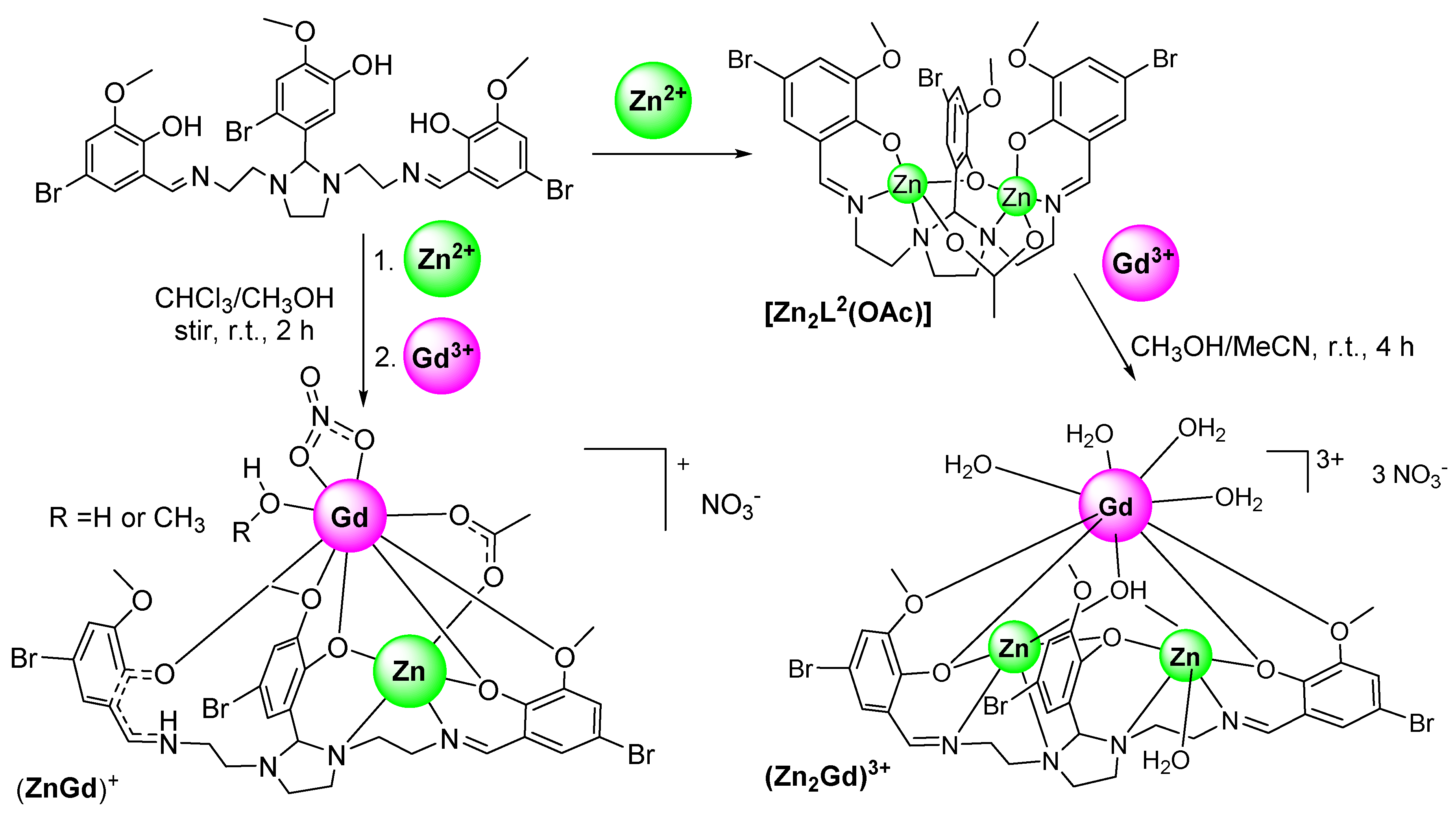
3.1. Synthetic Method

Mononuclear Gd (Figure 1) was readily obtained by template synthesis, as other analogous TbIII, DyIII, or ErIII mononuclear complexes containing this ligand [16], by simple mixing of triethylenetetramine, 2-hydroxy-5-methyl-1,3-benzenedicarboxaldehyde, and gadolinium nitrate in 1:3:1 molar ratios. After cooling only 24 h in the fridge, single crystals of Gd could be isolated. In contrast, multiple attempts to isolate a similar mononuclear gadolinium(III) complex derived from H3L were made without success. This was not surprising, as this type of three-armed ligand derived from triethylenetetramine and aromatic aldehydes is prone to yielding dinuclear lanthanoid complexes of the [Ln(L)]2 type, with a sandwich structure [42,43,44,45,46,47].
As commented, in this case, we were interested in obtaining a mononuclear gadolinium complex, and preferably with the lanthanoid ion only occupying the external compartment. Thus, the internal compartments of the ligand which are adequate for enclosing Zn2+ ions [36] remain uncoordinated, and the comparison with those containing one or two zinc ions could be more useful for the subsequent MRI study. In this sense, it is also remarkable that one water molecule is coordinated to the gadolinium(III) ion. This ligand was not chosen to prepare heteronuclear complexes, as no other Zn-Ln complexes could be obtained, apart from [Zn2Dy(L′)(NO3)3(OH)], and this complex does not include coordinated water molecules, only a bridging μ3-hydroxide ligand [16].
The synthetic method used to obtain new heteronuclear Gd-Zn complexes with H3L, was selected according to our previous experience with this three-armed ligand [15]. Thus, without surprise, direct reaction of H3L, Zn(OAc)2∙2H2O and Gd(NO3)3∙6H2O, both with 1:1:1 and 1:2:1 molar ratios, only led to obtaining ZnGd (Scheme 2), as it had also occurred when the same ligand had been combined with ErIII, TbIII, and DyIII [15].
As previously commented in the experimental section, the molecular structure of this heterodinuclear Zn-Gd complex could not be crystallographically determined, as all the single crystals obtained diffracted rather poorly. However, they diffracted enough to determine accurately the parameters of its unit cell. The values obtained for these latter one (vide supra) demonstrate that an almost total resemblance exists between these cell parameters and those previously reported for [ZnDy(HL)(NO3)(OAc)(MeOH)](NO3) [15]. In fact, their respective volumes are 4659 and 4654 Å3, respectively. This resemblance also occurs not only between their respective X-ray powder diffraction patterns, but also between their IR spectra (Figures S1 and S2 of the Supplementary Materials). Hence, they clearly appear to be isostructural. Furthermore, [ZnDy(HL)(NO3)(OAc)(MeOH)](NO3) is also equivalent to [ZnLn(HL)(NO3)(OAc)(H2O)](NO3) (Ln = Er or Tb) [15], being the four complexes obtained with the same synthetic method, also employed in this case, although the crystal parameters of the two latter ones are not comparable. Consequently, only some doubt could remain about the nature of the solvent molecule coordinated to the gadolinium(III) ion, which could be methanol or water, as proposed in (Scheme 2). A similar coincidence between cell parameters exist for the already mentioned isostructural compounds [Ln(H3L′)(NO3)(H2O)](NO3)2·complexes (Ln = Gd, Tb, Dy, Er) [16], and for other isostructural series even with different solvated molecules [48].
Likewise, after checking different synthetic routes, we had to employ the neutral homodinuclear metalloligand [Zn2(L)(OAc)], as starting material to prepare heterotrinuclear zinc-gadolinium complexes, by using a similar method to that previously employed to prepare heterotrinuclear complexes of the [Zn2Ln(L)(NO3)2(OAc)2(H2O)] type (Ln = Dy, Er) [15]. Thus, the result of mixing [Zn2(L)(OAc) with gadolinium(III) nitrate has finally allowed obtaining Zn2Gd (Figure 2). However, in spite of using a similar synthetic method, this complex does not contain any coordinated acetate or nitrate ligands, but a hydroxide (O1H) ligand connecting the three metal ions, while three nitrate anions are acting as simple counterions.

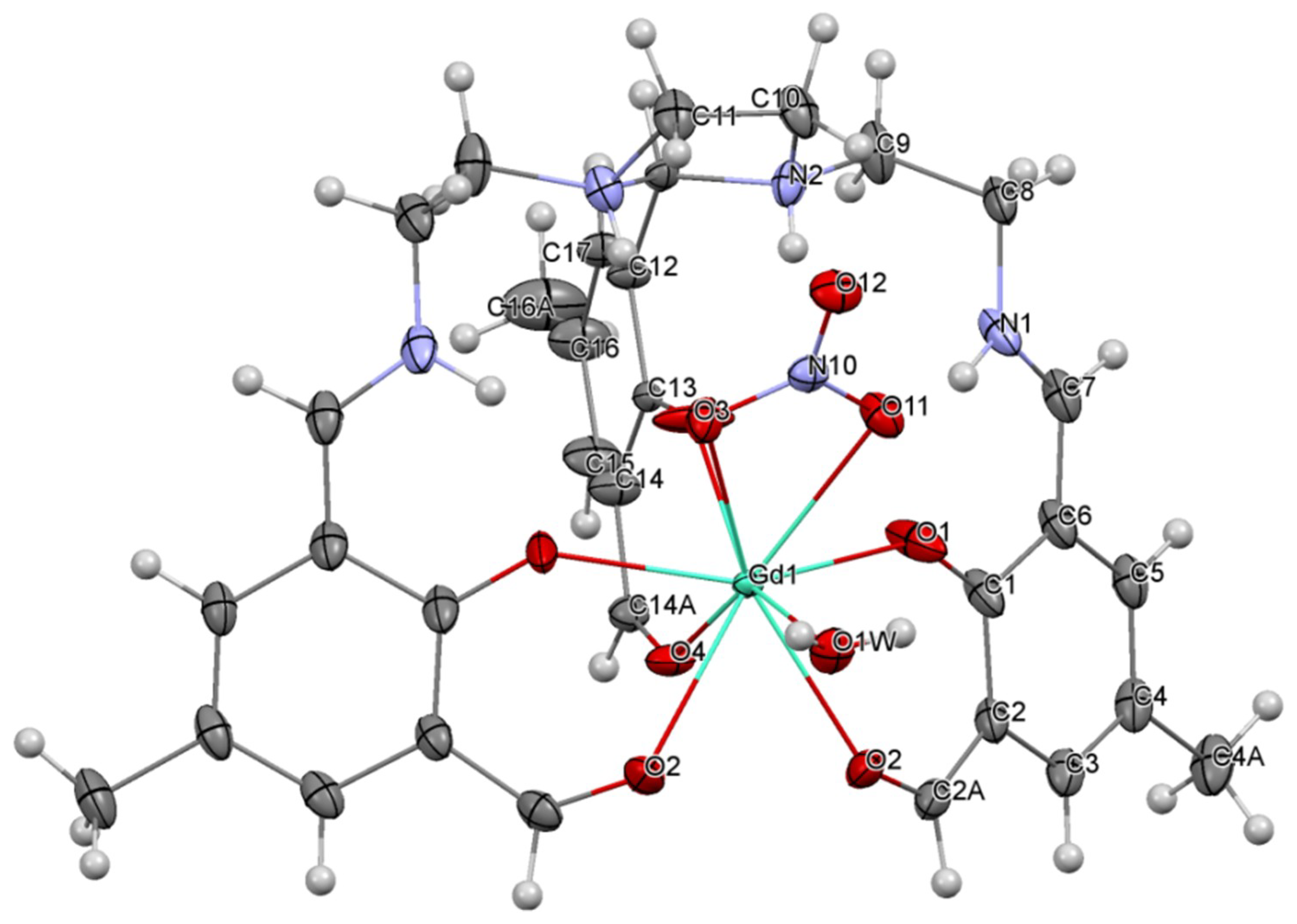
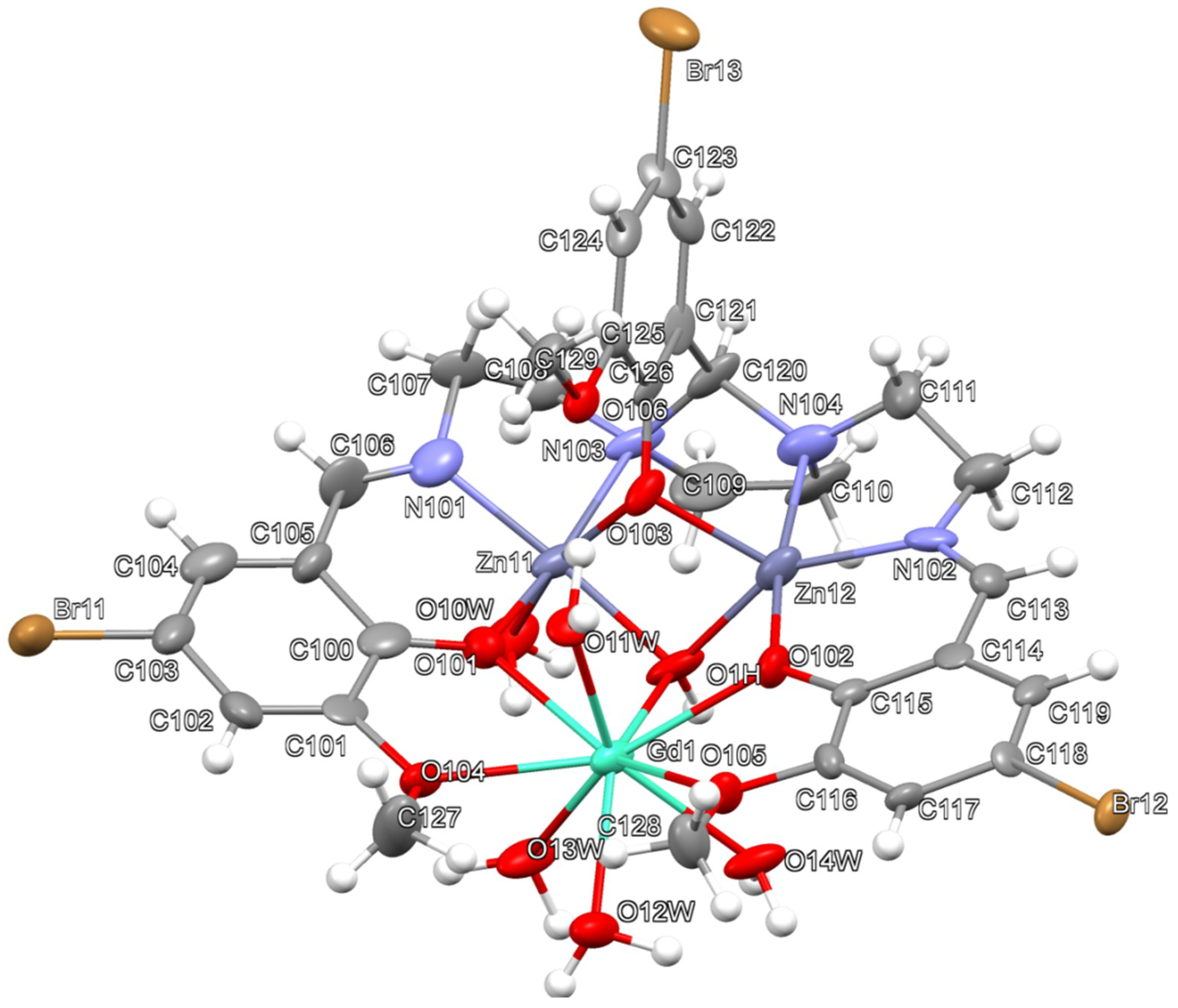
3.2. Coordination Environments of the Complexes

The main geometric parameters of Gd are collected in Table S2 of the Supplementary Materials. In the [Gd(H3L′)(NO3)(H2O)]2+ cation present in Gd (Figure 1), the three armed ligand is neutral, so totally protonated, and it is acting as hexadentate, by using only its six O donor atoms to bind the GdIII center. Accordingly, and as expected, the four the nitrogen atoms of H3L′ remain uncoordinated, but protonated in the complex, totally in the case of those of the external arms, or partially, since both imidazolidine N atoms are also holding an H atom with 50% occupation sites. Therefore, this indicates a keto more than phenol character for the O donor atoms of the three arms. This keto nature is confirmed by the C–O distances corresponding to these bonds [O(1)–C(1) and O(3)–C(13)] of about 1.29 Å (Table S2 of the Supplementary Materials) This tautomerism is not uncommon, and it can be also observed in its analogues [Ln(H3L′)(NO3)(H2O)](NO3)2 (Ln = Tb, Dy, Er)] [16], or those isostructural complexes similar to ZnGd {[ZnLn(HL)(NO3)(OAc)(HOX)](NO3)} (Ln = Dy, Er and Tb, X = H or Me) where one of the internal compartments also remains empty [15].
The coordination sphere of Gd3+ is completed by three additional oxygen atoms: two of them coming from a bidentate nitrate ligand, and one from a water molecule. This gives rise to an O9 coordination environment, considered as a ‘muffin’ according to calculations made with SHAPE software [49] (Figure 3).
The asymmetric unit of Zn2Gd contains two chemically comparable, but crystallographically inequivalent [Zn2Gd(L)(OH)(H2O)5]3+ cations (unit 1 and unit 2). Accordingly, just an ellipsoid diagram for unit 1 is shown in Figure 2, while unit 2 is shown in Figure S3 of the Supplementary Materials. Thus, there are four units of this complex in the triclinic unit cell of Zn2Gd. Main geometric parameters corresponding to this complex are listed in Table S3 of the Supplementary Materials.
Figure 2 shows that L3− is acting as trinucleating in Zn2Gd, with both internal N2O compartments accommodating two zinc(II) ions. This contrasts with the only zinc atom enclosed in the dinucleating HL2− entity present in ZnGd (Scheme 2), or in other equivalent complexes [ZnLn(HL)(NO3)(OAc)(ROH)](NO3) (Ln = Dy, Er, Tb and R = Me or H) [15], and of course, with the two empty internal compartments observed for Gd.
In contrast with the pentacoordinated zinc atoms of the symmetric [Zn2(L)(OAc)] precursor, only one of the zinc atoms remains pentacoordinated (Zn12 in unit 1 and Zn22 in unit 2), while the other ones are hexacoordinated. This change is probably favored by the substitution of the µ211 bridging acetate by a tiny µ3-OH bridge. This distortion also leads to a significant folding of the calculated planes formed by N,O,O,N donor sets to ca. 73.4°, when it was of only ca. 26.3° in [Zn2(L)(OAc)]. Furthermore, a water molecule occupies one of the apexes opposite to the central phenoxy group of L3−. The values of the Addison parameter τ [50] (0.38 for Zn12 and 0.43 for Zn22) are indicative of highly distorted square pyramid geometries, where the central phenol oxygen atom occupies the apical position. In spite of this distortion, all the bond distances and angles are within normal ranges [15,16,17,36]. The hexacoordinated zinc atoms display pseudo-octahedral geometries (Figure 3). Different coordination numbers for the two neighboring zinc ions are also present in other related heterotrinuclear complexes of the type [Zn2Ln(L)(OAc)2(NO3)2(H2O)] (Ln = Dy, Er), but all of them contrast with the two pseudo-octahedra found around the two zinc(II) ions present in the related [Zn2Ho(L)(ald)(HO)(H2O)3(MeCN)](NO3)2·EtOH [17].
This asymmetry related to the coordination environments of the two zinc atoms leads to this complex to be chiral. As a consequence, and despite the symmetry of the three-armed H3L ligand (Scheme 2), its central imidazolidine ring displays up to three asymmetric centers: both nitrogen atoms and the carbon atom that connects them. In both ellipsoid diagrams shown in Figure 2 and Figure S3 of the Supplementary Materials, we can see enantiomers only displaying S configurations for their stereocenters. Of course, although both units of the complex present in the asymmetric unit correspond coincidentally to the (S,S,S) enantiomer, the triclinic crystal is centrosymmetric, as it belongs to the crystal group P-1, and therefore it is racemic, so that the other two units also present in the unit cell are (R,R,R) enantiomers.
The above described [Zn2(L)(OH)(H2O)] fragment of Zn2Gd differs from the original [Zn2(L)(OAc)] metalloligand used as starting material, but it is also acting as a ligand towards the GdIII ion. This latter one is coordinated to the phenolate and methoxy O atoms of both external arms of the Schiff base, as well as to the hydroxide bridge. The coordination sphere of the gadolinium(III) ion is completed up to 9 by four water molecules, giving rise to a GdO9 environment (Figure 3). In this distorted GdO9 polyhedron, all the distances and angles are within their normal ranges [4,5,8,11,12,51], so this does not deserve further consideration.
The deviation of the coordination sphere with respect to an ideal nine-vertex polyhedron was calculated with the SHAPE software [49], and results indicate a muffin-like appearance, also near to a spherical capped square antiprism (Figure S2 and Table S4 in the Supplementary Materials. This muffin-like polyhedron that surrounds the gadolinium(III) ion shares one edge with each zinc pseudo-polyhedron, whilst these polyhedra around both zinc centers also share one edge (Figure 3), as occurs for [Zn2Ho(L)(ald)(HO)(H2O)3(MeCN)](NO3)2·EtOH [17].
The presence of this µ3-OH anion, which is absent in other complexes also derived from L3− and [Zn2(L)(OAc)] [11], lead to compare these complexes with [Zn2Dy(L′)(NO3)3(OH)] [16]. Thus, the tight µ3111-HO bridge leads to the three metal ions to appear as an isosceles triangle, with d(Zn···Zn), is ca. 3.0 Å, while the Zn···Ln distances are about 3.45 Å for both complexes. These intramolecular distances are similar to those found for complexes of the type [Zn2Ln(L)(OAc)2(NO3)2(H2O)] (Ln = DyIII and ErIII) [15], and for other Zn-Ln complexes with polycompartmental Schiff bases [8,9,17], and also with the 3-EtO-salen2− ligand [17,52,53]. By contrast, asymmetric heterodinuclear complexes of the type of ZnGd, this is {[ZnLn(HL)(NO3)(OAc)(HOX)](NO3)} [Ln = Dy, Er and Tb, X = H or CH3)], exhibit Zn···Ln distances of about 4.7 Å that are clearly longer.

3.3. Packing Schemes for the Complexes
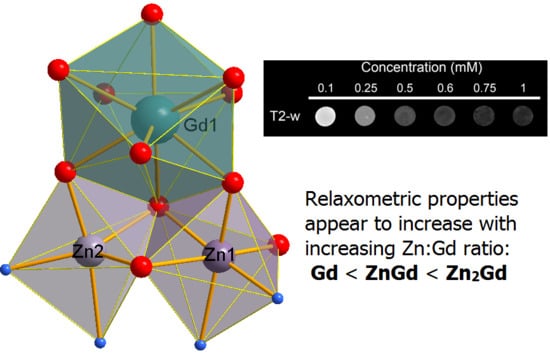
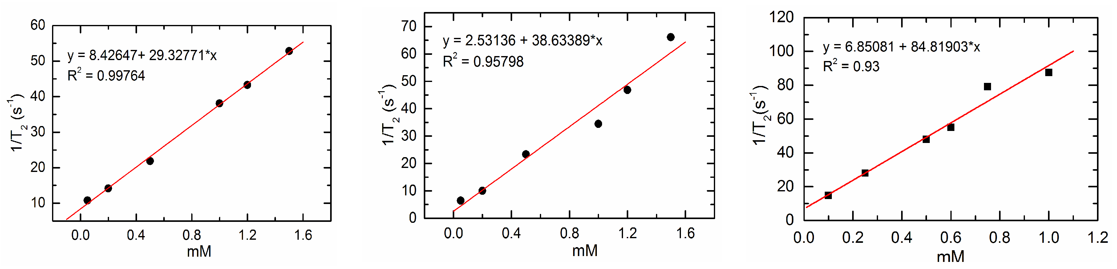
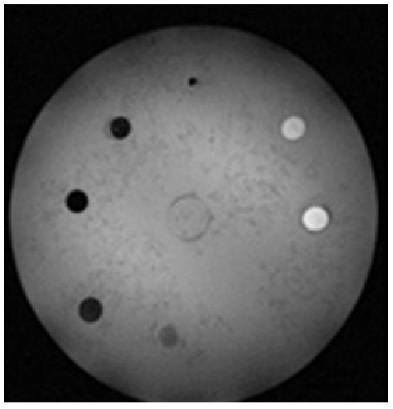
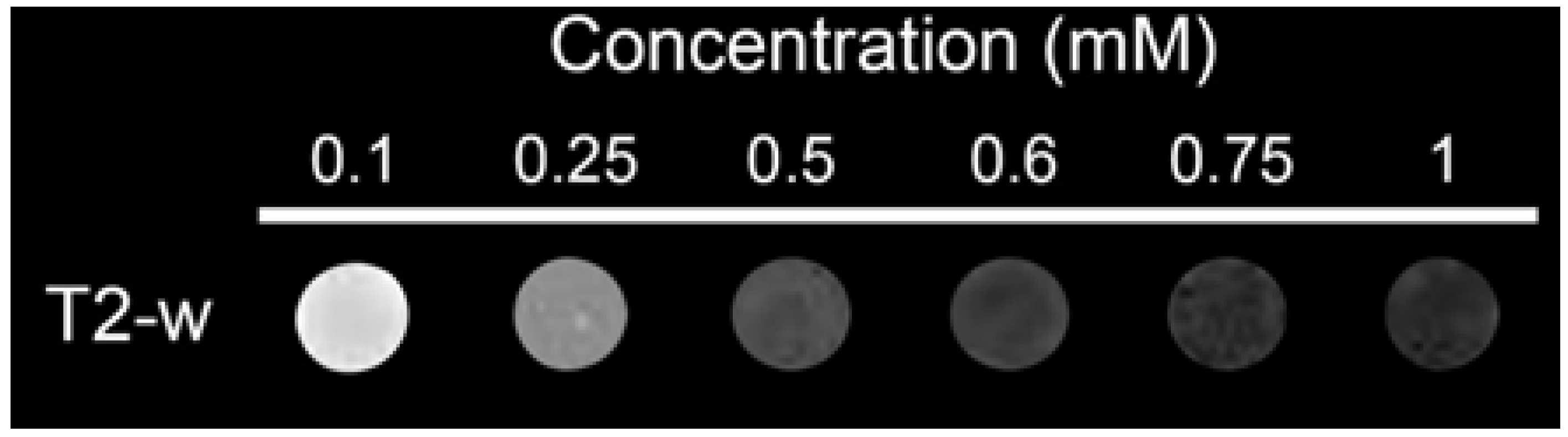
The H-bond scheme of Gd is very simple. The three-armed ligand, despite being totally protonated, displays these three H atoms involved in intramolecular bonds related to imine-amine tautomerism (Figure 1). Hence, the spatial arrangement of the ligand is curiously equivalent to that shown when it is fully deprotonated and with all its compartments occupied. Only the intermolecular H bonds are connecting the coordinated water molecule to both nitrate counterions.
Contrasting with this latter scheme, the heterotrinuclear Zn2Gd is forming a particularly intricate H bonding scheme (Table S5 and Figure S5 of the Supplementary Materials). Thus, since the quality of the diffraction data is not optimal, and in order to simplify this study, we will focus our attention mostly on classic H-bonds. In addition to classic O–H···O and some O–H···N bonds, several bifurcations, and many C–H···A interactions (A = O or Br) have been also detected (Table S5 of the Supplementary Materials).
It is evident that L3−, as other related three-armed ligands, has an enwrapping character, so this partially prevents an intermolecular propagation of classic H bonds. Despite this, the coordinated water molecules and the hydroxide anion are effective H donors, so that an expansion of multiple classic O–H···O bonds occurs, and as it has been observed for [Zn2Ho(L)(ald)(HO)(H2O)3(MeCN)](NO3)2·EtOH [17], this propagation curiously occurs practically with a predominately 2D arrangement, as Figure 4 shows.
The frontal part of the L3− ligands with the N and O donor atoms shows not only a coordinating character that encloses metal ions, but also a more hydrophilic character than its rear side [54]. In fact, this rear side, which shoes a more clearly hydrophobic nature, with the C–H bonds of imidazolidine, ethylene chains, and aromatic rings pointing towards the outside. This predominance of C–H bonds to form the surface of these layers (Figure 4 and Figure S4 of the Supplementary Materials) could influence on its solubility, as it can lead to forming extended hydrophobic surfaces that have been previously observed [17,54]. In this particular case only a few water molecules are occluded in the middle of these layers by means of an H-bond involving a solvated water molecule (O2w) and a nitrate counterion (Figure 4).

3.4. MRI Studies

The potentiality of crude solids of the three gadolinium complexes, this is: Gd′, ZnGd, and Zn2Gd′, as potential MRI contrast agents was evaluated. Thus, both longitudinal r1 and transversal r2 relaxivities were measured, and the obtained results are summarized in Table 1, while Figure 5 shows the plots of 1/T2 vs. concentration of the species.
As Gd′ is soluble in water, the longitudinal r1 relaxivity of a water solution of Gd′ was measured at 22 °C and 9.4 T, yielding a value of 0.71 mM−1 s−1 (Figure 5). This value demonstrates that, in spite of the nonacoordination displayed by the Gd3+ ion in this complex, and the presence of a water molecule in its inner sphere, even fixed by classic hydrogen bonds [55] Gd′ shows a low r1 relaxation and, accordingly, it is not appropriate as contrast agent.
With regard to the heteronuclear Zn-Gd complexes, the relaxometric properties of ZnGd and Zn2Gd′ as T1 agents were also studied, but with a significant handicap, as both complexes are scarcely soluble in water. Consequently, the experiments were performed in methanol.
For ZnGd, the r1 value of 4.90 mM−1 s−1 (Table 1) extracted from methanol solutions is comparable with those found for other commercial agents [56], so it may be considered as a potential T1 contrast agent. Nevertheless, the r2 value is 38.63 mM−1 s−1 (Figure 5) and, therefore, the T1/T2 (r2/r1) ratio is 7.9, a value that strongly differs from 1. As a consequence, this precludes the use of ZnGd as a positive MRI contrast [27]. However, the quite high r2 value found for this heteronuclear Zn-Gd complex, in connection to the T1/T2 ratio higher than 6 (a condition for being used as a T2 contrast agent), appears to indicate that it is more suitable as a negative MRI contrast, while the most usual T2 agents were based on magnetic iron oxide nanoparticles [29]. This behavior as negative contrast appears to be confirmed by in vitro MRI experiments with agarose gel phantoms. Thus, as Figure 6 shows, the T2-weighted phantoms show that darker images can be obtained by increasing the complex concentration.
Similarly, the ability of Zn2Gd′ to act as potential MRI CA was also tested in methanol. In this case, the value obtained for r1 is 7.14 mM−1 s−1, which represents about a 150% of the relaxation time for the solution containing ZnGd. This result appear to suggest that an increasing Gd:Zn molar ratio in the complex could improve its relaxometric properties. The r1 relaxivity value found of Zn2Gd′ (7.14 mM−1 s−1) is even higher than those values reported for some classical commercial T1 CAs [56]. However, as occurred before, calculated r2 value is even higher (84.83 mM−1 s−1), while the T1/T2 ratio of 11.88 clearly shows that instead of a T1 contrast, Zn2Gd′ could be proposed as a potential candidate for T2 CA.
At this point, it should be noted that all the classical T2 contrast agents based on superparamagnetic iron oxide nanoparticles (SPIONs) [57] have been recently forbidden, and that, currently, Ferumoxytol [58] is the only Food and Drug Administration-approved SPION that is being used as an MRI contrast agent [59]. It should be mentioned that calculated relaxivities for Ferumoxytol are r1 38 mM−1 s−1 and r2 83 mM−1 s−1 at 0.47 T [60]. Accordingly, the r2 value of 84.83 mM−1 s−1 for Zn2Gd′ is even greater than the one reported for the only approved SPION MRI species.
The ability of Zn2Gd′ to act as a negative contrast has also been checked in vitro and the T2-weighted phantom shows a progressive darkening when the concentration of complex increases (Figure 7).
As a result of the MRI studies performed, it seems that the presence of zinc intrinsically ligated into a gadolinium complex could notably enhance the relaxometric properties of the compound, given that the heterodinuclear ZnGd shortens both T1 and T2 relaxation times. It must be noted that ZnGd is supposed to only contain a coordinated water molecule, as occurring for Gd′. This behavior is even enhanced in the case of the heterotrinuclear Zn2Gd′ complex, but in this case, there are four coordinated water molecules to the external gadolinium ion, and this fact could also contribute to increase these r values. In any case, the r1 and r2 values increase in the order Gd′ < ZnGd < Zn2Gd′, what could indicate that the higher the Zn:Gd molar ratio in the sample, the greater the relaxation times are.
The r1 factors found for ZnGd and Zn2Gd′ are comparable or even greater than those for commercial contrast agents. Nevertheless, the T1/T2 ratio is in both cases even higher than 6, what indicates that they could not be used as positive, but maybe as negative contrast agents. The r2 value for Zn2Gd′ could be comparable to that of the only SPION approved as a T2 contrast.
It cannot be ignored that the heteronuclear Zn-Gd complexes described herein are rather insoluble in water, and, accordingly, they cannot be effective MRI agents. Nevertheless, it must be also taken into account that classical T2 contrast agents have been mostly forbidden, due to their toxicity and fatal anaphylactic reactions. In addition, most of the T1 CAs are also nowadays considered as potentially toxic, and consequently European Agency of Medicines is dealing with the suspension of all the classical commercial intravenous linear probes [25]. Consequently, and especially nowadays, there is a need for contributions for more efficient CAs with improved relaxivity.
Although, we are presenting herein some complexes based on linear ligands with low water solubility, a useful contribution of this work could be the presence of zinc ions in this agents accompanying to typical metal ions as gadolinium(III). This presence appears to increase the relaxation times. Although many zinc(II) responsive probes have been reported since 2001 [31], to the best of our knowledge, only two heteronuclear Zn-Gd complexes has been tested as potential contrast agents until now as T1 agents, but not as potential negative contrasts [61,62]. Accordingly, the findings of this work could suppose an incipient contribution in the search for new CAs: the potential use of heteronuclear Zn-Gd complexes as T2 contrast agents.

4. Conclusions

The ligands H3L and H3L′, with differentiated compartments for 3d and 4f metal ions, allow isolating the mononuclear [Gd(H3L′)(H2O)(NO3)](NO3)2·2H2O (Gd), heterodinuclear {[ZnGd(HL2)(NO3)(OAc)(CH3OH)](NO3)}∙3H2O (ZnGd), and heterotrinuclear {[Zn2Gd(L2)(OH)(H2O)5](NO3)3}∙0.75CH3CN·3CH3OH (Zn2Gd) complexes. Gd could be prepared by a template method, and is totally symmetric, but the synthesis of the heteronuclear complexes needed the previous preparation of the free ligand, or even the of the metalloligand [Zn2(L)(OAc)] as a precursor. Zn2Gd is chiral due to the asymmetry of the different coordination environments around the two zinc atoms.
Despite the presence of multiple and varied potential donors and acceptors for H bonding, the packing scheme of Gd is very simple, but that of Zn2Gd—which is mostly based on classic O–H···O bonds, is intricate and basically bidimensional—and with a rather hydrophobic surface, which could affect its solubility in water.
The crystal structures of mononuclear Gd and heteronuclear Zn2Gd demonstrate that both GdO9 cores contain at least one water molecule coordinated to the GdIII ion. The relaxometric properties of these two compounds and that of ZnGd were studied, with clearly different results. Thus, Gd′·yield low r1 and r2 parameters, without any interest as potential contrast agent. These r values notably increase in the sequence Gd′·< ZnGd < Zn2Gd′, suggesting that the relaxometric properties could improve as the Zn:Gd ratio increases. The r1 and r2 values, as well as the T1/T2 ratios indicate that Zn2Gd′ could be relevant as a T2 CA, given that its r2 value is even greater than that reported for the only approved SPION T2 agent. Therefore, although the low solubility in water of the complexes described herein prevents their use as CAs, the findings summarized could be worthy for future design and search for new CAs, proposing heteronuclear Zn-Gd as potential candidates for T2 MRI contrast agents.

Supplementary Materials

The following are available online at https://0-www-mdpi-com.brum.beds.ac.uk/2073-4352/8/11/431/s1. Supplementary crystallographic data for this paper have been deposited at Cambridge Crystallographic Data Center (CCDC 1563554 and 1570865) and can be obtained free of charge via www.ccdc.cam.ac.uk/conts/retrieving.html. An additional document contains: Table S1: X ray crystallographic data for Gd and Zn2Gd; Table S2: Main geometric parameters for Gd; Table S3: Main geometric parameters for Zn2Gd; Table S4: Continuous SHAPE measures calculations for Gd and Zn2Gd; Figure S1: Comparison of the XRD patterns for ZnGd and [ZnDy(HL)(NO3)(OAc)(MeOH)](NO3) 1.25MeOH·0.25H2O; Figure S2: IR spectra for ZnGd′ and [ZnDy(HL)(NO3)(OAc)(MeOH)](NO3)·1.25MeOH·0.25H2O; Figure S3: Ellipsoid diagram for a second [Zn2Gd(L)(OH)(H2O)5]3+ unit present in Zn2Gd; Figure S4: Coordination polyhedra for the GdIII centre present in unit 2 of Zn2Gd; Figure S4. Coordination polyhedra for the GdIII centre present in unit 2 of Zn2Gd; Figure S5: Sticks view of the asymmetric unit of Zn2Gd showing the multiple H-bonds between cations, anions and occluded solvent molecules; Figure S6: Space-filled views of portions of the 2D-H-bonded layers formed by Zn2Gd.

Author Contributions

The manuscript was written through contributions of all authors: methodology, M.F. and J.S.-M.; XRD analysis, J.C.-V. and A.M.G.-D.; MRI analysis, P.T.; laboratory investigation, J.C.-V. and J.S.-M.; writing—original draft preparation, A.M.G.-D. and J.S.-M.; writing—review and editing, A.M.G.-D., J.S.-M. and M.F.; project administration, M.F. and J.S.-M. All authors have given approval to the final version of the manuscript.

Funding

Financial support from Ministerio de Economía y Competitividad (MINECO, CTQ2014-56312-P) is gratefully acknowledged.

Acknowledgments

Julio Corredoira-Vázquez acknowledges Xunta de Galicia for his Ph.D. fellowship.

Conflicts of Interest

The authors declare no conflict of interest.

References

  1. Woodruff, D.N.; Winpenny, R.E.P.; Layfield, R.A. Lanthanide single-molecule magnets. Chem. Rev. 2013, 113, 5110–5148. [Google Scholar] [CrossRef] [PubMed]
  2. Osa, S.; Kido, T.; Matsumoto, V.; Re, N.; Pochaba, A.; Mrozinski, J. A Tetranuclear 3d–4f Single Molecule Magnet: [CuIILTbIII(hfac)2]2. J. Am. Chem. Soc. 2004, 126, 420–421. [Google Scholar] [CrossRef] [PubMed]
  3. Papatriantafyllopoulou, C.; Wernsdorfer, W.; Abboud, K.A.; Christou, G. Mn21Dy Cluster with a Record Magnetization Reversal Barrier for a Mixed 3d/4f Single-Molecule Magnet. Inorg. Chem. 2011, 50, 421–423. [Google Scholar] [CrossRef] [PubMed]
  4. Moreno Pineda, E.; Chilton, N.F.; Tuna, F.; Winpenny, R.E.P.; McInnes, E.J.L. Systematic Study of a Family of Butterfly-Like {M2Ln2} Molecular Magnets (M = MgII, MnIII, CoII, NiII, and CuII; Ln = YIII, GdIII, TbIII, DyIII, HoIII, and ErIII). Inorg. Chem. 2015, 54, 5930–5941. [Google Scholar] [CrossRef] [PubMed]
  5. Biswas, S.; Bag, P.; Das, S.; Kundu, S.; van Leusen, J.; Kögerler, P.; Chandrasekhar, V. Heterometallic [Cu2Ln3] (Ln = DyIII, GdIII and HoIII) and [Cu4Ln2] (Ln = DyIII and HoIII) Compounds: Synthesis, Structure, and Magnetism. Eur. J. Inorg. Chem. 2017, 1129–1142. [Google Scholar] [CrossRef]
  6. Bag, P.; Chakraborty, A.; Rogez, G.; Chandrasekhar, V. Pentanuclear Heterometallic {MnIII2Ln3} (=Gd, Dy, Tb, Ho) Assemblies in an Open-Book Type Structural Topology: Appearance of Slow Relaxation of Magnetization in the Dy(III) and Ho(III) Analogues. Inorg. Chem. 2014, 53, 6524–6533. [Google Scholar] [CrossRef] [PubMed]
  7. Vignesh, K.R.; Langley, S.K.; Murray, K.S.; Rajaraman, G. Exploring the Influence of Diamagnetic Ions on the Mechanism of Magnetization Relaxation in {CoIII2LnIII2} (Ln = Dy, Tb, Ho) “Butterfly” Complexes. Inorg. Chem. 2017, 56, 2518–2532. [Google Scholar] [CrossRef] [PubMed]
  8. Amjad, A.; Madalan, A.M.; Andruh, M.; Caneschi, A.; Sorace, L. Slow relaxation of magnetization in an isostructural series of zinc–lanthanide complexes: An integrated EPR and AC susceptibility study. Chem. Eur. J. 2016, 22, 12849–12858. [Google Scholar] [CrossRef] [PubMed]
  9. Akine, S.; Taniguchi, T.; Nabeshima, T. Helical Metallohost-Guest Complexes via Site-Selective Transmetalation of Homotrinuclear Complexes. J. Am. Chem. Soc. 2006, 128, 15765–15774. [Google Scholar] [CrossRef] [PubMed]
  10. Costes, J.P.; Titos-Padilla, S.; Oyarzabal, I.; Gupta, T.; Duhayon, C.; Rajaraman, G.; Colacio, E. Effect of ligand substitution around the DyIII on the SMM properties of dual-luminescent Zn–Dy and Zn–Dy–Zn complexes with large anisotropy energy barriers: A combined theoretical and experimental magnetostructural study. Inorg. Chem. 2016, 55, 4428–4440. [Google Scholar] [CrossRef] [PubMed]
  11. Das, S.; Bejoymohandas, K.S.; Dey, A.; Biswas, S.; Reddy, M.L.P.; Morales, R.; Ruiz, E.; Titos-Padilla, S.; Colacio, E.; Chandrasekhar, V. Amending the Anisotropy Barrier and Luminescence Behavior of Heterometallic Trinuclear Linear [MIILnIIIMII] (LnIII = Gd, Tb, Dy; MII = Mg/Zn) Complexes by Change from Divalent Paramagnetic to Diamagnetic Metal Ions. Chem. Eur. J. 2015, 21, 6449–6464. [Google Scholar] [CrossRef] [PubMed]
  12. Meng, Y.S.; Jiang, S.D.; Wang, B.W.; Gao, S. Understanding the Magnetic Anisotropy towards Single-Ion Magnets. Acc. Chem. Res. 2016, 49, 2381–2389. [Google Scholar] [CrossRef] [PubMed]
  13. Yue, S.T.; Wei, Z.Q.; Wang, N.; Liu, W.J.; Zhao, X.; Chang, L.M.; Liu, Y.L.; Mo, H.H.; Cai, Y.P. Three novel microporous 3D heterometallic 3d–4f coordination polymers: Synthesis, crystal structures and photoluminescence properties. Inorg. Chem. Commun. 2011, 14, 1396–1399. [Google Scholar] [CrossRef]
  14. Wang, Y.M.; Wang, Y.; Wang, R.X.; Qiu, J.Q.; Chi, Y.X.; Jin, J.; Niu, S.Y. Syntheses, structures and photophysical properties of a series of Zn-Ln complexes. J. Phys. Chem. Solids 2017, 104, 221–227. [Google Scholar] [CrossRef]
  15. Fondo, M.; Corredoira-Vázquez, J.; García-Deibe, A.M.; Sanmartín-Matalobos, J.; Herrera, J.M.; Colacio, E. Designing Ligands to Isolate ZnLn and Zn2Ln Complexes: Field-Induced Single-Ion Magnet Behavior of the ZnDy, Zn2Dy, and Zn2Er. Inorg. Chem. 2017, 56, 5646–5656. [Google Scholar] [CrossRef] [PubMed]
  16. Fondo, M.; Corredoira-Vázquez, J.; Herrera-Lanzós, A.; García-Deibe, A.M.; Sanmartín-Matalobos, J.; Herrera, J.M.; Colacio, E.; Nuñez, C. Improving the SMM and luminescence properties of lanthanide complexes with LnO9 cores in the presence of ZnII: An emissive Zn2Dy single ion magnet. Dalton Trans. 2017, 46, 17000–17009. [Google Scholar] [CrossRef] [PubMed]
  17. Corredoira-Vázquez, J.; Fondo, M.; Sanmartín-Matalobos, J.; García-Deibe, A.M. 2D Supramolecular Structure for a Chiral Heterotrinuclear ZnII2HoIII Complex through Varied H-Bonds Connecting Solvates and Counterions. Proceedings 2018, 2, 1114. [Google Scholar] [CrossRef]
  18. Fondo, M.; Corredoira-Vázquez, J.; García-Deibe, A.; Sanmartín-Matalobos, J.; Iglesias, R.; Taboada, P. A heteronuclear ZnGd complex as a potential contrast agent for magnetic resonance imaging. In Proceedings of the 20th International Electronic Conference on Synthetic Organic Chemistry, 1–30 November 2016; MDPI AG: Basel, Switzerland, 2016. [Google Scholar]
  19. Major, J.L.; Parigi, G.; Luchinat, C.; Meade, T.J. The synthesis and in vitro testing of a zinc-activated MRI contrast agent. Proc. Natl. Acad. Sci. USA 2007, 104, 13881–13886. [Google Scholar] [CrossRef] [PubMed] [Green Version]
  20. Esqueda, A.C.; López, J.A.; Andreu-de-Riquer, G.; Alvarado-Monzón, J.C.; Ratnakar, J.; Lubag, A.J.M.; Sherry, A.D.; León-Rodríguez, L.M.D. A new gadolinium-based MRI zinc sensor. J. Am. Chem. Soc. 2009, 131, 11387–11391. [Google Scholar] [CrossRef] [PubMed]
  21. Boros, E.; Gale, E.M.; Caravan, P. MR imaging probes: Design and applications. Dalton Trans. 2015, 44, 4804–4818. [Google Scholar] [CrossRef] [PubMed]
  22. Ye, D.; Pandit, P.; Kempen, P.; Lin, J.; Xiong, L.; Sinclair, R.; Rutt, B.; Rao, J. Redox-triggered self-assembly of gadolinium-based MRI probes for sensing reducing environment. Bioconj. Chem. 2014, 25, 1526–1536. [Google Scholar] [CrossRef] [PubMed]
  23. Leone, L.; Ferrauto, G.; Cossi, M.; Botta, M.; Tei, L. Optimizing the relaxivity of MRI probes at high magnetic field strengths with binuclear GdIII complexes. Front. Chem. 2018, 6, 158. [Google Scholar] [CrossRef] [PubMed]
  24. Nicolay, K.; Strijkers, G.; Grüll, H. Gd-Containing Nanoparticles as MRI Contrast Agents. In The Chemistry of Contrast Agents in Medical Magnetic Resonance Imaging, 2nd ed.; Merbach, A.E., Helm, L., Tóth, E., Eds.; John Wiley & Sons Ltd.: Hoboken, NJ, USA, 2013; pp. 449–487. ISBN 9781119991762. [Google Scholar]
  25. Helm, L. Optimization of gadolinium-based MRI contrast agents for high magnetic-field applications. Future Med. Chem. 2010, 2, 385–396. [Google Scholar] [CrossRef] [PubMed]
  26. Fries, P.H.; Belorizky, E. Electronic Spin Relaxation and Outer-Sphere Dynamics of Gadolinium-Based Contrast Agents. In The Chemistry of Contrast Agents in Medical Magnetic Resonance Imaging, 2nd ed.; Merbach, A.E., Helm, L., Tóth, E., Eds.; John Wiley & Sons Ltd.: Hoboken, NJ, USA, 2013; pp. 277–309. ISBN 9781119991762. [Google Scholar]
  27. Amoroso, A.J.; Pope, S.J.A. Using lanthanide ions in molecular bioimaging. Chem. Soc. Rev. 2015, 44, 4723–4742. [Google Scholar] [CrossRef] [PubMed]
  28. European Medicines Agency. PRAC Confirms Restrictions on the Use of Linear Gadolinium Agents; EMA/424715/2017. Available online: https://www.ema.europa.eu/documents/referral/gadolinium-article-31-referral-prac-confirms-restrictions-use-linear-gadolinium-agents_en.pdf (accessed on 15 November 2018).
  29. Tu, C.; Osborne, E.A.; Louie, A.Y. Activatable T1 and T2 magnetic resonance imaging contrast agents. Ann. Biomed. Eng. 2011, 39, 1335–1348. [Google Scholar] [CrossRef] [PubMed]
  30. Heffern, M.C.; Matosziuk, L.M.; Mead, T.J. Lanthanide probes for bioresponsive imaging. Chem. Rev. 2014, 114, 4496–4539. [Google Scholar] [CrossRef] [PubMed]
  31. De Leon-Rodriguez, L.M.; Lubag, A.J.; Malloy, C.R.; Martinez, G.V.; Gillies, R.J.; Sherry, A.D. Responsive MRI agents for sensing metabolism in vivo. Acc. Chem. Res. 2009, 42, 948–957. [Google Scholar] [CrossRef] [PubMed]
  32. Hanaoka, K.; Kikuchi, K.; Urano, Y.; Nagano, T. Selective sensing of zinc ions with a novel magnetic resonance imaging contrast agent. J. Chem. Soc. Perkin Trans. 2001, 2, 1840–1843. [Google Scholar] [CrossRef]
  33. Bonnet, C.S.; Caillé, F.; Pallier, A.; Morfin, J.F.; Petoud, S.; Suzenet, F.; Tóth, E. Mechanistic studies of Gd3+-based MRI contrast agents for Zn2+ detection: Towards rational design. Chem. Eur. J. 2014, 20, 10959–10969. [Google Scholar] [CrossRef] [PubMed]
  34. Yu, J.; Martins, A.F.; Preihs, C.; Clavijo-Jordan, V.; Chirayil, S.; Zhao, P.; Wu, Y.; Nasr, K.; Kiefer, G.E.; Sherry, A.D. Amplifying the sensitivity of zinc(II) responsive MRI contrast agents by altering water exchange rates. J. Am. Chem. Soc. 2015, 137, 14173–14179. [Google Scholar] [CrossRef] [PubMed]
  35. Regueiro-Figueroa, M.; Gündüz, S.; Patinec, V.; Logothetis, N.K.; Esteban-Gómez, D.; Tripier, R.; Angelovski, G.; Platas-Iglesias, C. Gd3+-based magnetic resonance imaging contrast agent responsive to Zn2+. Inorg. Chem. 2015, 54, 10342–10350. [Google Scholar] [CrossRef] [PubMed]
  36. Fondo, M.; García-Deibe, A.M.; Ocampo, N.; Sanmartín, J.; Bermejo, M.R. Influence of some reaction conditions on the obtaining of tetra- and dinuclear zinc complexes of some Schiff bases derived from 2,6-diformyl-4-alkyl-phenols. Polyhedron 2008, 27, 2585–2594. [Google Scholar] [CrossRef]
  37. Sheldrick, G.M. SADABS, Area-Detector Absorption Correction; Siemens Industrial Automation, Inc.: Madison, WI, USA, 2001. [Google Scholar]
  38. Sheldrick, G.M. SHELXT—Integrated space-group and crystal-structure determination. Acta Cryst. 2015, A71, 3–8. [Google Scholar] [CrossRef] [PubMed]
  39. Sheldrick, G.M. Crystal structure refinement with SHELXL. Acta Cryst. 2015, C71, 3–8. [Google Scholar] [CrossRef]
  40. Schneider, C.A.; Rasband, W.S.; Eliceiri, K.W. NIH Image to ImageJ: 25 years of image analysis. Nat. Methods 2012, 9, 671. [Google Scholar] [CrossRef] [PubMed]
  41. Trekker, J.; Leten, C.; Struys, T.; Lazenka, V.V.; Argibay, B.; Micholt, L.; Lambrichts, I.; Van Roy, W.; Lagae, L.; Himmelreich, U. Sensitive in vivo cell detection using size-optimized superparamagnetic nanoparticles. Biomaterials 2014, 35, 1627–1635. [Google Scholar] [CrossRef] [PubMed]
  42. Yang, L.W.; Liu, S.; Wong, E.; Retting, S.J.; Orvig, C. Complexes of Trivalent Metal Ions with Potentially Heptadentate N4O3 Schiff Base and Amine Phenol Ligands of Varying Rigidity. Inorg. Chem. 1995, 34, 2164–2178. [Google Scholar] [CrossRef]
  43. Howell, R.C.; Spence, K.V.N.; Kahwa, I.A.; Williams, D.J. Structure and luminescence of the neutral dinuclear lanthanide(III) complexes [{Ln(api)}2] {H3api = 2-(2-hydroxyphenyl)-1,3-bis[4-(2-hydroxy phenyl)-3-azabut-3-enyl]-1,3-imidazolidine}. J. Chem. Soc. Dalton Trans. 1998, 2727–2734. [Google Scholar] [CrossRef]
  44. Chakraborty, J.; Thakurta, S.; Pilet, G.; Ziessel, R.F.; Charbonnière, L.J.; Mitra, S. Syntheses, Crystal Structures and Photophysical Properties of Two Doubly μ-Phenoxo-Bridged LnIII (Ln = Pr, Nd) Homodinuclear Schiff Base Complexes. Eur. J. Inorg. Chem. 2009, 2009, 3993–4000. [Google Scholar] [CrossRef]
  45. Nematirad, M.; Gee, W.J.; Langley, S.K.; Chilton, N.F.; Moubaraki, B.; Murray, K.S.; Batten, S.R. Single molecule magnetism in a μ-phenolato dinuclear lanthanide motif ligated by heptadentate Schiff base ligands. Dalton Trans. 2012, 41, 13711–13715. [Google Scholar] [CrossRef] [PubMed]
  46. Zhao, L.; Wu, J.; Ke, H.; Tang, J. Three dinuclear lanthanide(III) compounds of a polydentate Schiff base ligand: Slow magnetic relaxation behaviour of the DyIII derivative. CrystEngComm 2013, 15, 5301–5306. [Google Scholar] [CrossRef]
  47. Xie, Q.F.; Huang, M.L.; Chen, Y.M. Bis{μ-1,3-bis[2-(5-bromo-2-oxidobenzylideneamino)ethyl]-2-(5-bromo-2-oxidophenyl)-1,3-imidazolidine}dineodymium(III) N,N-dimethylformamide hexasolvate. Acta Cryst. 2009, E65, m1660. [Google Scholar] [CrossRef] [PubMed]
  48. Mylonas-Margaritis, I.; Maniaki, D.; Mayans, J.; Ciammaruchi, L.; Bekiari, V.; Raptopoulou, C.P.; Psycharis, V.; Christodoulou, S.; Escuer, A.; Perlepes, S.P. Mononuclear Lanthanide(III)-Salicylideneaniline Complexes: Synthetic, Structural, Spectroscopic, and Magnetic Studies. Magnetochemistry 2018, 4, 45. [Google Scholar] [CrossRef]
  49. Llunell, M.; Casanova, D.; Cirera, J.; Alemany, P.; Alvarez, S. SHAPE: Program for the Stereochemical Analysis of Molecular Fragments by Means of Continuous Shape Measures and Associated Tools; University of Barcelona: Barcelona, Spain, 2010. [Google Scholar]
  50. Addison, A.W.; Rao, T.N.; Reedijk, J.; Van Rijk, J.; Verschoor, C.G. Synthesis, structure, and spectroscopic properties of copper(II) compounds containing nitrogen–sulphur donor ligands; the crystal and molecular structure of aqua[1,7-bis(N-methylbenzimidazol-2′-yl)-2,6-dithiaheptane]copper(II) perchlorate. J. Chem. Soc. Dalton Trans. 1984, 1349–1356. [Google Scholar] [CrossRef]
  51. Anastasiadis, N.C.; Kalofolias, D.A.; Philippidis, A.; Tzani, S.; Raptopoulou, C.P.; Psycharis, V.; Milios, C.J.; Escuer, A.; Perlepes, S.P. A family of dinuclear lanthanide(III) complexes from the use of a tridentate Schiff base. Dalton Trans. 2015, 44, 10200–10209. [Google Scholar] [CrossRef] [PubMed]
  52. Maeda, M.; Hino, S.; Yamashita, K.; Kataoka, Y.; Nakano, M.; Yamamurac, T.; Kajiwara, T. Correlation between slow magnetic relaxation and the coordination structures of a family of linear trinuclear Zn(II)–Ln(III)–Zn(II) complexes (Ln = Tb, Dy, Ho, Er, Tm and Yb). Dalton Trans. 2012, 41, 13640–13648. [Google Scholar] [CrossRef] [PubMed]
  53. Wong, W.K.; Liang, H.; Wong, W.Y.; Cai, Z.; Lib, K.F.; Cheah, K.W. Synthesis and near-infrared luminescence of 3d-4f bi-metallic Schiff base complexes. New J. Chem. 2002, 26, 275–278. [Google Scholar] [CrossRef]
  54. García-Deibe, A.M.; Fondo, M.; Corredoira-Vázquez, J.; Fallah, M.S.E.; Sanmartín-Matalobos, J. Hierarchical assembly of antiparallel homochiral sheets formed by hydrogen-bonded helixes of a trapped-valence CoII/CoIII complex. Cryst. Growth Des. 2017, 17, 467–473. [Google Scholar] [CrossRef]
  55. Boros, E.; Srinivas, R.; Kim, H.K.; Raitsimring, A.M.; Astashkin, A.V.; Poluektov, O.G.; Niklas, J.; Horning, A.D.; Tidor, B.; Caravan, P. Intramolecular hydrogen bonding restricts Gd–aqua-ligand dynamics. Angew. Chem. Int. Ed. 2017, 56, 5603–5606. [Google Scholar] [CrossRef] [PubMed]
  56. Kuda-Wedagedara, A.N.W.; Allen, M.J. Enhancing magnetic resonance imaging with contrast agents for ultra-high field strengths. Analyst 2014, 139, 4401–4410. [Google Scholar] [CrossRef] [PubMed] [Green Version]
  57. Wang, Y.X.J. Superparamagnetic iron oxide based MRI contrast agents: Current status of clinical application. Quant. Imaging Med. Surg. 2011, 1, 35–40. [Google Scholar] [CrossRef] [PubMed]
  58. Khurana, A.; Nejadnik, H.; Chapelin, F.; Lenkov, O.; Gawande, R.; Lee, S.; Gupta, S.N.; Aflakian, N.; Derugin, N.; Messing, S.; et al. Ferumoxytol: A new, clinically applicable label for stem cell tracking in arthritic joints with MRI. Nanomedicine 2013, 8, 1969–1983. [Google Scholar] [CrossRef] [PubMed]
  59. Thakor, A.S.; Jokerst, J.V.; Ghanouni, P.; Campbell, J.L.; Mittra, E.; Gambhir, S.S. Clinically approved nanoparticle imaging agents. J. Nucl. Med. 2016, 57, 1833–1837. [Google Scholar] [CrossRef] [PubMed]
  60. Simon, G.H.; Von Vopelius-Feldt, J.; Fu, Y.; Schlegel, J.; Pinotek, G.; Wendland, M.F.; Chen, M.H.; Daldrup-Link, H.E. Ultrasmall supraparamagnetic iron oxide-enhanced magnetic resonance imaging of antigen-induced arthritis: A comparative study between SHU 555 C, ferumoxtran-10, and ferumoxytol. Investig. Radiol. 2006, 41, 45–51. [Google Scholar] [CrossRef]
  61. Schmitt, J.; Heitz, V.; Sour, A.; Bolze, F.; Kessler, P.; Flamigni, L.; Ventura, B.; Bonnet, C.S.; Toth, E. A theranostic agent combining a two-photon-absorbing photosensitizer for photodynamic therapy and a gadolinium(III) complex for MRI detection. Chem. Eur. J. 2016, 22, 2775–2786. [Google Scholar] [CrossRef] [PubMed]
  62. Schmitt, J.; Jenni, S.; Sour, A.; Heitz, V.; Bolze, F.; Pallier, A.; Bonnet, C.S.; Toth, E.; Ventura, B. A porphyrin dimer–GdDOTA conjugate as a theranostic agent for one- and two-photon photodynamic therapy and MRI. Bioconj. Chem. 2018. ahead of print. [Google Scholar] [CrossRef] [PubMed]
Scheme 1. Three-armed Schiff base ligands used in this work.
Crystals 08 00431 sch001
Scheme 2. Simplified synthetic routes used to prepare the heterodi- and heterotrinuclear complexes: ZnGd and Zn2Gd. The structure of Zn2Gd is based on its crystal structure, and that of ZnGd reflects its analogy to similar compounds X-ray characterised (see text). Solvents have been omitted for simplicity.
Crystals 08 00431 sch002
Figure 1. Ellipsoid view of a [Gd(H3L′)(NO3)(H2O)]2+ cation present in the unit cell of Gd. Only the asymmetric unit has been labelled. Counterions and solvated molecules have been omitted for clarity.
Crystals 08 00431 g001
Figure 2. Ellipsoid view of one of the two [Zn2Gd(L)(OH)(H2O)5]3+ cations present in the asymmetric unit of Zn2Gd. This can be considered as unit 1, so all the atoms contain a 1 as first figure in the numbering scheme. This figure shows an (S,S,S) enantiomer of this chiral complex. A figure of the other unit has been included in the supporting information, and it is chemically equivalent.
Crystals 08 00431 g002
Figure 3. Pseudo-polyhedra formed by the coordination environments of the metal centers present in Gd (left), and in one of the cations of Zn2Gd (unit 1, right). For heterotrinuclear complexes the two zinc(II) ions corresponding to (S,S,S) enantiomers have been also included.
Crystals 08 00431 g003
Figure 4. Balls (metals) and sticks (non-metals) view of the H-bonded layers formed by Zn2Gd showing the multiple H-bonds between cations, anions and coordinated or occluded solvent molecules. Significant interactions are represented as discontinuous light blue lines. Only metal atoms have been labelled for clarity.
Crystals 08 00431 g004
Figure 5. Plots of 1/T2 vs. concentration of the following species: (left) Gd′; (middle) ZnGd; and (right) Zn2Gd′.
Crystals 08 00431 g005
Figure 6. T2-weighted MR image for six different increasing concentrations (clockwise) of ZnGd (0.05, 0.2, 0.5, 1, 1.2, and 1.5 mM), with the reference at the top center.
Crystals 08 00431 g006
Figure 7. T2-weighted MR image for Zn2Gd′.
Crystals 08 00431 g007
Table 1. Relaxivities for the complexes per GdIII ion.
Compoundr1 (mM−1 s−1)r2 (mM−1 s−1)
Gd′0.7129.33
ZnGd4.9038.63
Zn2Gd′7.1484.82

