Next Article in Journal
Genetic Diversity and Population Structure of Wild Beets (Beta spp.) from the Western Iberian Peninsula and the Azores and Madeira Islands
Next Article in Special Issue
Morphology and Molecular Phylogeny of Fuscheriides baugilensis sp. nov. (Protozoa, Ciliophora, Haptorida) from South Korea
Previous Article in Journal
Perceptions of Local Inhabitants towards Land Management Systems Used in the Rainforest Area of Ecuador: An Evaluation Based on Visual Rating of the Main Land Use Types
Previous Article in Special Issue
New Records of Cubozoan and Scyphozoan Jellyfish from Sabah Waters, Malaysia
 
 
Comment published on 9 March 2022, see Diversity 2022, 14(3), 200.
Article

Two New Species of the Genus Longipedia Claus, 1863 (Copepoda: Harpacticoida: Longipediidae) from Korea, with an Update and a Key to Species †

1
Division of Biomedical Engineering & Biotechnology, College of Science & Technology, Mokwon University, Daejeon 35349, Korea
2
Department of Taxonomy and Systematics, National Marine Biodiversity Institute of Korea, Seocheon 33662, Korea
*
Author to whom correspondence should be addressed.
LSID [urn:lsid:zoobank.org:pub:511245F0-A0D4-4D0A-AEE0-BF73F8B7E6A3].
Submission received: 19 October 2021 / Revised: 15 November 2021 / Accepted: 16 November 2021 / Published: 19 November 2021
(This article belongs to the Collection Collection of Experts’ Researches on Aquatic Life (CEREAL))

Abstract

:
Benthic harpacticoids were collected from Korean waters. Two species were identified as members of the genus Longipedia Claus, 1863, because they have an extremely elongated distal segment of the P2 endopod. Longipedia koreana sp. nov. is morphologically most closely related to L. nichollsi Wells, 1980 and L. scotti Sars, 1903, but it can clearly be distinguished from both species based on the following morphological characteristics: P1 coxa with strong spinules near the outer margin and the distal element being much bigger than the proximal elements, P2 coxa with a small inner seta on the anterior surface, P4 exopod first segment without an inner element, and the P5 with a rectangular exopod (more than 3.5 times as long as wide). L. ulleungensis sp. nov. is similar to L. brevispinosa Gurney, 1927, L. spinulosa Itô, 1981, and L. weberi Scott A., 1909. However, L. ulleungensis sp. nov. is characterized by the P2 coxa with a reduced inner seta, the P4 exopod second segment without an inner seta, and the anal operculum with a long median projection, a single spine, and a group of outer spines on each side. In a molecular analysis using the mitochondrial cytochrome c oxidase subunit I (COI) and 18S ribosomal RNA (18S rRNA) genes, the inter-specific variation was 22.525–23.102% and 1.325–1.382% of COI and 18S rRNA between the two new species, respectively. A key to the family Longipediidae is provided herein.

1. Introduction

The genus Longipedia Claus, 1863 is the sole genus in the family Longipediidae Boeck, 1865. Within the Order Harpacticoida Sars, 1903, Longipedia can be easily distinguished because it has a characteristic P2 endopod with an extremely elongated distal segment. However, it is not easy to accurately classify species within the genus because each species is defined by very detailed characteristics [1]. Therefore, the full descriptions of the species must be carefully examined for identification in the genus Longipedia.
The genus Longipedia is widely distributed in marine sublittoral habitats, in sediments mixed with sand and mud, sometimes caught on macroalgae and in marine plankton [2]. Longipedia species are generally marine, but some species were reported from brackish waters: L. corteziensis Gómez, 2001 from a coastal lagoon in northwestern Mexico and L. thailandensis Chullasorn & Kangtia, 2008 from a brackish water treatment pond in the inner Gulf of Thailand. Two new species, L. koreana sp. nov. and L. ulleungensis sp. nov., were also found in marine sublittoral zones.
Longipedia has been most frequently reported in Europe, North America, as well as in East Asia including Japan and Korea, and relatively few species have been reported from the southern hemisphere, e.g., L. nichollsi Wells, 1980 from Australia, L. santacruzensis Mielke, 1979 from Galapagos Islands. Moreover, species of the genus Longipedia have a very wide range of distribution. For example, L. coronata Claus, 1863 was found in Iceland, the Suez Canal (Egypt), and South Korea since it was first discovered and published in Helgoland (Germany) in the North Sea and Naples (Italy) in the Mediterranean Sea. This is probably because the Longipedia species inhabits shallow depths, so they might have been transported from one region to another by ships operating along major trading ports and routes [3].
Until now, six species of the family Longipediidae have been reported in East Asia; Longipedia andamanica Wells, 1980 from Andaman Islands [1], Bay of Bengal [4], and Japan [5]; L. coronata from all over Europe including Iceland, the Mediterranean Sea, and the Suez Canal [1], and Korea [6]; L. kikuchii Itô, 1980 from Japan [Itô, 1980], the Bay of Bengal, and Singapore [1]; L. spinulosa Itô, 1981 from Japan [3]; L. thailandensis from Thailand [4]; and L. weberi Scott A., 1909 from the Indian Sea [1], Japan [7], and Korea [8].
During a survey of benthic copepods from sublittoral zones in Korea, several specimens of the family Longipediidae were collected. Here, these two new species are described and an updated identification key to species of the family is provided.

2. Material and Methods

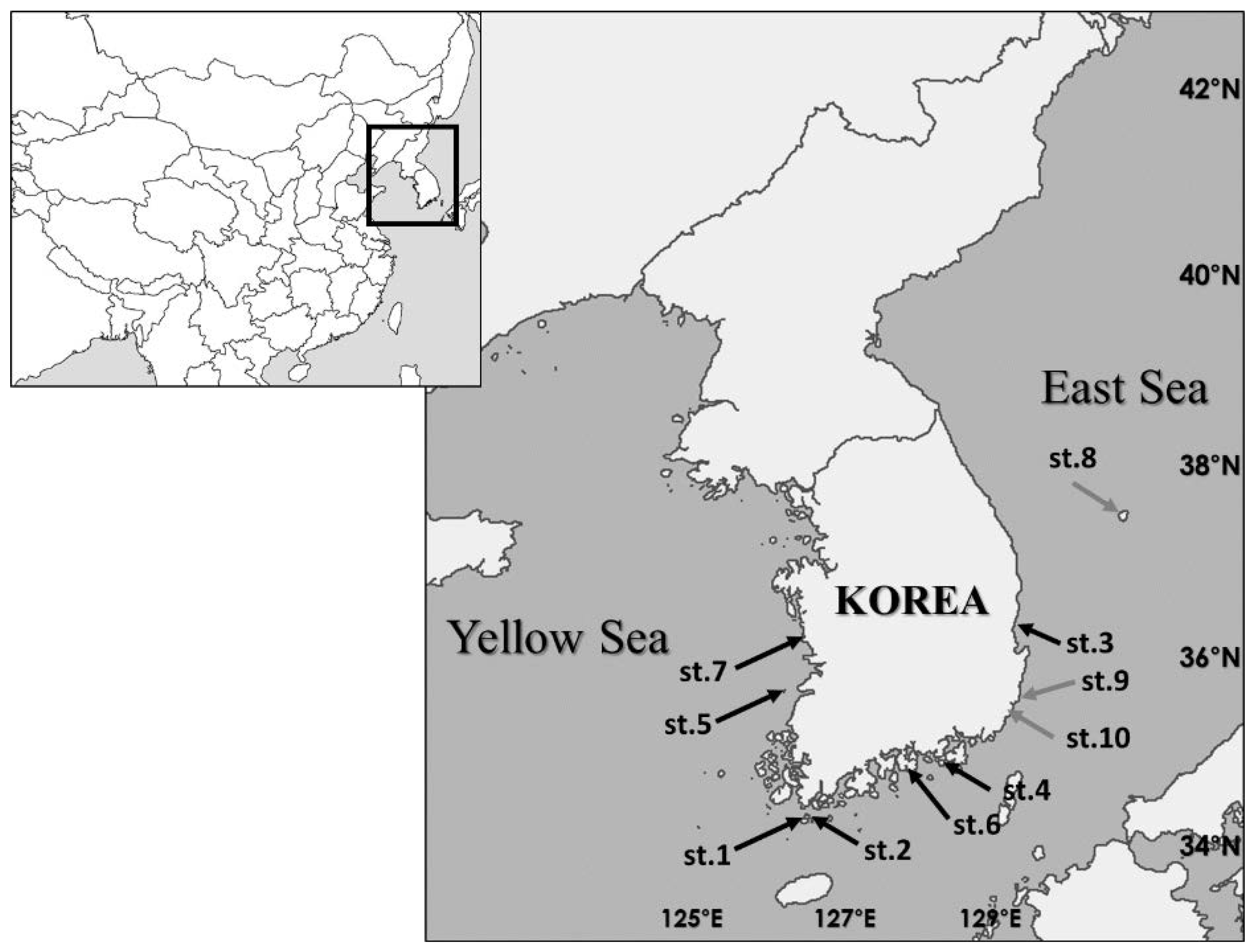
2.1. Sampling Locations

Sediment samples were collected by a grab sampler or SCUBA diving, and fixed with 95% ethanol (Figure 1, Table 1). Copepods were extracted from the sediments by the Ludox centrifugation method [9] and fixed in 99% ethanol. Harpacticoid specimens were sorted from the copepod samples with the aid of a Leica M80 stereomicroscope. The sorted specimens were refrigerated at −20 °C until DNA extraction. All species were morphologically described after DNA extraction.

2.2. DNA Extraction and Sequencing

Each specimen was rinsed in distilled water and then transferred to a 1.5 mL tube that contained 20 mL of Proteinase K and 180 mL of ATL buffer for non-destructive DNA extraction [10]. Next, the DNA extraction process was performed according to the protocol provided by Qiagen. (DNeasy Blood and Tissue Kit, Qiagen, Hilden, Germany). Both COI and 18Sr RNA sequences were amplified from the sample DNAs using an AccuPower HotStart PCR PreMix (Bioneer, Daejeon, Korea). Primers and PCR conditions are shown in Table 2. PCR products were sequenced in both directions using an ABI PRISM 3730XL Analyzer (Macrogen Inc., Daejeon, Korea). Sequences were assembled using Geneious 10.1.3 (Biomatters, Auckland, New Zealand) [11]. Pairwise distances were calculated using the Tamura and Nei distance model [12] in Geneious 10.1.3. The sequences from GenBank were aligned using the Muscle algorithm integrated in Geneious 10.1.3 [13].

2.3. Morphological Characterization

Harpacticoids specimens were dissected in lactic acid and the dissected parts were mounted on slides in lactophenol as mounting medium. Preparations were sealed with transparent nail varnish. All drawings were prepared using a camera lucida on a Leica DM 2500 differential interference contrast microscope.
The descriptive terminology is adopted from Huys et al. [14]. Abbreviations used in the text are A1, antennule; A2, antenna; ae, aesthetasc; exp, exopod; enp, endopod; P1–P6, first to sixth thoracopod; and exp (enp)-1 (2, 3) to denote the proximal (middle, distal) segment of a ramus. Scale bars in figures are indicated in μm.

3. Results

Order Harpacticoida Sars, 1903.
Family Longipediidae Boeck, 1865.
Genus Longipedia Claus, 1863.

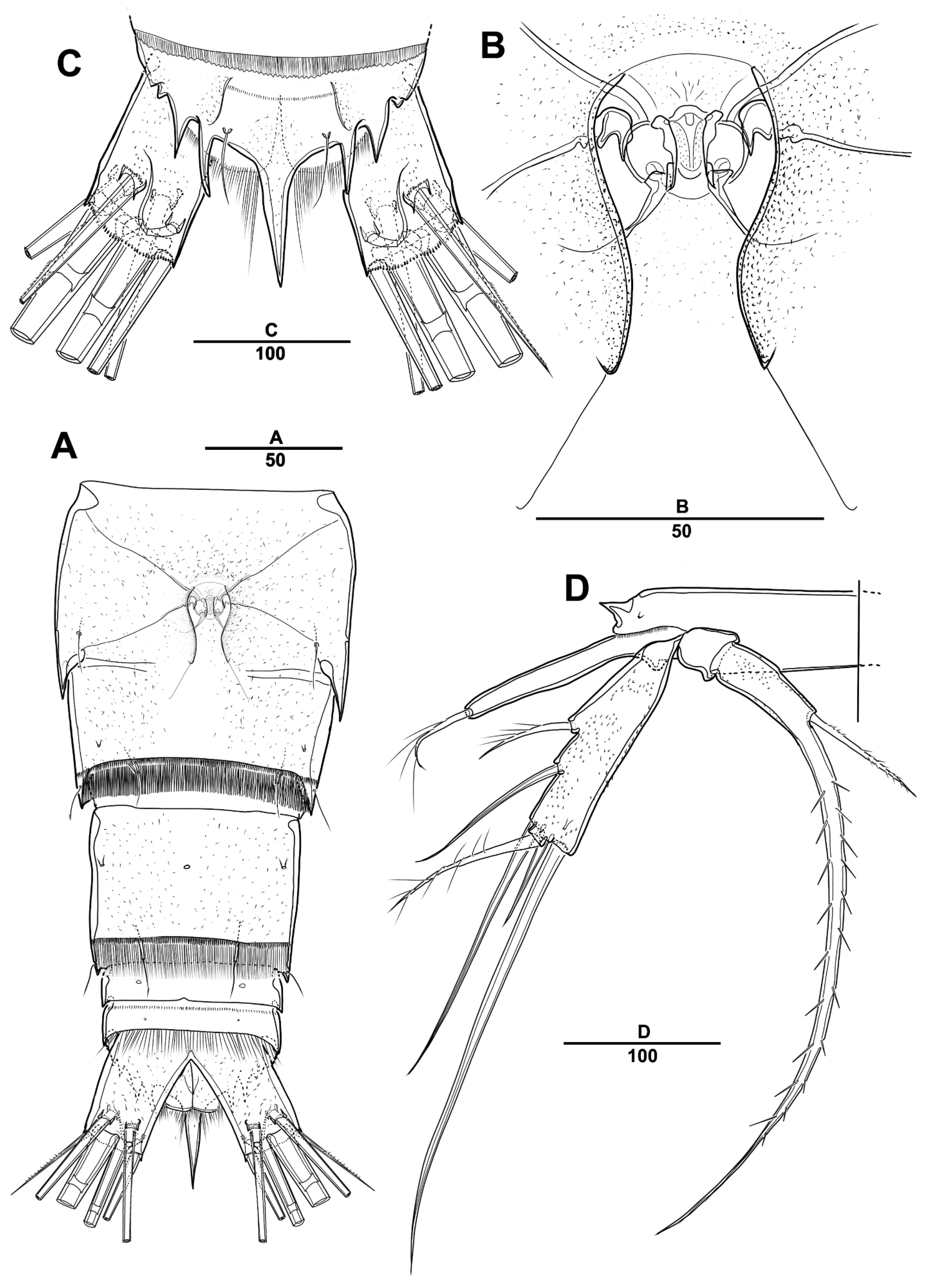
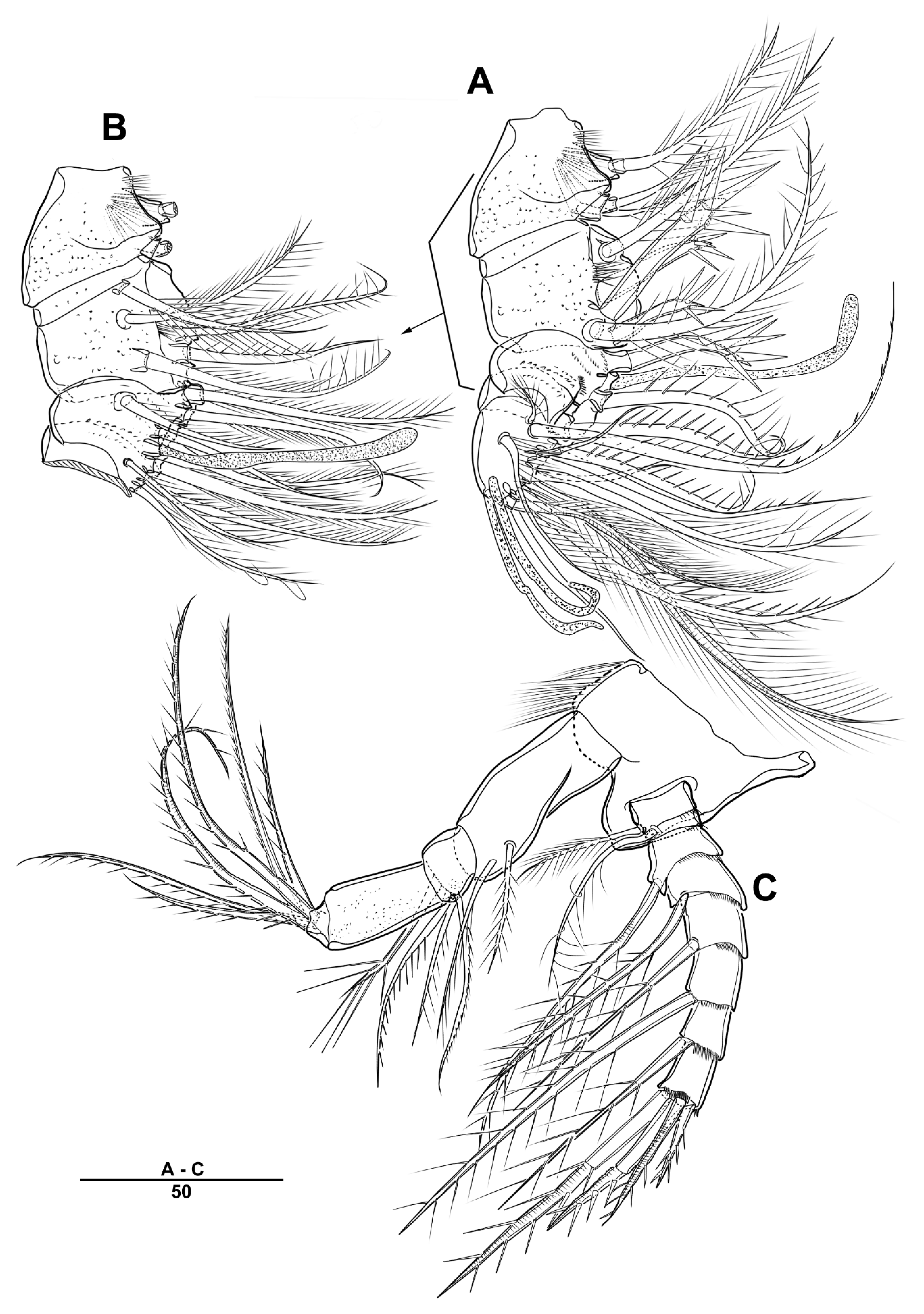
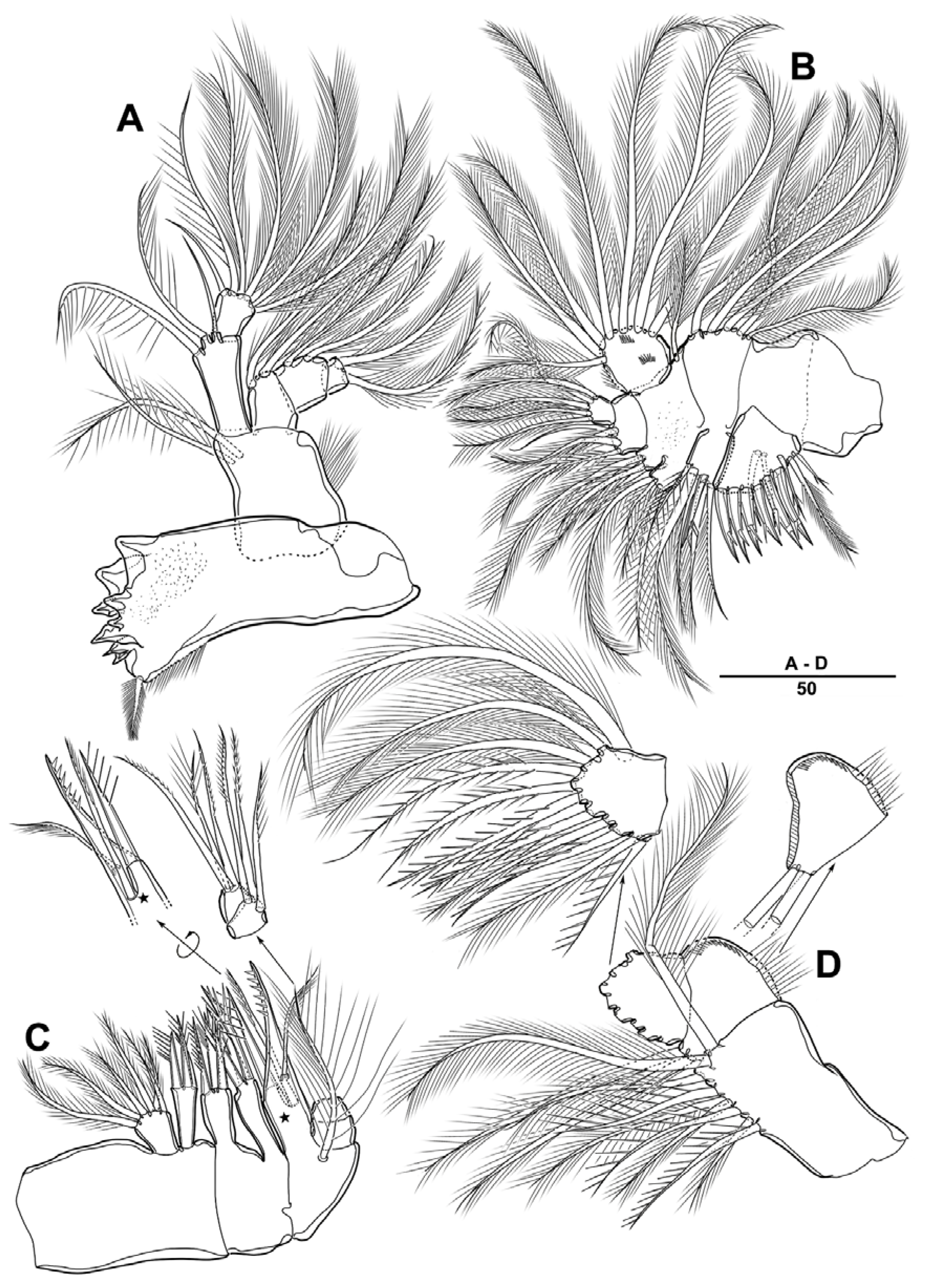
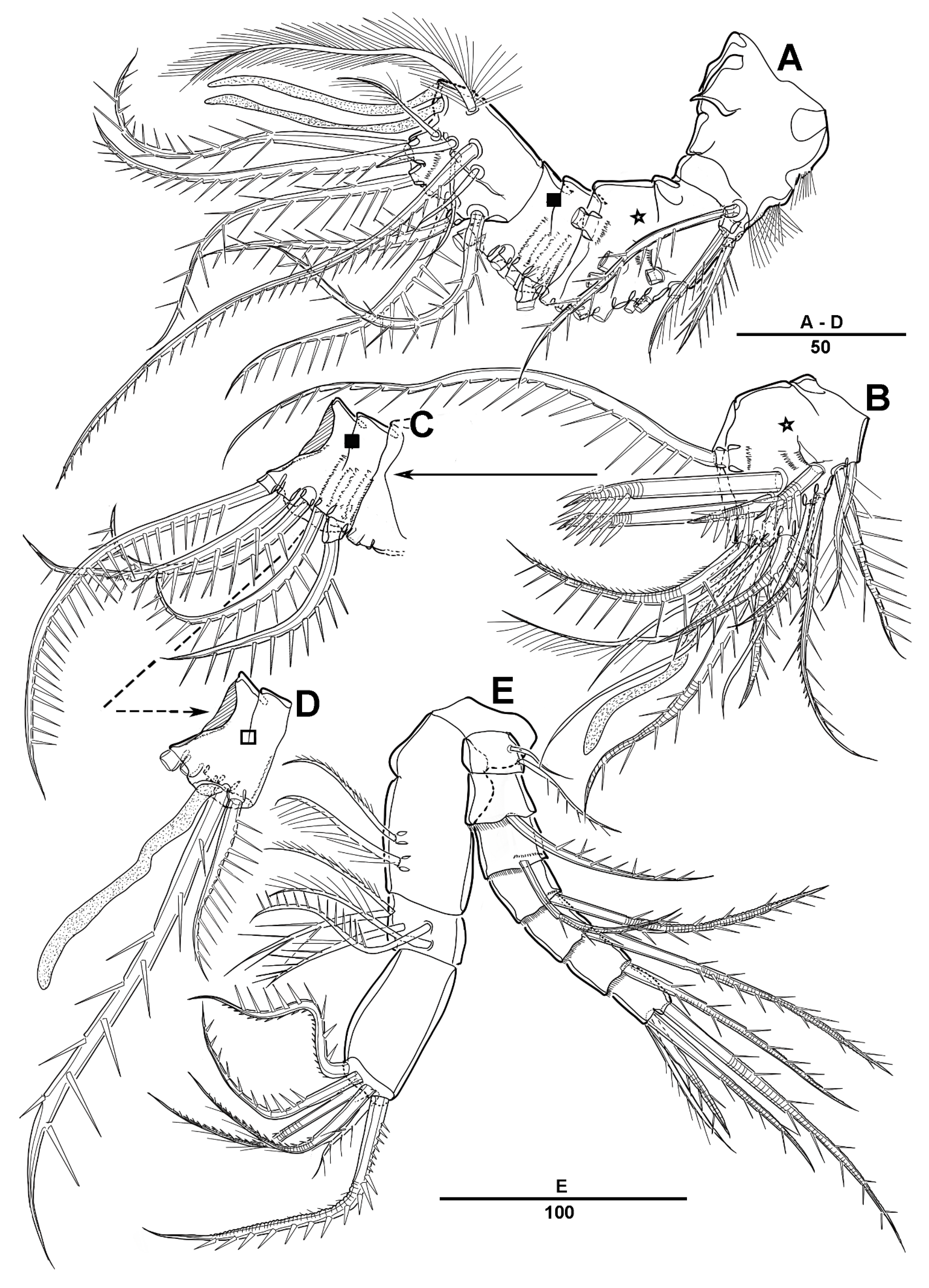
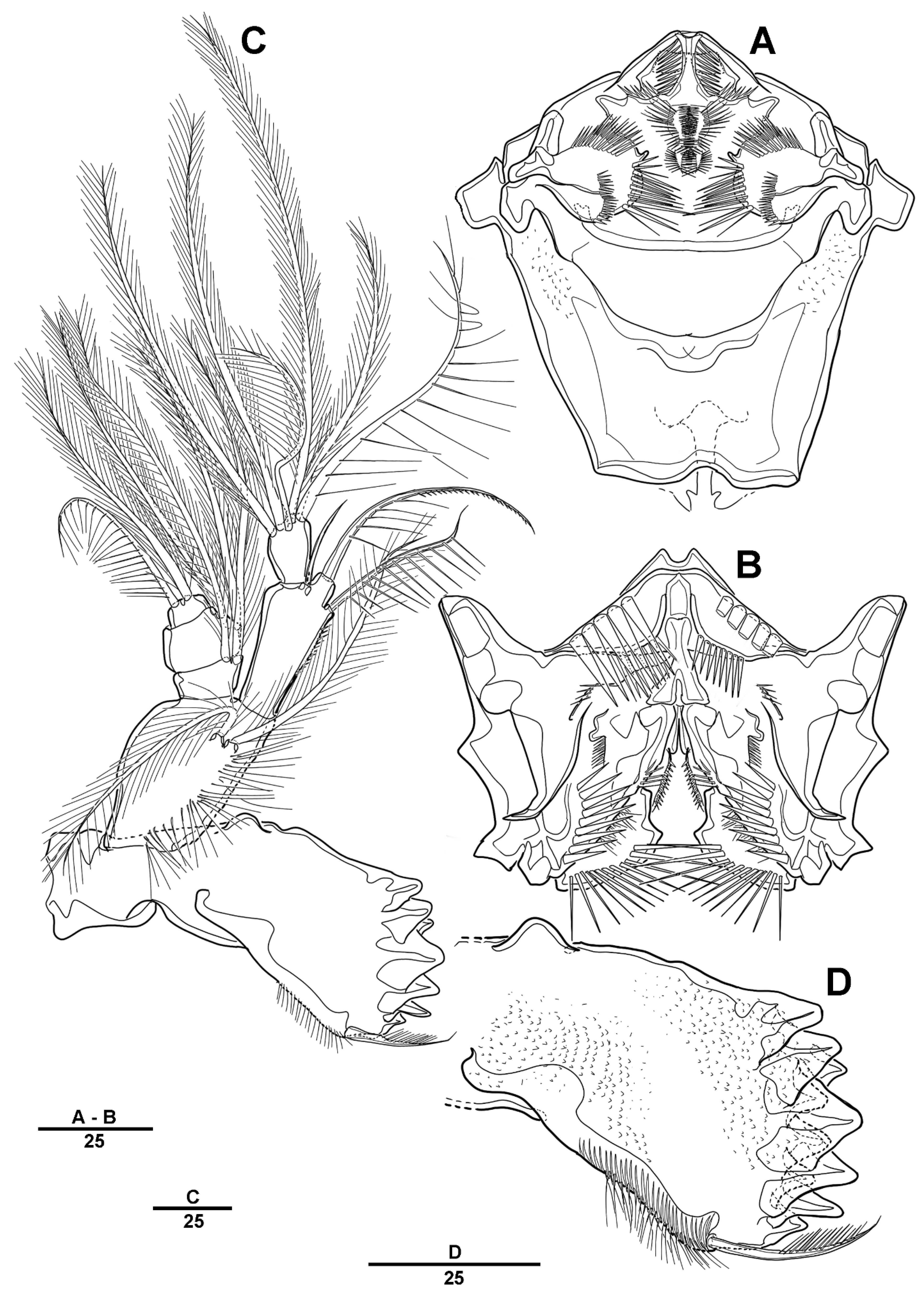
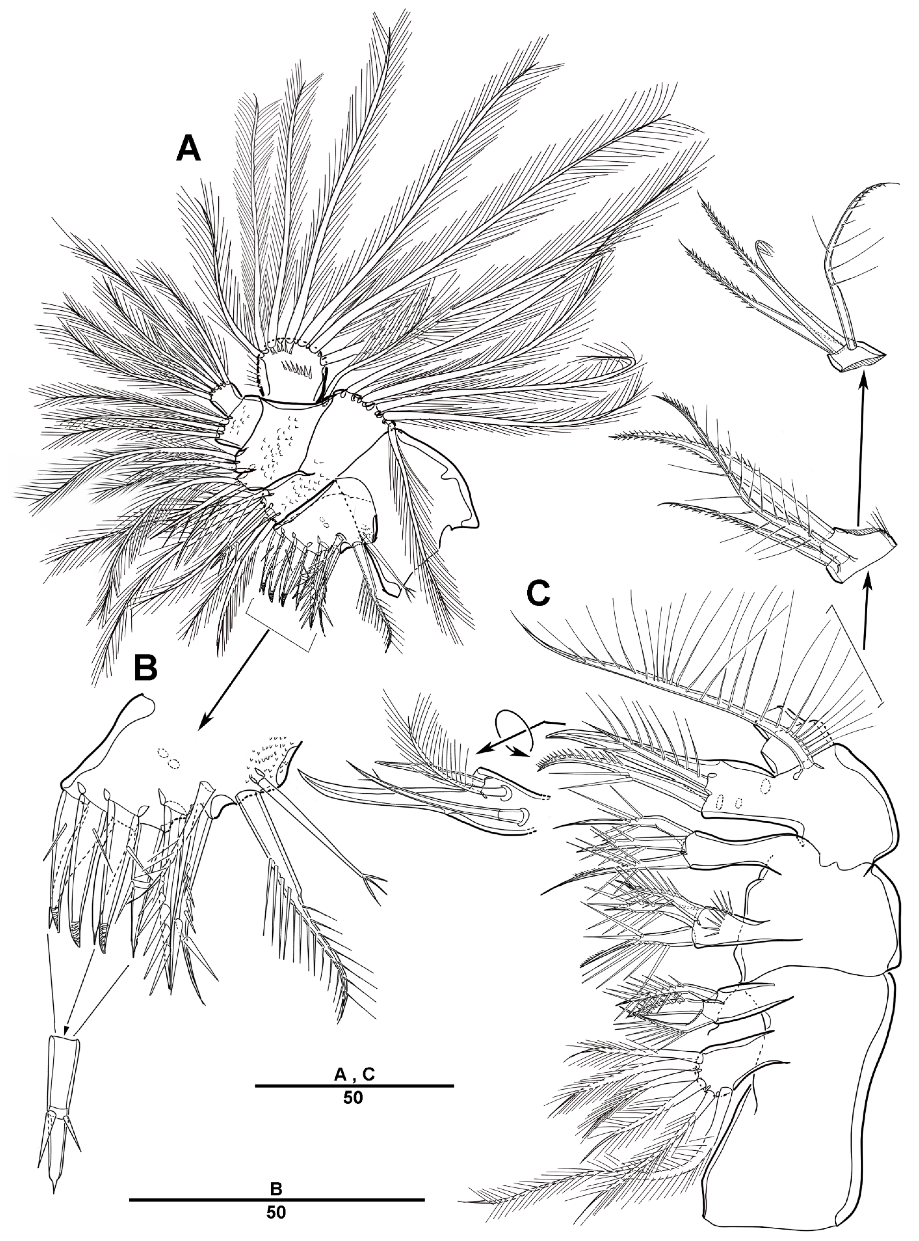
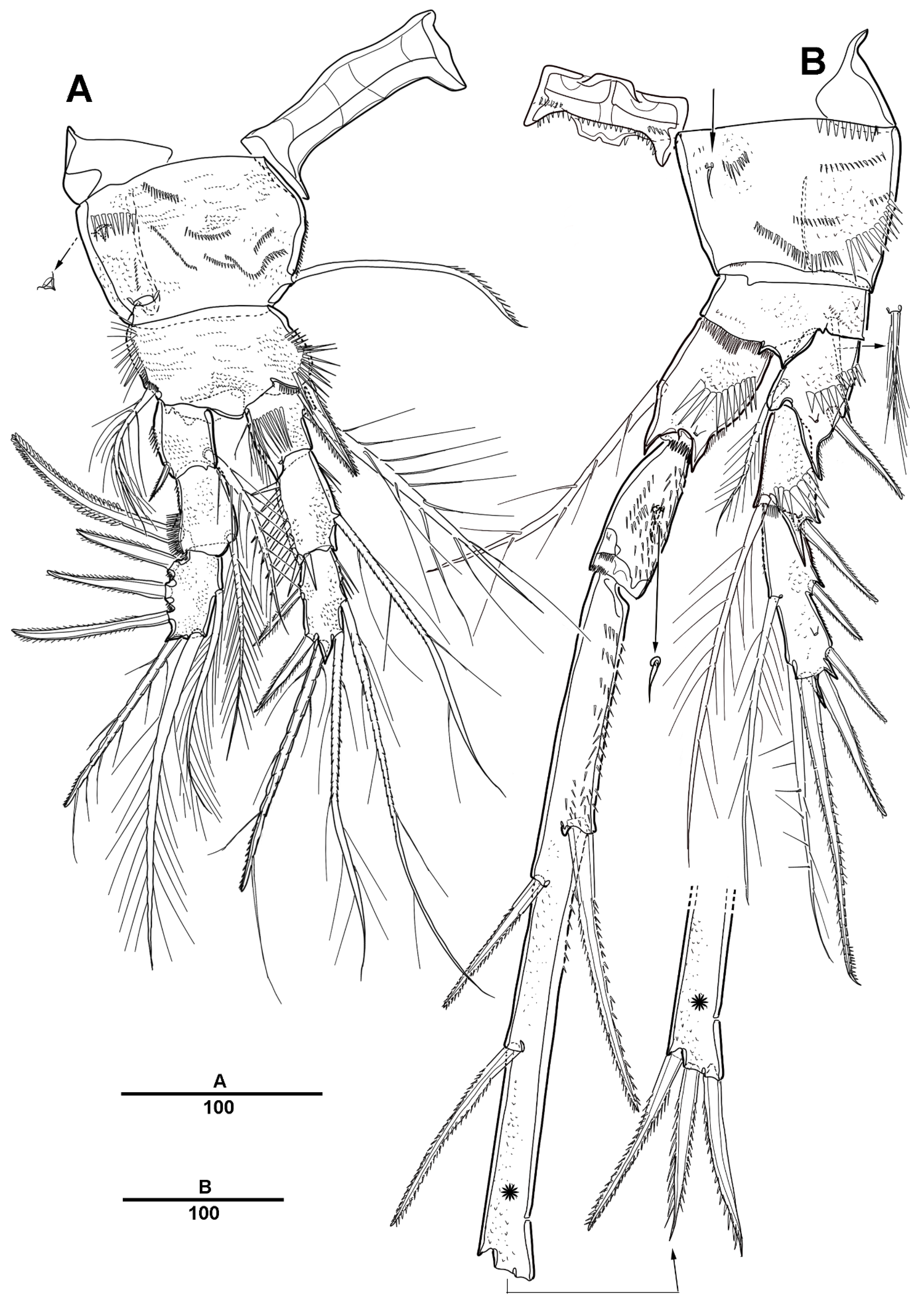
3.1. Longipedia koreana sp. nov.

LSID [urn:lsid:zoobank.org:act:29A2E160-76B5-49C7-B964-0B8CC17804DB]
Type locality. Area of algalculture, Nohwado Island, Jeollanam-do, Korea (34°13′34.68″ N, 126°31′35.37″ E).
Material examined. Holotype 1♀ (MABIK CR00248546) dissected on 11 slides. Paratype 1♂ (MABIK CR00248545) dissected on 10 slides. Detailed specimen and collection information is shown in Table 1.
Etymology. The species name refers to the type locality of the new species, Republic of Korea.
Genbank accession number. Mitochondrial cytochrome c oxidase subunit I gene (OK333363>–OK333374); 18S ribonucleic acid gene (OK335989–OK335997, OK339010).
Female. Total body length of 1132 µm (n = 9; range: 989–1203 µm), measured from the anterior margin of the rostrum to the posterior margin of the caudal rami. Largest width measured at the posterior margin of the cephalic shield: 300 µm. Habitus (Figure 2A,B) elongated, fusiform. Prosome slightly laterally compressed. Urosome slightly narrower than prosome.
Cephalothorax gradually tapering anteriorly, dorsal surface with sensilla as shown in Figure 2A, and posterior margin with finely striated hyaline frill. Rostrum bell-shaped, prominent, defined at base, with two small subdistal sensilla. Pedigerous somites with pores and sensilla as figured. Urosome (Figure 2A,B or Figure 3A) five-segmented, comprising P5-bearing somite, genital double-somite, and three free abdominal somites. Genital double-somite (Figure 3A) subdivided by chitinous suture dorsally and laterally, but shells completely fused; first somite with a pair of large ventrolateral processes, with sensilla along the posterior margin; second somite with comb-like hyaline frill. Genital apparatus (Figure 3B) is condensed into a compact median genital field, single copulatory pore located within the midventral complex. Anal somite (Figure 3C) with a transverse row of spinules ventrally. Anal operculum prominent, with strong median projection and four pairs of subsidiary lateral projections, and a pair of sensilla. Caudal ramus (Figure 2C) about twice as long as broad, with seven setae: seta I bare; seta II bare; seta III slightly pinnate; seta IV pinnate and long; seta V (Figure 2A,C,D) sparsely pinnate, longest; seta VI bare and slender; seta VII triarticulate at base and bare.
Antennule (Figure 4A,B) five-segmented. Segment 1 with long setules around the anterior margin; segment 2 with incompletely separated segments; third and fourth segment with one aesthetasc; last segment with two aesthetascs. Armature as follows: 1-[3], 2-[10], 3-[8+ae], 4-[5+ae], and 5-[11+2 ae].
Antenna (Figure 4C). Basis with long setules on the abexopodal margin. Exopod eight-segmented, few spinules present around the distal corner; first to seventh segments each with one seta, last segment with four spiniform setae. Endopod 3-segmented, enp-1 with two, enp-2 with four, and enp-3 with six setae.
Mandible (Figure 5A) with well-developed gnathobase. Basis with two setae inserted at the distal margin. Palp well-developed, biramous. Endopod 2-segmented, enp-1 with three, enp-2 with six setae. Exopod 3-segmented, each segment bearing two setae.
Maxillule (Figure 5B). Praecoxal arthrite with seven distal spines, two lateral setae, one lateral bipinnate seta, and two naked setae on the anterior surface. Coxo-endite with five elements, and epipodite represented by five setae. Basal proximal endite and distal endite with four setae each; basal exite represented by one small seta. Endopod indistinctly two-segmented, with one bare and three plumose setae on the proximal segment and five setae on the distal segment. Exopod l-segmented, with seven setae.
Maxilla (Figure 5C) comprising praecoxa, coxa, basis, and endopod. Praecoxa with two endites, proximal endite with six plumose setae, and distal endite with three spiniform elements. Coxa with two endites, each with three spinulose elements. Basis forming a strong spiniform process with three pinnate elements, and with one plumose seta on the anterior surface. Endopod two-segmented, each segment with three setae.
Maxilliped (Figure 5D) phyllopodial. Syncoxa with nine setae on the inner margin. Basis triangular, with two plumose setae on the inner margin, and with a row of setules along the outer margin. Endopod with 11 setae.
Swimming legs 1–4 (Figure 6 and Figure 7) with intercoxal sclerites and praecoxae, both without ornamentation. Coxae and bases with surface ornamentations of spinules and secretory pore as figured. All swimming legs with three-segmented exopods and endopods.
P1 (Figure 6A). Intercoxal sclerite and praecoxa unarmed. Coxa large, with spinular rows on the anterior surface, with strong spinules near the outer margin, and with a long pinnate inner seta. Basis with one outer plumose seta and one inner strong pinnate spine. Exp-1 with one outer spine and one long plumose inner seta; exp-2 with one outer strong pinnate spine and one plumose inner seta, and with spinules distal and outer margins; and exp-3 with three outer pinnate spines, one distal seta, and two inner setae. Endopod longer than exopod; enp-1 with inner plumose seta; enp-2 with one long inner plumose seta, and with spinules on distal and outer margins; and enp-3 with two long plumose inner setae, one well-developed seta, and two pinnate outer spines.
P2 (Figure 6B,C). Coxa large, ornamented with several rows of spinules, and armed with one small seta near the inner edge. Basis with one outer seta, with small spinules at the base of the endopod. Exp-1 and exp-2 with one outer spine and one plumose inner seta, and with spinules on the distal and outer margins each; exp-3 with two inner setae, one terminal pinnate spine, and three outer spines. Endopod about 2.4 times as long as three exopodite segments combined; enp-1 with inner plumose seta; enp-2 with several spinules on the anterior surface, with two tiny setae on the posterior surface (arrowed in Figure 6C); and enp-3 extremely elongated, about 15 times longer than it is broad, with two pinnate strong inner spines, one pinnate spine and two saw-like spines distally, and one pinnate outer spine.
P3 (Figure 7A). Coxa large, with one inner spine, with several rows of spinules. Basis with one outer seta, and with minute spinules along the outer margin. Exp-1 and exp-2 with one outer spine and one plumose inner seta, and with spinules on the distal and outer margins each; exp-3 with two plumose inner setae, two distal strong spines, and two outer pinnate spines. Endopod longer than exopod; enp-1 with one inner seta; enp-2 with spinules on the outer margins, and with two inner plumose setae; enp-3 with two long plumose inner setae, one plumose seta and pinnate spine terminally, and two outer spines.
P4 (Figure 7B,C). Coxa large, with few rows of spinules, and with one pinnate inner spine. Basis with one small seta. Exp-1 with spinules outer margin, and with one outer spine; exp-2 with one outer spine and one pinnate inner spine, setules on the inner margin and spinules on the outer margin; exp-3 with one inner spine, long pinnate spine inner side and small spine outer side at the terminal, and two outer pinnate spines. Enp-1 with a row of spinules on the inner proximal corner, and with one pinnate inner seta; enp-2 with one very small seta (arrowed in Figure 7B) and one spine; enp-3 with two distal and two inner pinnate spines.
Armature formula for swimming legs:
ExopodEndopod
P11.1.2131.1.212
P21.1.2131.2.231
P31.1.2221.2.222
P40.1.1221.2.022
P5 (Figure 3D) with separate endopod, exopod, and basis. Basis with outer setophore carrying one seta. Exopod 1-segmented, rectangular, about 3.6 times as long as wide, with six setae, the innermost the longest. Endopod 2-segmented; enp-1 without armature; enp-2 with two setae, the outermost pinnate, forming a whiplash-like structure, the innermost small and pinnate.
Male. Unknown.

3.2. Longipedia ulleungensis sp. nov.

LSID [urn:lsid:zoobank.org:act:CFBD6868-4DB6-44E1-AFB8-944C11DCC548]
Type locality. Ulleungdo Island, Gyeongsangbuk-do, Korea (37°29′13.14″ N, 130°55′19.62″ E).
Material examined. Holotype 1♀ (MABIK CR00248543) dissected on 10 slides. Paratype 1♀ (MABIK CR00248544) dissected on nine slides, 3♀♀ in 99% alcohol. Detailed specimen and collection information is shown in Table 1.
Etymology. The species name refers to the type locality, Ulleungdo Island in the East Sea of Korea.
Genbank accession number. Mitochondrial cytochrome c oxidase subunit I gene (OK333375–OK333377); 18S ribonucleic acid gene (OK339011–OK339013).
Female. Total body length of 1327 µm (n = 5; range: 1202–1362 µm), measured from the anterior margin of the rostrum to the posterior margin of the caudal rami. Largest width measured at the posterior margin of the cephalic shield: 409 µm. Body fusiform (Figure 8A,B). Prosome slightly laterally compressed. Urosome narrower than prosome.
Cephalothorax and free thoracic somites (Figure 8A,B or Figure 9A) furnished with sensilla. Sensillar pattern and tube pores on the cephalothorax and body somites are as figured. Rostrum defined at base, bell-shaped, with two sensilla either side of apex. Pedigerous somites with pores and sensilla as figured.
Urosome (Figure 8A,B or Figure 10A) consists of the fifth pedigerous somite, genital double-somite, and three free urosomites. Genital double-somite (Figure 10A) with a pair of triangular epimeral lappets on its anterior half and with dorsolateral suture; posterior half with well-developed hyaline frill on the posterior margin. Genital apparatus (Figure 10B) compact, comprising fused genital apertures and a single copulatory pore, located within the midventral complex. First and second free urosomites with posterior margins coarsely serrated both dorsally and ventrally. Anal somite (Figure 9B or Figure 10A) with a transverse row of spinules dorsally and ventrally. Anal operculum prominent, with long median projection and a single strong lateral projection, a group of outer spines on each side, and a pair of sensilla. Caudal ramus (Figure 9C) about 1.4 times as long as broad, with seven setae.
Antennule (Figure 11A–D) four-segmented. Segment 1 with long setules around the anterior margin; segment 2 and 3 with one aesthetasc each, segment 3 with rows of spinules on the anterior surface; and segment 4 with two aesthetascs. Armature as follows: 1-[3], 2-[17+ae], 3-[7+ae], and 4-[12+2ae].
Antenna (Figure 11E) biramous, with separate coxa and basis. Exopod seven-segmented; exp-1 with one bare and one pinnate seta; exp-2 to exp-6 with one spiniform seta each; and exp-6 with one pinnate and three spiniform setae. Endopod three-segmented, enp-1 with two setae; enp-2 with five setae; and enp-3 with six spiniform setae.
Labrum (Figure 12A) with a sophisticated and symmetrical pattern of tiny spinular rows; paragnaths (Figure 12B) well-developed lobes with spinular rows, and with long setules along the anterior margin as figured.
Mandible (Figure 12C,D) with well-developed gnathobase bearing several multicuspidate teeth around the distal margin and one pinnate seta at the dorsal corner, and covered with minute denticles. Basis with two plumose setae. Palp well-developed, biramous. Endopod 2-segmented; enp-1 with two pinnate and one bare seta; enp-2 with six setae. Exopod three-segmented, each segment bearing two setae.
Maxillule (Figure 13A,B). Praecoxal arthrite with three geniculated, three articulated, and two pinnate distal spines, laterally with one spiniform and one trifid seta, and with one pinnate seta on the anterior surface. Coxal endite with five setae and one spine, epipodite with five plumose setae. Basal proximal and distal endite with three setae each; basal exite represented by one seta. Endopod 2-segmented; enp-1 with three plumose setae; enp-2 with five setae. Exopod l-segmented, with seven setae.
Maxilla (Figure 13C). Praecoxa with two endites, proximal endite with six plumose setae, and distal endite with three spine-like elements. Coxa with two endites, each with three spinulose elements. Allobasis forming a curved claw with three pinnate elements, and with one plumose seta on the anterior surface. Endopod two-segmented; first segment with one bipinnate spine, one unipinnate fine seta, and one biplumose seta, second segment with four setae.
Maxilliped (Figure 9D) phyllopodial. Syncoxa with 10 setae along the inner margin and two setae on the anterior surface. Basis with two setae on the inner margin. Endopod with 11 plumose setae.
Swimming legs 1–4 (Figure 14 and Figure 15) with intercoxal sclerites and praecoxae. Coxae and bases with surface ornamentations of spinules and a secretory pore as figured. Coxa large, with spinular rows on the anterior surface, with a cone-like structure near the outer edge except for P2. All swimming legs with three-segmented exopods and endopods.
P1 (Figure 14A). Intercoxal sclerite and praecoxa unarmed. Coxa large, with a long pinnate inner seta. Basis with one outer plumose seta and one inner strong pinnate spine. Exp-1 with one small outer spine and one inner plumose seta; exp-2 with one outer strong pinnate spine and one inner seta, and with spinules on the outer margin; exp-3 with one inner seta, two distal setae, and three outer pinnate spines. Endopod slightly longer than exopod; enp-1 with one long inner seta; enp-2 with one inner plumose seta, and with long spinules on the outer margin; enp-3 with two plumose inner setae, one pinnate distal seta, and two pinnate outer spines.
P2 (Figure 14B). Praecoxa small, triangular. Coxa large, ornamented with several rows of spinules, and armed with one tiny seta near the inner edge. Basis with one outer pinnate seta, with spinules at the distal edge near the base of the endopod. Exp-1 and exp-2 with one outer spine and one plumose inner seta, with spinules on the distal and outer margins each, and the distal corner of exp-1 and exp-2 produced; exp-3 with two inner setae, one terminal pinnate spine, and three outer spines. Endopod about 2.4 times as long as the entire exopod; enp-1 with one inner plumose seta; enp-2 with several spinules on the anterior surface, with one tiny seta on the posterior surface; enp-3 extremely elongated, about 14.5 times longer than wide, with two pinnate strong inner spines, three strong distal spines, and one pinnate outer spine.
P3 (Figure 15A,B). Coxa large and rectangular, with one strong inner spine, and with several rows of spinules. Basis with one outer plumose seta, and with minute spinules close to the joint with the endopod. Exp-1 and exp-2 with one outer spine and one plumose inner seta, with a large projection at the outer distal corner, and with spinules near the distal inner edge; exp-3 with two inner setae, two distal spines, and two outer spines. Enp-1 and enp-2 with strong spinules on the outer margins, and with a large projection on the outer distal corner; enp-1 with one inner seta; enp-2 with two plumose inner setae; enp-3 with two long inner setae, one distal spine, and two outer spines.
P4 (Figure 15C). Coxa with rows of spinules, and with one strong inner spine. Basis with one small outer seta, and with spinules along the distal margin. Exp-1 with spinules on the outer margin, with one outer spine and one small bare inner seta, and with a large projection at the outer distal corner; exp-2 with one outer pinnate spine; exp-3 with one inner seta, with two distal pinnate spines, and with two outer pinnate spines. Enp-1 with one pinnate inner seta, with a row of long setules on the anterior surface, and with a row of spinules at the proximal corner; enp-2 with one small bare seta and one spine; enp-3 with two distal, and two inner pinnate spines.
Armature formula for swimming legs:
ExopodEndopod
P11.1.1231.1.212
P21.1.2131.1.231
P31.1.2221.2.212
P41.0.1221.2.022
P5 (Figure 10C). Exopod and endopod separate from the basis. Baseoendopod with a long outer setophore bearing one plumose basal seta. Exopod 1-segmented, apex wider than base, about 2.4 times as long as wide, with several spinules on the anterior surface, and with six setae, the innermost longest about 4.5 times as long as the exopod. Endopod one-segmented, with two setae, one fused to the segment forming a whiplash-like element.
Male. Unknown.

3.3. DNA Sequences of Two New Species

In regards to pairwise distances (Tamura–Nei distance) among the 607 bp COI sequences, L. koreana sp. nov. exhibited intra-specific variation of 0–1.155%, and inter-specific distances of 22.525–23.102% were observed between two new species. L. ulleungensis sp. nov. exhibited intra-specific variation of 0.413–0.578% (Table S1).
In regards to the 18S rRNA sequences among 1736 bp, intra-specific variations of 0% were observed in L. koreana sp. nov.; however, 0.058% was observed in L. ulleungensis sp. nov. Inter-specific distances of 1.325–1.382% were observed between the two new species (Table S2).

4. Discussion

The family Longipediidae was initially established by Boeck [15] under the name “Afdeling Longipedina” as the primary group of Harpacticidae. However, several authors [2,16,17] mistakenly attributed the family Longipediidae to Sars, 1903, causing confusion. However, these inaccuracies were pointed out and corrected by Wells [1].
Khodami et al. proposed a new order Canuelloida Khodami, MacArthur, Blanco-Bercial and Martinez Arbizu, 2017, including Canuellidae Lang, 1944 and Longipediidae Boeck, 1865; however, this article was retracted by the authors in 2020, due to inaccuracies in data and problems with data processing. Although the World of Copepods database [18] still shows the classification of the order Canuelloida based on the withdrawn paper, the family Longipediidae should be returned to belonging to the order Harpacticoida [19].
According to the database of World of Copepods, 22 species belonging to the family Longipediidae have been reported [18]. However, the species list should be modified according to the above. Wells [1] recognized 10 valid species within the genus Longipedia in his revision (L. americana Wells, 1980, L. andamanica, L. brevispinosa Gurney, 1927, L. coronata, L. helgolandica Klie, 1949, L. kikuchii, L. minor Scott T. & Scott A., 1893, L. nichollsi, L. scotti Sars, 1903, and L. weberi). In addition, he placed four species as incertae sedis in this genus (L. ferox Krichagin, 1877, L. mourei Jakobi, 1954, L. pirgos Apostolov, 1972, and L. pontica Krichagin, 1877). Furthermore, he synonymized two species, L. australica Nicholls, 1941 and L. longispina Monard, 1928 with L. scotti.
Since then, four species, Longipedia spinulosa [20], L. corteziensis [21], L. thailandensis [22], and L. gonzalezi Schizas, Dahms, Kantia, Corgosinho & Galindo, 2015 [23], have been added to this genus. Additionally, Gómez [21] elevates L. helgolandica santacruzensis Mielke, 1979 to species rank. Lastly, Longipedia paguri Müller, 1884 is a misspelling of Longipedina paguri Müller, 1884.
Gómez [21] proposed the helgolandica species group of the genus Longipedia based on the reduction of the inner spine of P4 enp-1 to a setiform element. L. helgolandica, L. americana, L. santacruzensis, and L. corteziensis belong to this group, and later, Schizas et al. [23] described the new species L. gonzalezi from Puerto Rico as the helgolandica group.
Schizas et al. [23] suggested that the characteristics of L. gonzalezi, which can be distinguished from L. americana, are (1) the sharp projection situated on the P2 basis, between the endopod and exopod (blunt projection in L. amaricana), and (2) the absence of spinules on P3 and P4 coxa (presence in L. amaricana). However, according to the redescriptions of L. americana by Onbé [24], the sharp protrusion is observed on the P2 basis. Therefore, it is difficult to find any significant differences between two species. So, we propose to treat that L. gonzalezi as a junior subjective synonym of L. americana.
Until now, most of the important characters for determining the species of copepods have been morphological features. The best way to describe a new species is to secure samples at different times, even in different regions, and report both sexes of the same species together. However, as in this study, it may not be easy for one species to secure both sexes at the same time or location. Therefore, if one sex is not continuously found, DNA sequences could provide important information in finding and classifying the other sex in the future. Furthermore, Schizas et al. [23] argued that Longipedia is a perfect genus to apply molecular sequences to corroborate morphological classification because of its high degree of morphological conservatism. Therefore, species identification using DNA sequences is very effective, and sometimes essential. In this study, we secured accurate sequence information using the non-destructive method [10].
NCBI has registered two species of COI sequences in the genus Longipedia, L. coronata, and L. kikuchii, and one species that was not analyzed at the species level (Table S3). As a result of calculating the COI pairwise distances based on 583 bp in Longipedia, there was a difference of up to 30.875%. This is higher than the 22.34% distance in the genus Diosaccus [10] and lower than 34.0% in the genus Delavalia [25]. These results indicate that the COI distance that determines the species may vary by genus.
Both new species are placed in the genus Longipedia Claus, 1863 because they have an extremely elongated distal segment of the P2 endopod, and have the outer seta representing the basal exite of the maxillule. Longipedia koreana sp. nov. has a morphological similarity to L. nichollsi and L. scotti based on the combination of three characters: (1) first segment of P4 endopod with a spiniform inner element, (2) first segment of the P2 endopod with a unguiform projection on the corner of the anterior surface, and (3) second segment of the P2 endopod with two tiny setae on the posterior surface. However, L. koreana sp. nov. can be differentiated from the congeners by (1) P1 coxa with strong spinules near the outer margin, and the distal element much larger than the proximal elements, whereas L. scotti has similar sized spinules, (2) P2 coxa with small inner seta on the anterior surface while with well-developed inner seta in L. nichollsi, (3) P4 exp-1 without armature element while with a minute inner seta in L. scotti and L. nichollsi, and (4) P5 exopod rectangular, and at least 3.5 times as long as wide (1.3 times in L. scotti and 3.0 times in L. nichollsi).
Based on previous descriptions by Wells [1], Longipedia ulleungensis sp. nov. is similar to L. brevispinosa, L. spinulosa Itô, 1981, and L. weberi, and they share the following characters: P2 endopod first segment with a claw-shaped projection on the corner of the anterior surface, P2 endopod second segment with one tiny seta on the posterior surface, and P5 exopod triangular. However, L. ulleungensis sp. nov. can be distinguished from its congeners by (1) P2 coxa with very reduced inner seta, (2) P4 exopod second segment without inner seta, and (3) anal operculum with a long median projection, and a single spine and group of outer spines on each side.
A key to the species of the family Longipediidae is provided. It is amended from Huys et al. [14] and Wells [26].
1
P4 enp-1 with setiform inner element ……………… helgolandica species-group… 2
-
P4 enp-1 with spiniform inner element ………………………………………………………… 5
2
P2 coxa with well-developed inner seta; P5 exopod rectangular …………………………………………………………………………… Longipedia helgolandica Klie, 1949
-
P2 coxa with small or slender inner seta; P5 exopod triangular, apex wider than base ……………………………………………………………………………………3
3
P5 baseoendopod not articulated ………………………………L. americana Wells, 1980
-
P5 baseoendopod articulated, with 2-segment endopodal lobe ……………………4
4
P1 basis with long setules on distal edge; P2 coxa with very reduced inner seta ……………………………………………………………… L. corteziensis Gómez, 2001
-
P1 basis distal edge smooth; P2 coxa with slender and relatively longer inner seta ……………………………………………… L. santacruzensis Mielke, 1979
5
P2 enp-1 with no projection on distal corner of anterior surface …………………………………………………………………… L. minor Scott T. & Scott A., 1893
-
P2 enp-1 with unguiform projection on corner of anterior surface ……………………………………………………………………………………………………………………… 6
6
P2 enp-2 without armature element on the posterior surface …………………………………………………… L. thailandensis Chullasorn & Kangtia, 2008
-
P2 enp-2 with one tiny seta on the posterior surface …………………………………… 7
-
P2 enp-2 with two tiny setae on the posterior surface …………………………………… 9
7
P4 exp-2 without inner seta; P2 coxa with much reduced inner seta; anal operculum with a long median projection (much longer than lateral projections), and a single spine and group of outer spines on each side……………………………………………………………….. L. ulleungensis sp. nov
-
P4 exp-2 with inner seta; P2 coxa with well-developed inner seta; anal operculum with a short median projection (no longer than lateral projections), and two spines on each side … L. brevispinosa Gurney, 1927
-
P4 exp-2 with inner seta; P2 coxa with well-developed inner seta; anal operculum with a long median projection (much longer than lateral projections), and two spines on each side ………………………………………… 8
8
P5 exopod about three times as long as wide; P2 enp-3 with a terminaldentate spine (median apical spine with a large tooth about halfway along) ……………………………………………………………………………… L. spinulosa Itô, 1981
-
P5 exopod about twice as long as wide; P2 enp-3 with serrated apicalspines ………………………………………………………………………… L. weberi Scott A., 1909
9
P2 coxa with small or reduced inner seta, or without inner seta ………………… 10
-
P2 coxa with well-developed inner seta ………………………………………………………11
10
P2 coxa without inner seta; P4 exp-1 with a minute inner seta; P5 exopod about twice as long as wide; P5 endopod with well-developed inner seta ……………………………………………… L. andamanica Wells, 1980
-
P2 coxa with very small inner seta, originated from the posterior surface; P4 exp-1 with a minute inner seta; P5 exopod at most 1.3 times as long as wide; P5 endopod with small and bare inner seta…………………………………………………………………………………………… L. scotti Sars, 1903
-
P2 coxa with small inner seta on anterior surface; P4 exp-1 without armature element; P5 exopod at least 3.5 times as long as wide; P5endopod with well-developed inner seta………………………… L. koreana sp. nov.
11
P1 coxa distal spinule much longer than the proximal spinule; P2 enp-3 median terminal spine with a large tooth about halfway along its length; P5 exopod about three times as long as wide……………………………………………………………………………………… L. nichollsi Wells, 1980
-
P1 coxa distal spinule much longer than the proximal spinule; P2 enp-3 median terminal spine with a large tooth about halfway along, or P2 enp-3 with three terminal spinulose claws; P5 exopod about twice as long as wide ……………………………………………… L. kikuchii Itô, 1980
-
P1 coxa with row of similar sized setules on outer side; P2 enp-3 with three terminal spinulose spines; P5 exopod about twice as long as wide ………………………………………………………………… L. coronata Claus, 1863

Supplementary Materials

The following are available online at https://0-www-mdpi-com.brum.beds.ac.uk/article/10.3390/d13110590/s1, Table S1: Pairwise distances (Tamura-Nei distance) between COI sequences from two new species, Table S2: Pairwise distances (Tamura-Nei distance) based on 1736 bp between 18SrRNA sequences from two new species, Table S3. Pairwise distances (Tamura-Nei distance) based on 583 bp between COI sequences from genus Longipedida.

Author Contributions

Conceptualization, H.W.B. and J.B.; Data curation, H.M.; Funding acquisition, H.W.B. and J.B.; Investigation, H.M.; methodology, H.W.B. and J.B.; Project administration, J.B.; Visualization, H.M.; writing—original draft, H.W.B.; writing—review & editing, H.W.B. and J.B. All authors have read and agreed to the published version of the manuscript.

Funding

This research was funded by Ministry of Ocean and Fisheries and National Marine Biodiversity Institute of Korea (2021M00100).

Institutional Review Board Statement

Not applicable.

Informed Consent Statement

Not applicable.

Data Availability Statement

Not applicable.

Acknowledgments

Thank you to Cheol Yu and Seo Woo Son for helping us with the sorting of copepods from sediment.

Conflicts of Interest

The authors declare no conflict of interest.

References

  1. Wells, J.B.J. A revision of the genus Longipedia Claus (Crustacea: Copepoda: Harpacticoida). Zool. J. Linn. Soc. 1980, 70, 103–189. [Google Scholar] [CrossRef]
  2. Boxshall, G.A.; Halsey, S.H. An Introduction to Copepod Diversity; Ray Society: London, UK, 2004; pp. 1–966. [Google Scholar]
  3. Hulme, P.E. Trade, transport and trouble: Managing invasive species pathways in an era of globalization. J. Appl. Ecol. 2009, 46, 10–18. [Google Scholar] [CrossRef]
  4. Ganapati, P.N.; Shanthakumari, K. The systematics and distribution of planktonic copepods in the Lawson’s Bay, Waltair. J. Mar. Biol. Assoc. India 1961, 3, 6–18. [Google Scholar]
  5. Itô, T. A new subspecies of Longipedia andamanica Wells from the Pacific coast of Japan, with reference to the morphology of L. coronata Claus (Copepoda: Harpacticoida). Publ. Seto Mar. Biol. Lab. 1985, 30, 307–324. [Google Scholar] [CrossRef] [Green Version]
  6. Kim, J.G. Taxonomic Study on the Harpacticoids (Crustacea: Copepoda: Harpacticoida) from Marine and Brackish Waters in Korea. Doctoral Dissertation; Chosun University: Gwangju, Korea, 2017. [Google Scholar]
  7. Itô, T. Two species of the genus Longipedia Claus from Japan, with reference to the taxonomic status of L. weberi previously reported from Amakusa, southern Japan (Copepoda: Harpacticoida). J. Nat. Hist. 1980, 14, 17–32. [Google Scholar] [CrossRef]
  8. Kim, S.H. Invertebrate fauna of Korea. In Arthropoda: Maxillopoda: Copepoda: Harpacticoida. Marine Harpacticoida II; National Institute of Biological Resources: Incheon, Korea, 2013; Volume 21, Number 27; pp. 1–113. [Google Scholar]
  9. Burgess, R. An improved protocol for separating meiofauna from sediments using colloidal silica sols. Mar. Ecol. Prog. Ser. 2001, 214, 161–165. [Google Scholar] [CrossRef]
  10. Lim, B.J.; Bang, H.W.; Moon, H.; Back, J. Integrative description of Diosaccus koreanus sp. nov. (Hexanauplia, Harpacticoida, Miraciidae) and integrative information on further Korean species. ZooKeys 2020, 927, 1–35. [Google Scholar] [CrossRef] [PubMed]
  11. Kearse, M.; Moir, R.; Wilson, A.; Stones-Havas, S.; Cheung, M.; Sturrock, S.; Buxton, S.; Cooper, A.; Markowitz, S.; Duran, C.; et al. Geneious Basic: An integrated and extendable desktop software platform for the organization and analysis of sequence data. Bioinformatics 2012, 28, 1647–1649. [Google Scholar] [CrossRef] [PubMed]
  12. Tamura, K.; Nei, M. Estimation of the number of nucleotide substitutions in the control region of mitochondrial DNA in humans and chimpanzees. Mol. Biol. Evol. 1993, 10, 512–526. [Google Scholar] [PubMed]
  13. Edgar, R.C. MUSCLE: Multiple sequence alignment with high accuracy and high throughput. Nucleic Acids Res. 2004, 32, 1792–1797. [Google Scholar] [CrossRef] [PubMed] [Green Version]
  14. Huys, R.; Gee, J.M.; Moore, C.G.; Hamond, R. Marine and Brackish Water Harpacticoid copepods. In Synopses of the British Fauna (New Series); Field Studies Council: Shrewsbury, UK, 1996; Part 1; pp. 4–19. [Google Scholar]
  15. Boeck, A. Oversigt over de ved Norges Kyster jagttagne Copepoder henhörende til Calanidernes, Cyclopidernes og Harpactidernes Familier. Forhandlinger i Videnskabsselskabet i Kristiania 1865, 1864, 226–282. [Google Scholar]
  16. Lang, K. Monographie der Harpacticiden; Håkan Ohlssons Boktryckeri: Lund, Sweden, 1948; pp. 1–1682. [Google Scholar]
  17. Siegfried, S. Phylogeny of Harpacticoida (Copepoda): Revision of “Maxillipedasphalea” and Exanechentera; Cuvillier Verlag: Göttingen, Germany, 2003; pp. 1–259. [Google Scholar]
  18. Walter, T.C.; Boxshall, G.; World of Copepods database. Longipedia Claus. 1863. Available online: http://www.marinespecies.org/copepoda/aphia.php?p=taxdetails&id=115403 (accessed on 18 October 2021).
  19. Mikhailov, K.V.; Ivanenko, V.N. Low support values and lack of reproducibility of molecular phylogenetic analysis of Copepoda orders. Arthropoda Sel. 2021, 30, 39–42. [Google Scholar] [CrossRef]
  20. Itô, T. Descriptions and records of marine harpacticoid copepods from Hokkaido, VIII. J. Fac. Sci. Hokkaido Univ. 1981, 22, 422–450. [Google Scholar]
  21. Gómez, S. Longipedia corteziensis sp. nov. (Copepoda, Harpacticoida, Longipediidae) from a coastal lagoon in northwestern Mexico, with the definition of the helgolandica species-group of the genus Longipedia Claus, 1863. Hydrobiologia 2001, 453/454, 483–496. [Google Scholar]
  22. Chullasorn, S.; Kangtia, P. Longipedia thailandensis sp. nov. (Copepoda, Harpacticoida) from a brackish water treatment pond, Samut Sakhon, Thailand. Crustaceana 2008, 81, 207–225. [Google Scholar]
  23. Schizas, N.V.; Dahms, H.U.; Kangtia, P.; Corgosinho, P.H.; Galindo Estronza, A.M. A new species of Longipedia Claus, 1863 (Copepoda: Harpacticoida: Longipediidae) from Caribbean mesophotic reefs with remarks on the phylogenetic affinities of Polyarthra. Mar. Biol. Res. 2015, 11, 789–803. [Google Scholar] [CrossRef]
  24. Onbé, T. The developmental stages of Longipedia americana (Copepoda: Harpacticoida) reared in the laboratory. J. Crustac. Biol. 1984, 4, 615–631. [Google Scholar] [CrossRef]
  25. Yeom, J.; Lee, W. A new species of the genus Sarsamphiascus Huys, 2009 (Copepoda: Harpacticoida: Miraciidae) from a sublittoral zone of Hawaii. PeerJ 2020, 8, e8506. [Google Scholar] [CrossRef] [PubMed] [Green Version]
  26. Wells, J.B. An annotated checklist and keys to the species of Copepoda Harpacticoida (Crustacea). Zootaxa 2007, 1568, 1–872. [Google Scholar] [CrossRef] [Green Version]
Figure 1. The type localities and the sampling locations (st.1: Nohwado Island, Jeollanam-do, Korea; st.8: Ulleungdo Island, Gyeongsangbuk-do, Korea; black arrows: sampling station of Longipedia koreana sp. nov.; gray arrows: sampling station of Longipedia ulleungensis sp. nov.).
Diversity 13 00590 g001
Figure 2. Longipedia koreana sp. nov. holotype female. (A), habitus, dorsal; (B), habitus, lateral; (C), left caudal ramus, dorsal; (D), the basis of the inner terminal seta (V). Scale bars indicate length in µm.
Diversity 13 00590 g002
Figure 3. Longipedia koreana sp. nov. holotype female. (A), urosome, ventral; (B), genital field, ventral; (C), anal somite and caudal rami, dorsal; (D), P5. Scale bars indicate length in µm.
Diversity 13 00590 g003
Figure 4. Longipedia koreana sp. nov. holotype female. (A), antennule; (B), opposite armature of the third and fourth segment of antennule; (C), antenna. Scale bar indicates length in µm.
Diversity 13 00590 g004
Figure 5. Longipedia koreana sp. nov. holotype female. (A), mandible; (B), maxillule; (C), maxilla; (D), maxillped. Scale bar indicates length in µm.
Diversity 13 00590 g005
Figure 6. Longipedia koreana sp. nov. holotype female. (A), P1; (B), P2; (C), second segment of P2 endopod, posterior. Scale bars indicate length in µm.
Diversity 13 00590 g006
Figure 7. Longipedia koreana sp. nov. holotype female. (A), P3; (B), P4; (C), first segment of P4 endopod. Scale bars indicate length in µm.
Diversity 13 00590 g007
Figure 8. Longipedia ulleungensis sp. nov. holotype female. (A), habitus, dorsal; (B), habitus, lateral. Scale bar indicates length in µm.
Diversity 13 00590 g008
Figure 9. Longipedia ulleungensis sp. nov. holotype female. (A), distal margin of the cephalothorax and first free thoracic somite, dorsal; (B) anal somite and caudal rami, dorsal; (C), caudal ramus, ventral; (D), maxilliped. Scale bars indicate length in µm.
Diversity 13 00590 g009
Figure 10. Longipedia ulleungensis sp. nov. holotype female. (A), urosome, ventral; (B), genital field, ventral; (C), P5. Scale bars indicate length in µm.
Diversity 13 00590 g010
Figure 11. Longipedia ulleungensis sp. nov. holotype female. (A), antennule, anterior; (B), second segment of antennule, anterior; (C), third segment of antennule, anterior; (D), third segment of antennule, posterior; (E), antenna. Scale bars indicate length in µm.
Diversity 13 00590 g011
Figure 12. Longipedia ulleungensis sp. nov. holotype female. (A), labrum; (B), paragnath; (C), mandible; (D), mandibular gnathobase. Scale bars indicate length in µm.
Diversity 13 00590 g012
Figure 13. Longipedia ulleungensis sp. nov. holotype female. (A), maxillule; (B), praecoxal arthrite of maxillule; (C), maxilla. Scale bars indicate length in µm.
Diversity 13 00590 g013
Figure 14. Longipedia ulleungensis sp. nov. holotype female. (A), P1; (B), P2. Scale bars indicate length in µm.
Diversity 13 00590 g014
Figure 15. Longipedia ulleungensis sp. nov. holotype female. (A), P3; (B), first segment of P3 exopod anterior; (C), P4. Scale bars indicate length in µm.
Diversity 13 00590 g015
Table 1. Information of collection and specimens in this study.
Species NameDateLocalityMethods (Depth)Specimen Nos.
L. koreana sp. nov.29 August 2018St. 1: N 34°13′34.68″ E 126°31′35.37″Grab (20 m)MABIK CR00248546
MABIK CR00248533
MABIK CR00248534
MABIK CR00248535
MABIK CR00248536
St. 2: N 34°12′15.82″ E 126°32′50.87″Grab (8 m)MABIK CR00248532
MABIK CR00248539
MABIK CR00248540
21 November 2018St. 3: N 36°22′11.69″ E 129°24′28.73″Scuba diving (15 m)MABIK CR00248531
MABIK CR00248545
27 June 2019St. 4: N 34°47′7″ E 128°27′32.42″Grab (15 m)MABIK CR00248537
26 September 2019St. 5: N 35°34′16.74″ E 126°16′49.94″Grab (9 m)MABIK CR00248538
21 October 2020St. 6: N 34°42′58.0″ E 127°59′29.0″Scuba diving (9 m)MABIK CR00248541
13 May 2021St. 7: N 36°13′18.0″ E 126°31’32.0”Beach (intertidal)MABIK CR00248542
L. ulleungensis sp. nov.25 July 2017St. 8: N 37°29′13.14″ E 130°55′19.62″Scuba diving (15 m)MABIK CR00248543
26 April 2018St. 9: N 35°28′47.52″ E 129°25′38.88″Grab (10 m)MABIK CR00248544
MABIK CR00248530
27 April 2018St. 10: N 35°20′53.9″ E 129°20′19.5″Grab (10 m)MABIK CR00248528
MABIK CR00248529
Table 2. Primer sequences and PCR conditions used in the present study.
GenePrimerPrimer SequencePCR Condition
COILCO149
HCO2198
GGTCAACAAATCATAAAGATATTGG
TAAACTTCAGGGTGACCAAAAAATCA
94 °C, 300 s; 40 cycles × (94 °C, 60 s; 48 °C, 120 s; 72 °C, 180 s; 72 °C, 600 s)
18S rRNA18S F1
18S R9
18S F2
18S F3
18S F4
18S R6
18S R7
18S R8
TACCTGGTTGATCCTGCCAG
GATCCTTCCGCAGGTTCACCTAC
CCTGAGAAACGGCTRCCACAT
GYGRTCAGATACCRCCSTAGTT
GGTCTGTGATGCCCTYAGATGT
TYTCTCRKGCTBCCTCTCC
GYYARAACTAGGGCGGTATCTG
ACATCTRAGGGCATCACAGACC
94 °C, 300 s; 40 cycle × (94 °C, 30 s; 50 °C, 30 s; 72 °C, 60 s); 72 °C, 420 s
Internal primers, 18S F2–F4 and 18S R6–R8, were used for primer walking to sequence over 1700 bps
Publisher’s Note: MDPI stays neutral with regard to jurisdictional claims in published maps and institutional affiliations.

